HAL Id: hal-03138518
https://hal.sorbonne-universite.fr/hal-03138518
Submitted on 11 Feb 2021
HAL is a multi-disciplinary open access
archive for the deposit and dissemination of
sci-entific research documents, whether they are
pub-lished or not. The documents may come from
teaching and research institutions in France or
abroad, or from public or private research centers.
L’archive ouverte pluridisciplinaire HAL, est
destinée au dépôt et à la diffusion de documents
scientifiques de niveau recherche, publiés ou non,
émanant des établissements d’enseignement et de
recherche français ou étrangers, des laboratoires
publics ou privés.
Pierre Bay, Guillaume Lebreton, Alexis Mathian, Pierre Demondion, Cyrielle
Desnos, Juliette Chommeloux, Guillaume Hekimian, Nicolas Bréchot, Ania
Nieszkowska, Matthieu Schmidt, et al.
To cite this version:
Pierre Bay, Guillaume Lebreton, Alexis Mathian, Pierre Demondion, Cyrielle Desnos, et al.. Outcomes
of severe systemic rheumatic disease patients requiring extracorporeal membrane oxygenation. Annals
of Intensive Care, SpringerOpen, 2021, 11 (1), �10.1186/s13613-021-00819-3�. �hal-03138518�
RESEARCH
Outcomes of severe systemic rheumatic
disease patients requiring extracorporeal
membrane oxygenation
Pierre Bay
1, Guillaume Lebreton
2, Alexis Mathian
3,4, Pierre Demondion
2, Cyrielle Desnos
1,
Juliette Chommeloux
1, Guillaume Hékimian
1, Nicolas Bréchot
1, Ania Nieszkowska
1, Matthieu Schmidt
1,5,
Fleur Cohen‑Aubart
3,4, Pascal Leprince
2, Charles‑Edouard Luyt
1,5, Zahir Amoura
3,4, Alain Combes
1,5and Marc Pineton de Chambrun
1,3,4,5*Abstract
Background: Systemic rheumatic diseases (SRDs) are a group of inflammatory disorders that can require intensive care unit (ICU) admission because of multiorgan involvement with end‑organ failure(s). Critically ill SRD patients requiring extracorporeal membrane oxygenation (ECMO) were studied to gain insight into their characteristics and outcomes.
Methods: This French monocenter, retrospective study included all SRD patients requiring venovenous (VV)‑ or venoarterial (VA)‑ECMO admitted to a 26‑bed ECMO‑dedicated ICU from January 2006 to February 2020. The primary endpoint was in‑hospital mortality.
Results: Ninety patients (male/female ratio: 0.5; mean age at admission: 41.6 ± 15.2 years) admitted to the ICU received VA/VV‑ECMO, respectively, for an SRD‑related flare (n = 69, n = 38/31) or infection (n = 21, n = 10/11). SRD was diagnosed in‑ICU for 31 (34.4%) patients. In‑ICU and in‑hospital mortality rates were 48.9 and 51.1%, respectively. Nine patients were bridged to cardiac (n = 5) or lung transplantation (n = 4), or left ventricular assist device (n = 2). The Cox multivariable model retained the following independent predictors of in‑hospital mortality: in‑ICU SRD diagnosis, day‑0 Simplified Acute Physiology Score (SAPS) II score ≥ 70 and arterial lactate ≥ 7.5 mmol/L for VA‑ECMO–treated patients; diagnosis other than vasculitis, day‑0 SAPS II score ≥ 70, ventilator‑associated pneumonia and arterial lac‑ tate ≥ 7.5 mmol/L for VV‑ECMO–treated patients.
Conclusions: ECMO support is a relevant rescue technique for critically ill SRD patients, with 49% survival at hospital discharge. Vasculitis was independently associated with favorable outcomes of VV‑ECMO–treated patients. Further studies are needed to specify the role of ECMO for SRD patients.
Keywords: Systemic rheumatic disease, Extracorporeal membrane oxygenation, Intensive care unit, Vasculitis, Systemic lupus erythematosus, Connective tissue disease, Acute respiratory distress syndrome, Cardiogenic shock
© The Author(s) 2021. This article is licensed under a Creative Commons Attribution 4.0 International License, which permits use, sharing, adaptation, distribution and reproduction in any medium or format, as long as you give appropriate credit to the original author(s) and the source, provide a link to the Creative Commons licence, and indicate if changes were made. The images or other third party material in this article are included in the article’s Creative Commons licence, unless indicated otherwise in a credit line to the material. If material is not included in the article’s Creative Commons licence and your intended use is not permitted by statutory regulation or exceeds the permitted use, you will need to obtain permission directly from the copyright holder. To view a copy of this licence, visit http://creat iveco mmons .org/licen ses/by/4.0/.
Introduction
Systemic rheumatic diseases (SRDs) are a group of inflammatory disorders (including connective tissue diseases, rheumatic disorders, vasculitides, sarcoidosis, adult-onset Still’s disease…) involving more than one organ and often requiring immunosuppressant therapy [1]. They share common characteristics: multiorgan
Open Access
*Correspondence: marc.dechambrun@gmail.com
1 Service de Médecine Intensive‑Réanimation, Hôpital La Pitié–Salpêtrière,
Sorbonne Université, Assistance Publique‑Hôpitaux de Paris (APHP), Paris, France
involvement responsible for end-organ failures; specific treatments causing immunosuppression and infectious complications; and are rare entities with challenging diagnoses and diagnostic difficulties. Outcomes of SRD patients requiring ICU admission remain unclear, with 16%–33% reported in-ICU mortality [1–4].
Extracorporeal membrane oxygenation (ECMO) is a rescue technique used to temporarily replace the heart and/or lung functions of the most severe patients [5, 6]. It may serve as a bridge-to-recovery or a bridge-to-organ transplantation for patients with treatment-refractory heart and/or lung failure(s).
We undertook this study to determine the outcome and identify in-hospital mortality associated factors of criti-cally ill SRD patients receiving ECMO.
Methods Patients
We retrospectively reviewed the prospectively con-stituted ECMO database of our 26-bed ICU to iden-tify adult SRD patients who received, between January 2006 and February 2020, venoarterial (VA)-ECMO and/ or venovenous (VV)-ECMO for heart and/or lung end-organ failure(s). SRD were identified searching in all medical charts a large number of keywords referring to SRD including: systemic rheumatic disease; connective tissue disease; lupus; systemic sclerosis; scleroderma; antiphospholipid; myositis; inflammatory myopathy; Sharp; Sjögren; Gougerot; rheumatoid arthritis; spon-dylarthritis; vasculitides; Goodpasture; antineutrophil cytoplasmic antibodies; proteinase 3; myeloperoxidase; Henoch-Schönlein; sarcoidosis; Still’s disease; eosino-philia; myasthenia; neuromyelitis optica… Our ter-tiary ICU is an ECMO-referral center for Greater Paris. Patients with the following SRDs were considered for inclusion: connective tissue diseases, vasculitides, sar-coidosis, nonmalignant eosinophilia-related disorders, adult-onset Still’s disease and other organ-specific auto-immune diseases with more than one organ involved.
ECMO implantation
The detailed surgical procedure for femoral–femoral VA-ECMO or femoral–jugular VV-VA-ECMO placement was described previously [7–9]. Briefly, trained cardiovascu-lar surgeons performed all procedures in-ICU at bedside or in the cardiac angiography room because of patient’s hemodynamic instability. Femoral and/or jugular ves-sels were cannulated after limited cut-down using the Seldinger technique and, for VA-ECMO, an additional 7 French catheter was systematically inserted distally into the femoral artery to prevent severe leg ischemia. For highly unstable patients diagnosed with refractory car-diogenic shock or acute respiratory distress syndrome
(ARDS) in other hospitals, our institution’s Mobile ECMO Retrieval Team traveled rapidly to primary-care hospitals with a portable ECMO system, installed the device before refractory multiorgan failure or ARDS occurred, and then transported the patient to our ter-tiary-care center [10].
Study endpoints
The primary endpoint was in-hospital mortality, defined as death during the hospital stay consecutive to the first ICU admission and before the patient’s discharge to home. The secondary outcomes included ECMO wean-ing: bridge-to-recovery, bridge-to-transplantation (lung or cardiac) and bridge-to-long term ventricle assist device.
Data collection
The following information was collected on stand-ardized forms: epidemiological parameters; SRD clinical, biological and therapeutic history; clinical mani-festations; laboratory findings; ECMO type, indication and complication(s); Survival after Veno-Arterial ECMO (SAVE) [11] and Respiratory Extracorporeal Membrane Oxygenation Survival Prediction (RESP) [12] scores, that are survival predictors in VA-ECMO and VV-ECMO patients, respectively; in-ICU treatments; organ-support treatments; SRD-specific treatments introduced in the ICU; ECMO-weaning status; bridge-to-transplantation or left ventricular assist device (LVAD); ICU complica-tions; vital status, transplantation status at ICU and hos-pital discharges and at last follow-up.
Statistical analyses
Results for categorical variables, expressed as number (%), were compared with χ2 tests; those for continuous
variables, expressed as mean ± standard deviation or median [25–75th percentile interquartile range (IQR)], were compared using Student’s t-test or Wilcoxon’s rank test. Normality of continuous variable distribution was assessed with the Shapiro–Wilk test; when not normal, Wilcoxon’s rank test was used for comparisons.
First, patients’ characteristics (laboratory findings, in-ICU organ-failure treatment(s), SRD-specific mani-festations and treatment(s), complications and out-comes) were subjected to descriptive analysis. Next, the mean/median values and frequencies of patients’ char-acteristics were compared according to the primary endpoint for the entire population and in the following subgroups: flare-related admission and VA/VV-ECMO. Then, for each subgroup, a Cox proportional hazards model, including the variables associated with the pri-mary endpoint (entry threshold: P < 0.05), was run using backward-stepwise variable elimination (exit threshold:
P > 0.10). Continuous variable were dichotomized to
using the cut-offs with the best association with the pri-mary endpoint in univariable Cox proportional hazards model. All potential explanatory variables included in the multivariable analyses were subjected to ity analysis with a correlation matrix. When colinear-ity was found (variance inflation factor > 5), only one of the two variables could be included the model. Sta-tistical significance was defined as P < 0.05. Analyses were computed with IBM SPSS Statistics v22.0 software (IBM Corp, Armonk, NY).
Ethical considerations
The database is registered with the
“Commis-sion Nationale de l’Informatique et des Libertés”
(2217847v0). In accordance with the ethical standards of our hospital’s institutional review board, the Com-mittee for the Protection of Human Subjects, and French law, written informed consent was not needed for demographic, physiological and hospital-outcome data analyses, because this observational study did not modify existing diagnostic or therapeutic strategies; however, patients were informed of their inclusion in the study.
Results
General characteristics
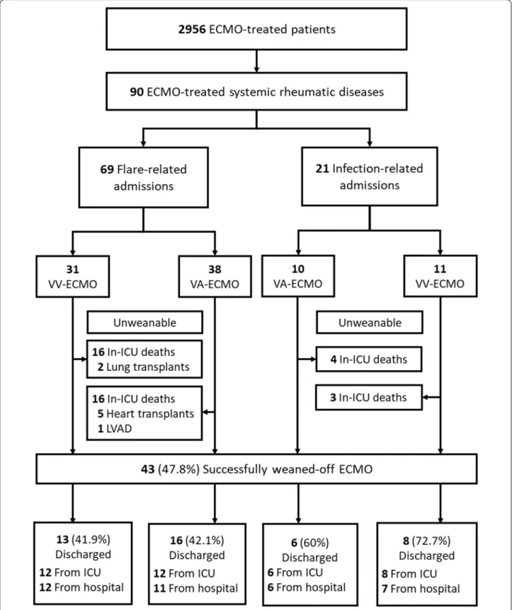
During the study period, 90 SRD patients requiring ICU admission (male/female ratio: 0.5; mean age at ICU admission: 41.6 ± 15.2 years) received VV-ECMO (n = 42, 46.7%) or VA-ECMO (n = 48, 53.3%). Their demograph-ics and the SRD characteristdemograph-ics are detailed in Table 1 and Additional file 1: Table S1. SRD was diagnosed in-ICU for 34.4% patients. The main diagnoses were: connective tissue disease (57.8%), vasculitis (11.1%), rheumatic dis-orders (11.1%) and sarcoidosis (5.6%). The organs most frequently affected pre-admission were: lung (47.8%), joints (38.9%), skin, heart and kidney. Before ICU admis-sion, 47.8% of the patients took corticosteroids regularly and 36.7% immunosuppressants. Three-quarters were admitted for an SRD flare and about one-quarter for an infection. The flow chart reports patients’ outcomes according to the reason for admission and ECMO hook-up (Fig. 1). In-ICU mortality, in-hospital mortality and in-hospital mortality/LVAD/ transplantation rates were: 48.9, 51.1 and 60.0%.
The main ECMO complications were cannula-related infection, insertion-site hemorrhage and limb ischemia. In-ICU–acquired infections occurred in 65.6% of patients; their sites and pathogens are reported in Addi-tional file 1: Table S2.
Uni‑ and multivariable analyses of in‑hospital mortality‑associated factors
Nonsurvivors, compared to survivors, were less quickly admitted to the ICU after symptom onset and hospital admission, had more frequent SRD heart involvement before admission, higher day-0 SAPS II and SOFA scores, lower RESP and SAVE scores, and more frequently received vasopressors and renal replacement therapy in ICU (Table 2). Nonsurvivors also had more frequent in-ICU–acquired infections, especially fungal, and ECMO
Table 1 Characteristics of 90 SRD patients given ECMO support
Continuous variables are expressed as mean ± standard deviation or median [interquartile range]; categorical variables are expressed as n (%)
ICU intensive care unit, ANCA antineutrophil cytoplasm antibodies, ECMO extracorporeal membrane oxygenation, SRD systemic rheumatic disease
* Three missing data
† One each: myasthenia gravis, neuromyelitis optica, multicentric Castleman’s
disease, autoimmune thrombocytopenic purpura or inflammatory bowel disease
§ Methotrexate n = 15, azathioprine n = 13, mycophenolate mofetil n = 9,
cyclophosphamide n = 8, rituximab n = 6, tumor necrosis factor-inhibitor n = 6, calcineurin inhibitors n = 5, tocilizumab n = 2
Variables Value
Women 60 (66.7)
Body mass index, kg/m2 26.3 ± 6.8
Age at ICU admission, years 41.6 ± 15.2 Systemic rheumatic diseases
Diagnosis in the ICU 31 (34.4) Diagnosis‑to‑ICU interval*, months 93 (25–132) Connective tissue diseases 52 (57.8)
Systemic lupus erythematosus 22 (24.4) Idiopathic inflammatory myositis 12 (13.3) Antiphospholipid syndrome 12 (13.3) Systemic sclerosis 5 (5.6) Mixed connective tissue disease 5 (5.6) Sjögren’s syndrome 3 (3.3) Rheumatic disorders 10 (11.1) Vasculitides 10 (11.1) Goodpasture’s syndrome 3 (3.3) ANCA‑associated 5 (5.6) Small‑vessel 1 (1.1) IgA‑associated 1 (1.1) Sarcoidosis 5 (5.6)
Nonmalignant eosinophilia‑related diseases 4 (4.4) Adult‑onset Still’s disease 4 (4.4)
Others† 5 (5.6)
Pre‑ICU Specific treatment(s)
Corticosteroids 43 (47.8)
Immunosuppressant(s)§ 33 (36.7)
Flare‑related admission 69 (76.7) Infection‑related admission 21 (23.3)
Fig. 1 Flow chart of the 90 patients with systemic rheumatic disease requiring extracorporeal membrane oxygenation. ICU intensive care unit, LVAD left ventricular assist device, VA/VV-ECMO venoarterial/venovenous‑extracorporeal membrane oxygenation
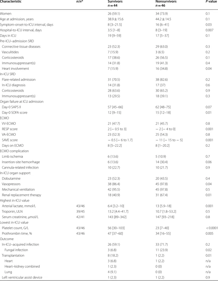
Table 2 In-ICU characteristics and outcomes of the 90 ECMO-treated SRD patients: hospital survivors vs. nonsurvivors
Characteristic n/n* Survivors
n = 44 Nonsurvivorsn = 46 P value
Women 26 (59.1) 34 (73.9) 0.1
Age at admission, years 38.9 ± 15.6 44.2 ± 14.5 0.1
Symptom‑onset‑to‑ICU interval, days 8 [3–21.5] 16 [6–41] 0.03
Hospital‑to‑ICU interval, days 3.5 [1–8] 8 [3–19] 0.007
Days in ICU 19 [9–59] 17 [5–37] 0.1
Pre‑ICU–admission SRD
Connective tissue diseases 23 (52.3) 29 (63.0) 0.3
Vasculitides 7 (15.9) 3 (6.5) 0.2 Corticosteroids 17 (38.6) 26 (56.5) 0.1 Immunosuppressant(s) 14 (31.8) 19 (41.3) 0.4 Heart involvement 7 (15.9) 16 (34.8) 0.04 In‑ICU SRD Flare‑related admission 31 (70.5) 38 (82.6) 0.2 In‑ICU diagnosis 14 (31.8) 17 (37) 0.6 Corticosteroids 28 (63.6) 30 (65.2) 0.9 Immunosuppressant(s) 13 (29.5) 18 (39.1) 0.3
Organ failure at ICU admission
Day‑0 SAPS II 57 [45–66] 62 [48–75] 0.07
Day‑0 SOFA score 12 [9–15] 15 [12–18] 0.01
ECMO VV‑ECMO 21 (47.7) 21 (45.7) 0.8 RESP score 2 [− 0.5 to 3] − 2 [− 4 to 0] 0.001 VA‑ECMO 23 (52.3) 25 (54.3) 0.8 SAVE score − 0.5 [− 6 to 1.7] − 11 [− 15 to − 5] 0.001 Days on ECMO 8 [5–22.2] 8 [1–20.2] 0.2 ECMO complication Limb ischemia 6 (13.6) 5 (10.9) 0.7 Insertion‑site hemorrhage 6 (13.6) 14 (30.4) 0.06 Cannula‑related infection 10 (22.7) 10 (21.7) 0.9
In‑ICU organ support
Dobutamine 23 (52.3) 20 (43.5) 0.4
Vasopressors 38 (86.4) 45 (97.8) 0.04
Mechanical ventilation 42 (95.5) 45 (97.8) 0.5
Renal replacement therapy 18 (40.9) 31 (67.4) 0.01
Highest in‑ICU value
Arterial lactate, mmol/L 43/46 6.4 [3.2–10] 13 [5.9–18] 0.001
Troponin, ULN 39/45 13.2 [4.4–41.7] 10.7 [1.8–53.2] 0.5
Serum creatinine, µmol/L 42/41 143 [89–342] 147 [93–218] 0.8
Lowest in‑ICU value
Platelet count, G/L 43/46 56 [30–103] 23 [7–40] < 0.0001 Prothrombin time, % 43/46 47 [37–60] 34 [16–55] 0.005 Outcome In‑ICU–acquired infection 26 (59.1) 33 (71.7) 0.2 Fungal infection 3 (6.8) 11 (23.9) 0.02 Transplantation 8 (18.2) 1 (2.2) 0.01 Heart 3 (6.8) 1 (2.2) n/a
Heart–kidney combined 1 (2.3) 0 (0) n/a
Lung 4 (9.1) 0 (0) n/a
insertion-site hemorrhages. The frequencies of flare-related admissions, in-ICU SRD diagnoses and VA/VV-ECMO percentages were not different for the two groups. The Cox proportional hazards model univariable and multivariable analyses for the 90 SRD patients (Table 3) retained: pre-admission SRD heart involvement; day-0 SAPS II score ≥ 70; arterial lactate ≥ 7.5 mmol/L and
bilirubin ≥ 125 µmol/L, as independently associated with in-hospital mortality.
Uni‑ and multivariable analyses of in‑hospital mortality‑associated factors: flare‑related admissions
Among the 69 flare-related admissions: 44.9% patients received VV-ECMO and 55.1% VA-ECMO, 21 could be
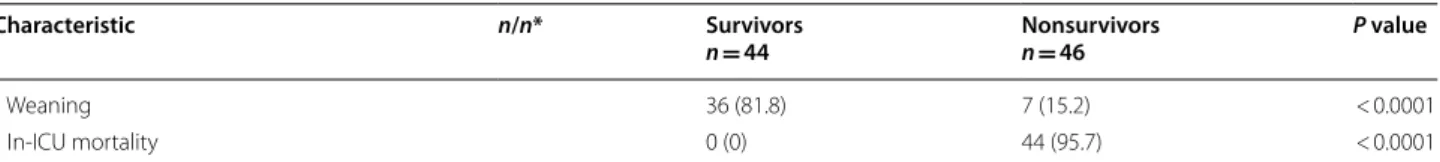
Table 2 (continued)
Characteristic n/n* Survivors
n = 44 Nonsurvivorsn = 46 P value
Weaning 36 (81.8) 7 (15.2) < 0.0001
In‑ICU mortality 0 (0) 44 (95.7) < 0.0001
Continuous variables are expressed as mean ± standard deviation or median [interquartile range] and compared with Student’s t-test or Wilcoxon’s rank test; categorical variables are expressed as n (%) and compared with χ2 tests
ICU intensive care unit, LVEF left ventricle ejection fraction, RESP Respiratory Extracorporeal Membrane Oxygenation Survival Prediction, SAPS Simplified Acute Physiology Score, SAVE Survival after Veno-Arterial ECMO, SOFA Sequential Organ-Failure Assessment, SRD systemic rheumatic disease, VA-/VV-ECMO venoarterial/ venovenous-extracorporeal membrane oxygenation, ULN upper limit of normal value, VTI velocity–time integral
* Numbers of survivor/nonsurvivor data available
Table 3 Univariable and multivariable analyses of factors associated with in-hospital mortality for the 90 ECMO-treated SRD patients
Bold values indicates statistically significant in multivariable analysis
The multiple Cox proportional hazards model used backward-stepwise variable elimination (with variable exit threshold set at P > 0.10). All potential explanatory variables included in the multivariable analyses were subjected to colinearity analysis with a correlation matrix. Variables associated with one another were not included in the model. Statistical significance was defined as P < 0.05
ICU intensive care unit, SAPS Simplified Acute Physiology Score, SOFA sequential organ failure assessment, SRD systemic rheumatic diseases, VA-ECMO venoarterial-extracorporeal membrane oxygenation
Factor Univariable analysis multivariable analysis
HR 95% CI P value HR 95% CI P value
Age ≥ 40 years 1.4 0.8–2.5 0.3
Women 1.5 0.8–2.9 0.2
Pre‑admission SRD lung involvement 0.5 0.3–0.9 0.04 0.8 0.4–1.6 0.6 Pre‑admission SRD heart involvement 1.7 0.9–3.2 0.08 2.9 1.5–5.8 0.001 Corticosteroids before admission 1.7 0.9–3.1 0.07 1.8 0.9–3.3 0.052 Immunosuppressants before admission 1.3 0.7–2.3 0.4
In‑ICU SRD diagnosis 1.0 0.6–1.9 0.9
Day‑0 SAPS II ≥ 70 2.7 1.5–4.9 0.001 2.7 1.4–5.1 0.003
Day‑0 SOFA score ≥ 16 2.8 1.6–5.1 < 0.0001
Flare‑related admission 1.4 0.6–3.0 0.7 VA‑ECMO 1.3 0.7–2.4 0.3 In‑ICU corticosteroids 0.8 0.4–1.5 0.5 In‑ICU immunosuppressant(s) 1.0 0.5–1.8 0.9 Vasopressors 5.3 0.7–38.6 0.1 2.7 0.3–21.0 0.3 Mechanical ventilation 1.8 0.2–13.2 0.5
Renal replacement therapy 2.2 1.2–4.0 0.01 0.6 0.2–1.4 0.2
ICU‑acquired infection 1.0 0.6–2.0 0.9
Highest in‑ICU value
Arterial lactate ≥ 7.5 mmol/L 3.2 1.7–5.9 < 0.0001 2.8 1.4–5.3 0.002
Bilirubin ≥ 125 µmol/L 2.3 1.2–4.3 0.007 2.0 1.0–3.9 0.04
Lowest in‑ICU value
weaned-off ECMO and 10 were bridged-to-transplant (n = 8) or -LVAD (n = 2). Nonsurvivors, compared to survivors, had more frequent SRD heart involvement before admission, higher day-SOFA scores, lower RESP and SAVE scores, and more frequently received vaso-pressors and renal replacement therapy in ICU (Table 4).
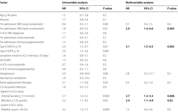
Similarly to the whole cohort, the Cox proportional hazards model univariable and multivariable analyses (Table 5) retained: pre-admission SRD heart involvement, day-0 SAPS II score ≥ 70, arterial lactate ≥ 7.5 mmol/L and bilirubin ≥ 125 µmol/L, as independently associated with in-hospital mortality.
Uni‑ and multivariable analyses of VA‑ECMO–associated in‑hospital mortality factors
Among the 48 VA-ECMO patients, 23 (47.9%) survived to hospital discharge. Nonsurvivors, compared to survi-vors, more frequently had SRD heart involvement before admission, higher day-0 SAPS II and SOFA scores, lower left ventricular ejection fraction before cannulation and SAVE scores, more frequently received in-ICU vasopres-sors and renal replacement therapy, and more frequently experienced in-ICU cardiac arrest (Additional file 1: Table S3).
The Cox proportional hazards model univariable and multivariable analyses for the 48 patients given VA-ECMO support (Additional file 1: Table S4) retained: in-ICU SRD diagnosis, day-0 SAPS II score ≥ 70 and arterial lactate ≥ 7.5 mmol/L as independently associated with in-hospital mortality.
Uni‑ and multivariable analyses of VV‑ECMO–associated in‑hospital mortality‑associated factors
Among the 42 patients receiving VV-ECMO, 21 (50.0%) survived to hospital discharge. Nonsurvivors, compared to survivors, were less quickly admitted to the ICU after symptom onset and hospital admission; had vasculitis less frequently, lower RESP scores and more in-ICU– acquired infections, especially ventilator-associated pneumonia and invasive fungal infection (Additional file 1: Table S5).
The Cox proportional hazards model univariable and multivariable analyses for these 42 patients (Addi-tional file 1: Table S6) retained: vasculitis, day-0 SAPS II score ≥ 70, ICU-acquired ventilator-associated pneumo-nia and arterial lactate ≥ 7.5 mmol/L, as independently associated with in-hospital mortality.
Discussion
SRDs are heterogeneous diseases, whose severe organ involvement may lead to end-organ failure requiring ICU admission. Their rarity makes diagnoses sometimes dif-ficult and management of critically ill patients a delicate
undertaking. When end-organ lung or heart failure occurs, the capacity to recover is uncertain, especially for chronic SRD involvement, even with the latest therapeu-tic innovations. ECMO is an emerging rescue therapy, whose indications for VV [13] or VA hook-up [14] have not yet been clearly delineated. Data are urgently awaited to support or refute the indication of ECMO for SRD patients.
Herein, we report the largest series of ECMO-treated, severely ill SRD patients. Available literature is scarce, other than multiple case reports, and ECMO use was anecdotical in previous populations: 6 (1.6%) patients in the study by Dumas and colleagues [2], 6 (7.3%) and 3 (3.1%) in the largest ICU studies on antineutrophil cyto-plasm antibody-associated vasculitides [15, 16]. A signifi-cant number of the 62 (11.8%) ECMO-treated patients in Larcher and colleagues’ recent paper [3] were managed in our center and are also included herein, however this study did not specifically addressed the characteristics, management and outcomes of ECMO-treated patients and, therefore, does not duplicate the results of the pre-sent study.
Our analyses identified several new findings. Unlike previous studies, most of our patients were admitted for an SRD flare and only a quarter for an infection. This inverse proportion reflects bias related to the population for which ECMO is indicated: a small percentage of bac-terial/viral pneumonias require VV-ECMO implantation and few infections (mainly severe septic shock) will need VA-ECMO cannulation. At the same time, the number of patients admitted for their first SRD manifestation was particularly high: third of our patients vs. one-tenth in previous reports [2, 3]. While those admissions for infection had usually been associated with worse outcomes, the in-hospital survival rates of our flare and infection patients were similar. Some classical, ICU-prognostic factors were not associated with in-hospital mortality, particularly: age, mechanical ventilation, vaso-pressor use and renal replacement therapy. That finding probably reflects the stringent selection of our patients and the very high level of in-ICU organ support that most of them received.
Our in-hospital mortality was significantly higher than previously reported. Indeed, the main series of criti-cally ill SRD patients reported 16–21% in-ICU [2, 4] and 20–43% in-hospital–mortality rates [1, 3, 17–21]. How-ever, our patients were obviously more severely ill, as shown by their higher median day-0 SOFA scores and day-0 SAPS II, respectively: 13.5 vs. 5–7.2 [2, 3, 19] and 59 vs. 29–45 [3, 4, 21]. Moreover, our in-hospital–mor-tality rate was similar to those of ESLO patients: ~ 43% VV-ECMO–treated [12] and ~ 58% VA-ECMO–treated [11].
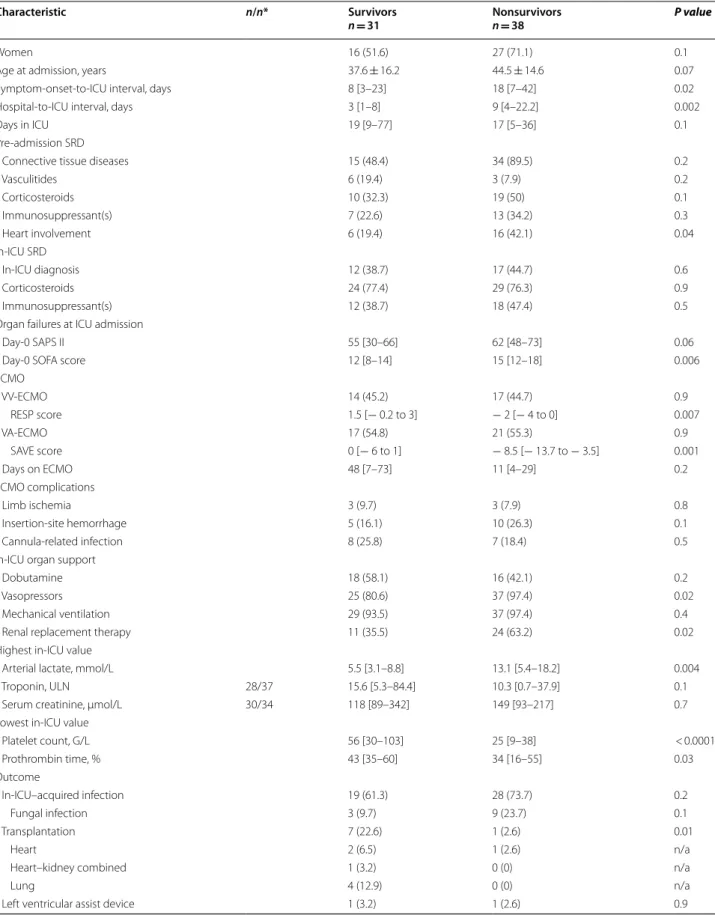
Table 4 In-ICU characteristics and outcomes of the 69 ECMO-treated SRD flare patients: hospital survivors vs. nonsurvivors
Characteristic n/n* Survivors
n = 31 Nonsurvivorsn = 38 P value
Women 16 (51.6) 27 (71.1) 0.1
Age at admission, years 37.6 ± 16.2 44.5 ± 14.6 0.07
Symptom‑onset‑to‑ICU interval, days 8 [3–23] 18 [7–42] 0.02
Hospital‑to‑ICU interval, days 3 [1–8] 9 [4–22.2] 0.002
Days in ICU 19 [9–77] 17 [5–36] 0.1
Pre‑admission SRD
Connective tissue diseases 15 (48.4) 34 (89.5) 0.2
Vasculitides 6 (19.4) 3 (7.9) 0.2 Corticosteroids 10 (32.3) 19 (50) 0.1 Immunosuppressant(s) 7 (22.6) 13 (34.2) 0.3 Heart involvement 6 (19.4) 16 (42.1) 0.04 In‑ICU SRD In‑ICU diagnosis 12 (38.7) 17 (44.7) 0.6 Corticosteroids 24 (77.4) 29 (76.3) 0.9 Immunosuppressant(s) 12 (38.7) 18 (47.4) 0.5
Organ failures at ICU admission
Day‑0 SAPS II 55 [30–66] 62 [48–73] 0.06
Day‑0 SOFA score 12 [8–14] 15 [12–18] 0.006
ECMO VV‑ECMO 14 (45.2) 17 (44.7) 0.9 RESP score 1.5 [− 0.2 to 3] − 2 [− 4 to 0] 0.007 VA‑ECMO 17 (54.8) 21 (55.3) 0.9 SAVE score 0 [− 6 to 1] − 8.5 [− 13.7 to − 3.5] 0.001 Days on ECMO 48 [7–73] 11 [4–29] 0.2 ECMO complications Limb ischemia 3 (9.7) 3 (7.9) 0.8 Insertion‑site hemorrhage 5 (16.1) 10 (26.3) 0.1 Cannula‑related infection 8 (25.8) 7 (18.4) 0.5
In‑ICU organ support
Dobutamine 18 (58.1) 16 (42.1) 0.2
Vasopressors 25 (80.6) 37 (97.4) 0.02
Mechanical ventilation 29 (93.5) 37 (97.4) 0.4
Renal replacement therapy 11 (35.5) 24 (63.2) 0.02
Highest in‑ICU value
Arterial lactate, mmol/L 5.5 [3.1–8.8] 13.1 [5.4–18.2] 0.004
Troponin, ULN 28/37 15.6 [5.3–84.4] 10.3 [0.7–37.9] 0.1
Serum creatinine, µmol/L 30/34 118 [89–342] 149 [93–217] 0.7
Lowest in‑ICU value
Platelet count, G/L 56 [30–103] 25 [9–38] < 0.0001 Prothrombin time, % 43 [35–60] 34 [16–55] 0.03 Outcome In‑ICU–acquired infection 19 (61.3) 28 (73.7) 0.2 Fungal infection 3 (9.7) 9 (23.7) 0.1 Transplantation 7 (22.6) 1 (2.6) 0.01 Heart 2 (6.5) 1 (2.6) n/a
Heart–kidney combined 1 (3.2) 0 (0) n/a
Lung 4 (12.9) 0 (0) n/a
Nine (10%) patients with refractory heart (n = 5) or lung (n = 4) failure could be successfully bridged to emergency transplantation. While urgent cardiac trans-plantation in patients under VA-ECMO is frequent, lung transplantation of unlisted patients on VV-ECMO
is unusual. We advocate that SRD patients, especially young patients, who could not be weaned-off ECMO, should be considered for heart or lung transplantation.
SRD diagnosis, corticosteroid and immunosuppres-sant use before ICU admission or thereafter were not
Continuous variables are expressed as mean ± standard deviation or median [interquartile range] and were compared with Student’s t-test or Wilcoxon’s rank test; categorical variables are expressed as n (%) and were compared with χ2 tests
ICU intensive care unit, LVEF left ventricle ejection fraction, RESP Respiratory Extracorporeal Membrane Oxygenation Survival Prediction, SAPS Simplified Acute Physiology Score, SAVE Survival after Veno-Arterial ECMO, SOFA Sequential Organ-Failure Assessment, SRD systemic rheumatic disease, ULN upper limit of normal value, VA-/VV-ECMO venoarterial/venovenous extracorporeal membrane oxygenation, VTI velocity–time integral
* Numbers of survivor/nonsurvivor data available
Table 4 (continued)
Characteristic n/n* Survivors
n = 31 Nonsurvivorsn = 38 P value
Weaning 23 (74.2) 6 (15.8) < 0.0001
In‑ICU mortality 0 (0) 37 (97.4) < 0.0001
Table 5 Univariable and multivariable analyses of factors associated with in-hospital mortality of the 69 ECMO-treated SRD flare patients
Bold values indicates statistically significant in multivariable analysis
The multiple Cox proportional hazards model used backward-stepwise variable elimination (with the variable exit threshold set at P > 0.10). All potential explanatory variables included in the multivariable analyses were subjected to colinearity analysis with a correlation matrix. Variables associated with one another were not included in the model. Statistical significance was defined as P < 0.05
ICU intensive care unit, SAPS Simplified Acute Physiology Score, SOFA Sequential Organ Failure Assessment, SRD systemic rheumatic diseases, VA-ECMO venoarterial-extracorporeal membrane oxygenation
Factor Univariable analysis Multivariable analysis
HR 95% CI P value HR 95% CI P value
Age ≥ 40 years 1.5 0.7–2.8 0.2
Women 1.7 0.8–3.4 0.1
Pre‑admission SRD lung involvement 0.6 0.3–1.1 0.08 0.7 0.4–1.5 0.4 Pre‑admission SRD heart involvement 1.8 0.9–3.5 0.07 2.9 1.4–6.0 0.003
In‑ICU SRD diagnosis 1.1 0.6–2.0 0.8
Pre‑admission corticosteroids 1.7 0.9–3.1 0.1 Pre‑admission immunosuppressant(s) 1.5 0.8–2.9 0.2
Day‑0 SAPS II ≥ 70 2.4 1.2–4.7 0.01 3.1 1.5–6.5 0.002
Day‑0 SOFA ≥ 16 2.9 1.5–5.6 0.001
Symptom‑onset‑to‑ICU interval ≥ 10 days 1.6 0.8–3.1 0.2
VA‑ECMO 1.2 0.6–2.2 0.6
In‑ICU corticosteroids 0.7 0.4–1.6 0.5
In‑ICU immunosuppressant(s) 0.9 0.5–1.7 0.8
Vasopressors 5.9 0.8–43.0 0.08 2.8 0.3–21.7 0.3
Mechanical ventilation 1.9 0.3–14.2 0.5
Renal replacement therapy 2.1 1.1–4.0 0.03 0.5 0.2–1.4 0.2
ICU‑acquired infection 1.0 0.5–2.2 0.9
Highest In‑ICU value
Arterial lactate ≥ 7.5 mmol/L 2.7 1.4–5.2 0.004 2.7 1.3–5.3 0.006
Bilirubin ≥ 125 µmol/L 2.2 1.1–4.5 0.02 2.4 1.1–4.9 0.02
Lowest in‑ICU value
associated with in-hospital mortality for the entire population. However, specific SRD heart involvement known before ICU admission was associated with poorer outcomes, independently of VA-ECMO can-nulation. That finding underlines the impact of heart sequalae from previous SRD flares on these patients’ prognoses. Conversely, patients receiving VA-ECMO support for the first SRD manifestation had poorer out-comes, underscoring the severity of SRD myocardial involvement.
Importantly, for the VV-ECMO–treated subgroup, a vasculitis diagnosis was strongly and independently associated with favorable outcomes. Their intra-alveo-lar hemorrhages were usually quickly reversible under specific regimens combining corticosteroids, rituxi-mab/cyclophosphamide and plasma exchanges. Our results strongly support the use of VV-ECMO for these patients.
Corticosteroid and immunosuppressant administra-tion can be associated with in-ICU–acquired infecadministra-tion, especially for patients on ECMO. Indeed, our series’ infection frequency was high, but rates were simi-lar for infection vs. fsimi-lare admissions, despite the latter having more frequently received corticosteroids and immunosuppressant(s). The rates of ventilator-asso-ciated pneumonia and bloodstream infections were in accordance with those in the ESLO database for VV-ECMO–treated patients [22]. Invasive fungal infections were particularly high (15%) and ventilator-associated pneumonia was independently associated with in-hos-pital mortality of VV-ECMO–treated patients, suggest-ing that careful attention should be paid to infectious complications in these profoundly immunosuppressed patients.
Our study has limitations and strengths. First, despite its retrospective, observational design, many patients had rare diseases requiring a still evolving and relatively rarely used rescue technique. Second, patient inclu-sion lasted > 14 years, meaning inevitable heterogeneity of diagnoses and management, but most patients were included during the last decade. Third, it is likely that ECMO support was declined for some SRD patients that were considered unfit to endure such an aggressive technique. The mortality rates herein reported should, therefore, be extrapolated with caution as they refer to an highly selected population of patients. Lastly, the main analysis considered VA- and VV-ECMO patients jointly. The reasons for ICU admission and ECMO cannulation, and the characteristics, management and outcomes of these patients obviously differ. We acknowledge that such an analysis might confound the results and their inter-pretation. However, the analysis aimed to present a com-prehensive, real-life picture of ECMO treatment of SRD
patients, with separate analyses of VA- and VV-ECMO subgroups thereafter.
Conclusion
ECMO is a relevant rescue technique for critically ill SRD patients, with 49% survival to hospital discharge. Vasculitis was independently associated with a favora-ble outcome of VV-ECMO–cannulated patients. Fur-ther studies are needed to specify the role of ECMO for SRD patients.
Supplementary Information
The online version contains supplementary material available at https ://doi. org/10.1186/s1361 3‑021‑00819 ‑3.
Additional file1: Table S1. Supplementary characteristics of 90 SRD Patients Given ECMO Support. Table S2. Microbiological Findings for In‑ ICU–Acquired Infections of the 90 ECMO‑Treated SRD Patients. Table S3. In‑ICU Characteristics and Outcomes of the 48 VA‑ECMO–Treated SRD Patients: Hospital Survivors vs. Nonsurvivors. Table S4. Univariable and Multivariable Analyses of Factors Associated with In‑Hospital Mortality for the 48 VA‑ECMO–Treated SRD Patients. Table S5. In‑ICU Characteristics and Outcomes of the 42 VV‑ECMO–Treated SRD Patients: Hospital Survi‑ vors vs. Nonsurvivors. Table S6. Univariable and Multivariable Analyses of Factors Associated with In‑Hospital Mortality for the 42 VV‑ECMO–Treated SRD Patients (DOCX 58 KB)
Abbreviations
ARDS: Acute respiratory distress syndrome; CI: Confidence interval; HR: Hazard ratio; ICU: Intensive care unit; IQR: 25–75Th percentile interquartile range; LVAD: Left ventricle assist device; RESP: Respiratory Extracorporeal Membrane Oxygenation Survival Prediction; SAPS: Simplified Acute Physiology Score; SAVE: Survival after Veno‑Arterial ECMO; SOFA: Sequential Organ‑Failure Assessment; SRD: Systemic Rheumatic Diseases; VA‑ECMO: Venoarterial‑ extracorporeal membrane oxygenation; VV‑ECMO: Venovenous extracorporeal membrane oxygenation.
Acknowledgements
None.
Authors’ contributions
PB and MPdC contributed to study design, data collection, statistical analysis conduction and interpretation, manuscript writing and final approval. CEL and AC contributed to study design, statistical analysis interpretation, manuscript writing and final approval. All other authors collected data. MPdC is the study guarantor.
Funding
None.
Availability of data and materials
All data generated or analyzed during the study are included in this published article and the its supplementary information files.
Ethics approval and consent to participate
The database is registered with the “Commission Nationale de l’Informatique
et des Libertés” (2217847v0). In accordance with the ethical standards of our
hospital’s institutional review board, the Committee for the Protection of Human Subjects, and French law, written informed consent was not needed for demographic, physiological and hospital‑outcome data analyses, because this observational study did not modify existing diagnostic or therapeutic strategies; however, patients were informed of their inclusion in the study.
Consent to publication
Competing interests
The authors declare that they have no competing interests.
Author details
1 Service de Médecine Intensive‑Réanimation, Hôpital La Pitié–Salpêtrière,
Sorbonne Université, Assistance Publique‑Hôpitaux de Paris (APHP), Paris, France. 2 Service de Chirurgie Cardiothoracique, Hôpital La Pitié–Salpêtrière,
Institut de Cardiologie, Sorbonne Université, APHP, Paris, France. 3 Service
de Médecine Interne 2, Institut E3M, Sorbonne Université, Hôpital La Pitié– Salpêtrière, 47–83, Boulevard de L’Hôpital, 75651 Paris Cedex 13, France.
4 Centre de Référence National Lupus Systémique, Syndrome Des Anticorps
Anti‑Phospholipides Et Autres Maladies Auto‑Immunes Systémiques Rares, Paris, France. 5 Institut de Cardiométabolisme Et Nutrition (ICAN), Sorbonne
Université, INSERM, UMRS_1166‑ICAN, Paris, France. Received: 18 September 2020 Accepted: 29 January 2021
References
1. Godeau B, Boudjadja A, Dhainaut JF, et al. Outcome of patients with systemic rheumatic disease admitted to medical intensive care units. Ann Rheum Dis. 1992;51:627–31. https ://doi.org/10.1136/ard.51.5.627. 2. Dumas G, Géri G, Montlahuc C, et al. Outcomes in critically ill
patients with systemic rheumatic disease: a multicenter study. Chest. 2015;148:927–35. https ://doi.org/10.1378/chest .14‑3098.
3. Larcher R, Pineton de Chambrun M, Garnier F, et al. One‑year outcome of critically ill patients with systemic rheumatic disease: a multicenter cohort study. Chest. 2020. https ://doi.org/10.1016/j.chest .2020.03.050. 4. Faguer S, Ciroldi M, Mariotte E, et al. Prognostic contributions of the
underlying inflammatory disease and acute organ dysfunction in criti‑ cally ill patients with systemic rheumatic diseases. Eur J Intern Med. 2013;24:e40‑44. https ://doi.org/10.1016/j.ejim.2012.11.018.
5. Combes A, Brodie D, Bartlett R, et al. Position paper for the organization of extracorporeal membrane oxygenation programs for acute respiratory failure in adult patients. Am J Respir Crit Care Med. 2014;190:488–96. https ://doi.org/10.1164/rccm.20140 4‑0630C P.
6. Abrams D, Garan AR, Abdelbary A, et al. Position paper for the organiza‑ tion of ECMO programs for cardiac failure in adults. Intensive Care Med. 2018;44:717–29. https ://doi.org/10.1007/s0013 4‑018‑5064‑5. 7. Combes A, Hajage D, Capellier G, et al. Extracorporeal membrane oxy‑
genation for severe acute respiratory distress syndrome. N Engl J Med. 2018;378:1965–75. https ://doi.org/10.1056/NEJMo a1800 385.
8. Combes A, Leprince P, Luyt C‑E, et al. Outcomes and long‑term quality‑of‑ life of patients supported by extracorporeal membrane oxygenation for refractory cardiogenic shock. Crit Care Med. 2008;36:1404–11. https ://doi. org/10.1097/CCM.0b013 e3181 6f7cf 7.
9. Aissaoui N, Luyt C‑E, Leprince P, et al. Predictors of successful extracor‑ poreal membrane oxygenation (ECMO) weaning after assistance for refractory cardiogenic shock. Intensive Care Med. 2011;37:1738–45. https ://doi.org/10.1007/s0013 4‑011‑2358‑2.
10. Beurtheret S, Mordant P, Paoletti X, et al. Emergency circulatory support in refractory cardiogenic shock patients in remote institutions: a pilot
study (the cardiac‑RESCUE program). Eur Heart J. 2013;34:112–20. https :// doi.org/10.1093/eurhe artj/ehs08 1.
11. Schmidt M, Burrell A, Roberts L, et al. Predicting survival after ECMO for refractory cardiogenic shock: the survival after veno‑arterial‑ECMO (SAVE)‑score. Eur Heart J. 2015;36:2246–56. https ://doi.org/10.1093/eurhe artj/ehv19 4.
12. Schmidt M, Bailey M, Sheldrake J, et al. Predicting survival after extracor‑ poreal membrane oxygenation for severe acute respiratory failure. The Respiratory Extracorporeal Membrane Oxygenation Survival Prediction (RESP) score. Am J Respir Crit Care Med. 2014;189:1374–82. https ://doi. org/10.1164/rccm.20131 1‑2023O C.
13. Combes A, Bréchot N, Luyt C‑E, Schmidt M. What is the niche for extra‑ corporeal membrane oxygenation in severe acute respiratory distress syndrome? Curr Opin Crit Care. 2012;18:527–32. https ://doi.org/10.1097/ MCC.0b013 e3283 57f09 0.
14. Pineton de Chambrun M, Bréchot N, Combes A. Venoarterial extracorpor‑ eal membrane oxygenation in cardiogenic shock: indications, mode of operation, and current evidence. Curr Opin Crit Care. 2019;25:397–402. https ://doi.org/10.1097/MCC.00000 00000 00062 7.
15. Demiselle J, Auchabie J, Beloncle F, et al. Patients with ANCA‑associated vasculitis admitted to the intensive care unit with acute vasculitis manifestations: a retrospective and comparative multicentric study. Ann Intensive Care. 2017;7:39. https ://doi.org/10.1186/s1361 3‑017‑0262‑9. 16. Kimmoun A, Baux E, Das V, et al. Outcomes of patients admitted to inten‑
sive care units for acute manifestation of small‑vessel vasculitis: a multi‑ center, retrospective study. Crit Care. 2016;20:27. https ://doi.org/10.1186/ s1305 4‑016‑1189‑5.
17. Godeau B, Mortier E, Roy PM, et al. Short and longterm outcomes for patients with systemic rheumatic diseases admitted to intensive care units: a prognostic study of 181 patients. J Rheumatol. 1997;24:1317–23. 18. Moreels M, Mélot C, Leeman M. Prognosis of patients with systemic rheu‑
matic diseases admitted to the intensive care unit. Intensive Care Med. 2005;31:591–3. https ://doi.org/10.1007/s0013 4‑005‑2563‑y.
19. Beil M, Sviri S, de la Guardia V, et al. Prognosis of patients with rheumatic diseases admitted to intensive care. Anaesth Intensive Care. 2017;45:67– 72. https ://doi.org/10.1177/03100 57X17 04500 110.
20. Heijnen T, Wilmer A, Blockmans D, Henckaerts L. Outcome of patients with systemic diseases admitted to the medical intensive care unit of a tertiary referral hospital: a single‑centre retrospective study. Scand J Rheumatol. 2016;45:146–50. https ://doi.org/10.3109/03009 742.2015.10673 29.
21. Brünnler T, Susewind M, Hoffmann U, et al. Outcomes and prognostic fac‑ tors in patients with rheumatologic diseases admitted to the ICU. Intern Med. 2015;54:1981–7. https ://doi.org/10.2169/inter nalme dicin e.54.4283. 22. Abrams D, Grasselli G, Schmidt M, et al. ECLS‑associated infections in
adults: what we know and what we don’t yet know. Intensive Care Med. 2020;46:182–91. https ://doi.org/10.1007/s0013 4‑019‑05847 ‑z.
Publisher’s Note
Springer Nature remains neutral with regard to jurisdictional claims in pub‑ lished maps and institutional affiliations.