HAL Id: inserm-00826557
https://www.hal.inserm.fr/inserm-00826557
Submitted on 27 May 2013HAL is a multi-disciplinary open access archive for the deposit and dissemination of sci-entific research documents, whether they are pub-lished or not. The documents may come from teaching and research institutions in France or abroad, or from public or private research centers.
L’archive ouverte pluridisciplinaire HAL, est destinée au dépôt et à la diffusion de documents scientifiques de niveau recherche, publiés ou non, émanant des établissements d’enseignement et de recherche français ou étrangers, des laboratoires publics ou privés.
Emery-Dreifuss muscular dystrophy, laminopathies, and
other nuclear envelopathies.
Gisèle Bonne, Susana Quijano-Roy
To cite this version:
Gisèle Bonne, Susana Quijano-Roy. Emery-Dreifuss muscular dystrophy, laminopathies, and other nuclear envelopathies.. Handb Clin Neurol, Elsevier B.V., 2013, 113, pp.1367-76. �10.1016/B978-0-444-59565-2.00007-1�. �inserm-00826557�
Emery-Dreifuss Muscular dystrophy. Laminopathies and other nuclear envelopathies
Gisèle Bonne1 and Susana Quijano-Roy2
Affiliations
1
Gisèle Bonne, PhD
- Inserm, UMR S974, Paris, F-75013, France;
- Université Pierre et Marie Curie-Paris 6, UM 76; CNRS, UMR7215; Institut de Myologie, IFR14, Paris, F-75013, France;
- AP-HP, Groupe Hospitalier Pitié-Salpêtrière, U.F. Cardiogénétique et Myogénétique Moléculaire, Service de Biochimie Métabolique, Paris, F-75013, France
Tel: +33 1 42 16 57 23 (or 17) Fax: +33 1 42 16 57 00 E-mail: g.bonne@institut-myologie.org http://www.institut-myologie.org/ 2 Susana Quijano-Roy MD, PhD
Praticien Hospitalier, Neurologie Pédiatrique.
- AP-HP, Pole de Pédiatrie. Centre de Référence de Maladies neuromusculaires GNMH, Hôpital
Universitaire Raymond Poincaré, F-92380 Garches, France.
- CIC-IT - Faculté de Médecine Paris Ile de France Ouest - Université Versailles Saint Quentin en Yvelines UVSQ – France.
- Inserm, UMR S974, Paris, F-75013, France;
- Directeur du laboratoire d’EMG, Service des Explorations fonctionnelles, Hôpital Necker
Enfants Malades, F-75015 Paris, France.
Tel: +33 1 47 10 78 90 (or 79 00) Fax: +33 1 47 10 76 43
ABSTRACT
Nuclear envelopathies or laminopathies are a group of hereditary diseases caused by mutations
of genes that encode proteins of the nuclear envelope such as lamins and emerin. Concerning
skeletal muscle, there is a spectrum of phenotypes of onset from birth to adult age. L-CMD is a
severe congenital muscular dystrophy characterized either by the absence of motor acquisitions
or by a striking loss of head support (dropped-head syndrome), associated with respiratory
failure. The Emery-Dreifuss muscular dystrophy (EDMD) has a later onset and typically shows
the triad of slowly progressive scapulo-peroneal muscular weakness, elbow joint contractures and
cardiac disease. EDMD is genetically heterogeneous (LMNA, EMD, FHL1) and inheritance may
be variable (X-linked, autosomal dominant or recessive). Other than skeletal muscle disorders,
LMNA has also been involved in a cardiomyopathy with cardiac conduction disease, in an axonal neuropathy and, more surprisingly, in lipodystrophy and a wide spectrum of premature aging
syndromes (progeria, mandibula acral dysplasia, restrictive dermopathy). It is still not clear how a
single gene is responsible for such heterogeneous spectrum of conditions, but mutations in other
genes implicated in the processing or maturation of nuclear lamins have been also found and the
INTRODUCTION
The nuclear envelopathies are a rapidly expanding group of human hereditary diseases caused
by mutations of genes that encode proteins of the nuclear envelope. The most frequent and best
known form is the Emery-Dreifuss muscular dystrophy (EDMD), a skeletal myopathy that
typically presents between mid-childhood and the second decade of life with slowly progressive
muscular weakness, joint contractures and cardiac disease. This nosological entity is genetically
heterogeneous and inheritance may be variable. An X-linked form was first studied by Emery &
Dreifuss more than 40 years ago (1966), and the eponymous association (EDMD) for this
condition was proposed late in the seventies by Rowland et al. but it was not until 1994 that the
gene STA, now called EMD, encoding emerin was identified as the cause of the disease (Bione et
al., 1994). In 1999, another gene linked to the nuclear envelope encoding lamins A and C, LMNA,
was found to be mutated in a series of patients with the same clinical features described for the
X-linked form but males and females were equally affected, showing a dominant transmission
(Bonne et al., 1999). Later on, mutations of LMNA were also reported in a family with an EDMD
phenotype, this time inherited as a recessive autosomal trait (di Barletta et al., 2000). Since then,
the spectrum of conditions has been extraordinarily enlarged, from a congenital muscular
dystrophy with severe paralytic or rapidly progressive picture (Quijano-Roy et al., 2008) to a
limb girdle muscular dystrophy with adult onset and much milder weakness (Muchir et al., 2000).
LMNA has also been involved in a form of isolated cardiomyopathy associated with cardiac conduction disease (Fatkin et al., 1999) and in an axonal form of hereditary neuropathy (De
Sandre-Giovannoli et al., 2002). More surprising has been the identification of this gene in a
number of non-neuromuscular disorders including lipodystrophy syndromes and a wide spectrum
of premature aging syndroms ranging from mandibula acral dysplasia to restrictive dermopathy
or maturation of nuclear lamins have been also found in some of these diseases. The fact that the
largest spectrum of phenotypes observed in nuclear envelopathies is mostly linked to lamin A/C
gene defects, has lead to the emergence of the more extended term of laminopathies (Worman
and Bonne, 2007). Intense research in the field is taking place and is currently generating huge
amounts of data and revealing the extraordinary complexity of the molecular and
physiopathologic mechanisms in these diseases. However, it is still not clear how a single gene is
responsible for such heterogeneous spectrum of conditions. The occurrence of modifying factors
or genes is highly suspected and has been occasionally proven. This and other investigations are
helping in the better understanding of nuclear envelopathies, opening the possibility for the
identification of new genes and the investigation of new therapeutic approaches. Work is in
progress and allows expecting new and promising developments of these fascinating and
complex conditions in the next coming years.
THE NUCLEAR ENVELOPE
The nuclear lamina is a network of lamin polymers, a fibrous layer that is embedded in the
nucleoplasmic side of the inner nuclear membrane and provides an interface between the nuclear
envelope and the genetic material inside the nucleus. The lamina consists of intermediate
filaments called lamins and comprises A-type (lamins A and C) and B-type (lamins B1 and B2)
lamins, and in human both A- and B-type lamins are known to cause diseases (Worman and
Bonne, 2007). Lamins interact with chromatin as well as with other proteins of the inner nuclear
membrane (lamina-associated proteins [LAPs] and emerin) through various binding sites (Figure
1).
Emerin, encoded by the EMD gene, is a transmembrane protein of the inner nuclear membrane
that interacts with A-type lamins. EMD gene located on chromosome Xq28, is 2327 bp in length
nuclear envelope but also expressed in other cell compartments in some tissues. The main disease
in humans known to be caused by defects in emerin is the X-linked form of Emery-Dreifuss
muscular dystrophy (XL-EDMD); although a few rare cases of limb-girdle muscular dystrophy
and isolated cardiac diseases have been also published. So far, 131 different mutations in the
EMD gene have been reported in 314 individuals and both genetic and clinical details are available in mutation database UMD-EMD that is maintained at the website address
www.umd.be/EMD/.
Lamins A and C are A-type lamins both derived by alternative splicing from a same gene,
LMNA located on chromosome 1q21. LMNA encompass 25 kb and contains 12 exons. A-Type lamins are not only present at the nuclear envelope but also in the nucleoplasm. They are
expressed only in differentiated cells and appear in the course of development. In contrast to
emerin, mutations in the LMNA gene have been reported to be associated with a number of
different phenotypes (see Table 1) (for review see Worman and Bonne, 2007). A mutation
database of LMNA mutations is available and constantly updated (www.umd.be/LMNA/), with
more than 360 different mutations reported in 1732 individuals so far. The clinical diversity from
a single causative gene is quite striking and the association between individual LMNA mutations
and different phenotypes is not fully understood. Genotype-phenotype correlations are not always
evident and marked inter- and intrafamilial clinical diversity has been reported (Bécane et al.,
2000; Bonne et al., 2000). To explain the variety of severity associated with laminopathies, the
coexistence of another co-inherited modifying gene or the effect of single nucleotide
polymorphism should be considered. In this line we recently identilied a modifier locus that may
modulate the age at onset of myopathic symptoms (Granger et al., 2011). Digenism has also been
identified in families with AD-EDMD and simultaneous mutations in LMNA and other genes
PATHOPHYSIOLOGY
It seems difficult to explain how defects in nuclear membrane proteins may cause so different
diseases as muscle dystrophy, cardiac or nerve diseases, as well as lipodystrophy and premature
aging syndromes. For emerin (and presumably lamins A/C), to function properly the protein must
be correctly localized to the nuclear membrane. Any defect of the nuclear membrane could
interfere with satellite cell function and thereby skeletal muscle regeneration because emerin
appears to be important in the organization of the nuclear membrane during cell division
(Holaska, 2008). As for other disorders with pathophysiology implicating the nucleus, cell death
and apoptosis may play an important role in pathogenesis. Concerning laminopathies, to explain
the extremely heterogeneous phenotypic consequences, several mechanism are proposed,
including mechanical stress, altered gene expression and accumulation of toxic prelamin A
(Worman et al., 2009). Enhanced nuclear fragility may affect in particular mechanically stressed
tissues such as cardiac or skeletal muscle. On the other hand, since lamin A/C and
lamin-associated polypeptides physically interact with histones, chromatin, and transcription factors,
altered gene expression may contribute to the pathogenesis of laminopathies. There is increasing
evidence that the nuclear lamins A/C are crucially involved in the spatial organization of
chromatin, gene regulation and signal transduction at the cellular level. The premature aging
syndromes constitute a phenotypic continuum sharing a common physiopathologic mechanism in
rapport with accumulation of a precursor (prelamin A) that stays abnormally farnesylated. This
may be the consequence of mutations in LMNA or in genes leading to defective posttranslational
processing of prelamin A (for a review see Navarro et al., 2006).
CLINICAL SPECTRUM
Striated muscle disorders
Globally, laminopathies affecting the striated muscles seem to constitute a continuous spectrum
of successive phenotypes (Figure 2). There is a strong correlation between age of onset and the
resulting phenotype in single patients. Overall, it appears that early prenatal onset may be
associated with lethal foetal akinesia, late prenatal onset with severe L-CMD, onset before 1 year
with dropped-head L-CMD, onset in childhood or young adulthood with classic EDMD, later
onset with LGMD1B, and finally the end of the sprectrum where no skeletal muscle involvement
is noted (Quijano-Roy et al., 2008). The cardiac involvement seems to be a common feature in
the course of the disease, independently of the form..
Lamin-related Congenital muscular dystrophy (L-CMD)
This recently described form of CMD is at the severe end of the spectrum of the striated muscle
laminopathies (Quijano-Roy et al., 2008). Some patients were initially reported as severe EDMD
patients, as they present with the typical humero-peroneal distribution of weakness and muscle
wasting. However, the early onset, the progressive course, the extreme severity of presentation
with some patients not achieving even head or trunk control and the absence of some hallmarks
of the EDMD triad at early stages (elbow contractures, cardiac symptoms) are atypical features
not easily recognisable as EDMD. Myopathic or dystrophic changes are seen in the muscle
biopsies. Merosin and other routine immunostaining markers of congenital muscular dystrophies
are usually normal. The serum CK levels are often increased but rarely more than 4 or 5 folds the
normal values. Overall, the phenotype observed is so distinct that, in the absence of tissular,
immunohistochemical or biochemical specific markers, diagnosis is based in the clinical findings.
Two groups of severity are distinguished: a subgroup of patients with early severe onset that have
very poor spontaneous movements and motor development (Fig. 2a), and another subset with
initially milder disease, who are able to sit or walk and present with progressive neck weakness
presentation there is a strikingly similar pattern of muscle involvement. All children have a
progressive course with an initial rapid decline in cervical/axial strength followed by a period of
slower progression or stasis. Progressive restrictive respiratory insufficiency is a major
complication and may require continuous mechanical ventilation, particularly in the most early
and severe form. In our reported series of 15 patients (Quijano-Roy et al., 2008), respiratory
failure was universal within the 2 first years of life in the severe group and arose before the age of
8 years in many children in the dropped-head group. Thus, these patients need close monitoring
of respiratory function and gas exchange, especially after the onset of progressive motor decline.
Cardiac involvement was rarely observed and was often subclinical in this series with most
children under the age of 10, but one patient with dropped head syndrome died unexpectedly at
age 3 years and another child with a more severe onset who never acquired trunk support
presented rythm disturbances at 7 years of age. Routinary cardiac tests to survey cardiac function
and rythm abnormalities regularly are therefore highly recommended from diagnosis
Emery-Dreyfuss Muscular Dystrophy (EDMD)
The overall prevalence of EDMD is not known, but the X-linked form is estimated to be
1:100,000. Hopkins and Warren (1992) estimated EDMD to be the third most prevalent muscular
dystrophy, after the two dystrophinopathies (Duchenne muscular dystrophy and Becker muscular
dystrophy). Most patients present autosomal-dominant EDMD due to a heterozygote mutation in
the LMNA gene and a lesser proportion have the X-linked form, while the autosomal recessive
transmission has only been reported in a family with a severe phenotype (di Barletta et al., 2000).
Germinal mosaicism in the LMNA gene is possible and has to be distinguished from a recessive
trait in families with several siblings affected (Bonne et al., 1999). However, in more than 60% of
EDMD cases, no mutations are detected in EMD or LMNA genes. Recently, in a search for new
number of unrelated patients with XL-EDMD (Gueneau et al., 2009). In this study, 28% of
EDMD patients carried a mutation in LMNA, 8% in EDM.and less than 2% in FHL1. Although
FHL1 proteins are not nuclear envelope proteins, FHL1, A-type lamins and emerin share some
functional features that will need to be further explored in the future.
Clinically, the typical EDMD phenotype is characterized by early joint contractures affecting
selectively the Achilles tendons, the elbows and the neck extensor muscles. A rigid spine
syndrome is the consequence of the progressive development of spinal cervico-dorsal and lumbar
contractures (Fig. 2c, d). Muscle atrophy and weakness show also a very distinct pattern, with a
humero peroneal distribution. Initially, muscle involvement in proximal muscles of upper
extremities is predominantly proximal, while the weakness and wasting is mostly distal in the
lower limbs. Although weakness later extends to the scapular and pelvic limb girdle musculature,
course is slowly progressive or static and patients usually do not develop profound motor or
respiratory dysfunction. However, EDMD is a potentially very severe condition due to the
cardiac involvement that usually arises after the second decade of life and may lead to sudden
death from heart block or due to progressive cardiac failure. Sudden cardiac death may be the
first manifestation of the disorder (Bécane et al., 2000). The most frequent heart abnormalities are
the conduction defects, ranging from sinus bradycardia, prolongation of the PR interval on
electrocardiography to complete heart block. Atrial paralysis is almost pathognomonic of EDMD.
Atrial arrhythmias (extrasystoles, atrial fibrillation, flutter) and ventricular arrhythmias
(extrasystoles, ventricular tachycardia) are frequent (van Berlo et al., 2005). Besides arrhythmia,
a dilated cardiomyopathy may also occur in the course of the disease. Cardiac symptoms include
palpitations, presyncope and syncope, poor exercise tolerance and congestive heart failure.
AD-EDMD and XL-EDMD have similar, but not identical, neuromuscular and cardiac
involvements (Bécane et al., 2000; Bonne et al., 2000; di Barletta et al., 2000). In XL-EDMD,
joint contractures are usually the first sign, whereas in AD-EDMD, joint contractures appear after
the onset of muscle weakness. The progression of muscle wasting is usually slow in the first three
decades of life, after which it becomes more rapid. Loss of ambulation can occur in AD-EDMD,
but is rare in XL-EDMD (Bonne et al., 2000). In AD-EDMD, the risk of ventricular
tachyarrhythmia and dilated cardiomyopathy manifested by left ventricular dilation and
dysfunction is higher than in XL-EDMD (Bécane et al., 2000). Individuals are at risk for cerebral
emboli and sudden death. A generalized dilated cardiomyopathy often occurs in the later stages
of the disease.
AR-EDMD are extremely rare. So far only one family and an isolated patient have been reported
with a homozygous mutation in LMNA (di Barletta et al., 2000) (Jimenez-Escrig et al. 2011). The
isolated patient experienced difficulties when started walking at age 14 months, had severe joint
contractures and loss walking at 5 years and by age 40 years he had severe and diffuse muscle
wasting but cardiac evaluation revealed no abnormalities. In the recently reported family which
was diagnosed using new techniques of exome sequencing (Jimenez-Escrig et al. 2011), the four
affected siblings presented later in life, with a limb-girdle progressive muscular dystrophy of
onset in the first to third decades of life. In addition, their father and his sister presented with
syncopes due to severe rhythm disturbances after the age of 70 requiring both a pacemaker.
Severity of weakness was variable among the siblings, but was progressive and they also
developed neck, elbow and Achilles contractures in the course of the disease. Subclinical cardiac
rhythm disturbances were detected in all in the fourth decade, after the homozygous LMNA
cardiac involvement seems to occur later. Further identification of recessive families will address
this issue better.
Genetics: In the case of XL-EDMD, a complete deletion of the gene can result from an inversion
within the Xq28 region and, in fact, almost a quarter of cases carry this inversion although not all
with the deletion of the EMD gene. Almost all mutations are null mutations (stop, splice site
mutation or out-of frame deletion/insertion) and result in a complete absence of the emerin on
both Western blotting and immunohistochemistry. Rare cases with a reduced amount of the
protein (due to a rare missense mutation) may have a milder phenotype (Manilal et al., 1998;
Bonne et al., 2003). Concerning LMNA mutations leading to EDMD, more than 80% are
missense mutations leading most probably to the production of mutant proteins. The remaining
LMNA mutations are nonsense or out-of-frame mutations and less frequently splice site mutations (see www.umd.be/LMNA/). These LMNA mutations lead to truncated proteins that are most
certainly degraded (Bécane et al., 2000). Concerning FHL1, mutations in this gene mainly lead to
truncated proteins and thus to absence or highly reduced FHL1 proteins (Gueneau et al., 2009).
However, there is still a great proportion of EDMD patients for whom the genetic defect remains
unknown.
Diagnosis: For the X-linked form, as emerin is ubiquitously expressed and most EMD mutations
lead to the absence of emerin, it is possible to analyze the expression of emerin by
immunoflurescence (IF) and/or by western blot (WB) not only in muscle (IF, WB) but also in
various tissues easy to sample such as exfoliative buccal cells (IF), lymphocytes or
lymphoblastoid cell lines (WB), and skin fibroblasts (IF, WB). In female carriers of XL-EDMD,
emerin is absent in varying proportions in nuclei due to variable level of X chromosome
normal or reduced amount of emerin. As for FHL1, so far reduced or absence of FHL1 have been
essentially tested in muscle tissues by WB and/or IF (Gueneau et al., 2009). In individuals with
AD-EDMD, emerin and FHL1 are normally expressed.
Concerning laminopathies, diagnosis is mainly based in clinical features because other
complementary tests, i.e. histology, IF, WB, are usually not specific. Serum CK levels are
moderately increased, especially at the beginning of the disease (Bonne et al., 2000).
Electromyogram (EMG) shows often myopathic features with normal nerve conduction studies,
but neuropathic signs have been described in patients with LMNA and EMD mutations. Muscle
histopathology shows nonspecific myopathic or dystrophic changes, including variation in fibre
size, increased number of internal nuclei, increase in endomysial connective tissue, and necrotic
fibres. Inflammatory features have been observed especially in severe or progressive patients
(Quijano-Roy et al., 2008). Electronic microscopy may reveal specific alterations in the nuclear
architecture (Fidzianska and Hausmanowa-Petrusewicz, 2003). Muscle biopsy is now rarely
performed for diagnostic purposes because of the lack of specificity of the dystrophic changes
observed and the absence of immunostaining abnormalities on immunodetection for lamins A/C
in AD-EDMD. Muscle MRI of the lower limbs in dominant LMNA mutations shows
involvement of glutei, vasti, adductor longus and magnus, semimembranosus and the long head
of the biceps femoralis muscles. In particular, a very severe and selective abnormal signal in
vastus lateralis is often observed (Mercuri et al., 2002). In L-CMD, severe diffuse involvement
sparing head and often forearm and psoas muscles are prominent features (Quijano-Roy et al.,
2008). These findings may be useful to distinguish from other myopathies with overlapping
clinical symptoms but different patterns of involvement, especially of the COLVI related
myopathies [Ullrich congential muscular dystrophy (UCMD) and Bethlem myopathy (BM)].
It is an autosomal dominant form of limb-girdle muscular dystrophy associated with
atrioventricular conduction defect and LMNA mutation (Muchir et al., 2000) (Fig. 2e-h)
Dilated Cardiomyopathy with conduction defects (CMD1A or DCM-CD)
An autosomal dominant form of dilated cardiomyopathy with cardiac conduction defects has
been described in which no skeletal muscle involvement are present (Fatkin et al., 1999; Bécane
et al., 2000).
Differential diagnosis in muscle striated laminopathies
Making a diagnosis in those patients with advanced disease is usually easy because of the distinct
and recognizable clinical picture, although there is clinical overlap with other muscular
dystrophies and myopathies. In fact, in early stages patients may not show specific features, and
complementary investigations (histology, immunohistochemistry, CK levels, muscle imaging)
may be needed before molecular studies are intended. Patients with marked elbow contractures
may be difficult sometimes to distinguish from the COLVI-related disorders, Ullrich congenital
muscular dystrophy (UCMD) and Bethlem myopathy (BM). In this setting, dosage of CK levels
and muscle MRI help to orientate the diagnosis. A severe and progressive course in a child with
increased CK levels and no cognitive impairment may resemble a congenital muscular dystrophy
due to mutations in FKRP, but L-CMD patients lack the muscle pseudohypertrophy and facial
weakness typically observed in FKRP-related CMD (MDCIC), and increment in CK is usually
less marked. The development of multiple contractures may be seen in merosin-deficient and
UCMD patients, but different localization of the muscle and joint involvement and specific
immunohistochemical and phenotypic markers (striking brain white matter changes and distal
hyperlaxity respectively) are useful in distinguishing these disorders.
• EMD. The majority of EMD mutations are null mutations that result in complete absence of emerin expression in nuclei; however, intra- and interfamilial variability in the severity of the
phenotype associated with null mutations may be observed. The few missense mutations that
have been identified are associated with decreased or normal amounts of emerin and result in a
milder phenotype (www.umd.be/EMD/).
• LMNA. In L-CMD, so far, LMNA mutations reported arise all de novo, whereas, in EDMD, de novo mutations were found in up to 76% of the cases and LGMD1B cases are mostly familial (Bonne et al., 2000; Bonne et al., 2003). Certain mutations are only identified in L-CMD patients,
which suggest a particular severe pathogenicity for these changes (Quijano-Roy et al., 2008). In
contrast, in EDMD and LGMD patients there is not a clear genotype-phenotype correlation
(Bonne et al., 2003) and, in fact, marked intra- and interfamilial variability is observed for the
same LMNA mutation, not only in severity, but also in the pattern of muscular or cardiac
involvement (Bécane et al. 2000; Bonne et al. 2000; Brodsky et al. 2000). Thus, in a given family
the same mutation can cause AD-EDMD, LGMD1B or isolated DCM-CD (Bécane et al., 2000;
Brodsky et al., 2000). Interestingly, severe and variable pictures have been reported in different
individuals of a large family with cosegregation of mutations in both EMD and LMNA (i.e.
CMT2, CMT2- EDMD, and isolated cardiomyopathy) (Ben Yaou et al., 2007). Furthermore,
extreme phenotypic diversity and low penetrance has been observed in patients with certain
missense mutations in the LMNA gene. For example, R644C has been found in patients with mild
to severe myopathy, arthrogryposis with myocardiopathy, motor neuropathy, limb girdle muscle
weakness, dilated cardiomyopathy atypical progeria, left ventricular hypertrophy, lipodystrophy,
insulin resistance and focal segmental glomerulosclerosis (Rankin et al., 2008).
Evaluations recommended following initial diagnosis in patients with myopathy are mainly
orthopaedic (spinal X-rays, assessment of spinal and joint contractures), neurological (motor
function and muscle testing), respiratory (spirometry, blood gases, sleep studies) and cardiac
(ultrasound, 24h Holter-ECG). These last aspects are particularly important, since respiratory and
cardiac complications may be subclinical and life-threatening from very early in life and should
be studied at least annually. Concerning pulmonary follow-up, night studies are recommended
when vital capacity on supine position is under 60% of theoretic values due to the risk of
nocturnal hypoventilation.. Other examinations are dependant of the severity of muscle disease
and secondary complications. Although no swallowing and facial involvement is relevant even in
severe congenital cases, some patients may require gastrostomy due to difficulties in feeding,
frequent hypoglycaemias, failure to thrive and hypotrophy that are multifactorial and often more
related to respiratory or cardiac insufficiency than to digestive problems. Metabolic
complications such as insulin resistance and diabetes are likely to happen more frequently in
LMNA mutated patients than in the general population.
At present, no etiological treatment is available and therefore, therapy is basically preventive
and/or symptomatic. In patients with myopathy, orthopaedic treatment to minimize progression
of joint contractures and spinal deformity are often required. Orthosis may be useful, for example
night cast to fight against Achilles tendon tightness and trunk or neck bracing for severe trunk or
neck hypotonia or scoliosis. They may prevent joint surgery for tenotomy or delay spinal fusion
until spinal growth is finished (scoliosis). Mechanical aids will be necessary in cases with severe
phenotype or progressive course (canes, walkers, orthesis, wheelchairs). Cardiac treatments
including antiarrhythmic drugs, cardiac pacemaker and implantable cardioverter defibrillator
(ICD) are used in patients with arrhythmias, AV conduction disorders and congestive heart
Bonne et al., 2000). Progressive restrictive respiratory insufficiency requiring nocturnal or
continuous mechanical ventilation is a constant complication in children with the congenital form
(L-CMD) and often in those patients with EDMD and onset is early in the first decade of the life.
Prevention: Regular physical therapy and trunk and limb orthosis are important in patients with myopathy symptoms or joint contractures. In certain cases with subclinical arrythmias, cardiac
defibrillators have been shown to reduce mortality by detecting life-threatening events that were
reverted (Meune et al., 2006). Thromboembolic complications in case of decreased left
ventricular function or atrial arrhythmias may be prevented by using antithromboembolic drugs
(Boriani et al., 2003).
Testing of relatives at risk is highly recommended in AD-EDMD, LGMD1B and DCM-CD
because of the incomplete penetrance of the cardiac disease at young age. Cardiac evaluation is
recommended for female carriers of an EMD mutation (Bonne et al., 2003).
Disorders of the peripheral nerve
CMT2B1. An autosomal recessive form of axonal Charcot-Marie-Tooth disease has been
described, with the founder mutation p.Arg298Cys (De Sandre-Giovannoli et al., 2002) (see
Charcot-Marie-Tooth type 2). Other mutations have also been associated with axonal motor
neuropathy (p.R644C, Rankin et al., 2008). In addition, autosomal dominant forms associating
CMT2 with other phenotypic features have been described as case reports: CMT2 associated with
muscular dystrophy, cardiomyopathy and leukonychia or with myopathy (for review see Worman
and Bonne, 2007).
Premature aging disorders
The premature aging syndromes constitute a phenotypic continuum ranging from the Mandibulo
They share a common feature which is the accumulation of prelamin A (normal or truncated) that
stays abnormally farnesylated. These entities may be the consequence not only of LMNA but also
of mutations in the gene FACE1/ZMPSTE24 which encodes a determinant enzyme responsible
for maturation of the prelamin A (Navarro et al., 2006).
Hutchinson-Gilford progeria syndrome (HGPS) is a severe and fatal developmental disorder
characterized by severe growth retardation, usually associated to skeletal alterations (osteolyses,
osteoporosis), marked amyotrophy, lipodystrophy, skin atrophy with sclerodermatous focal
lesions and alopecia. Affected children present with severe atherosclerosis. Cognitive functions
are fully preserved. Death occurs at the mean age of 13.5 years, mostly due to myocardial
infarction. HGPS is in most cases due to de novo dominant mutations at codon 608 of LMNA
gene, which introduce a cryptic splice site leading to a truncated protein that lacks the major site
of posttranslational modification necessary for correct maturation of prelamin A into lamin A.
Therefore, this pre-protein is stocked in the nuclear envelope as a incompletely processed
farnelsylated precursor which exerts toxic functions (Navarro et al., 2006).
Mandibulo Acral Dysplasia (MAD) is an autosomal recessive disorder characterized by growth
retardation, postnatal onset of craniofacial anomalies with mandibular hypoplasia, progressive
acral osteolysis, and skin changes including mottled pigmentation, skin atrophy, and
lipodystrophy affecting the face as well as the extremities. Some patients show progeroid features
such as thin nose, sparse, brittle hair and sclerodermatous (stiff and parched) skin and may have a
severe progressive glomerulopathy. Owing to its slowly progressive course, the syndrome has
been recognized in adults, and paediatric case reports are scarce. They may present metabolic
complications due to insulin resistance and diabetes. MAD is most often due to founder
reported with mutations in the ZMPSTE24 gene, some of them with progeroid features (Navarro
et al., 2006).
Restrictive Dermopathy (RD) is a perinatal lethal genodermatosis, mainly characterized by
intrauterine growth retardation, tight and rigid skin, prominent superficial vessels,
micrognathism, bone mineralization defects and multiple joint contractures. RD represent the
most severe end of the spectrum of laminopathies and is due either to a p.G608G LMNA mutation
(Navarro et al., 2004) or to ZMPSTE24 mutations leading to loss of function of this enzyme
(Navarro et al., 2006).
Other phenotype variants were reported completing the continuum of premature aging syndrome
(table 1 and for a review Navarro et al., 2006).
Other Disorders
Autosomal dominant Dunnigan type of familial partial lipodystrophy (FPLD)
Lipodystrophies represent a group of diseases characterized by altered body fat repartition and
major metabolic alterations with insulin resistance. Dunnigan syndrome (FPLD) is a genetic form
of partial lipodystrophy inherited in an autosomal dominant trait, characterized by subcutaneous
lipoatrophy but preserved or increased fat at the level of face and neck. The majority of FPLD
cases are caused by mutations in the LMNA gene affecting codon Arg482, leading to several
amino acid substitutions (Bonne et al., 2003; www.umd.be/LMNA/). Other associated features of
FPLD are muscular hypertrophy, hyperandrogenism, acanthosis nigricans, hepatomegaly with
steatosis and at the biological level, marked hypertriglyceridaemia, low HDL cholesterol, insulin
resistance and altered glucose tolerance or diabetes. These signs occur after puberty and are more
marked in females. Partial lipodistrophy has been reported also in patients with mutations in
REFERENCES
Bécane HM, Bonne G, Varnous S, Muet al (2000). High incidence of sudden death with conduction system and myocardial disease due to lamins A and C gene mutation. Pacing Clin Electrophysiol 23: 1661-1666.
Ben Yaou R, Toutain A, Arimura T, Deet al (2007). Multitissular involvement in a family with LMNA and EMD mutations: Role of digenic mechanism? Neurology 68: 1883-1894.
Bione S, Maestrini E, Rivella S, et al (1994). Identification of a novel X-linked gene responsible for Emery-Dreifuss muscular dystrophy. Nature Genet 8: 323-327.
Bonne G, Ben Yaou R, Beroud C, (2003). 108th ENMC International Workshop, 3rd Workshop of the MYO-CLUSTER project: EUROMEN, 7th International Emery-Dreifuss Muscular Dystrophy (EDMD) Workshop, 13–15 September 2002, Naarden, The Netherlands. Neuromusc Disord 13: 508-515.
Bonne G, Di Barletta MR, Varnous S, et al (1999). Mutations in the gene encoding lamin A/C cause autosomal dominant Emery-Dreifuss muscular dystrophy. Nature Genet 21: 285-288. Bonne G, Mercuri E, Muchir A, et al (2000). Clinical and molecular genetic spectrum of
autosomal dominant Emery Dreifuss muscular dystrophy due to mutations of the lamin A/C gene. Ann Neurol 48: 170-180.
Boriani G, Gallina M, Merlini L, et al (2003). Clinical relevance of atrial fibrillation/flutter, stroke, pacemaker implant, and heart failure in Emery-Dreifuss muscular dystrophy: a long-term longitudinal study. Stroke 34: 901-908.
Brodsky GL, Muntoni F, Miocic S, et al (2000). Lamin A/C gene mutation associated with dilated cardiomyopathy with variable skeletal muscle involvement. Circulation 101: 473-476. De Sandre-Giovannoli A, Chaouch M, Kozlov S, et al (2002). Homozygous defects in LMNA,
encoding lamin A/C nuclear-envelope proteins, cause autosomal recessive axonal neuropathy in human (Charcot- Marie-Tooth Disorder Type 2) and mouse. Am J Hum Genet 70: 726-736.
di Barletta MR, Ricci E, Galluzzi G, et al (2000). Different mutations in the LMNA gene cause autosomal dominant and autosomal recessive Emery-Dreifuss muscular dystrophy. Am J Hum Genet 66: 1407-1412.
Emery AEH and Dreifuss FE (1966). Unusual type of benign X-linked muscular dystrophy. J Neurol Neurosurg Psychiat 29: 338-342.
Fatkin D, MacRae C, Sasaki T, et al (1999). Missense Mutations in the Rod Domain of the Lamin A/C Gene as Causes of Dilated Cardiomyopathy and Conduction-System Disease. N Engl J Med 341: 1715-1724.
Fidzianska A and Hausmanowa-Petrusewicz I (2003). Architectural abnormalities in muscle nuclei. Ultrastructural differences between X-linked and autosomal dominant forms of EDMD. J Neurol Sci 210: 47-51.
Granger B, Gueneau L, Drouin-Garraud V, Pedergnana V, Gagnon F, Ben Yaou R, Tezenas du Montcel S, and Bonne G. (2011). Modifier locus of the skeletal muscle involvement in Emery-Dreifuss muscular dystrophy. Hum Genet 129(2): 149-159.
Gueneau L, Bertrand AT, Jais JP, et al (2009). Mutations of the FHL1 gene cause Emery-Dreifuss muscular dystrophy. Am J Hum Genet 85: 338-353.
Holaska JM (2008). Emerin and the nuclear lamina in muscle and cardiac disease. Circ Res 103: 16-23.
Hopkins LC and Warren S (1992). Emery-Dreifuss muscular dystrophy. in Handbook of Clinical Neurology: Myopathies (ed. L.P. Rowland and S. DiMauro), pp. 145-160. Elsevier Science, Amsterdam.
Jimenez-Escrig A, Gobernado I, Garcia-Villanueva M, Sanchez-Herranz A. (2011) Autosomal recessive Emery-Dreifuss muscular dystrophy caused by a novel mutation (R225Q) in the laminA/C gene identified by exome sequencing, in press.
Manilal S, Recan D, Sewry CA, et al (1998). Mutations in Emery-Dreifuss muscular dystrophy and their effects on emerin protein expression. Hum Mol Genet 7: 855-864.
Mercuri E, Counsell S, Allsop J, et al (2002). Selective muscle involvement on magnetic resonance imaging in autosomal dominant Emery-Dreifuss muscular dystrophy. Neuropediatrics 33: 10-14.
Meune C, Van Berlo JH, Anselme F, et al (2006). Primary prevention of sudden death in patients with lamin A/C gene mutations. N Engl J Med 354: 209-210.
Muchir A, Bonne G, van der Kooi AJ, et al (2000). Identification of mutations in the gene encoding lamins A/C in autosomal dominant limb girdle muscular dystrophy with atrioventricular conduction disturbances (LGMD1B). Hum Mol Genet 9: 1453-1459.
Muntoni F, Bonne G, Goldfarb LG, et al (2006). Disease severity in dominant Emery Dreifuss is increased by mutations in both emerin and desmin proteins. Brain 129: 1260-1268.
Navarro C, De Sandre-Giovannoli A, Bernard R, et al (2004). Lamin A and ZMPSTE24 (FACE-1) defects cause nuclear disorganisation and identify restrictive dermopathy as a lethal neonatal laminopathy. Hum Mol Genet 13: 2493-2503.
Navarro CL, Cau P and Levy N (2006). Molecular bases of progeroid syndromes. Hum Mol Genet 15 Spec No 2: R151-161.
Novelli G, Muchir A, Sangiuolo F, Helbling-Leclerc A, D'Apice MR, Massart C, Capon, F, Sbraccia P, Federici M, Lauro R et al. (2002). Mandibuloacral dysplasia is caused by a mutation in LMNA-encoding lamins A/C. Am J Hum Genet 71(2): 426-431.
Quijano-Roy S, Mbieleu B, Bonnemann CG, et al (2008). De novo lmna mutations cause a new form of congenital muscular dystrophy. Ann Neurol 64: 177-186.
Rankin J, Auer-Grumbach M, Bagg W, et al (2008). Extreme phenotypic diversity and nonpenetrance in families with the LMNA gene mutation R644C. Am J Med Genet A 146A: 1530-1542.
van Berlo JH, de Voogt WG, van der Kooi AJ, et al (2005). Meta-analysis of clinical characteristics of 299 carriers of LMNA gene mutations: do lamin A/C mutations portend a high risk of sudden death? J Mol Med 83: 79-83.
Worman HJ and Bonne G (2007). "Laminopathies": a wide spectrum of human diseases. Exp Cell Res 313: 2121-2133.
Worman HJ, Fong LG, Muchir A, et al (2009). Laminopathies and the long strange trip from basic cell biology to therapy. J Clin Invest 119: 1825-1836.
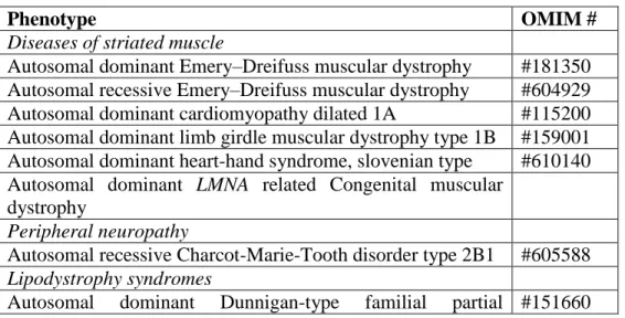
TABLE 1.- List of lamin A/C gene related diseases.
Phenotype OMIM #
Diseases of striated muscle
Autosomal dominant Emery–Dreifuss muscular dystrophy #181350 Autosomal recessive Emery–Dreifuss muscular dystrophy #604929 Autosomal dominant cardiomyopathy dilated 1A #115200 Autosomal dominant limb girdle muscular dystrophy type 1B #159001 Autosomal dominant heart-hand syndrome, slovenian type #610140 Autosomal dominant LMNA related Congenital muscular
dystrophy
Peripheral neuropathy
Autosomal recessive Charcot-Marie-Tooth disorder type 2B1 #605588 Lipodystrophy syndromes
lipodystrophy
Autosomal dominant lipoatrophy with diabetes, hepatic steatosis, hypertrophic cardiomyopathy and leukomelanodermic papules
#608056
Premature aging disorders
Autosomal recessive mandibuloacral dysplasia #248370 Autosomal dominant Hutchinson-Gilford progeria syndrome #176670 Autosomal dominant atypical Werner Syndrome #277700 Autosomal dominant restrictive dermopathy lethal #275210 Arthropathy, tendinous calcinosis and progeroid features #611618
FIGURES and LEGENDS
Figure 1. Model of the location of nuclear lamins and their interaction with nearby localized
proteins. Lamins bind directly to various integral membrane proteins of the inner nuclear
membrane (LBR, LAP2, emerin, MAN1, nesprins-1 and -2), but also to several proteins localised
within the nuclear matrix (BAF, Rb, SREBP1, histone proteins) as well as DNA, and thereby
mediate association with a scala of interacting structural proteins, linking the cytoplasm to the
nuclear interior. Question marks indicate suggested but not yet proven interactions. Reproduced
Figure 2. Clinical spectrum of the striated laminopathies: congenital muscular dystrophy (A,
B); Emery-Dreifuss muscular dystrophy (C,D); limb-girdle muscular dystrophy (E-H) (A)
Severe L-CMD.- Two year-old boy with absent motor acquisitions, continuous mechanical
ventilation, talipes and knee contractures. (B) Dropped head syndrome L-CMD.- Eight
year-old boy who lost walking and developed marked cervical weakness but dorsal spinal stiffness and
hyperextension. (C and D) EDMD boy with joint contractures (elbows, ankles) and diffuse
muscle wasting of humeroperoneal predominance. (E-H) LGMD1B.- Clinical features (E, F) and
muscle MRI findings (G,H) in the pelvic and thigh regions. Prominent involvement of vasti and
biceps femoris muscles with moderate affected gluteus maximus, adductor longus and major
muscles. Pictures C-H were kindly provided by Prof. Bruno Eymard (Groupe Hospitalier
Pitié-Salpêtrière, Paris, France).
A B C D E F