HAL Id: pasteur-02863037
https://hal-pasteur.archives-ouvertes.fr/pasteur-02863037
Submitted on 9 Jun 2020
HAL is a multi-disciplinary open access
archive for the deposit and dissemination of
sci-entific research documents, whether they are
pub-lished or not. The documents may come from
teaching and research institutions in France or
abroad, or from public or private research centers.
L’archive ouverte pluridisciplinaire HAL, est
destinée au dépôt et à la diffusion de documents
scientifiques de niveau recherche, publiés ou non,
émanant des établissements d’enseignement et de
recherche français ou étrangers, des laboratoires
publics ou privés.
Distributed under a Creative Commons Attribution| 4.0 International License
Pseudomonas aeruginosa Lung Infection
Fatima Benmohamed, Mathieu Medina, Yongzheng Wu, Sophia Maschalidi,
Gregory Jouvion, Laurent Guillemot, Michel Chignard, Bénédicte Manoury,
Lhousseine Touqui
To cite this version:
Fatima Benmohamed, Mathieu Medina, Yongzheng Wu, Sophia Maschalidi, Gregory Jouvion, et al..
Toll-Like Receptor 9 Deficiency Protects Mice against Pseudomonas aeruginosa Lung Infection. PLoS
ONE, Public Library of Science, 2014, 9 (3), pp.e90466. �10.1371/journal.pone.0090466�.
�pasteur-02863037�
Toll-Like Receptor 9 Deficiency Protects Mice against
Pseudomonas aeruginosa
Lung Infection
Fatima BenMohamed1,2, Mathieu Medina1,2, Yong-Zheng Wu1,2, Sophia Maschalidi3, Gregory Jouvion4, Laurent Guillemot1,2, Michel Chignard1,2, Be´ne´dicte Manoury3, Lhousseine Touqui1,2*
1 Unite´ de De´fense Inne´e et Inflammation, Institut Pasteur, Paris, France, 2 INSERM, U.874, Paris, France, 3 INSERM, U.1013, Paris, France, 4 Unite´ d’histopathologie humaine et mode`les animaux, Institut Pasteur, Paris, France
Abstract
Pseudomonas aeruginosa is an opportunistic pathogen involved in nosocomial infections. While a number of studies have demonstrated the roles of TLR2, TLR4 and TLR5 in host defense againt P. aeruginosa infection, the implication of TLR9 in this process has been overlooked. Here, we show that P. aeruginosa DNA stimulates the inflammatory response through TLR9 pathway in both a cell line and primary alveolar macrophages (AMs). This activation requires asparagine endopeptidase-and endosomal acidification. Interestingly, TLR9-/-mice resisted to lethal lung infection by P. aeruginosa, compared to WT C57BL/6 mice. The resistance of TLR9-/-mice to P. aeruginosa infection was associated with: (i) a higher ability of TLR9-/-AMs to kill P. aeruginosa; (ii) a rapid increase in the pro-inflammatory cytokines such as TNFa, IL-1b and IL-6 production; and (iii) an increase in nitric oxide (NO) production and inductible NO synthase expression in AMs. In addition, inhibition of both IL-1b and NO production resulted in a significant decrease of P. aeruginosa clearance by AMs. Altogether these results indicate that TLR9 plays a detrimental role in pulmonary host defense toward P. aeruginosa by reducing the AMs clearance activity and production of IL-1b and NO necessary for bacteria killing.
Citation: BenMohamed F, Medina M, Wu Y-Z, Maschalidi S, Jouvion G, et al. (2014) Toll-Like Receptor 9 Deficiency Protects Mice against Pseudomonas aeruginosa Lung Infection. PLoS ONE 9(3): e90466. doi:10.1371/journal.pone.0090466
Editor: Samithamby Jeyaseelan, Louisiana State University, United States of America Received October 8, 2013; Accepted February 3, 2014; Published March 4, 2014
Copyright: ß 2014 BenMohamed et al. This is an open-access article distributed under the terms of the Creative Commons Attribution License, which permits unrestricted use, distribution, and reproduction in any medium, provided the original author and source are credited.
Funding: This work was supported by ANR (ANR 2010 MIDI 008 01) and the Association Vaincre la Mucoviscidose (RF20120600716). The funders had no role in study design, data collection and analysis, decision to publish, or preparation of the manuscript.
Competing Interests: The authors have declared that no competing interests exist. * E-mail: touqui@pasteur.fr
Introduction
Pulmonary infections represent a major cause of mortality by infection in the world [1]. In immuno-compromised patients, exposition of respiratory tract to infectious pathogens results in serious and life-threatening diseases [2]. Pseudomonas aeruginosa, a Gram-negative bacteria, is an important cause of pulmonary infections in immuno-compromised patients and in patients with cystic fibrosis [3,4].
The ability of the host to respond to invading pathogens is in part attributed to a family of receptors called toll-like receptors (TLRs), which recognize conserved pathogen associated molecular patterns (PAMPs) present in microbes [5]. TLRs-PAMPs interac-tions initiate innate immune defence against pulmonary pathogens [5]. Previous studies have demonstrated the protective role of TLR2, TLR4 and TLR5 in host defence following lung infection with P. aeruginosa [6,7,8,9]. However, whether or not TLR9 plays a protective or deleterious role in host defence against P. aeruginosa remained to be determined. TLR9 plays an essential role in activating innate immunity by recognizing CpG specific motifs present in microbial DNA [10]. In the absence of stimulation, TLR9 is retained in the endoplasmic reticulum [10]. Upon activation, TLR9 relocates to the endo-lysosomal compartment, allowing the recruitment of the adaptor molecule MyD88 and stimulation of its subsequent signaling pathways [11]. In dendritic cells, TLR9 is activated after its proteolytic cleavage by asparagine endopeptidase (AEP) and cathepsins [12]. A recruitment and a
boost in AEP activity, which was induced shortly after TLR9 activation, has been shown to promote TLR9 cleavage and correlated with an increased acidification in endosomes and lysosomes [12].
The present studies have been undertaken to investigate whether the TLR9 pathway plays a role in host defence against P. aeruginosa pulmonary infection. We report that TLR9-/- mice exhibit a significant resistance to lethal infection following pulmonary infection with P. aeruginosa compared to wild type (WT) mice. The apparent resistance to P. aeruginosa was associated with an improvement of pulmonary bacterial clearance, a better killing of bacteria by AMs and increased productions of IL-1b and nitric oxide (NO), two inflammatory mediators involved in bacteria killing. Taken together these findings suggest that TLR9 down-regulates the innate immune response against P. aeruginosa. As a consequence, the absence of TLR9 leads to the improvement of the clearance of P. aeruginosa in the lungs and increased mouse survival.
Materials and Methods
Mice were housed in the Pasteur institute animal facilities accredited by the French Ministry of Agriculture and European regulations (EC Directive 86/609, French Law 2001-486 issued on June 6, 2001). Protocols were approved by the veterinary staff of the Institut Pasteur animal facility (Permit number 04.146) and
were performed in compliance with NIH Animal Welfare Insurance #A5476-01 issued on 31/07/2012.
Reagents
P. aeruginosa LPS serotype 10, purchased from Sigma-Aldrich (Saint Quentin Fallavier, France) was purified by gel-filtration chromatography and used at 1mg/ml. CpG oligonucleotide, 59-TGA CTG 59-TGA ACG TTC GAG ATG A-39 purified by HPLC was purchased from Tebu-Bio (Le Perray, France) and used at 1mg/ml. Bafilomycin A1 and concanamycin B were obtained from Sigma-Aldrich (Saint Quentin Fallavier, France) and used at 10 nM. MV026630 is an acyloxymethyl ketone specific inhibitor of AEP and used at 50mM as previously decrypted [13]. ODN-2088, the specific TLR9 antagonist and its control, ODN C were purchased from Invivogen (Toulouse, France) and both are used at 1mg/ml. N-Methyl-L-arginine acetate salts (L-NMMA) was purchased from Sigma-Aldrich (Saint Quentin Fallavier, France) and used at 100 nM. IL-1b receptor antagonist (IL-1bRA) was purchased from Peprotech (Neuilly-Sur-Seine, France) and used at 200 ng/ml.
Bacterial strain and growth conditions
The wild-type strain PAK, a commonly studied P. aeruginosa strain, was obtained from S. Lory (Harvard Medical School, Boston, MA), as originally isolated by D. Bradley (Memorial University of Newfoundland, St. John’s, Canada). This strain of P. aeruginosa is known to contain and express a full complement of virulence factors, including pili, flagella, the type II secreted enzymes, exotoxin A, elastases and phospholipases and the type III secreted exoenzymes S, T, and Y [14]. The DPscf is a PAK mutant with a deletion of pscF gene (a major needle protein of the type III secretion system (T3SS), defective in T3SS. PAK and its mutant were prepared as previously reported [6]. Briefly, bacteria were grown overnight in Luria-Bertani broth then transferred to fresh medium and grown for 4–5 h to mid-log phase. The cultures were centrifuged at 40006g for 15 min. The bacterial pellet was diluted in its original volume and the OD adjusted to give the approximate desired inocula. The inocula were verified by serial 10-fold dilutions of the bacterial suspensions and plating on Luria-Bertani agar.
Mouse strains
TLR9 deficient mice [15], backcrossed to the C57BL/6 background for 10 generations, were provided by S. Akira (Osaka University, Japan). Mice were fed normal mouse chow and water ad libitum and were bred and housed under standard conditions with air filtration.
Mouse infections and analyses of lung inflammation and bacterial clearance
Mice were anesthetized by i.m. with a mixture of ketamine (40 mg/Kg) and xylazine (8 mg/Kg) and infected intranasally with WT PAK strain at 107colony-forming unit (CFU) per mouse, as described previously [8]. Briefly, after anesthetization, mice were held by ears and 50ml of the inoculum ((PAK 107CFU/ mouse) diluted in PBS) were gradually released into the nostrils (25ml in each nostril) with the help of a micropipette. The rate of release was adjusted to allow mice to inhale the inoculum without trying to form bubbles. Mice were held in the hanging position for another couple of minutes till their breathing gradually returned to normal. The control mice were inoculated intranasally with 50ml of PBS. Survival experiments, collection of bronchoalveolar lavage
fluids (BALs) and cell counts in BALs were performed as previously described [8].
Culture of mouse AMs and MHS cell line
Mouse primary AMs were isolated as described previously in our laboratory [9]. Briefly, primary cells were isolated from mice after lung washing with PBS. Cells were plated in complete RPMI medium supplemented with 1% sodium pyruvate, 200 mM L-glutamine, 10% (v/v) fetal calf serum, 100 UI/ml penicillin, 100mg/ml streptomycin, 2.5 mg/l glucose and buffered with 25 mM HEPES. After 2 h, medium was removed and cells were incubated overnight with fresh medium. Mouse AM cell line MHS (CRL-2019; ATCC) was plated in the same complete medium RPMI. Cells were stimulated with CpG 1mg/ml, or P. aeruginosa DNA 25mg/ml, genomic DNA from lung (eDNA) 25mg/ml or LPS 1mg/ml for 24 hours. Primary AMs were infected with PAK, 1 MOI in free medium without serum and antibiotics for 2 or 4 hours. After infection, cells were centrifuged (80 g, 4 min, 4uC) to increase the adherence between cells and bacteria. For the time 24 hours, the cells were infected with PAK, 5 MOI for one hour and then the bacteria were removed and the cells were incubated for 24 hours in culture-free medium. Conditioned media (200mL) were collected after 2, 4 or 24 hours of incubation, centrifuged (400 g, 5 min, 4uC) to remove bacteria and stored at 220uC.
Pseudomonas aeruginosa DNA isolation
DNA was extracted from PAK strain using QIAGEN Genomic-tip 500 (Qiagen, Courtaboeuf, France). To remove contaminating lipopolysaccharide (LPS), the DNA was passed through a Detoxi-Gel Endotoxin Removing Resin (Thermo Scientific, Rockford, USA), resulting in DNA preparations with low level of LPS (,1 pg/mg DNA). LPS levels were measured using the Limulus amebocyte lysate kit (Lonza, Basel, Switzerland). The P.a. DNA was used at 25mg/ml. The genomic DNA was extracted from lung and used at 25mg/ml as a control.
Histological Studies
Mice were infected with 16107CFU of PAK strain and euthanized with pentobarbital 17 hours post-infection. The lungs were then fixed in formol for 48 h, sectioned, and stained with hematoxylin-eosin.
Analyses of bacterial clearance by AMs
The bacterial clearance was performed as previously described in our laboratory [9]. Briefly, AMs were isolated from mice and infected for 4 h with PAK at MOI of 0.1. CFU were quantified in AMs supernatants and cell lysates pooled together and expressed in percentages using the following formula: (CFU counts recovered without AMs 2 CFU counts recovered after AM infection) 6100. In certain experiments, AMs were infected for 4 h with a luminescent PAK strain at an MOI 10 and then bacteriolytic activity analysed by measuring the luminescence intensity in supernatants of AMs as detailed below.
Analyses of bacterial phagocytosis by AMs
Bacterial phagocytosis was performed as previously described in our laboratory [9]. A total of 56105 AMs were infected with bacteria (MOI = 10) for 1 h. Free and adherent bacteria were removed by washing cells with PBS and were killed with tobramycin treatment (40mg/mL; 30 min). Then, the cells were washed and lysed in H2O containing 0.1% Triton X-100. The
number of bacteria in lysates was determined by counting CFU on LB agar plate. The percentage of relative phagocytosis index was
assessed as follows: (CFU counts in mutant PAK-treated cells/ CFU counts in WT PAK-treated cells) 6100.
Assays of cytokines and NO production
Murine KC, TNFa, IL-6 and IL-1b concentrations in cell culture supernatants and BAL fluids were determined using
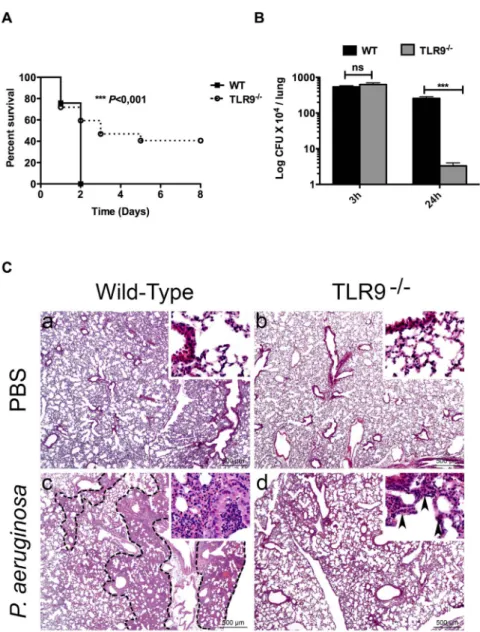
Figure 2. Resistance of TLR9-/-mice to lethal lung infection byP. aeruginosa. (A) groups of WT and TLR9-/-female mice (n = 10 in each
group), were inoculated intranasally with P. aeruginosa (PAK strain) at 107cfu/mouse. Animal survival was determined up to 8 days post-infection. All WT mice died 2 days following lung infection with PAK. Near 40% of TLR9-/-mice resisted to death up to 8 days post infection. (B) 3 and 24 hours
post-infection, the bacterial load was determined in lungs from both WT and TLR9-/-mice. (C), Histopathological analysis revealed different lesion profiles between WT and TLR9-/-mice. Control PBS lungs do not display any histological lesion in both WT and TLR9-/-mice (a,b). In contrast, 17 hours
post-infection with P. aeruginosa, (c) WT mice displayed a multifocal to coalescing well-delineated inflammatory lesion (dotted line), characterised by an infiltration of neutrophils and macrophages resulting in a complete loss of alveolar spaces and a filling of bronchiolar and alveolar spaces by inflammatory cells and cell debris (inset). (d) TLR9-/-mice also displayed an inflammatory lesion characterised by an infiltration of neutrophils and macrophages, but not well-delineated, and sparing the general alveolar structure (inset, black arrowheads). Data 6 SEM and are representative of three independent experiments *** P,0,001; ns: not significant.
doi:10.1371/journal.pone.0090466.g002
Figure 1. Stimulation of AMs withP.a. DNA requires endosomal acidic pH and AEP activation. (A) Show the levels of TNFa (left panel) and IL-6 (right panel) secreted by MHS unstimulated (Control, C) or stimulated with either P.a. DNA, eDNA (negative control) or with CpG (positive control) for 24 hours. (B) Shows TNFa levels produced by WT or TLR9-/-AMs stimulated with P.a. DNA, CpG for 24 hours. (C and D) Show TNFa levels
produced by MHS pre-treated for one hour with Bafilomycin A (Bafilo) or concanamycin B (CCB) and stimulated with P.a. DNA, CpG, or LPS. (E) Shows TNFa levels produced by MHS pre-treated with the AEP inhibitor MV026630 (MVO) 1 hour and stimulated with CpG or P.a. DNA. (F) Shows TNFa levels produced by WT and AEP-/-AMs stimulated with CpG, P.a. DNA or LPS for 24 hours. Data represent means 6 SEM and are representative of
three independent experiments *** P,0,001 vs C (control), ** P,0,01 ns = not significant. doi:10.1371/journal.pone.0090466.g001
DuoSet ELISA assay kits (R&D Systems, Lille, France) with TMB peroxidase substrate (Eurobio, Les Ulis, France). NO production was determined by colorimetric assay based on the Griess reaction [16] using Griess reagent kit (Invitrogen, Saint Aubin, France).
NF-kB reporter assay
MHS cells were plated at a density of 36104cells/ml in 60-mm tissue culture plates 48 hours prior to transfection with an NF-kB reporter plasmid. The reporter plasmid consisted of a luciferase encoding gene controlled by an NF-kB-responsive promoter sequence. Transfection was accomplished by the use of Gene juice from Merck-Millipore (Saint-Quentin-en-Yvelines, France).
Figure 3. Deletion of TLR9 is associated with an early increase in lung inflammation. BALs from WT and TLR9-/-mice (n = 5 for each group)
were collected 3 and 24 hours infection. AMs and PMN number recruited by the lung were determined 3 hours (A) and 24 hours (B) post-infection. KC, TNFa, IL-1b and IL-6 released in BALs were determined 3 hours (C) and 24 hours (D) post-post-infection. Data represent means 6 SD and are representative of three independent experiments. *** P,0,001, ** P,0,01, * P,0,05. ns = not significant, nd = not detected.
doi:10.1371/journal.pone.0090466.g003
Figure 4. Deletion of TLR9 leads in increased bacterial killing by AMs without interfering with bacterial phagocytosis. (A and B) WT and TLR9-/-AMs were infected for 4 hours with PAK (MOI = 0.1) and bacterial killing was quantified as indicated in M&M.B) One hour before infection, the cells were stimulated with ODN2088 (1 mg/ml), a specific TLR9 antagonist, or its control ODN (ODN C, 1 mg/ml). (C) WT and TLR9-/-AMs were
infected with PAK strain (MOI = 1) for 4 hours before collection of cell-free supernatants. The latter were then incubated with a luminescent PAK strain (PAKLux, MOI = 10) followed by killing assay, as indicated in M&M. As a positive control, incubation of PAK-Lux with tobramycin led to a total inhibition of bacterial growth (D) bacterial phagocytic activity was measured in AMs of both WT and TLR9-/-mice as indicated in M&M. Results are means 6 SEM of three independent experiments. ***P,0.001, **P,0.01, * P,0,05.
Transfected cells were plated at 105/well in RPMI with 10% of SVF and incubated for 24 hours before stimulation. The cells were then stimulated with LPS, CpG or LPS/CpG for 8 hours. NF-kB
transcriptional activity was assessed by measuring the luciferase activity of cellular lysates and normalized to lysate protein contents. Luciferase activity was quantified by measuring
chemi-Figure 5. TLR9-/-AMs exhibit increased cytokine secretion followingP. aeruginosainfection. (A) WT and TLR9-/-AMs were infected for 2 or
4 hours with PAK (MOI = 1). (B) WT and TLR9-/-AMs were infected for one hour with PAK (MOI = 5) and then the bacteria were removed and the cells were incubated for 24 hours. TNFa, IL-1b and IL-6 levels released in culture supernatants were determined. (C) TLR9-/-AMs were infected for 4 hours
with PAK or its DPscf mutant (MOI = 1) and TNFa, IL-1b and IL-6 levels released in culture supernatants were determined. (D) Killing assay was performed in WT and TLR9-/-AMs incubated either with PAK or its DPscf mutant. Results are means 6 SEM of three independent experiments.
***P,0.001, **P,0.01, * P,0,05. doi:10.1371/journal.pone.0090466.g005
luminescence in cell lysates using luciferase assay system from Promega according to the manufacturer’s specifications.
Western Blot
After incubation, MHS cells were lysed in buffer [5 mM EDTA, 150 mM NaCl, 1% Triton X-100, 50 mM Tris HCl (pH 7.4)] with anti-proteases (Roche Diagnostics). 20mg of protein lysates were run in a 12% SDS/PAGE, transferred onto PVDF membranes, and probed with antibodies directed against mouse ERK1/2 or its phoshorylated form (Santa Cruz Biotechnology).
Real-time PCR
RT-PCR was performed using an ABI 7900 RT-PCR detection system (Applied Biosystems, Foster City, CA) in 10ml reactions that contained 1ml of diluted cDNA, 300 nM each of forward and reverse primer, and SYBR Green PCR Master Mix (Fisher scientific, Illkirch, France). The primer for murine HPRT has been described in our recent study [17]. Other primers were designed using the Oligo Explorer 1.1. 2 software, murine TLR2 (Fw:59-GTTTCTGATGGTGAAGGTTG-39; Rv:59-GCTGAAGAG-GACTGTTATGG-39), murine TLR4 (Fw:59- AAATGCCAG-GATGATGC-39; Rw:59-AGGGACTTTGCTGAGTTTC-39), murine TLR5 (Fw:59- TTCAGACGGCAGGATAG -39; Rw:59-AAGATTGGGCAGGTTTC-39), murine TLR7 (Fw:59-CCACA-GGCTCACCCATACTTC-39; Rv:59-GGGATGTCCTAGGTG-GTGACA-39), murine TLR9 (Fw:59-GCACAGGAGCGGTGAA-GGT-39; Rw:59-GCAGGGGTGCTCAGTGGAG-39), murine iNOS (Fw:59- CCC TCC TGA TCT TGTGTT GGA -39; Rv:59- CAA CCC GAG CTC CTG GAA -39).
Luminescence Activity
PAKLux growth was evaluated using an EGNG Berthold luminometer. As a positive control of bactericidal activity, PAKLux was cultured with tobramycin (40mg/mL), and the resultant was presented as % of growth vs. PAKLux at T0.
Statistical analyses
Data were represented as means 6 SEM. and compared using the unpaired Student’s t test for the experiments with only two groups. For the experiments with three groups or more, one way ANOVA test was used followed by Bonferroni as a secondary test. For survival curve log-lank (Mantel-cox) test was used to evaluate the significance between groups. P values less than 0.05 are considered significant.
Results
P. aeruginosa DNA stimulates TLR9 in AMs and requires endosomal acidic pH and AEP activation
We first assessed whether DNA isolated from P. aeruginosa (P.a. DNA) induces innate immune response through TLR9 signalling
pathway in both AMs cell line (MHS) and primary AMs. Our results showed that P.a. DNA as well as CpG (the positive control) stimulated TNFa and IL-6 production by MHS (Fig. 1A). As a negative control, endogenous DNA (eDNA) extracted from mouse lung had no effect on cytokines production (Fig. 1A). Significant levels of TNFa, induced by P.a. DNA and CpG, were also detected in the supernatants of AMs derived from WT mice, but were abrogated in AMs from TLR9-/- mice (Fig. 1B). Both concanamycin B and bafilomycin A, which increase endosomal pH, inhibited TNFa production triggered by P.a. DNA and CpG in MHS (Fig. 1C, 1D), suggesting that TLR9 stimulation requires endosomal acidic pH. As a control of specificity, neither concanamycin B nor bafilomycin A compounds had an effect on LPS-induced TNFa production (Fig. 1C). In addition, MV026630, an AEP inhibitor, reduced P.a. DNA- and CpG-induced TNFa production by MHS (Fig. 1E). We verified that MV026630 inhibited AEP activity in these cells (Fig. S1). The dependence of TLR9 on AEP activation was also confirmed using AMs from AEP-/-mice. TNFa production induced by P.a. DNA was significantly reduced in AMs derived from AEP-/- in comparison to AMs from WT mice (Fig.1F). No effect of AEP on TNFa production following stimulation with LPS was observed (Fig. 1F). These results suggest that activation of TLR9 by P. aeruginosa DNA in AMs requires endosomal acidic pH and AEP activation.
TLR9 deficiency is associated with increased mouse survival and bacterial clearance during pulmonary infection with P. aeruginosa
We next examined the role of TLR9 in host defence against pulmonary P. aeruginosa infection. We infected WT and TLR9 -/-mice (n = 10) intranasally with a lethal dose of PAK strain of P. aeruginosa and then monitored animal survival for 8 days. As expected, 100% mortality in WT mice was observed within two days post-infection (Fig. 2A). However, unexpectedly, 40% of mice lacking TLR9 resisted to death up to 8 days post-infection (Fig. 2A). We next examined whether the difference in survival between WT and TLR9-/-mice was due to differences in bacterial clearance in the airways. At the early time point (3 hours) post-infection the same load of bacteria was found in WT and TLR9 -/-mice lung (Fig. 2B). However, at 24 hours post-infection, the bacterial load was significantly decreased in TLR9-/-compared to WT mice (Fig. 2B). This suggests that the increased survival of TLR9-/-mice was likely due to increased bacterial clearance in their lungs. This increase was not associated to changes in the pulmonary expression of TLR2, TLR4 or TLR5 in TLR9-/-mice (Fig. S2). Indeed, no significant difference was observed in the levels of TLR2, TLR4 and TLR5 mRNA expression in both lungs and AMs isolated from WT vs. TLR9-/-mice (Fig. S2A and B). TLR7 expression was significantly increased in TLR9-/-compared
Figure 6. TLR9-/-AMs exhibit an increase in NO production and iNOS expression followingP. aeruginosainfection. (A and B) NO
production and iNOS mRNA levels were measured in AMs 4 hours post-infection with PAK (MOI = 1). (C) Killing assay was performed in WT AMs after inhibition of NO production by L-NMMA (100 nM) one hour before infection for 4 hours with PAK (MOI = 1). (D) Nitrite production in WT AMs pre-treated with L-NMMA for 1 hour and infected for 4 hours (MOI = 1) with PAK. (E) NO production in WT AMs pre-pre-treated for 1 hour with IL-1bRA (200 ng/ml) before infection for 4 hours with PAK (MOI = 1). (F) IL-1b production in WT AMs pre-treated with L-NMMA (100 nM) for one hour before infection for 4 hours with PAK (MOI = 1). (G) WT and TLR9-/-AMs were stimulated with LPS (50 ng/ml) for 4 hours. (H) MHS cells were stimulated with
CpG (1 mg/ml), LPS (1 mg/ml) or CpG+LPS for 24 hours. TNFa levels released in culture supernatants were determined. (I) MHS cells were transfected with NF-kB luciferase reporter plasmid for 24 hours and then stimulated with CpG, LPS or CpG+LPS for additional 8 hours, at the same concentrations as in (H). The luciferase activity was then measured in cell lysates by luciferase assay. (J) MHS cells were stimulated with CpG (1 mg/ml) and/or LPS (1 mg/ml) for one hour. After reaction, total proteins were extracted and ERK1/2 total levels and ERK1/2 phosphorylation were analysed by Western Blotting using specific antibodies. (K) shows the quantifications of the blots presented in the figure (J). Results are means 6 SEM of three independent experiments. *** P,0,001, **P,0.01, * P,0.05.
doi:10.1371/journal.pone.0090466.g006
to WT lung, but expression of this receptor had no effect on mouse mortality by P. aeruginosa pulmonary infection (Fig. S2A, C).
Histopathological analysis revealed no histological lesion in the lung of WT and TLR9-/- instilled with PBS (Fig. 2Ca,b). In contrast, 17 hours post-infection with P. aeruginosa, different lesion profiles were identified between WT and TLR9-/-mice. Most WT mice displayed a multifocal to coalescing well-delineated inflam-matory lesion, centred on bronchioles and secondary extending to alveoli, leading to a complete loss of alveolar spaces, filled by neutrophils and macrophages (Fig. 2Cc). These lesions coincided with deaths of WT mice that began as early as day 1 post-infection. However, most TLR9-/-mice, displayed a more diffuse and poorly delineated inflammatory lesion, characterised by infiltration of bronchiolar and alveolar walls and spaces by neutrophils and macrophages with preservation of the general alveolar structure (Fig. 2Cd). These results suggest that TLR9 exerts a deleterious effect on mouse survival and airways clearance of P. aeruginosa
TLR9 modulates the host innate responses induced by P. aeruginosa pulmonary infection
Given the increased bacterial clearance and resistance to death observed in TLR9-/- mice, we investigated the mechanisms by which TLR9-/- mice control P. aeruginosa infection. We first examined lung recruitment of inflammatory cells after P. aeruginosa infection. Three hours post-infection, significantly higher numbers of AMs were found in BALs of both WT and TLR9-/- mice compared to PBS-treated mice (Fig. 3A). However, no significant differences were observed in AMs number between WT and TLR9-/-mice (Fig. 3A). A marked increase in the PMNs number was observed in BALs of WT and TLR9-/-mice at 24 hours after infection. At this time point, both AMs and PMN numbers were significantly lower in TLR9-/-compared to WT mice (Fig. 3B). To test whether the innate immune response against P. aeruginosa was increased in TLR9-/- mice, we measured inflammatory cytokines levels in BALs. Three hours post-infection, a significant increase in KC, IL-1b, TNFa, and IL-6 levels was detected in BALs from TLR9-/-compared to WT mice (Fig. 3C). However,
at 24 hours after infection, these levels were detected in BALs of TLR9-/-mice at much lower levels compared to those observed in BALs of WT mice (Fig. 3D). These findings suggest that TLR9 modulates the host innate responses induced by P. aeruginosa pulmonary infection.
AMs isolated from TLR9-/-exhibit increased P. aeruginosa killing and cytokines production
In order to determine the mechanisms by which P. aeruginosa was cleared more efficiently in TLR9-/-lung, we isolated AMs from both WT and TLR9-/-mice and studied the up-take and bacteria killing by these cells. Interestingly, TLR9-/-AMs infected with P. aeruginosa exhibited increased bacteria killing (,40%) in compar-ison with WT AMs (,20%) (Fig. 4A). This TLR9-dependent bacterial killing was supported by the fact that the TLR9 specific antagonist, ODN2088, induced a significant increase of bacterial killing in MHS cells (Fig. 4B). We then assessed whether this increase of bacteria killing was mediated by a bactericidal extracellular activity, present in cell supernatants, and/or by intracellular killing. Conditioned media collected from non-infected AMs from WT or TLR9-/- mice had no bactericidal effect (Fig. 4C). However, media from AMs infected with PAK exhibited increased bactericidal activity without significant differ-ence between WT and TLR9-/-AMs (Fig. 4C). In another series of experiments, the bacterial phagocytic test indicated that
deletion of TLR9 had no effect on bacterial phagocytosis (Fig. 4D). In a subsequent step, we examined the potential involvement of inflammatory cytokines in the increase of P. aeruginosa killing by TLR9-/- AMs. A rapid increase of TNFa production was observed in TLR9-/-AMs compared to WT AMs, at both 2 and 4 hours post-infection (Fig. 5A). This increase was also observed at 24 hours after infection (Fig. 5B). The inflammatory cytokines, IL-6 and IL-1b were not detected at 2 hours post-infection but their levels increased in TLR9-/-AMs, at 4 hours and 24 hours post-infection (Fig. 5A, B). As expected, these cytokines were not detected in non-infected cells (Fig. 5A, B). Given that IL-1b is known to play a crucial role in intracellular bacterial killing [9], we examined whether this cytokine was associated to the observed increase of bacterial killing in TLR9 -/-AMs. We used a PAK mutant (DPscf), deficient in the type III secretion system (T3SS), which is unable to induce IL-1b production in WT AMs [9]. TLR9-/- AMs infection with the DPscf mutant induced both TNFa and IL-6 but failed to induce IL-1b (Fig. 5C). Our result indicates that the DPscf mutant was remarkably resistant to killing by both WT and TLR9-/- AMs suggesting that IL-1b was essential for the induction of bacterial clearance (Fig. 5D). These data suggest that AMs from TLR9 -/-mice exhibit increased inflammatory cytokines production and killing of P.aeruginosa.
IL-1b induced bacteria killing in part through the increase of nitric oxide in lung of TLR9-/-mice
Nitric oxide (NO) produced by host immune cells plays a major role in innate immunity due to its ability to kill a broad range of microorganisms [18,19]. Following infection with P. aeruginosa, a significant increase in both NO production and the inducible nitric oxide synthase (iNOS) mRNA was detected in AMs derived from TLR9-/-compared to WT mice (Fig. 6A, B). Pharmacological inhibition of iNOS by L-NMMA significantly decreased P. aeruginosa killing by AMs (Fig. 6C). L-NMMA inhibitor completely abrogated NO production by AMs (Fig. 6D). Interestingly, treatment of AMs with IL-1bRA, an antagonist of IL-1b receptor, significantly decreased NO production in AMs following P. aeruginosa infection (Fig. 6E). Conversely, IL-1b production by AMs was abrogated by L-NMMA (Fig. 6F) in parallel to the observed decrease of P. aeruginosa killing (Fig. 6C). These results indicate that TLR9-/- AMs exhibit increased NO associated to
enhanced bacterial killing.
Stimulation of TLR9 with CpG reduces TLR4 signaling in AMs
The cytokines increase observed in lung of infected TLR9 -/-mice and their AMs suggests that TLR9 in our model probably limites the inflammatory response induced by others TLRs like TLR4. LPS is one of the major actors involved in the induction inflammatory response against P. aeruginosa in the lung, through the activation of TLR4 [6,7]. This led us to examine the effect of the TLR9 agonist CpG on LPS-induced AM activation. Our results showed that AMs from TLR9-/- mice produce higher TNFa levels compared to WT AMs under LPS stimulation (Fig. 6G). In addition, TLR9 activation with CpG reduced the extent of inflammation induced by LPS in MHS cells through a decrease of NF-kB activation (Fig. 6H, I). The increase of NF-kB activation was associated with a significant decrease in ERK1/2 phosphorylation (Fig. 6J and K). These results suggest that TLR9 may affect the inflammatory response through inhibition of NF-kB activity.
Discussion
In the present study we established that TLR9 plays a detrimental role in lung defence against P. aeruginosa. Indeed, we showed that TLR9-/- mice exhibited increased survival and efficient pulmonary clearance of P. aeruginosa in comparison with control mice. This unexpected detrimental effect led us to investigate the underlying cellular and molecular mechanisms by which TLR9 modulated the lung inflammatory response following P. aeruginosa infection. We found that TLR9-/- mice displayed increased levels of airways cytokines at the early stage of P. aeruginosa infection, compared to infected WT mice, leading to the improvement of P. aeruginosa clearance by lungs of TLR9-/-mice. The improvement of airways bacterial clearance resulted in the attenuation of the intensity of late pulmonary inflammation accompanied with reduced airways histological lesions in TLR9-/-mice, 24 h after the initiation of infection.
In an in vitro model of murine bone marrow-derived macro-phages, TLR9 has been shown to down-regulate the immune response against Candida albicans [20]. However, in a mouse model of pneumonia, TLR9 exhibited a protective effect against the Gram-negative bacterium, Klebsiella pneumonia [21,22]. In that study, TLR9-/- mice displayed significantly increased mortality following intra-tracheal infection with Klebsiella pneumonia [21,22]. This was associated to impaired bacterial clearance and activation of type I cytokine production. On the same line, it was demonstrated that TLR9 played a protective role in airway infection with Streptococcus pneumonia [23]. Thus, it is likely that the role of TLR9 in the early immune response may vary depending of the animal model and the pathogen used. This difference can be explained by the extent of TLR9 activation depending on the composition of the genomic DNA in different bacteria. The efficient anti-inflammatory role of TLR9 in our model may be due to the high GC content (66,6%) and hence abundance of unmethylated CpG motifs in P. aeruginosa DNA [24]. Given that AMs are well known to contribute to the early innate defence in the lungs by phagocytosing pathogens [25], we examined the regulation by TLR9 of the inflammatory response and P. aeruginosa killing by these cells. Our results indicated that TLR9 deficiency increased the inflammatory cytokine productions by isolated AMs. This increase was not a transient phenomenon as it was observed at both the early stage (2 and 4 hours) and late stage (24 hours) following infection of these cells by P. aeruginosa. This led to an increase in the killing activity of AMs toward P. aeruginosa and promoted the production by these cells of IL-1b and NO, two inflammatory mediators involved in bacterial killing. The enhanced bactericidal activity of AMs may explain the improve-ment of P. aeruginosa clearance in airways by TLR9-/-mice. The enhanced bacterial clearance was not due to an increased bacterial uptake by TLR9-/-AMs, suggesting that in these cells TLR9 did not modulate the expression of receptors involved in bacterial uptake. The observed increase of cytokines production by AMs of TLR9-/-mice suggested that, in spite of the ability of P. aeruginosa DNA to stimulate TLR9, the latter exerted an anti-inflammatory effect when AMs were stimulated by the whole bacterium. It is likely that TLR9 down-regulated the activation of AMs by other PAMPs of P. aeruginosa such as LPS (the specific ligand of TLR4) and that the removal of TLR9 led to an increased AMs activation by LPS. Inhibition of NF-kB translocation appears to be one of the mechanisms by which TLR9 may impair AM activation by other PAMPs. Indeed, our studies showed that TLR9 down-regulated NF-kB activation by LPS but the mechanisms involved in this down-regulation are still unclear. Our findings showed that CpG abolished induction of TNFa production in AMs stimulated by
LPS from P. aeruginosa by interfering with NF-kB and ERK1/2 signalling pathways. It has been shown that TLR9 activation by DNA of various Lactobacillus species inhibited H2O2-induced IkB-alpha degradation and NF-kB translocation to the nucleus [26]. These findings suggest that TLR9 down-regulated inflam-matory reaction by inhibiting NF-kB signalling pathways.
The cytokine increase observed at the early stage of lung infection with P. aeruginosa in TLR9-/-mice suggested that TLR9 limited the inflammatory response induced by P. aeruginosa via other TLRs. Indeed, P. aeruginosa has been shown to stimulate lung inflammation via TLR4 and TLR5-dependent process [6,7]. In this regard, it was demonstrated that activation of TLR9 with CpG limited TLR4 signalling in enterocytes leading to the reduction of the extent of intestinal inflammation [27,28]. In addition, it has been shown that the inhibition of TLR9 conferred protection from liver injury in ischemia/reperfusion model [29].
We next examined the relationship between increased bacterial clearance of P. aeruginosa by AMs of TLR9-/- mice and IL-1b production by these cells. Our results showed that a PAK mutant (DPscf), unable to induce IL-1b production by AMs, was remarkably resistant to killing by TLR9-/- AMs, compared to what we previously observed with WT AMs [9]. This finding together with the increased IL-1b production by TLR9-/-AMs confer a key role to IL-1b in the increased ability of these cells to kill P. aeruginosa. On the other hand, our results showed that P. aeruginosa induced increased iNOS transcription and NO produc-tion in part via an IL-1b-dependent mechanism. This cytokine is known to stimulate NO production through the increase of iNOS mRNA transcription [30]. Microbicidal effect of NO is mediated by a direct interaction of NO with the DNA repair system of pathogens, by damaging their membrane lipids, or by modulating the host immune response [31,32,33]. These mechanisms may potentially play a role in the modulation of P. aeruginosa killing by TLR9 in AMs. Our findings are in agreement with previous studies showing the implication of IL-1b in the induction of NO production. Indeed, It has been shown that IL-1b induced NO production in murine macrophages [34] and human A549 epithelial cells through the activation of the iNOS expression [35,36].
In summary, this study reports, for the first time, that TLR9 down-regulates the innate immune response against P. aeruginosa and that the absence of TLR9 leads to an early increase in the inflammatory response. As a consequence, this leads to the improvement of the clearance of P. aeruginosa in the lungs and increased mouse survival. The apparent enhancement in the ability of TLR9-/-mice to eliminate P. aeruginosa seems to be due to the fact that TLR9 deletion improves AMs killing of this bacterium by increasing the production of IL-1b and NO, two inflammatory mediators necessary for P. aeruginosa killing by AMs. Our findings would help design future therapeutic strategies, based on TLR9 inhibition, to control P. aeruginosa-induced pneumonia.
Supporting Information
Figure S1 MV026630 inhibits AEP activity in CpG stimulated MHS cells. Cells were incubated with MV026630 50mM for 1 hour before and during stimulation with CpG 1mg/ml for 24 hours. AEP activity was measured as indicated in M&M. ** P,0,01.
(TIFF)
Figure S2 Comparative levels of TLRs mRNA expression in lungs of WT vs. TLR9-/-mice. (A, B) TLR2, 24, 25, 27 and 29 mRNA levels expressed in the lung or AMs from WT vs. TLR9-/-mice was determined, as indicated in M&M. (C) WT and
TLR7-/-mice (n = 10 in each group) were inoculated intranasally with P. aeruginosa 107CFU. Mice survival was determined for up to 4 days post-infection. Data represent means 6 SEM and are representative of three independent experiments *** P,0,001; ns: not significant.
(TIFF)
Acknowledgments
We are grateful to Dr. Noelle Doyen (Institut Pasteur, France) for providing TLR92/2 and AEP2/2 mice. We thank Dr. Delphyne
Descamps for helpful advices for the studies of P. aeruginosa killing and uptake by AMs, Clement Bellanger his for technical help.
Author Contributions
Conceived and designed the experiments: LT FB MM YW. Performed the experiments: FB MM YW SM LG GJ. Analyzed the data: LT FB MM YW BM GJ MC. Contributed reagents/materials/analysis tools: MC BM GJ. Wrote the paper: FB LT.
References
1. Mizgerd JP (2008) Acute lower respiratory tract infection. N Engl J Med 358: 716–727.
2. Martin TR, Frevert CW (2005) Innate immunity in the lungs. Proc Am Thorac Soc 2: 403–411.
3. Lau GW, Hassett DJ, Britigan BE (2005) Modulation of lung epithelial functions by Pseudomonas aeruginosa. Trends Microbiol 13: 389–397.
4. Bals R, Hiemstra PS (2004) Innate immunity in the lung: how epithelial cells fight against respiratory pathogens. Eur Respir J 23: 327–333.
5. Janeway CA Jr, Medzhitov R (2002) Innate immune recognition. Annu Rev Immunol 20: 197–216.
6. Ramphal R, Balloy V, Jyot J, Verma A, Si-Tahar M, et al. (2008) Control of Pseudomonas aeruginosa in the lung requires the recognition of either lipopolysaccharide or flagellin. J Immunol 181: 586–592.
7. Raoust E, Balloy V, Garcia-Verdugo I, Touqui L, Ramphal R, et al. (2009) Pseudomonas aeruginosa LPS or flagellin are sufficient to activate TLR-dependent signaling in murine alveolar macrophages and airway epithelial cells. PLoS One 4: e7259.
8. Balloy V, Verma A, Kuravi S, Si-Tahar M, Chignard M, et al. (2007) The role of flagellin versus motility in acute lung disease caused by Pseudomonas aeruginosa. J Infect Dis 196: 289–296.
9. Descamps D, Le Gars M, Balloy V, Barbier D, Maschalidi S, et al. (2012) Toll-like receptor 5 (TLR5), IL-1beta secretion, and asparagine endopeptidase are critical factors for alveolar macrophage phagocytosis and bacterial killing. Proc Natl Acad Sci U S A 109: 1619–1624.
10. Latz E, Schoenemeyer A, Visintin A, Fitzgerald KA, Monks BG, et al. (2004) TLR9 signals after translocating from the ER to CpG DNA in the lysosome. Nat Immunol 5: 190–198.
11. Hemmi H, Akira S (2005) TLR signalling and the function of dendritic cells. Chem Immunol Allergy 86: 120–135.
12. Sepulveda FE, Maschalidi S, Colisson R, Heslop L, Ghirelli C, et al. (2009) Critical role for asparagine endopeptidase in endocytic Toll-like receptor signaling in dendritic cells. Immunity 31: 737–748.
13. Loak K, Li DN, Manoury B, Billson J, Morton F, et al. (2003) Novel cell-permeable acyloxymethylketone inhibitors of asparaginyl endopeptidase. Biol Chem 384: 1239–1246.
14. Verma A, Arora SK, Kuravi SK, Ramphal R (2005) Roles of specific amino acids in the N terminus of Pseudomonas aeruginosa flagellin and of flagellin glycosylation in the innate immune response. Infect Immun 73: 8237–8246. 15. Hemmi H, Takeuchi O, Kawai T, Kaisho T, Sato S, et al. (2000) A Toll-like
receptor recognizes bacterial DNA. Nature 408: 740–745.
16. Kleinbongard P, Rassaf T, Dejam A, Kerber S, Kelm M (2002) Griess method for nitrite measurement of aqueous and protein-containing samples. Methods Enzymol 359: 158–168.
17. Ben Mohamed F, Garcia-Verdugo I, Medina M, Balloy V, Chignard M, et al. (2012) A crucial role of Flagellin in the induction of airway mucus production by Pseudomonas aeruginosa. PLoS One 7: e39888.
18. Fang FC (2004) Antimicrobial reactive oxygen and nitrogen species: concepts and controversies. Nat Rev Microbiol 2: 820–832.
19. Assis MC, Freitas C, Saliba AM, AP DAC, Simao TA, et al. (2006) Up-regulation of Fas expression by Pseudomonas aeruginosa-infected endothelial
cells depends on modulation of iNOS and enhanced production of NO induced by bacterial type III secreted proteins. Int J Mol Med 18: 355–363. 20. Kasperkovitz PV, Khan NS, Tam JM, Mansour MK, Davids PJ, et al. (2011)
Toll-like receptor 9 modulates macrophage antifungal effector function during innate recognition of Candida albicans and Saccharomyces cerevisiae. Infect Immun 79: 4858–4867.
21. Bhan U, Lukacs NW, Osterholzer JJ, Newstead MW, Zeng X, et al. (2007) TLR9 is required for protective innate immunity in Gram-negative bacterial pneumonia: role of dendritic cells. J Immunol 179: 3937–3946.
22. Bhan U, Ballinger MN, Zeng X, Newstead MJ, Cornicelli MD, et al. (2010) Cooperative interactions between TLR4 and TLR9 regulate interleukin 23 and 17 production in a murine model of gram negative bacterial pneumonia. PLoS One 5: e9896.
23. Albiger B, Dahlberg S, Sandgren A, Wartha F, Beiter K, et al. (2007) Toll-like receptor 9 acts at an early stage in host defence against pneumococcal infection. Cell Microbiol 9: 633–644.
24. Labaer J, Qiu Q, Anumanthan A, Mar W, Zuo D, et al. (2004) The Pseudomonas aeruginosa PA01 gene collection. Genome Res 14: 2190–2200. 25. Gwinn MR, Vallyathan V (2006) Respiratory burst: role in signal transduction
in alveolar macrophages. J Toxicol Environ Health B Crit Rev 9: 27–39. 26. Hiramatsu Y, Satho T, Irie K, Shiimura S, Okuno T, et al. (2013) Differences in
TLR9-dependent inhibitory effects of H(2)O(2)-induced IL-8 secretion and NF-kappa B/I NF-kappa B-alpha system activation by genomic DNA from five Lactobacillus species. Microbes Infect 15: 96–104.
27. Sodhi C, Levy R, Gill R, Neal MD, Richardson W, et al. (2011) DNA attenuates enterocyte Toll-like receptor 4-mediated intestinal mucosal injury after remote trauma. Am J Physiol Gastrointest Liver Physiol 300: G862–873.
28. Lee J, Mo JH, Katakura K, Alkalay I, Rucker AN, et al. (2006) Maintenance of colonic homeostasis by distinctive apical TLR9 signalling in intestinal epithelial cells. Nat Cell Biol 8: 1327–1336.
29. Bamboat ZM, Balachandran VP, Ocuin LM, Obaid H, Plitas G, et al. (2009) Toll-like receptor 9 inhibition confers protection from liver ischemia-reperfusion injury. Hepatology 51: 621–632.
30. Kwon S, George SC (1999) Synergistic cytokine-induced nitric oxide production in human alveolar epithelial cells. Nitric Oxide 3: 348–357.
31. Fang FC (1997) Perspectives series: host/pathogen interactions. Mechanisms of nitric oxide-related antimicrobial activity. J Clin Invest 99: 2818–2825. 32. Parratt JR (1998) Nitric oxide in sepsis and endotoxaemia. J Antimicrob
Chemother 41 Suppl A: 31–39.
33. Darling KE, Evans TJ (2003) Effects of nitric oxide on Pseudomonas aeruginosa infection of epithelial cells from a human respiratory cell line derived from a patient with cystic fibrosis. Infect Immun 71: 2341–2349.
34. Xie QW, Kashiwabara Y, Nathan C (1994) Role of transcription factor NF-kappa B/Rel in induction of nitric oxide synthase. J Biol Chem 269: 4705–4708. 35. Taylor BS, de Vera ME, Ganster RW, Wang Q, Shapiro RA, et al. (1998) Multiple NF-kappaB enhancer elements regulate cytokine induction of the human inducible nitric oxide synthase gene. J Biol Chem 273: 15148–15156. 36. Spitsin SV, Koprowski H, Michaels FH (1996) Characterization and functional
analysis of the human inducible nitric oxide synthase gene promoter. Mol Med 2: 226–235.