HAL Id: tel-00918966
https://tel.archives-ouvertes.fr/tel-00918966
Submitted on 16 Dec 2013
HAL is a multi-disciplinary open access
archive for the deposit and dissemination of
sci-entific research documents, whether they are
pub-lished or not. The documents may come from
teaching and research institutions in France or
abroad, or from public or private research centers.
L’archive ouverte pluridisciplinaire HAL, est
destinée au dépôt et à la diffusion de documents
scientifiques de niveau recherche, publiés ou non,
émanant des établissements d’enseignement et de
recherche français ou étrangers, des laboratoires
publics ou privés.
mouse models, Interferon-α therapy and clonal
architecture
Salma Hasan
To cite this version:
Salma Hasan. JAK2V617F-positive Myeloproliferative Neoplasms : KI mouse models, Interferon-α
therapy and clonal architecture. Human health and pathology. Université Paris Sud - Paris XI, 2013.
English. �NNT : 2013PA11T078�. �tel-00918966�
UNIVERSITE PARIS-SUD
ÉCOLE DOCTORALE : Cancérologie
Laboratoire : INSERM U1009
Hématopoïèse Normales et pathologiques
DISCIPLINE Cancérologie
THÈSE DE DOCTORAT
Soutenue le 27/11/2013
par
Salma HASAN
JAK2
V617F
-positive Myeloproliferative Neoplasms: KI mouse
models, interferon-α therapy and clonal architecture
Directeur de thèse : Dr. Jean-Luc VILLEVAL (DR2-INSERM U1009, IGR, Villejuif) Composition du jury :
Président du jury : Dr. Sandra PELLEGRINI (DR2-CNRS-Institut Pasteur, Paris) Rapporteurs : Dr. Chloé JAMES (AHU CHU-INSERM U1034, Bordeaux)
Pr. Radek SKODA (Professeur-University Hospital Basel, Suisse) Examinateurs : Pr. Christian AUCLAIR (Professeur-ENS Cachan)
‘Tis a common proof,
That lowliness is young ambition’s ladder,
Whereto the climber-upward turns
His face …
“Thankfulness brings you to the place where the Beloved lives” -Rumi
hough only my name appears on the cover of this dissertation, a major research project like this is never the work of anyone alone. It is a pleasure to convey my gratitude to these great many people, who in their different ways, have made my graduate experience the one that I will cherish forever…
I must, foremost, extend my acknowledgements to my thesis committee.
I am grateful to Dr. Sandra Pellegrini for being the president of my thesis committee.
I am thankful to Dr. Chloé James and Pr. Radek Skoda for, in the midst of all their activities, they accepted to be the reviewers of my dissertation.
Equally, I am grateful to Pr. Christian Auclair and Pr. Stefan Constantinescu for their precious time they have generously allotted for my PhD defense being examiners.
This jury in itself is a compliment for me!
I owe my deepest gratitude to Pr. Eric Solary for accepting me in his lab. More than that, I appreciate your friendly, ever-welcoming and motivating attitude. I thank you for all the continuous help, discussions and guidance whenever I was in need.
My sincere thanks to my PhD supervisor Dr. Jean-Luc Villeval! His guidance for the selection of final theme for this research and his thought-provoking and constructive criticism helped me to accomplish this journey. I wish one day I could reproduce your rational scientific thinking and skilled writing.
I owe my profound gratitude to Dr. William Vainchenker for his unconditional support. Scientific discussions with you have triggered and nourished my intellectual maturity that I will benefit from, for a long time to come…
Catherine Lacout I owe you a heart-felt gratitude for the uncountable things you have done for me without
mention! Thanks for introducing me to the world of mice. Thanks a million for your practical advices, encouragement and moral support in day-to-day affairs during these long four years.
Comment je vais faire sans toi Catherine ?!
Thanks Isabele Plo! I cherished ‘The 46/1’ project… the way we tried to make people understand that 46/1 is not as boring/complicated as it appears!
I feel obliged to give a special mention to Nathalie Droin for her generous help on IKAROS project and beyond. Thank you! I hope one day we will figure out the exact number of existing IKAROS isoforms on earth…
I would like to extend my humble acknowledgments to Isabelle Godin: for making me discover le joli collier de
perles, Hana, Fawzia, Najet, Yunhua, Monika: for generous donations of antibodies, Antonio, JPLC: for help with RT-PCR, Remi, François and Stephane who in one way or other played a role in my development as a scholar.
I think of Paule and Rahma for their help in administrative issues.
Thank you Philippe and Yann for your help in realization of my colorful experiments.
I am thankful to Dr. Patrick Gonin and his team for the animal care.
It is a pleasure to convey my gratitude to the clinical sample collaborators: Dr. Nicole Casadevall, Dr. Jean
Jacques Kiladjian, Dr. Bruno Cassinat, Dr. Michaela Fontenay, Dr. Christine Dosquet, Dr. Christine Chomienne and Fanny Fava. And also all the patients who have accepted to be a part of this study
deserve a particular thanks.
Thanks Iléana for being my best buddy in the lab and outside!
Thanks Debeurme for sharing your optimism!! Your support! And also thanks for Prism, Flow Jo, ELISA… hein hein…
Pr. Langlois, Joseph and Elodie deserve special mention for French lessons. Thanks Olivier (from copy/paste of
an image… to cytometry problems… to the cloning of a construct), Céline, Pauline, Sofiane, Xenia, Sarah,
Florence (pour les p’tits RDV convivial), Larissa, YanYan, Dominique, Caroline, Idinath, Nathalie B, Rudolph, Gaëlle, Marc, Aline M, Hind, Fabrizia, Barbara, Jane, Hajer, Hayat, Vladimir, Jia Jia, Hélène, Afaq, Emna, Siham, Kristell, Kahia, Aline B, Lise, Morgane, Alberta, Marie, Julien, Claudine and Niccolò.
Thanks for all the good time we spent! Thanks Farooq for being the one with whom I could share my taste of poetry, readings, politics back home and more… Thanks Fayeza FK and Elia Shayan for your priceless friendship! Shariq (crazy outings!!), Jai and AQSW. Best of luck Dr. Beke! (Bientôt bientôt t’auras ton bureau.. ton l’ordinateur).
Where would I be without my family?
Mummy and Abbu, words fail to express how much gratitude I owe you! More than infinite!!
Loads of prayers, constant concern and unconditional love… I have always evidenced an intangible presence of yours!
Thanks Sara, Asif Bhai, Erum Apa, Naheed Bhabhi and Ali for encouraging me in all my persuits! My dearest kiddies Zerlush, Uroosa, Unsa and Murtajiz, thanks for being the reason to laugh!
Zerlush, I hope one day I will satisfy all your curiosities about my life in France out of that little skype
window…
Thanks Adeeb for your love, affection and persistent confidence in me!
Equally cheerful I thank everybody who was important to the successful realization of this dissertation, as well as expressing my apology that I could not mention personally one by one. To you my sincere acknowledgments!
Mummy
&
Abbu
RESUME ... 1
LIST OF FIGURES AND TABLES ... 3
LIST OF ABBREVIATIONS ... 6
INTRODUCTION ... 10
1. Hematopoiesis – an overview ... 11 1.1. Definition ... 11 1.2. Hematopoietic hierarchy ... 11
1.3. Lineage selection – a guide to hematopoietic road map ... 12
1.4. Homeostasis - self renewal, cell cycle and apoptosis ... 13
2. Myeloproliferative neoplasms ... 14
2.1. Myeloid neoplasms ... 14
2.2. Classification – from the age of Dameshek to the epoch of genome ... 14
2.3. Main etiological factors in MPN ... 15
2.4. Classic Ph- MPNs ... 17
2.4.1. Main molecular characteristics of Ph- MPN ... 17
2.4.1.1. Ph- MPNs are acquired clonal HSC disorders ... 17
2.4.1.2. Ph- MPNs are characterized by mature myeloid cell hyper proliferation ... 17
2.4.2. Main entities in Ph- MPN ... 18 2.4.2.1. Essential thrombocythemia ... 18 2.4.2.2. Primary myelofibrosis ... 20
3. The JAKV617F mutation ... 23
3.1. JAK2 as a signaling molecule ... 23
3.1.1. JAK2 structure and cytokine receptor interaction ... 23
3.1.2. Negative regulation of JAK2 signaling ... 25
3.1.3. Constitutive activation of JAK2 ... 26
3.1.4. JAK2V617F mutation and its downstream signalization in Ph- MPNs ... 29
3.1.4.1. JAK-STAT pathway ... 29
3.1.4.2. PI3K/AKT pathway ... 32
3.1.4.3. RAS/RAF/MEK/ERK pathway ... 32
3.1.4.4. JAK2 goes nuclear ... 33
3.1.4.5. JAK2 as a chaperon ... 34
3.2. Effect of JAK2V617F mutation ... 34
3.2.1. JAK2V617F homozygosity: double sin and other stories ... 34
3.2.2. One mutation three phenotypes: a million dollar question ... 35
3.2.2.1. JAK2V617F gene dosage theory ... 35
3.2.2.2. Differential STAT signaling ... 37
3.2.2.3. Host genetic variation ... 37
3.2.2.4. Other cell-intrinsic and extrinsic factors ... 38
3.2.2.5. Pre- or Peri-JAK2V617F events ... 39
3.2.3. JAK2V617F in HSC compartment ... 39
3.3. JAK2V617F mutation: Of Mice and MPN ... 41
3.3.1. Retroviral BMT mouse models ... 41
3.3.2. Transgenic mouse models ... 45
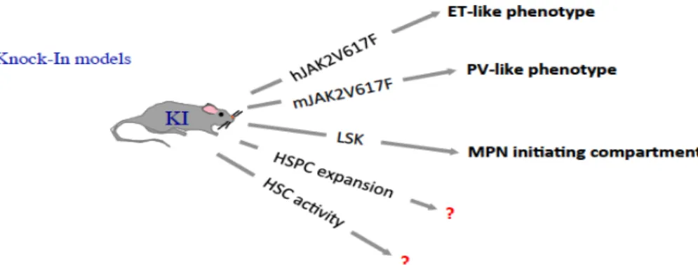
3.3.3. KI mouse models ... 48
3.4. Pre- JAK2V617F events: back to square one? ... 51
3.5. JAK2V617F clonal diversity: chaos is a friend of mine ... 53
4. Other molecular abnormalities in MPN – it’s complicated! ... 55
4.1. Genes involved in intracellular signaling ... 55
4.2. Genes involved in leukemic transformation ... 58
4.3. Genes involved in epigenetic regulation ... 60
4.4. Genes involved in RNA splice machinery ... 63
5. Treatments of MPN ... 64
5.1. Conventional approaches in management of MPN ... 64
5.1.1. Essential thrombocythemia ... 64
5.1.2. Polycythemia vera ... 65
5.1.3. Primary myelofibrosis ... 65
5.2. Investigational drugs in management of MPN ... 66
5.2.1. JAK2 inhibitors ... 66 5.2.1.1. SAR302503 (TG101348) ... 66 5.2.1.2. CYT387 ... 66 5.2.1.3. LY2784544 ... 67 5.2.2. Other drugs ... 67 5.2.2.1. Pomalidomide ... 67 5.2.2.2. HDAC inhibitor ... 67 5.2.2.3. HSP90 inhibitor ... 67 5.3. Interferon-alpha (IFN-α) ... 68
5.3.1. Interferon mediated signaling ... 68
5.3.2. Molecular mechanisms of IFN-α action ... 69
5.3.2.1. IFN-α in immunomodulation ... 69
5.3.2.2. Effect of IFN-α on cell proliferation ... 70
5.3.2.3. Anti-angiogenic effect of IFN-α ... 71
5.3.2.4. Effect of IFN-α on cellular apoptosis ... 72
5.3.3. IFN-α in MPN treatment ... 72
RESULTS ... 75
Result 1 ... 76
JAK2V617F-positive MPN: KI mouse models and IFN-α therapy ... 76
Approach ... 77
Objectives ... 77
A) Phenotype of JAK2V617F KI mice ... 78
Context ... 78
Results ... 78
Discussion ... 79
B) Effect of JAK2V617F KI on early hematopoietic cells / clonal dominance ... 80
Context ... 80
Results ... 80
Discussion ... 82
C) Effect of IFNa therapy ... 83
Context ... 83
Results ... 83
Discussion ... 84
Article 1 ... 85
Myeloproliferative neoplasm induced by constitutive expression of JAK2V617F in Knock-in mice ... 85
Article 2 ... 92
JAK2V617F expression in mice amplifies early hematopoietic stem cells and gives them a competitive advantage that is hampered by IFNα ... 92
Result 2 ... 124
JAK2V617F-positive MPN: Clonal architecture in PV patients ... 124
Context ... 125
Objectives and approach ... 126
Results and discussion ... 126
Article 3 ... 129
Use of the 46/1 haplotype to model JAK2V617Fclonal architecture in PV patients: clonal evolution and impact of IFNα treatment ... 129
DISCUSSION AND PERSPECTIVES ... 145
1. Can one mutation explain three phenotypes? ... 146
1. Does JAK2V617F modulate HSC activity? ... 147
3. Therapeutic potential of IFN-α in treatment of MPN ... 150
4. JAK2V617F clonal architecture ... 153
BIBLIOGRAPHY ... 155
LIST OF PUBLICATIONS ... 171
Articles ... 172 Protocols ... 172
LIST OF COMMUNICATIONS ... 173
English
This work concerns malignant myeloid hemopathies called classical BCR-ABL-negative Myeloproliferative Neoplasms (MPN) and include Polycythemia Vera (PV), Essential Thrombocythemia (ET) and Primary Myelofibrosis (PMF). They result from the transformation of a multipotent hematopoietic stem cell (HSC) with hyperproliferation but no blockade of differentiation. The most common molecular defect is the acquired point mutation JAK2V617F
resulting into the activation of the cytokine receptor/JAK2 pathway. We have developed a mouse constitutive and a conditional JAK2V617F knock-in (KI)
mouse models. These animals developed a disease mimicking human PV evolving into secondary MF. They also displayed an age dependent increase in the total numbers of early hematopoietic cells (phenotype LK, LSK and SLAM: LSK/CD48-/CD150+). Using In vivo competitive repopulation assays we demonstrated that cells from KI origin outcompeted their WT counterparts and that a low number of JAK2V617F
KI SLAM cells propagates the disease. These results show that the sole JAK2V617F mutation, without any additional mutations, is sufficient for disease phenotype and emergence. Using this KI
mouse model, we tested the effect of interferon-α (IFNα) treatment on MPN development. We found that IFNα treats the disease phenotype by blocking the propagation of early JAK2V617F
cells and eradicates disease-initiating cells, showing that IFNα could cure the disease in mice, as shown in some PV patients. Finally, we developed a new method combining the measurement of 46/1 SNPs and JAK2V617F
allele burdens in blood predicting the frequency of normal, heterozygous and homozygous JAK2V617F clones in PV patients. This study suggested that IFNα preferentially targets the homozygous
JAK2V617F
clone in PV patients suggesting a link between the levels of JAK2 signaling and the success of the IFNα response.
KEYWORDS: Myeloproliferative Neoplasms, JAK2V617F, HSC, Interferon- α, haplotype 46/1
Français
Ce travail concerne des hémopathies myéloïdes malignes appelés Néoplasmes Myéloprolifératifs qui incluent les Polyglobulies de Vaquez (PV), les Thrombocythémies Essentielles (TE) et les Myélofibroses Primaires (MFP). Ces maladies résultent de la transformation d’une cellule souche hématopoïétique (CSH) avec hyperprolifération mais sans blocage de différentiation. Leur défaut moléculaire le plus fréquent est la mutation JAK2V617F
résultant dans l’activation de la signalisation des récepteurs aux cytokines utilisant JAK2. Au cours de ce travail, nous avons développé un modèle murin « Knock-In » (KI) constitutif et conditionnel pour la mutation JAK2V617F
. Ces animaux développent une maladie mimant la PV humaine évoluant vers la MF secondaire. Ces animaux présentent augmentation en fonction de l’âge du nombre de cellules immatures (phénotypes Lin-, LSK et SLAM: LSK/CD48-/CD150+). Dans un système compétitifs in vivo nous montrons que les cellules KI ont un avantage prolifératif dés le stade CSH et qu'un faible nombre de CSH peuvent déclencher la maladie. Ces résultats suggèrent que la mutation JAK2V617F
seule est suffisante pour (1) le phénotype et (2) l'émergence de ces maladies. Nous avons aussi testé l'effet de l'interféron-α (IFNα) sur le développement des NMP en utilisant ces souris JAK2V617F KI. Nous montrons que l'IFNα traite le phénotype de la maladie en bloquant la propagation des cellules KI dés le
stade immature avec éradication des cellules souches néoplasiques, entraînant comme chez certains patients PV une rémission hématologique et aussi moléculaire. Enfin, en combinant l’analyse quantitative de l’haplotype 46/1 et de la mutation JAK2V617F
sur les cellules sanguines nous développons une nouvelle méthode prédictive de la fréquence des clones hétérozygotes et homozygotes JAK2V617F chez les patients PV. Cette étude suggère que l'IFNα cible préférentiellement le
clone homozygote JAK2V617F
et que sa réponse est fonction de l’intensité de la signalisation JAK2.
Introduction
Figure 1: Hematopoietic Pyramid ---12
Figure 2: 2008 WHO classification of myeloid neoplasm ---15
Figure 3: Main etiological factors in MPN ---16
Figure 4: Classic model of JAK2V617F-positive MPN ---20
Figure 5: Natural evolution of Ph- MPN ---21
Figure 6: Overlaps in clinical presentation and complications in classic Ph- MPN ---22
Figure 7: Structure of JAK2 protein ---23
Figure 8: JAKs and cytokine receptor ---24
Figure 9: JAK2 signaling ---26
Figure 10: Schematic representation of wildtype (WT) JAK2 and its activated fusion proteins found in various hematologic malignancies ---27
Figure 11: Schematic representation of JAK2 activating mutations ---28
Figure 12: JAK2V617F and aberrant downstream signaling pathways ---31
Figure 13: Nuclear role of JAK2 ---33
Figure 14: JAK2V617F homozygosity ---35
Figure 15: JAK2V617F phenotypic pleiotropy hypotheses ---38
Figure 16: Effect of JAK2V617F on hematopoietic cell populations ---40
Figure 17: Lesson from JAK2V617F retroviral BMT mouse models ---44
Figure 18: Lesson from JAK2V617F transgenic BMT mouse models ---47
Figure 19: Lesson from JAK2V617F Knock-In BMT mouse models ---50
Figure 20: Pre-JAK2V617F hits ---52
Figure 21: Schematic representation of JAK2V617F clonal diversity ---53
Figure 22: Signaling pathway defects involved in pathogenesis of MPN ---56
Figure 23: Genes involved in leukemic transformation and epigenetic modulations ---61
Figure 24: Approximate frequencies of recurrent mutations found in MPN and post-MPN AML ---63
Figure 25: Inteferon alpha mediated signaling ---69
Results
Figure 26: Onjectives to study JAK2V617F KI mouse models ---77Figure 28: Effect of JAK2V617F on HSC competitiveness ---81
Figure 29: JAK2V617F-positive SLAM minimum graft size ---82
Figure 30: Hematological response in JAK2V617F KI mouse model upon interferon alpha exposure ---83
Figure 31: Molecular response in JAK2V617F KI mouse model upon interferon alpha exposure ---84
Figure 32: Schematic representation of 46/1 haplotype and homologous mitotic recombination (HR) on chromosome 9 ---125
Figure 33: Proliferative profiling of JAK2V617F homozygous clone during hematopoietic differentiation ---127
Figure 34: Effect of interferon alpha exposure on JAK2V617F sub clones---128
Discussion
Figure 35: JAK2V617F priming hypothesis 1 ---152Figure 36: JAK2V617F priming hypothesis 2 ---153
Introduction
Table 1: JAK2V617F reteroviral BMT mouse models ---44Table 2: JAK2V617F transgenic mouse models ---47
2-HG = 2 hydroxyglutarate α-KG = Alpha ketoglutarate AML = Acute myeloid leukemia
AMMoL = Acute myelomonocytic leukemia Ana = Anagrelide
ASXL1 = Additional sex-comb like 1
Allo-SCT = Allogeneic stem cell transplantation ALL = Acute lymphoblastic leukemia
Bfn = Busulfan BM = Bone marrow
BFU-E = Blast forming unit-erythroid
BCR-ABL = Break point cluster-Abelson kinase bFGF = Basic fibroblast growth factor
CD = Cluster of differentiation CDK = Cyclin dependent kinase CFU-E = Colony forming unit-erythroid
CFU-GM = Colony forming unit-granulocyte macrophage CCR = Complete cytogenic remission
CHR = Complete hematologic remission CMR = Complete molecular remission CTL = Cytotoxic T cells
CMP = Common myeloid progenitor CLP = Common lymphoid progenitor CMML = Chronic myelomonocytic leukemia CML = Chronic myeloid leukemia
CEL = Chronic eosinophilic leukemia CNL = Chronic neutrophilic leukemia CSF3R = Colony stimulating factor 3 receptor CBL = Casitas B-lymphoma
CDKi = Cyclin dependent kinase inhibitor
CFU-GEMM = Colony forming unit-granulocyte erythrocyte macrophage megakaryocyte DC = Dendritic cells
DGS = De Guglielmo’s syndrome
DNMT3a = DNA (cytosine-5)-methyltransferase 3A EEC = Endogenous erythroid colonies
EZH2 = Enhancer of Zeste homology 2 ET = Essential thrombocythemia Epo = Erythropoietin
F = Phenylalanine
FGFR1 = Fibroblast growth factor receptor 1 G = Guanine
GHR = Growth hormone receptor
G-6PD = Glucose-6 phosphate dehydrogenase GMP = Granulocyte-macrophage progenitor
GM-CSF = Granulocyte macrophage-colony stimulating factor G-CSF = Granulocyte-colony stimulating factor
GAS = IFN-γ activated site hmC = Hydroxymethyl Cytosine
HP-α1 = Heterochromatin Protein-alpha 1 HSC = Hematopoietic stem cell
HU = Hydroxy Urea
HSPC = Hematopoietic stem and progenitor cells HR = Homologous recombination
IL = Interleukin IFNα = Interferon alpha
IGF-1 = Insulin-like growth factor receptor 1 ISG = Interferon stimulated gene
ISRE = IFN stimulated response element IGSF3 = IFN stimulated gene factor 3 IFN-γ = Interferon gamma
IFN-α = Interferon alpha
IKZF1 = IKAROS family zinc finger 1 IDH1/2 = Isocitrate dehydrogenase ½ JH = Janus homology
JAK2 = Just another kinase/Janus kinase 2 KI = Knock-In
KO = Knock-Out
KIT = Tyrosine-protein kinase LSK = Lineage-Sca1+cKit+
LT-HSC = Long term-hematopoietic stem cell LTC-IC = Long term culture-initiating cell LOH = Loss heterozygosity
LD = Linkage disequilibrium LK = Myeloid progenitor
MEP50 = Methylosome protein 50 MEP = Myelo-erythroid progenitor MPP = Multipotent progenitor MDS = Myelodysplastic syndrome MPD = Myeloproliferative disorder MPN = Myeloproliferative neoplasm MDM2 = Mouse double minute 2 homolog NGS = Next generation sequencing NAC = N-acetylcystein
NOD/SCID = Nonobese Diabetic/Severe Combined Immunodeficiency PRMT5 = Protein Arg methyltransferase 5
PV = Polycythemia vera
PRC2 = Polycomb repressive complex 2
PR-DUB = Polycomb repressive – deubiquitinase P3 = Proteinase 3
PMF = Primary myelofibrosis Ph = Philadelphia
PDGFRA = Platelet derived growth factor alpha PDGFRB = Platelet derived growth factor beta PIAS = Protein inhibitors of activated STAT PI3K = Phosphoinositol-3-Kinase
ROS = Reactive oxygen species RV = Retroviral
SM = Systemic mastocytosis
SOCS = Suppressor of cytokine receptor SRC = SCID repopulating cells
ST-HSC = Short term-hematopoietic stem cell SLAM = Signaling lymphocyte activation molecules SCA-1 = Stem cell antigen-1
T = Thymine
TGF-β = Transforming growth factor beta TET2 = Ten eleven translocation 2 T-reg = Regulatory T cells TG = Transgenic
Tpo = Thrombopoietin TF = Transcription factor UPD = Uniparental disomy V = Valine
WHO = World health organization WBC = White blood cell
WT = Wild type
XCIP = X chromosome inactivation pattern Y = Tyrosine
INTRODUCTION
1. Hematopoiesis – an overview
lood is the most highly regenerative tissue with approximately 1012 cells arising daily from adult
human bone marrow (BM) and lymphoid tissues. The cells in the hematopoietic system are continually generated from a low number of self-renewing cells called hematopoietic stem cells (HSCs). As multi-potent HSCs, at the apex of hematopoietic system, differentiate they give rise to progenitors, precursors and finally, terminally differentiated cells with increasing lineage-restricted capacity down the hierarchy. This lineage relationship between stem cell, progenitors and mature cells form a complex road map that is guided by strict interplay of chromatin remodeling, transcription factors, signaling molecules and cytokine receptors. In order to perpetuate as well as differentiate, to sustain the homeostasis in hematopoietic system, HSCs maintain a balance between self-renewal, differentiation, proliferation and apoptosis.
1.1. Definition
Hematopoiesis is the process of generating all functional effector blood cell types from HSC through successive differentiation events. These effector cells include at least 8 blood lineages i.e., erythrocytes, platelets, granulocyte, macrophages, dendritic cells, T cells, natural killer cells and B cells. Prenatally, hematopoiesis occurs in yolk sac then in liver and finally in BM while in adults it takes place primarily in BM and lymphoid tissues.
1.2. Hematopoietic hierarchy
HSC resides at the top of the hematopoietic pyramid. Murine HSC compartment is confined within lineage marker-negative, c-kit-positive and Sca-1-positive (LSK: Lin-c-kit+Sca-1+) population and represents a very
small fraction of cells of total BM with the capacity of self-renewal1,2. As the dormant HSC with most enriched
self-renewing potential enters into an active cycling state they acquire the expression of CD343. HSCs express
Thy1.1 (CD90) at very low levels and the expression of Flk2 (also called Flt3 or CD135) tyrosine kinase differentiates the long term HSC (LT-HSC: Thy1.1lowLin-c-kit+Sca-1+CD34-flk-) with strong self-renewal and
differentiating potential, from the short term HSC (ST-HSC: Thy1.1lowLin-c-kit+Sca-1+CD34+flk+) with limited
self-renewal and full multilineage differentiating potential4. Kiel et al. identified a more sophisticated set of
SLAM family cell surface receptors (CD150 and CD48) to distinguish between most primitive and highly purified LT-HSC (SLAM: Lin-cKit+Sca1+CD150+CD48-) from multipotent progenitors (MPP: Lin
-cKit+Sca1+CD150-CD48-). In mice these SLAM cells compose ~0.0058% of total BM and single SLAM cell injection into lethally irradiated mice revealed that every 1 out of 2.1 SLAM cells has a long term multilineage reconstitution capacity5. ST-HSCs generate MPPs (Lin-Thy1.1-cKit+Sca1+flk2highCD34+SLAM-)6 with retained
full lineage potential but limited self-renewal capacity. This appears to be the branching point in hematopoietic hierarchy giving rise to progenitors with differential and restricted differentiation capacity. MPPs give rise to two kinds of oligolineage-restricted progenitors with no self-renewal capacity: the common lymphoid progenitor (CLP: Lin-cKitlowSca1lowIL-7Rα+
) with lymphoid potential7 and the common myeloid progenitor within the Lin -ckit+(LK) population (CMP: Lin-ckit+
FcγRloCD34 +) with myeloid potential that eventually give rise to
megakaryocytic-erythroid progenitor (MEP: Lin-ckit+FcγRloCD34-) and common granulocyte-macrophage
progenitors (GMP: Lin-ckit+FcγRhiCD34+)8. The CLPs have the capability to produce further lineage-restricted
progenitors i.e., Pro-dendritic cells (DC), Pro-T cells, pro-natural killer cells (NK) and pro-B cells. Likewise, GMPs give rise to precursors that will eventually be able to produce granulocyte and macrophages. MEPs further differentiate to give megakaryocyte progenitors and erythroid precursors. Interestingly, both CLP and CMP carry the potential to generate pro-DC. This multilayered differentiation forms the base of hematopoietic pyramid: mature blood cells. Because these effector cells have very short life span, mature blood cell production is an ongoing process demanding the production of 1.5 X 10⁶ blood cells every second in an adult human Figure 1.
1.3. Lineage selection – a guide to hematopoietic road map
HSC differentiation is a unidirectional process in which it loses its pluripotency to assume the process of commitment. However, how the HSC makes decision whether to differentiate and acquire a lineage identity or to self-renew is largely unknown. This HSC differentiation to opt a lineage fate can be explained by different models. In ‘Stochastic model’9,10 it is assumed that the cell’s intrinsic genetic and epigenetic factors as well as
Figure 1. Hematopoietic pyramid At the apex of pyramid, long term-hematopoietic stem cell (LT-HSC) asymmetrically divides to give rise to self-renewable HSC and short term-HSC (ST-HSC), which in turn gives rise to multipotent progenitor (MPP). MPP differentiates into oligopotent common myeloid (CMP) and common lymphoid progenitors (CLP). CMP further engages to produce megakaryocyte-erythroid (MEP) and granulocyte-macrophage progenitors (GMP). These oligopotent progenitors give rise to specific lineage restricted precursors i.e., erythroid (Ep), megakaryocytic (Mkp), myeloid (myelo), monocytic (mono), Dendritic (Pro-DC), Natural killer (Pro-NK), B-cells (Pro-B) and T-cells (Pro-T) precursors that finally differentiate into mature effector cells to form the base of pyramid. As cells assume their lineage fate down the hierarchy their proliferative potential also increases. Colored panels in right show surface markers use to isolate hematopoietic stem and progenitor cells. Inspired by Bryder et al. (2006) and Keil et al. (2005).
external stimuli determines the cell’s fate ‘randomly’, say cytokines, cytokine receptors and transcription factors (TFs) play a permissive role rather than instructive. In contrast, ‘Determinism model’11 emphasizes on the
predetermined role of these factors and stimuli in HSC differentiation and lineage selection. As HSC differentiation is a highly complex stepwise process, for being conducive to differentiation process, it first enters in the state of ‘priming’ where chromatin remodeling allows the expression of lineage specific genes and repression of undesired genes12,13. These primed cells are now ready for the transcriptional response, expressing
positive and negative TFs, under the influence of external stimuli i.e., binding of cytokines to lineage specific cytokine receptors14,15,16. Cytokines and their cognate receptors finely tune the regulatory pathways that
eventually decide the fate of HSC.
1.4. Homeostasis - self renewal, cell cycle and apoptosis
Functional effector cells of hematopoietic system have very short life span demanding efficient homeostatic control mechanisms. In order to perpetuate as well as produce progeny, HSCs maintain a balance between self-renewal and differentiation through asymmetric cell division. However, the molecular mechanisms that control HSC self- renewal are poorly understood. Interestingly, several signaling pathways that are postulated to be involved in HSC self-renewal, like Hox genes17, Notch18, Sonic hedgehog and Wnt signaling pathways19,20, are
also hypothesized to be associated with oncogenesis emphasizing on the fact that normal and cancer stem cell share the ability to self-renew.
Most of the HSC resides in Go phase of cell cycle 21
. The minimal proliferative pressure on HSC protects them from mutagenic hazards of DNA replication and damage-inducing metabolic side products. As HSC quits self-renewing potential and enters into differentiation it gradually becomes mitotically active. As the proliferative advantage increases down the hierarchy this add an advantage of fine-tuning the homeostasis of a given effector cell type.
Like other cell types, HSCs are also subjected to apoptosis and programmed cell death. In fact, overexpression of oncogene bcl-2 prevented apoptosis, increased LT-HSC frequency and radio-resistance in transgenic mice22
showing the role of apoptosis in hematopoietic homeostasis.
Overall, hematopoiesis is a highly orchestrated process of producing and maintaining high turnover of all effector cell types from a single HSC under the control of intrinsic and extrinsic stimuli of hematopoietic syste
2. Myeloproliferative neoplasms
he concept of disease with excessive proliferation of blood cells was already conceived when Hippocrates, The Father Of Medicine, (460-370 BC) and Galen, the most prominent physician after Hippocrates, (129-200 AD) postulated the humoral theory. ‘Blood’ is one of the four humors (three
being phlegm: white blood cells, yellow bile: serum and black bile: deoxygenated red blood cells) and ‘plethora’ is the imbalance of humors with blood dominating over the others23. Since then, many studies to understand the
origin, evolution, diversity and complexity of blood malignancies have shown that this plethora is a result of alteration in genetic and epigenetic factors that perturb the key process such as self-renewal, proliferation and differentiation of cells.
2.1. Myeloid neoplasms
Myeloid neoplasms (MN) are clonal HSC disorders of myeloid cell hyper proliferation. MN can be categorized into three broad clinicopathological groups: acute myeloid leukemia (AML), myelodysplastic syndrome (MDS) and myeloproliferative neoplasm (MPN).
AML are characterized by proliferation of blast cells, principally in marrow and resulting in impaired production of normal blood cells e.g, acute myelomonocytic leukemia (AMMoL) with excess production of myeloblasts and monoblasts.
MDS, on the other hand, are characterized by ineffective hematopoiesis, dysmorphogenesis of myeloid cells and cytopenias e.g, refractory cytopenias with multilineage dysplasia (RCMD) that is characterized by cytopenias and dysplasia in more than one myeloid lineage and <1% blast cells in blood stream.
In contrast to AML and MDS, MPN are disorders with increased numbers of functional and terminally differentiated myeloid elements e.g, polycythemia vera in which red blood cells are over produced.
However, such classification is not precise; as some patients present with symptoms that overlap MPN and MDS and, they are assigned to ‘MPN/MDS overlap’ subgroup e.g, chronic myelomonocytic leukemia (CMML) with increased numbers of monocytes: characteristic of MPN and dysplasia of monocytes and granulocytes: characteristic of MDS.
Moreover, both MDS and MPN have a propensity to evolve into AML.
2.2. Classification – from the age of Dameshek to the epoch of genome
Chronic myeloid leukemia (CML), primary myelofibrosis (PMF), polycythemia vera (PV), Di Guglielmo’s syndrome (DGS) and essential thrombocythemia (ET) were already recognized in early 19th century as distinct disorders of hyper proliferation of three main myeloid lineages but predominate in one of them: the granulocytic lineage in CML, the megakaryocytic/granulocytic lineages in PMF, the erythroid lineage in PV and DGS and the megakaryocytic/platelet lineage in ET.
Proper classification of myeloproliferative disorder (MPD) began when in 1951 William Dameshek has highlighted the overlapping clinical and laboratory features of these entities. He argued that although erythrocytosis is the key characteristic of PV, PV is also presented with ‘pancytosis’, over production of erythroid, megakaryocytic and granulocytic lineages. Moreover, PV patients often display erythroblasts in
peripheral blood, BM fibrosis and splenomegaly mimicking PMF. Considering these disorders distinct yet closely related Dameshek grouped them for the first time under the term ‘myeloproliferative disorders’ (MPD)24.
Over the time, DGS was recognized as erythroid leukemia and the rest of the members remained under the umbrella of classic MPDs. In 1960, with the discovery of Philadelphia (Ph) chromosome25, classic MPDs were
further sub-grouped into Ph positive MPD (CML) and classic Ph negative MPD (PV, ET and PMF).
In 2008, the WHO has revised its classification26 of myeloid malignancies and coined the term
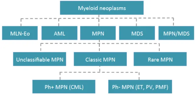
‘myeloproliferative neoplasm’ (MPN). Furthermore, it has sub-categorized the myeloid malignancies into 5 well-defined groups: 1-Myeloproliferative neoplasms (MPNs), 2-Myeloid and lymphoid neoplasms with eosinophilia and abnormalities of PDGFRA, PDGFRB, and FGFR1, 3-MDS, 4-MPN/MDS and 5-AML Figure 2.
2.3. Main etiological factors in MPN
MPNs are acquired and clonal myeloid malignancies of HSC origin with the over production of mature myeloid cells due to cytokine hypersensitivity. The group comprises of Ph positive CML, Ph negative MPN (PV, ET and PMF), less frequent non-classic MPN (CNL, CEL/NOS, and MCD) and unclassifiable MPN.
John Hughes Bennett has reported CML as a new clinical entity with hypertrophy of spleen and liver in 184527
and X-linked glucose-6-phosphate dehydrogenase (G-6PD) locus analysis demonstrated its clonal hematopoietic stem cell origin28. CML’s etiological factor, the fusion tyrosine kinase ‘BCR-ABL’29, resulting from a
Philadelphia translocation between chromosome 9 and 22 ; t(9;22)(q34;q11)25,30, was the first and long time
unique, constitutively active mutated kinase identified and characterized in MPNs. Since its discovery,
BCR-Figure 2. 2008 WHO classification of myeloid neoplasm Myeloid neoplasms are classified into five broad categories: 1- Myeloid and lymphoid neoplasms with eosinophilia and abnormalities of PDGFRA, PDGFRB and FGFR1 (MLN-Eo), 2- acute myeloid leukemia (AML), 3- myeloproliferative neoplasm (MPN), 4- myelodysplastic syndrome (MDS) and 5. MPN/MDS overlaps. MPN are further divided into rare MPN, classic MPN and unclassifiable MPN. Classic MPN encompass Philadelphia positive chronic myeloid leukemia (Ph+ CML) and Philadelphia negative (Ph-) MPN i.e., essential thrombocythemia (ET), polycythemia vera (PV) and primary myeloid fibrosis (PMF).
ABL was thoroughly studied in in-vitro and in-vivo systems and shown to transform mouse BM derived IL-3 dependent cells and to induce CML-like disease in retrovirally transduced BM transplantation mouse models31–33.
Following the discovery of oncogenic translocation in CML many other fusion tyrosine kinases including fusions of platelet derived growth factor beta (PDGFβ) in CMML34 and PDGFRα in SM and CEL35 were later
unrevealed. The fact that KIT tyrosine kinase is highly expressed in mast cells led to the discovery of KITD816V mutant allele in SM36. All these observations showed that MPNs are tyrosine kinase disorders.
Classic Ph- MPNs are closely linked diseases that were initially grouped on the basis of their shared clinical and laboratory features. Ph- MPNs also display deregulated clonal myeloid proliferation as observed in other MPNs harboring constitutively active tyrosine kinases. Inhibition of endogenous erythroid colonies (EEC) with imatinib mesylate37 (a tyrosine kinase inhibitor that also inhibits the kinase activity of ABL, KIT and PDGFR)
and JAK2 inhibitor AG49038 suggested a tyrosine kinase as an etiological factor involved in signalization of
myeloid cells. Moreover, because ET can transform into PV and PV into MF, their pattern of clinical evolution strongly suggested a common etiological factor involved in signaling through different receptors of myeloid lineage in all three Ph- MPNs. In 2005 several teams have published the discovery of a somatic guanine-to-thymine (G>T) mutation encoding a valine-to-phenylalanine substitution at position 617 (V617F) in the auto inhibitory JH2 (JAK homology 2) domain of non-receptor tyrosine kinase JAK2. The mutation JAK2V617F results
Figure 3. Main etiological factors in MPN A predominate terminally differentiated myeloid lineage is involved in each clinically distinct MPN. Activated alleles resulting in constitutive tyrosine kinase signaling are identified in each disorder. HSC: hematopoietic stem cell, BCR-ABL: Break point cluster region-Abelson tyrosine kinase, JAK2: Janus kinase, FIP1L1-PDGFR: FIP1-like-1-Platelet derived growth factor receptor, CSF3R: Colony stimulating factor 3 receptor, KIT: Tyrosine-protein kinase.
in the abrogation of auto inhibition leading to constitutive activation of kinase JAK239–43 (Structure and
functional consequeces of JAK2 are discussed in detail in chapter 3).
Recently point mutations in colony stimulating factor 3-receptor (CSF3R) gene have been identified in patients suffering either CNL or aCML. These mutations lead to preferential downstream kinase signaling through SRC family–TNK2 or JAK kinases and possess differential sensitivity to kinase inhibitors 44 Figure 3.
2.4. Classic Ph- MPNs
2.4.1. Main molecular characteristics of Ph- MPN
In past two decades, causative lesion in classic Ph+ CML and rare MPNs, like CMML, SM and CEL, were identified to be mutated tyrosine kinases. These constitutively active tyrosine kinases like BCR-ABL in CML and FIP1L1-PDGFRα in SM are found to activate several signaling pathways like STATs, MAPK and PI3k which lead to constitutively active cell survival, proliferation and differentiation pathways. Classic Ph- MPNs also share these features and the discovery of JAK2V617F in a tyrosine kinase JAK2 as a genetic cause in more
than half of Ph- MPN patients has provided a great deal of knowledge about molecular pathogenesis of this disease.
2.4.1.1. Ph- MPNs are acquired clonal HSC disorders
In 1970s, X-chromosome linked polymorphism studies in women have already demonstrated that Ph- MPN are clonal disorders with the involvement of multiple myeloid lineages and, therefore, have probable multipotent stem cells origin45–4748. The presence of mutant JAK2V617F allele in all the myeloid lineages supported the notion
that this clonal mutation has occurred at the apex of hematopoietic hierarchy and also, the absence of mutation in non-hematopoietic cells showed that mutation is somatically acquired40. Indeed, just after the discovery of JAK2V617F, Jamieson et al. investigated for the hematopoietic compartment that harbor the mutation using flow cytometry technique. They found that JAK2V617F is not only present in myeloid progenitors (CMP, GMP and
MEP) but also in HSC compartment (Lin-CD34+CD38-CD90+) of PV patients49.
However, this mutation is also reported in mature lymphoid lineage (B cells, NK cells and very rarely in T cells) in a subset of PV and PMF patients 66. This suggests, first, the cell that has acquired JAK2V617F mutation is capable of differentiating in all hematopoietic lineages and second, this mutation renders a downstream selective advantage unique to myeloid lineage, likely due to the use of JAK2 by cytokine receptors of these lineages. Interestingly, the subsets of JAK2V617F negative ET and PMF patients also exhibited clonal myelopoiesis pointing
towards the yet-unknown genetic alterations that phenocopy JAK2V617F and contribute to MPN pathogenesis52.
2.4.1.2. Ph- MPNs are characterized by mature myeloid cell hyper proliferation
JAK2 has shown to play a crucial role in pre- and post-natal hematopoiesis in mouse models53,54. Mice knockout
for JAK2 (JAK2-/-) are embryonically lethal at day 12.5 of gestation due to failure of definitive erythropoiesis. The phenotype of JAK2-/- embryos was comparable to that of EpoR null or Epo null mice, showing a non-redundant function of JAK2 in Epo-R signaling55. Moreover, fetal liver cells from JAK2-/- embryos failed to
respond IL-3, GM-CSF, IL-5 and Tpo, but not to G-CSF, in colony formation assays demonstrating the essential role of JAK2 in signalization via receptors of these cytokines. Furthermore, signaling of type II interferon
receptor (IFN-gamma) but not type I interferon receptor (Interferon-alpha) was abolished showing their dependence on JAK2.
Cytokine independent or hypersensitive growth of hematopoietic progenitors is a cardinal feature of ET, PV and PMF. Endogenous erythroid colonies (EECs)56,57 and endogenous megakaryocyte colonies (EMCs)58 are
evidenced in these patients along with the hypersensitivity to IGF-1, IL-3, GM-CSF, Epo and Tpo59–62. The
mutation JAK2V617F renders the tyrosine kinase JAK2 an authority to constitutively auto phosphorylate41 with
increased kinase activity43. To check whether this constitutive auto phosphorylation was responsible for hematopoietic transformation in PV patients, JAK2V617F was expressed in BaF3 and FDC-P1 primary cell lines,
both dependent on IL-3 for proliferation, but they were not resulted into cytokine independent growth. However, co-expression of EpoR, a homodimeric type I receptor, transformed these cell lines to cytokine independency39,41. Because the megakaryocytic and granulocytic lineages are also involved in Ph- MPNs and their proliferation is also mediated by homodimeric type I receptor it was worth investigating the role of JAK2V617F in signalization via Tpo receptor (Mpl) and granulocyte-macrophage colony stimulating factor
receptor (GCSFR). Indeed like EpoR, co-expression of JAK2V617F with Mpl or GCSFR transformed the BaF3 cell
line63. Overall, mutant tyrosine kinase JAK2V617F renders a proliferative advantage to myeloid cells via
signalization through homodimeric type I receptors: EpoR, Mpl and GCSFR, which are present on erythroid, megakaryocyte and granulocyte lineages, respectively.
Constitutive activation of JAK2 results in activation of downstream signaling pathways i.e., JAK-STAT, MAPK/ERK and PI3/AKT, which are crucial to cell survival and proliferation39,41–43,63.
2.4.2. Main entities in Ph- MPN 2.4.2.1. Essential thrombocythemia
Long before its recognition as Dameshek defined classic MPD; Emil Epstein and Alfred Goedel already formally described ET as ‘hemorrhagic thrombocythemia’ in 1934. XCIPs studies by polymerase chain reaction (PCR) demonstrated that, in a large proportion of patients, ET can occur as clonally heterogeneous disease64,65 and sometimes clonality is only restricted to megakaryocytic lineage65.
Clinically, ET is featured with vasomotor symptoms: thrombosis, headache, dizziness, syncope, acrocyanosis, tingling and visual changes, however, 25-35% cases are asymptomatic and are diagnosed accidently. In laboratory, ET is characterized by thrombocytosis and BM megakaryocyte hyperplasia with hyperlobated nuclei without associated erythrocytosis and leukoerythroblastosis. Megakaryocyte precursors show hypersensitivity to thrombopoietin (TPO)58. Mild splenomegaly may occur in about 40% of patients. JAK2V617F is the major
molecular lesion of ET with a frequency of 55% 39–43 Figure 4 and 5. Because this mutation is also present in
other MPNs, the 2008 WHO JAK2V617F positive ET diagnostic criteria requires the presentation of
thrombocytosis with the exclusion of other JAK2V617F positive MPNs diagnostic proofs.
Deregulation of megakaryocyte lineage in ET provided the rationale to screen thrombopoietin receptor (MPL), which led to the discovery of the mutations of codon 515 (MPLW515K, MPLW515L and MPLS505N) in minor sub-set of ET patients66–68. However, the etiology of large proportion of ET patients without JAK2 or MPL
The median age at diagnosis is 65-67 years but it can occur at any age with the disease incidence of 1.5/100,000 cases in the general population69. 5-6% ET evolve into PV after 2.5 years70 and 3% ET after 5 years, 8% ET
after 10 years and 15% ET after 15 years evolve into MF71. Transformation to AML is a life-threatening
complication in around 0.6-6% of cases72 Figure 6. Median survival is found to be 20 years in ET patients. Risk
of thrombosis and major hemorrhage is shown to be 6.6%73 and 0.33% patients /year74, respectively.
Polycythemia Vera
Initially, Louis Henri Vaquez, a French physician, described polycythemia as ‘maladie de Vaquez’ in 1892. Later on, in 1903, William Osler, a renowned physician, presented a descriptive landmark of this disease as chronic cyanosis, splenomegaly and erythrocytosis: a new clinical entity.
PV patients are often presented with headache, pruritus, vaso-occlusive symptoms like tinnitus, paresthesia and erythromelalgia, and in some patients, splenomegaly is present with its degree depending upon the stage of disease. High hematocrit (Hct), erythrocytosis and increased hemoglobin (Hb) are the Sine-qua-non of PV. BM is hyper cellular due to tri-lineage hyperplasia (RBC, granulocytes and megakaryocytes) with morphologically normal looking erythroid and myeloid precursors but hyper lobulated loosely clustered megakaryocytes. A variable increase in reticulin fibers is also observed specially over disease progression.
An important finding that erythroid progenitors from BM of PV patients are capable of growth without added exogenous erythropoietin (Epo)56, laid the basis of the idea that suspected etiological factor is likely to be present
in cytokine regulation. Mutation in JAK2, a tyrosine kinase down stream to erythropoietin receptor (EpoR) signaling, finally explained the molecular etiology in almost 95% of PV patients39–42 Figure 4 and 5. Moreover, chromosome 9 uniparental disomy (UPD 9), resulting in the duplication of mutant allele, is a common phenomenon in PV (~70%), in contrast to ET, patients75. Presence of JAK2V617F in majority of PV patients has influenced its diagnosis and it is reflected in the 2008 WHO diagnostic criteria guidelines. Other mutations leading to constitutive activation of signaling pathways are also discovered in exon 12 of JAK2 in JAK2V617F
negative PV patients with an overall frequency of around 3% in PV.
The annual incidence of PV is ~2/100,000 cases in general population69 and it can occur in all ages but risk increases over 60 years. Natural evolution of PV into MF is noted in 30% of patients after 10-12 years of diagnosis76. 2-5% of PV patients develop AML in the course of disease progression77. Although genotoxic
therapies increase the risk of AML transformation, it also occurs in treatment naïve patients emphasizing on its natural evolution Figure 5. PV patients bear the risk of thrombotic events specially over 60 year and with previous history of such complication78 Figure 6. Leukocytosis may impact thrombotic events maybe considered as an average risk factor79. Median survival approaches to 20 years in PV patients.
2.4.2.2. Primary myelofibrosis
In 1879, Gustav Heuck presented the pathologic description of primary myelofibrosis (PMF) in his two patients as massive splenomegaly, circulating nucleated red blood cells and increased numbers of morphologically abnormal leukocytes, marrow fibrosis, osteosclerosis and extensive extra medullary hematopoiesis27.
PMF patients may present with dyspnea, fatigue, night sweats, cachexia, fever and bleeding. More than 90% of patients present splenomegaly. However, 30% of patients are asymptomatic at the time of diagnosis. PMF patients may exhibit anemia, leukocytosis and/or thrombocytosis. Initial stage PMF, ‘pre-fibrotic stage’, is difficult to diagnose because of thrombocytosis that resembles ET. The sine-qua-non of myelofibrosis, whether
Figure 4. Schematic representation of JAK2V617F
-positive MPN The mutation JAK2V617F appears in hematopoietic stem
cell (HSC) and results in clonal proliferation of terminally mature myeloid cells. This mutation is found in three clinically distinct MPN i.e., essential thrombocythemia (ET), polycythemia vera (PV) and myelofibrosis (MF) that are characterized by the hyper proliferation of platelets, red blood cells (RBC) / neutrophils and neutrophils/megakaryocytes, respectively. However, a small proportion of ET may overlap PV phenotype depicting the fruste form of PV and PV can evolve into myelofibrosis by passing through a spent phase featuring some symptoms MF. Of note, PV is also characterized by hyperproliferation of granulocytes and platelets in contrast to erythrocytosis that is characterized by the sole hyperproliferation of erythroid cells. JAK2V617F heterozygosity is associated with ET and JAK2V617F homozygosity due to
chromosome 9 uniparental disomy (9p UPD) is associated with PV. JAK2V617F
allele burden increases across the spectrum of Ph- MPN i.e., lowest in ET and highest in MF. These disorders form a continuum where ET and PV depicts the chronic phase of disease and accelerated phase may encompass the transformation of ET and PV into myelofibrosis along with other manifestations such as increasing white blood cells (WBC), blasts and neutropenia and thrombocytopenia.
PMF or post ET/PV MF, is accumulation of reticulin fibers in marrow accompanied by hyper cellular BM with expanded left-shifted granulopoiesis and decreased erythropoiesis. Megakaryocyte hyperplasia is one of the major criteria in PMF diagnosis. BM harbors densely packed clustered of cytologically bizarre megakaryocytes with hypolobated nuclei. Peripheral blood smears show leukoerythroblastosis and anisopoikilocytosis with teardrop shaped red cells Figure 4.
PMF shares etiological factor of ET and PV i.e. JAK2V617F in around 50% of PMF patients Figure 5. Although
the discovery of JAK2V617F promised a unifying mechanism to be targeted effectively, recent molecular studies have identified several other molecular defects, for example in MPL, SOCS1, TET2, EZH2 and IDH1/2, which have provided further opportunity to comprehend the molecular underpinnings of PMF pathophysiology. However, still a large subset of PMF patients harbors a yet-unknown causative factor of disease. Clonal myeloproliferation in PMF is followed by secondary stage with stromal changes and abnormal cytokine profiles that are believed to provoke an inflammatory response and may contribute to the clinical phenotype, including bone marrow fibrosis, angiogenesis, extra medullary hematopoiesis, constitutional symptoms and cachexia80.
The annual incidence of PMF is 1.5 cases per 100,000 people and it commonly occurs over the age of 60 years with same frequency in both genders76. Around 15-30% of PMF patients may transform into AML77 Figure 5
and 6. Median survival is approximately 6 years. It was not established until recently that aged PMF patients (over 60 years) and those with leukocytosis and JAK2V617F are at an elevated risk of cardiovascular thrombotic events81. In a recent cytokine profiling study of PMF patients certain specific plasma cytokine signatures are
shown to be related with overall and leukemia free survival82.
As Dameshek speculated 64 years ago, these classical Ph- MPNs share core cardinal features and form a spectrum of closely related disease entities. Although erythrocytosis is the hallmark of PV, ET and PMF share thrombocytosis. Likewise, splenomegaly is a common finding in PMF but is also present in approximately 30% ET and PV patients. Marrow fibrosis is sine-qua-non of PMF but ET and PV patients may accumulate reticulin fiber over the natural course of disease evolution. Both ET and PV have the propensity to develop myelofibrosis and all three of them can transform into AML Figure 5.
Discovery of the mutation JAK2V617F in the epoch of genome has proved to be an important twist in the story of
Figure 5. Natural evolution of Ph- MPN Around 95% polycythemia vera (PV) patients and 50-60% essential thrombocythemia (ET) and myelofibrosis (MF) patients harbor JAK2V617F
mutation (VF). In the natural course of disease progression ET may evolve into PV and PV into secondary myelofibrosis and all three of them can directly transform into acute myeloid leukemia (AML). Value on each flash represents the approximate percentage of each type of progression.
Ph- MPN. The finding of JAK2V617F, on one hand, simplified the concept of relatedness of three diseases under
the umbrella of classic Ph- MPN and, on the other hand, raised the question of how one mutation can cause three distinct disease entities. JAK2V617F positive ET as compared to JAK2V617Fnegative ET present many features
resembling PV such as high hemoglobin, leukocytosis, BM erythropoiesis and granulopoiesis, low serum Epo levels and higher rates of venous thrombosis83 thus, JAK2V617Fpositive ET depicts the ‘fruste form’ of PV.
JAK2V617F homozygosity is an event mostly associated with PV as compared to ET suggesting the contribution of
JAK2V617Fallele burden in disease phenotype and evolution75,84. Evolution of PV is also stepwise with splenomegaly in absence of myelofibrosis, cytopenias of one or several cell lines, unabated erythrocytosis and myeloid metaplasia being the ‘spent phase’ of PV and a propensity to transform into secondary myelofibrosis or AML85.
In clinical studies, the JAK2V617F allele burden is found to increase across the spectrum of these three disease
entities, with the lowest level in ET and highest level during fibrotic transformation86,87. All these observations
suggest that JAK2V617Fpositive ET, PV and PMF form a continuum model being three stages of Ph- MPN:
chronic stage ET and PV precede PMF in the accelerated stage Figure 4. JAK2V617F provided a simple molecular
profile of Ph- MPNs, nevertheless, around 50% JAK2V617F negative ET and PMF demand a more complex
molecular signature predicting additional events in the pathogenesis and evolution of Ph- MPNs.
Figure 6. Overlaps in clinical presentation and complications in classic Ph- MPN Clinical features and complications are shared over the spectrum of Ph- MPN. Grey bars represent the proportion of specific features and complications found overlapping in essential thrombocythemia (ET), polycythemia vera (PV) and primary myelofibrosis (PMF). Leukocytosis often present in PV and PMF patients and cytopenia of some PMF patients is not represented. (Credit: Jean-Jacques Kiladjian, Hematology, 2012)
3. The JAK
V617Fmutation
3.1. JAK2 as a signaling molecule
ust another kinase (JAK) or Janus kinase, as later named after a roman God, is a family of non-receptor tyrosine kinases crucial for cytokine receptor, devoid of intrinsic kinase activity, mediating signal transduction of variety of hematopoietic and immune responses. Ligand binding to their cognate receptor induces auto/trans phosphorylation of JAKs, which in turn triggers a cascade of downstream signalizations that lead to the cell survival, differentiation and proliferative pathways. The phosphorylation process is tightly regulated and abnormalities that lead to constitutive phosphorylation result in deregulated myeloid cell proliferation, a hallmark of MPNs.
3.1.1. JAK2 structure and cytokine receptor interaction
JAK is one of ten families identified as non-receptor tyrosine kinases, comprising of four family members JAK1, JAK2, JAK3 and TYK2. In human, JAK1 gene is located on ch. 1p31.3, JAK2 on ch. 9p24, JAK3 on ch. 19p13.1 and TYK2 on ch. 19p13.2. They are large proteins of approximately 120-140 KDa. JAK1, JAK2 and TYK2 are ubiquitously expressed while JAK3 is restricted to hematopoietic tissues and they are localized to endosomes and beneath the plasma membrane in close proximity of their cognate receptors.
Structurally, JAKs have seven well-defined homologous domains (JH1-JH7) Figure 7.
JH1 domain at C-terminal possess all the characteristic features of catalytic tyrosine kinase including tyrosine residues in the activation loop region, the canonical GXGXXG motif in the nucleotide-binding loop and a conserved aspartic acid residue involved in the phosphotransfer reaction in the catalytic loop. The JH1 domain is activated upon phosphorylation of tyrosine 1007 (Y1007) situated in the activation loop of JAK2 protein88. Next
to JH1 domain is its highly homologous JH2 domain and is preseumed to be catalytically inactive due to lack of residues required for tyrosine kinase activity, therefore called pseudokinase or kinase-like domain. JHI and JH2 domain interaction studies predicted an auto regulation mechanism where JH2 inhibits JH1 tyrosine kinase activity89. However, recently it has been shown that it actually possess a kinase activity to phosphorylate two
negative regulatory site in JAK2: Ser523 and Tyr 570. Mutations that block JH2 activity, like JAK2V617F,
J
Figure 7. Structure of JAK2 protein The protein JAK2 encodes seven well-conserved Janus homology (JH) domains namely JH1-JH7. JH1 is a kinase domain required for phosphorylation of target proteins, JH2 is a pseudokinase domain that is implicated in tyrosine kinase auto-inhibition. Src-homology domain (SH2) interacts with diverse signaling intermediates and FERM domain which mediates receptor interaction. Y represents different tyrosine residues. Kinase domain is activated upon phosphorylation of tyrosine 1007 (Y1007).
indeed reduce phosphorylation of Tyr570 in MPN patient cells suggesting that loss of JH2 activity may contribute to the pathogenesis of MPN90. Moreover, crystal structure of JH2 domain revealed that V617F induces
a structural change so that JH2 adopts the fold of prototypical preotein kinase and bints to Mg-ATP. This configuration of JH2 helps trans-phosphorylation of JH1. These observations point towards JH2 as a novel therapeutic target in MPN treatment91.
JH3 and JH4 domain form SH2 domain homologous to Src-homology-2 domain. At N-terminal JH5-JH7 domains make FERM (Band-4.1, ezrin, radixin and meosin) domain that is implicated in mediating interactions with cytokines receptors. Moreover, FERM domain of JAK2 is also implicated in cell surface localization of Mpl and EpoR92,93.
The cytokine receptor superfamily is comprised of two types of single span membrane receptors: type I and type II, both lacking enzymatic activity in their cytosolic domains. Type I receptors share a conserved cysteine residue and WSXWS motif in their extracellular domain and can be further divided into four sub groups based on the architecture of their activated complex: homodimers (EpoR, Mpl, GCSFR, GHR, etc.), heterodimers sharing common beta chain (GM-CSF, IL-3, IL-5), multimeric receptors that contain the gp-130 signaling chain (IL-6R, IL-11R, etc.) and heterodimers containing the common gamma chain (IL-2R, IL-4R, IL7R, etc.). Type II receptors are activated as heterodimers (IFNR, IL-22R, IL-24R, IL-29R, etc.) Figure 8. One or more JAKs may interact with each receptor subunit by interacting through Box1 (proline-rich) and Box2 (hydrophobic and charged) sequences in the cytosolic domain of the receptor.
JAKs are constitutively present in the close proximity of the cytosolic domain of receptors. Ligand binding to their cognate receptor induces a change in receptor confirmation that facilitates JAK’s approach to the receptor. This positioning of receptor and JAKs results in auto and/or trans phosphorylation of two juxtapositioned JAKs
Figure 8. JAKs and cytokine receptors The cytokine superfamily is divided into type I and type II cytokine receptors. Type I receptors are further sub-divided into homodimeric receptors and receptor groups sharing βc, gp130 or γc subunits. JAK2 is almost exclusively activated by homodimeric and βc subunit sharing type I cytokine receptors. Type I cytokine receptors sharing gp130 or γc subunits may utilize various members of JAK family. Type II cytokine receptors activate JAK1, JAK2 and TYK2. (Credit: Vainchenker et al., Seminars in cell and developmental biology, 2008)