HAL Id: hal-01386881
https://hal.archives-ouvertes.fr/hal-01386881
Submitted on 6 Dec 2016
HAL is a multi-disciplinary open access archive for the deposit and dissemination of sci-entific research documents, whether they are pub-lished or not. The documents may come from teaching and research institutions in France or abroad, or from public or private research centers.
L’archive ouverte pluridisciplinaire HAL, est destinée au dépôt et à la diffusion de documents scientifiques de niveau recherche, publiés ou non, émanant des établissements d’enseignement et de recherche français ou étrangers, des laboratoires publics ou privés.
Anterior cervical interbody fusion using
polyetheretherketone cage filled with synthetic bone
graft in acute cervical spine injury
L. Hattou, X. Morandi, J. Lefebvre, P.-J. Le Reste, L. Riffaud, P.-L. Hénaux
To cite this version:
L. Hattou, X. Morandi, J. Lefebvre, P.-J. Le Reste, L. Riffaud, et al.. Anterior cervical interbody fusion using polyetheretherketone cage filled with synthetic bone graft in acute cervical spine in-jury. Orthopaedics and Traumatology - Surgery and Research, Elsevier, 2017, 103 (1), pp.61-66. �10.1016/j.otsr.2016.09.004�. �hal-01386881�
Anterior cervical interbody fusion using
polyetheretherketone cage filled with synthetic bone
graft in acute cervical spine injury
Lotfi Hattou, Xavier Morandi, Jean Lefebvre, Pierre-Jean Le Reste, Laurent Riffaud, Pierre-Louis Hénaux Department of Neurosurgery, Rennes University Hospital, CHU Pontchaillou, 2 rue Henri Le Guilloux,
35033, Rennes Cedex 9, France. .
Corresponding author: Pierre-Louis Hénaux
Department of Neurosurgery, Rennes University Hospital, CHU Pontchaillou, 2, rue Henri le Guilloux
35033 Rennes Cedex 9, France
E-mail : pierrelouis.henaux@chu-rennes.fr
Phone : 33 2 99 28 42 77 Fax : 33 2 99 28 41 80
Abstract
Purpose The aim of this study was to assess the interbody fusion rate for patients treated by
anterior cervical interbody fusion (ACIF) using polyetheretherketone (PEEK) cages filled with synthetic bone graft in acute cervical spine injury.
Materials and Methods Twenty-nine patients (mean age: 49 years) with monosegmental
instability due to cervical spine injury were followed. We assessed the rate of and time to interbody fusion at one-year follow-up. In case of secondary displacement, we analysed its causes and surgical management.
Results The rate of fusion was 86.2%. The mean time to fusion was 7.2 months. Interbody
fusion was observed at 3 months in 4 patients, at 6 months in 14 and at 1 year in 7. Four patients had secondary displacement within 3 months.
Conclusion ACIF with a PEEK cage filled with synthetic bone graft seems to be an
alternative to iliac crest bone graft with no morbidity related to the harvest site.
Keywords Cervical Spine, Trauma, Polyetheretherketone, Bone fusion, anterior cervical
Introduction
Acute spine injuries are frequent with an incidence of 19.4 cases per million inhabitants in France. It is estimated that there are more than 900 new cases per year (1). In Europe, cervical spine injuries represent 45% of spinal cord injuries (2). The most common causes are road accidents, falls and sports injuries. Subaxial cervical spine injuries affect mostly young patients. In contrast, elderly people have more upper cervical spine injuries (3). Subaxial spinal injuries may cause mechanical instability with bone fractures or disco-ligamentous disruption and can lead to nerve root injury or spinal cord compression (4, 5). The goal of treatment of unstable lesions is restoration of a normal spinal canal and decompression of the spinal cord (6). In case of a fracture with dislocation or translation, reduction is recommended first (7). Treatment is based on external immobilisation, anterior surgery with fusion (8) or posterior arthrodesis (9).
Introduced at the end of the fifties by Cloward, Smith and Robinson (10, 11), anterior cervical interbody fusion (ACIF) using a tricortical bone graft harvested from the iliac crest is often performed with a high rate of interbody fusion (8, 12, 13) in injuries. However, this method leads to harvest-site morbidity such as unsightly scars, infections, haematomas, prolonged pain, nerve injury, higher intraoperative blood loss, and fracture and could impair recovery (14, 15). Fusion with a cage filled with synthetic bone is the most used technique in cervical spondylosis (16, 17, 18, 19). In these cases, polyetheretherketone (PEEK) cages are the most common choice (20, 19, 17). In acute cervical spine injuries, interbody fusion with cages has rarely been reported (15, 21, 22). In these rare reports, cages are filled with autologous bone graft. To our knowledge, no study has reported the use of PEEK cages filled by synthetic bone graft.
The aim of this study was to assess the interbody fusion rate for patients with acute cervical spine injury treated by ACIF using PEEK cages filled with synthetic bone graft.
Materials
Materials
Thirty-four patients suffering from acute cervical spine injury with monosegmental instability were enrolled retrospectively between January 2009 and April 2014 in our study. Mean age at presentation was 52 years (range 19–88 years) with a sex ratio of 1:0.26. We analysed the patients’ preoperative neurological status, the type of fracture and the integrity of the disco-ligamentous soft tissue complex on cervical CT scan or/and MRI according to the Subaxial Injury Classification (SLIC) (Table 1) (4). This classification has been validated and is recommended (level I of evidence) for management of cervical spine injury (5). The SLIC system separates fractures according to their morphology in compression, distraction, rotation or translation. We added disc herniation and isolated facet fracture, which were not described specifically in this classification. The mean SLIC score was 5.5 (range 2–10). A score greater or equal to 5 is considered as severe and surgical treatment is recommended in these cases (4). The mean neurological score was 1.4 (range 0–4). Twenty-six patients had a neurological deficit and eight had no deficit. The mean Disco-Ligamentous Complex (DLC) score was 1.8 (range 0–2). The mean morphology score was 2.3 (range 0–4). Fifteen patients had a translation injury (eight biarticular and seven uniarticular) (Fig. 1); eight had an isolated facet fracture; eight had a distraction; four had a disc herniation and one had a vertebral body compression injury. C5-C6 level was the most frequently involved level (n=10), then C6-C7 (n=9), C3-C4 (n=5), C4-C5 (n=5), C7-T1 (n=4) and C2-C3 (n=1). In cases of dislocation, closed reduction was performed first under radioscopic control by pulling gently on the cervical spine with a slight extension. All patients included were treated by an ACIF using a modular PEEK cage filled with synthetic bone graft composed exclusively of hydroxyapatite (LDR medical MC+®) (Fig. 2) combined with an anchoring clip placed into the lower
vertebrae for almost of them (Fig. 3). We added an anterior locking plate to strengthen stabilization for all patients. Patients did not have postoperative immobilisation by cervical collar. Patients requiring an open reduction were excluded and had a posterior osteosynthesis. Cases with fracture or deformation of the vertebral body were also excluded because of technical constraints (insufficient height of the cage). The mean duration of surgery was 83 min (range 60–143 min). Concerning the height of PEEK cages, we used 4.5 mm for one patient, 5 mm for seven, 6 mm for 15, 7 mm for 10 and 7.5 mm for the last one. The mean
hospital stay was 10 days (range 4–46 days). Twentytwo patients were discharged home and seven were discharged to a rehabilitation centre. Patients who were discharged home stayed on average 7 days (range 4–13 days), whereas patients discharged to a rehabilitation centre stayed 19 days (range 9–46 days) (Table 2). Five patients died during hospitalisation due to respiratory failure or bedsore complications.
Method
The primary endpoint was defined as the rate of interbody fusion at one year. Interbody fusion was defined by visualisation of trabecular bone bridging across the bone-graft interface and absence of radiolucent gaps between the endplate and graft on anterior and lateral cervical X-rays for all patients and confirmed on a CT scan with a slice thickness of 1 or 1.25 mm for 8 of them. The secondary endpoint was the time to interbody fusion. The rate of interbody fusion was analysed independently by two senior neurosurgeons. Agreement between observers was evaluated using kappa statistics.
In case of default of fusion with secondary displacement, the causes and surgical management were analysed and described.
Results
Results are summarised in Table 2.
Interobserver agreement was determined as excellent (kappa=0.88). After reaching a
consensus, the rate of interbody fusion was 86.2% (25 patients among the 29 followed). The mean time to interbody fusion was 7.2 months (range 3–12 months). Interbody fusion was observed at 3 months in 4 patients, at 6 months in 14 and at 1 year in 7 (Figs.4 and 5). Four patients had no interbody fusion and had secondary displacement within 3 months. Case 5 (C6-C7) had an anterior displacement of the cage and the anterior plate at 1.5 month on the X-ray control. He had an isolated neck pain. We decided to perform a complementary surgery by posterior arthrodesis with laminar hooks from C4 toT1 (Fig.6). Case 17 (C6-C7) had an antelisthesis of C6 over C7 with a right unilateral dislocation three months after surgery. The patient was asymptomatic. We first applied a closed reduction with 4 kg traction for three days. Then, we performed a posterior arthrodesis with transarticular screws from C5 to C7. Case 20 (C7-T1) had anterior displacement of the cage and the anterior locking plate, leading to bilateral dislocation 3 months after surgery. This patient had a motor deficit being
improved. We performed a reduction by 6 kg traction followed by a posterior arthrodesis with transarticular screws from C6 to T1. Case 33 (C4-C5) had a poor reduction with C4-C5 bilateral dislocation. The patient was tetraplegic. An additional posterior arthrodesis was performed one month after surgery with transarticular screws from C3 to C5.
Discussion
ACIF with PEEK cages filled by synthetic bone graft and osteosynthesis with an anterior locking plate seems to be an interesting alternative to autologous bone graft. Fusion rate at one year for monosegmental cervical spine injury is satisfactory. Furthermore this technique makes it possible to avoid iliac crest harvest and its associated complications. Surgical treatment of cervical spine injuries includes anterior, posterior or combined approaches. Currently, this surgical management is a subject of controversy (6). Neither the anterior or the posterior approach has proved superior to the other and both are effective in acute cervical spine injuries (6). In Europe, the anterior approach seems to be the most commonly used (13). We focused on the anterior approach because it allows ventral decompression of the spinal cord under direct visualization and it offers preservation of soft tissue trophicity better than the posterior approach (6, 23). On the other hand, if open reduction of the facet dislocation is required, posterior arthrodesis remains the only alternative (6).
In 1958, Cloward introduced the anterior approach for the management of cervical spine injuries (10). The first studies evaluating this method showed poor outcomes that could be explained by the lack of stability without additional osteosynthesis by anterior plate (11, 24). Nowadays, reported fusion rates are high in cervical spine injuries. Laus et al. treated 20 patients with anterior decompression and interbody fusion with iliac bone grafts and reported a fusion rate of 100% with a mean time of 4.5 months (12). They used additional immobilisation with a Philadelphia cervical collar for 2.5 months after surgery. We did not add cervical immobilisation for our patients because we find it is uncomfortable, especially for elderly people. Kasimatis et al. studied 74 patients: 65 of them had an iliac bone graft and nine had a titanium cage filled with autologous bone graft. They reported a fusion rate of 90.5% (8). Woordworth et al. studied 17 cases treated by a single surgeon with autologous bone graft with a rate of fusion of 88.2% (23). In our study, all the neurosurgical team members performed this surgery in emergency situations. Our rate of fusion was 86.2% and is slightly lower than the rates reported in the literature. The use of an interbody cage in cervical spondylosis is a validated process with a high fusion rate (16, 17, 25). Using a cage decreases the rate of complications, as it does not require iliac harvesting (16). PEEK cages have the following advantages, such as their anatomical shape and their potential gain of height increasing foraminal height space to allow nerve root decompression (25); they are also
radiolucent and facilitate postoperative control to follow fusion (18); they are non-resorbable and have a modulus of elasticity similar to the bone (19).
In cervical spine injuries, Kandziora et al. compared the use of a titanium cage filled with autologous bone graft and additional osteosynthesis with anterior plate to the use of autologous tricortical iliac crest bone grafts (15). They reported no statistically significant difference in terms of clinical and radiological outcomes between these two techniques. However, among the 53 patients, there were complications for 14 of the 53 patients related to iliac crest harvesting. They reported a fusion rate of 76.9% at oneyear follow-up in the cage group. Nevertheless, the time to fusion seemed to be shorter for the iliac crest bone graft group (15). Delepine et al. used a PEEK cage filled with cancelous bone harvested percutaneously from the iliac bone associated with an anterior titanium plate, followed by immobilisation using a cervical collar for 2 to 3 months. They reported a good fusion rate for 26 out of 30 patients (86.7%) (21). Song et al. reported a good fusion rate within 3 months for 54 out of 58 cases (93.1%) treated by anterior fusion with a PEEK cage filled with cancelous bone. They also used postoperative immobilisation with a cervical collar for 6 weeks (22). No study evaluated the use of a cage filled with a synthetic bone graft of hydroxyapatite in the treatment of cervical spine injuries. We did not use any immobilisation after surgery, in contrast to most of the other teams (8, 12, 15, 21, 22). This element of postoperative management should be further studied as it might impact bone fusion.
For the few patients who did not fuse, secondary displacement occurred early, within three months following surgery. In the majority of cases, this complication was related to incomplete reduction prior to arthrodesis. Case 5 (C6-C7) had an anterior displacement of the cage and the anterior locking plate, probably due to a low height cage of 5 mm which could lead to unwanted motion. It might be more appropriate, as far as practicable, to use a higher cage of at least 6 mm. Some authors used heights of 7 or 8 mm (23). For case 17, the primary reduction was also incomplete and despite fusion there was secondary displacement with recurrent translation injury. Case 20 was technically difficult to treat because of a short neck and a diffuse idiopathic skeletal hyperostosis (Forestier Disease). He had secondary displacement with joint attachment without neurological complications. External and open reduction was impossible and he was treated by posterior arthrodesis with transarticular screws. Case 33 had a displacement related to incomplete reduction and an incompletely impacted cage. Also, it appeared that patients with displacement had a higher mean age than the rest of the population of patients in this study. In light of these data, it appears that the fusion failures are related to a default in surgical technique rather than to the use of synthetic
bone grafts: It is therefore important (1) to be certain that complete reduction has occurred before proposing an anterior approach; (2) to perform a good removal of endplates to increase surface fusion; (3) to use a high cage (6–7 mm) centered in the interbody space; (4) to also use an anchoring clip to increase the primary stability of the cage; (5) to add an anterior locking plate; (6) to insert screws in the vertebral body pointing towards the top and bottom. In one case, the anchoring clip was not used (case 23) and yet the fusion was obtained at one year. Even if spinal fusion is probably enhanced by anchoring clip, we think that anterior plate increase mainly the fusion. If there is any doubt in particular about the reduction of the facet joints, the posterior approach should be favoured. Even if quality of fusion is related to severity of the initial lesion, our policy for the most part is to begin with an anterior approach whenever it is possible. Rarely in severe cases, we add a contention by a cervical collar with a further close imaging follow up to detect early displacement. In case of secondary displacement, we perform a complementary posterior approach. Another benefit of anterior arthrodesis with a cage is economic. Indeed, the duration of surgery is reduced. In the end, the financial cost of a cage seems to be less expensive than the cost associated with donor site complications, which generally increase hospital stay (15,21).
The limitations of our study were its small sample size, its single neurological centre setting and its retrospective nature. The heterogeneity of the surgeons’ experience levels skews the outcome. There was also a bias related to the different vertebral levels injured. Indeed, it seems more difficult to operate a patient on C2-C3 segment because of the mandible obstructing approach, or a less accessible C7-T1 segment. Finally, our population was not homogeneous in terms of age, type of injury, neurological status and comorbidities.
Another limitation of our study is the method to assess spinal fusion. There is no widely-used classification for the cervical interbody fusion in the literature. Spinal fusion and diagnosis of pseudarthrosis require an association of clinical arguments and various imaging modalities. In this way, evaluate fusion rate using static x-rays is debatable. Dynamic x-rays, even if not systematically performed in routine, could be useful to evaluate fusion. It may help surgeons to diagnose a pseudarthrosis when excessive movement is observed. Nonetheless, lack of movement in a fused segment does not confirm fusion. Indeed, the instrumentation decreases motion and may overestimate spinal fusion. We can also use Thin slice CT scan which is more sensitive than x-rays and which remains the best non invasive modality of spinal fusion assessment (26).
Conclusion
ACIF with PEEK cages filled with synthetic bone seems to be an alternative to iliac crest bone grafts in acute cervical spine injury. This technique has the advantage of simplifying the surgery with a shorter duration of the procedure and of avoiding postoperative morbidity related to iliac harvesting. However, it is a preliminary study and it is necessary to conduct a prospective comparative study on a larger scale to confirm the interest of the PEEK cage and synthetic bone in cervical traumatic surgery.
References
1. Lee BB, Cripps RA, Fitzharris M, Wing PC The global map for traumatic spinal cord injury epidemiology: update 2011, global incidence rate. Spinal Cord 2014 ; 52:110–116.
2. Hasler RM, Exadaktylos AK, Bouamra O, Benneker LM, Clancy M, Sieber R et al. Epidemiology and predictors of spinal injury in adult major trauma patients: European cohort study. Eur Spine J 2011; 20:2174-2180.
3. Lieberman IH, Webb JK. Cervical spine injuries in the elderly. J Bone Joint Surg Br 1994; 76:877-881.
4. Vaccaro AR, Hulbert RJ, Patel AA, Fisher C, Dvorak M, Lehman RA et al. The subaxial cervical spine injury classification system: a novel approach to recognize the importance of morphology, neurology, and integrity of the disco-ligamentous complex. Spine 2007; 32:2365-2374.
5. Aarabi B, Walters BC, Dhali SS, Gelb DE, Hurlbert RJ, Rozelle CJ et al. Subaxial cervical spine injury classification systems. Neurosurgery 2013; 3:170-185.
6. Gelb DE, Aarabi B, Dhali SS, Hurlbert RJ, Rozelle CJ, Ryken TC et al. Treatment of subaxial cervical spinal injuries. Neurosurgery 2013 ; 3:187-192.
7. Hadley MN, Walters BC. Introduction to the guidelines for the management of acute cervical spine and spinal cord injuries. Neurosurgery 2013; 3:5-16.
8. Kasimatis GB, Panagiotopoulos E, Gliatis J, Tyllianakis M, Zouboulis P, Lambiris E. Complications of anterior surgery in cervical spine trauma : an overview. Clin Neurol Neurosurg 2009; 111:18-27.
9. Yukawa Y, Kato F, Ito K et al. Placement and complications of cervical pedicle screws in 144 cervical trauma patients using pedicle axis view techniques by fluoroscope. Eur Spine J 2009; 18:1293-1299.
10. Cloward RB. The anterior approach for removal of ruptured cervical disc. J Neurosurg 1958; 15:602-617.
11. Smith GW, Robinson RA. The treatment of certain cervical-spine disorders by anterior removal of the intervertebral disc and interbody fusion. J Bone Joint Surg Am 1958; 40:607-24.
12. Laus M, Pignatti G, Tigani D, Alfonso C, Giunti A. Anterior decompression and plate fixation in fracture dislocations of the lower cervical spine. Eur Spine J 1993 ; 2:82-88.
13. Lee SH, Sung JK. Unilateral lateral mass-facet fractures with rotational instability: new classification and a review of 39 cases treated conservatively and with single segment anterior fusion. J Trauma 2009; 66:758-767.
14. Dimitriou R, Mataliotakis GI, Angoules AG, Kanakaris NK, Giannoudis PV. Complications following autologous bone graft harvesting from the iliac crest and using the RIA: a systematic review. Injury 2011; 42:S3-15.
15. Kandziora F, Pflugmacher R, Scholz M, Schnake K, Putzier M, Khodadadyan- Klostermann C, et al. Treatment of traumatic cervical spine instability with
interbody fusion cages: a prospective controlled study with a 2-year follow-up. Injury 2005; 36 Suppl 2:B27-35.
16. Jacobs W, Willems PC, Kruyt M, van Limbeek J, Anderson PG, Pavlov P et al. Systematic review of anterior interbody fusion techniques for single- and double level cervical degenerative disc disease. Spine 2011; 36:E950-960.
17. Cho DY, Lee WY, Sheu PC, Chen CC. Cage containing a biphasic calcium phosphate ceramic (Triosite) for the treatment of cervical spondylosis. Surg Neurol 2005; 63:497-503.
18. Cho DY, Liau WR, Lee WY, Liu JT, Chiu CL, Sheu PC. Preliminary experience using a polyetheretherketone (PEEK) cage in the treatment of cervical disc disease. Neurosurgery 2002; 51:1343-1349.
19. Boakye M, Mummaneni PV, Garrett M, Rodts G, Haid R. Anterior cervical discectomy and fusion involving a polyetheretherketone spacer and bone morphogenetic protein. J Neurosurg Spine 2005; 2:521-525.
20. Hee HT, Kundnani V. Rationale for use of polyetheretherketone polymer interbody cage device in cervical spine surgery. Spine J 2010; 10:66-69.
21. Delepine F
,
Jund S, Schlatterer B, de Peretti F. Experience with Poly Ether Ether Ketone (PEEK) cages and locking plate for anterior cervical fusion in the treatment of spine trauma without cord injury. Rev Chir Orthop Reparatrice Appar Mot 2007; 93:789-797.22. Song KJ, Choi BW, Kim GH, Song JH. Usefulness of polyetheretherketone (PEEK) cage with plate augmentation for anterior arthrodesis in traumatic cervical spine injury. Spine J 2010; 10: 50-57.
23. Woodworth RS, Molinari WJ, Brandenstein D, Gruhn W, Molinari RW. Anterior cervical discectomy and fusion with structural allograft and plates for the treatment of unstable posterior cervical spine injuries. J Neurosurg Spine 2009; 10: 93-101. 24. Cloward RB. Treatment of acute fractures and fracture-dislocations of the cervical
spine by vertebral-body fusion. A report of eleven cases. J Neurosurg 1961; 18: 201 209.
25. Celik SE, Kara A, Celik S. A comparison of changes over time in cervical foraminal height after tricortical iliac graft or polyetheretherketone cage placement following anterior discectomy. J Neurosurg Spine 2007; 6:10-16.
26. Gruskay JA, Webb ML, Grauer JN. Methods of evaluating lumbar and cervical fusion. Spine J 2014 ; 14 : 531- 539.
Conflict of interest
Figure 1
CT scan (sagittal view) showing a right unilateral translation of C5-C6 with antelisthesis of C5 over C6 (case 2).
Figure 2
PolyEtherEtherKetone cage filled with synthetic bone graft (LDR medical MC+®).
RCOT 1721 1–7
ARTICLE IN PRESS
G Model
L. Hattou et al. / Revue de chirurgie orthopédique et traumatologique xxx (2016) xxx–xxx 3
Fig. 2. Cage en polyétheréthercétone avec le substitut osseux synthétique (matériel LDR medical MC+®).
Fig. 3. Radiographie du rachis cervical : a : de profil ; b : de face montrant une arthrodèse antérieure : cage en PEEK et fixation par ancre (flèches) et ostéosynthèse par plaque vissée antérieure (cas 6).
externes et donc opérés par voie postérieure. Les cas de fractures avec déformation du corps vertébral et surtout avec perte de hau-teur étaient aussi exclus pour difficultés techniques (hauhau-teur de cage insuffisante). Le temps moyen de chirurgie était de 83 min (extrêmes : 60–143 min). Concernant les hauteurs des cages en PEEK utilisées, elles étaient de : 4,5 mm pour un patient, 5 mm pour sept, 6 mm pour 15, 7 mm pour 10 et 7,5 mm pour le der-nier. Le temps moyen d’hospitalisation était de 10 jours (extrêmes : 4–46 jours). Vingt-deux patients ont regagné leur domicile, avec une moyenne d’hospitalisation de 7 jours (extrêmes : 4–13 jours) et sept ont été transférés en centre de rééducation, avec une moyenne d’hospitalisation de 19 jours (extrêmes : 9–46 jours) (Tableau 2). Cinq patients sont décédés de troubles cardio-respiratoires ou de complications de décubitus.
2.2. Méthode
Le critère de jugement principal était le taux de fusion osseuse obtenu à 1 an. La fusion osseuse était définie, sur les radiographies du rachis cervical de face et de profil, par la visualisation de ponts osseux au travers du greffon et l’absence de solution de continuité radio-transparente entre les plateaux vertébraux et ce greffon. Pour huit d’entre eux, la fusion osseuse a été confirmée par un scanner avec une épaisseur de coupe de 1 à 1,25 mm. Le critère de jugement secondaire était le délai nécessaire pour obtenir une fusion osseuse pour chaque patient. Le taux de fusion osseuse a été analysé indé-pendamment par deux neurochirurgiens seniors. La concordance inter-observateur a été évaluée en utilisant un test Kappa. Dans les cas de déplacement secondaire avec défaut de fusion, nous avons analysé les causes de ce défaut de fusion et décrit la prise en charge réalisée.
2.3. Résultats
Les résultats sont résumés dans leTableau 2.
La concordance inter-observateur était élevée (Kappa = 0,88). Après avoir abouti à un consensus, le taux de fusion osseuse était de 86,2 % (25 patients sur les 29 suivis). Le délai moyen de fusion osseuse était de 7,2 mois (extrêmes : 3–12 mois). Cette fusion était observée à 3 mois dans quatre cas, à 6mois dans 14 cas et à 1 an dans sept cas (Fig. 4 et 5). Quatre patients n’ont pas fusionné et ont eu un déplacement secondaire dans les 3 premiers mois. Le cas 5 (C6–C7) a eu un déplacement antérieur de la cage et de la plaque vissée à 1,5 mois sur les radiographies de contrôle. Il avait des cervical-gies isolées. Nous avons réalisé une chirurgie complémentaire par
Fig. 4. TDM du rachis cervical : a : coupe sagittale montrant une arthrodèse antérieure par cage en PEEK, fixation par ancre et plaque vissée antérieure. Contexte de fracture bi-isthmique de C2 (cas 10) ; b : coupe axiale montrant la fusion osseuse à 1 an.
110 111 112 113 114 115 116 117 118 119 120 121 122 123 124 125 126 127 128 129 130 131 132 133 134 135 136 137 138 139 140 141 142 143 144 145 146 147 148 149 150
Figure 3
Lateral (a) and anterior (b) X-rays showing anterior cervical fusion by a PEEK cage filled with synthetic bone graft combined with an anchoring clip (arrows) and an anterior locking plate (Case 16).
Figure 4
a Sagittal CT scan showing fusion with PEEK cage and synthetic bone graft with an anterior
locking plate for a bilateral isthmic fracture of C2 (Case 10).
b Axial CT scan showing bone fusion at one year.
RCOT 1721 1–7
ARTICLE IN PRESS
G Model
L. Hattou et al. / Revue de chirurgie orthopédique et traumatologique xxx (2016) xxx–xxx 3
Fig. 2. Cage en polyétheréthercétone avec le substitut osseux synthétique (matériel LDR medical MC+®).
Fig. 3. Radiographie du rachis cervical : a : de profil ; b : de face montrant une arthrodèse antérieure : cage en PEEK et fixation par ancre (flèches) et ostéosynthèse par plaque vissée antérieure (cas 6).
externes et donc opérés par voie postérieure. Les cas de fractures avec déformation du corps vertébral et surtout avec perte de hau-teur étaient aussi exclus pour difficultés techniques (hauhau-teur de cage insuffisante). Le temps moyen de chirurgie était de 83 min (extrêmes : 60–143 min). Concernant les hauteurs des cages en PEEK utilisées, elles étaient de : 4,5 mm pour un patient, 5 mm pour sept, 6 mm pour 15, 7 mm pour 10 et 7,5 mm pour le der-nier. Le temps moyen d’hospitalisation était de 10 jours (extrêmes : 4–46 jours). Vingt-deux patients ont regagné leur domicile, avec une moyenne d’hospitalisation de 7 jours (extrêmes : 4–13 jours) et sept ont été transférés en centre de rééducation, avec une moyenne d’hospitalisation de 19 jours (extrêmes : 9–46 jours) (Tableau 2). Cinq patients sont décédés de troubles cardio-respiratoires ou de complications de décubitus.
2.2. Méthode
Le critère de jugement principal était le taux de fusion osseuse obtenu à 1 an. La fusion osseuse était définie, sur les radiographies du rachis cervical de face et de profil, par la visualisation de ponts osseux au travers du greffon et l’absence de solution de continuité radio-transparente entre les plateaux vertébraux et ce greffon. Pour huit d’entre eux, la fusion osseuse a été confirmée par un scanner avec une épaisseur de coupe de 1 à 1,25 mm. Le critère de jugement secondaire était le délai nécessaire pour obtenir une fusion osseuse pour chaque patient. Le taux de fusion osseuse a été analysé indé-pendamment par deux neurochirurgiens seniors. La concordance inter-observateur a été évaluée en utilisant un test Kappa. Dans les cas de déplacement secondaire avec défaut de fusion, nous avons analysé les causes de ce défaut de fusion et décrit la prise en charge réalisée.
2.3. Résultats
Les résultats sont résumés dans leTableau 2.
La concordance inter-observateur était élevée (Kappa = 0,88). Après avoir abouti à un consensus, le taux de fusion osseuse était de 86,2 % (25 patients sur les 29 suivis). Le délai moyen de fusion osseuse était de 7,2 mois (extrêmes : 3–12 mois). Cette fusion était observée à 3 mois dans quatre cas, à 6mois dans 14 cas et à 1 an dans sept cas (Fig. 4 et 5). Quatre patients n’ont pas fusionné et ont eu un déplacement secondaire dans les 3 premiers mois. Le cas 5 (C6–C7) a eu un déplacement antérieur de la cage et de la plaque vissée à 1,5 mois sur les radiographies de contrôle. Il avait des cervical-gies isolées. Nous avons réalisé une chirurgie complémentaire par
Fig. 4. TDM du rachis cervical : a : coupe sagittale montrant une arthrodèse antérieure par cage en PEEK, fixation par ancre et plaque vissée antérieure. Contexte de fracture bi-isthmique de C2 (cas 10) ; b : coupe axiale montrant la fusion osseuse à 1 an.
110 111 112 113 114 115 116 117 118 119 120 121 122 123 124 125 126 127 128 129 130 131 132 133 134 135 136 137 138 139 140 141 142 143 144 145 146 147 148 149 150 RCOT 1721 1–7
ARTICLE IN PRESS
G ModelL. Hattou et al. / Revue de chirurgie orthopédique et traumatologique xxx (2016) xxx–xxx 3
Fig. 2. Cage en polyétheréthercétone avec le substitut osseux synthétique (matériel LDR medical MC+®).
Fig. 3. Radiographie du rachis cervical : a : de profil ; b : de face montrant une arthrodèse antérieure : cage en PEEK et fixation par ancre (flèches) et ostéosynthèse par plaque vissée antérieure (cas 6).
externes et donc opérés par voie postérieure. Les cas de fractures avec déformation du corps vertébral et surtout avec perte de hau-teur étaient aussi exclus pour difficultés techniques (hauhau-teur de cage insuffisante). Le temps moyen de chirurgie était de 83 min (extrêmes : 60–143 min). Concernant les hauteurs des cages en PEEK utilisées, elles étaient de : 4,5 mm pour un patient, 5 mm pour sept, 6 mm pour 15, 7 mm pour 10 et 7,5 mm pour le der-nier. Le temps moyen d’hospitalisation était de 10 jours (extrêmes : 4–46 jours). Vingt-deux patients ont regagné leur domicile, avec une moyenne d’hospitalisation de 7 jours (extrêmes : 4–13 jours) et sept ont été transférés en centre de rééducation, avec une moyenne d’hospitalisation de 19 jours (extrêmes : 9–46 jours) (Tableau 2). Cinq patients sont décédés de troubles cardio-respiratoires ou de complications de décubitus.
2.2. Méthode
Le critère de jugement principal était le taux de fusion osseuse obtenu à 1 an. La fusion osseuse était définie, sur les radiographies du rachis cervical de face et de profil, par la visualisation de ponts osseux au travers du greffon et l’absence de solution de continuité radio-transparente entre les plateaux vertébraux et ce greffon. Pour huit d’entre eux, la fusion osseuse a été confirmée par un scanner avec une épaisseur de coupe de 1 à 1,25 mm. Le critère de jugement secondaire était le délai nécessaire pour obtenir une fusion osseuse pour chaque patient. Le taux de fusion osseuse a été analysé indé-pendamment par deux neurochirurgiens seniors. La concordance inter-observateur a été évaluée en utilisant un test Kappa. Dans les cas de déplacement secondaire avec défaut de fusion, nous avons analysé les causes de ce défaut de fusion et décrit la prise en charge réalisée.
2.3. Résultats
Les résultats sont résumés dans leTableau 2.
La concordance inter-observateur était élevée (Kappa = 0,88). Après avoir abouti à un consensus, le taux de fusion osseuse était de 86,2 % (25 patients sur les 29 suivis). Le délai moyen de fusion osseuse était de 7,2 mois (extrêmes : 3–12 mois). Cette fusion était observée à 3 mois dans quatre cas, à 6mois dans 14 cas et à 1 an dans sept cas (Fig. 4 et 5). Quatre patients n’ont pas fusionné et ont eu un déplacement secondaire dans les 3 premiers mois. Le cas 5 (C6–C7) a eu un déplacement antérieur de la cage et de la plaque vissée à 1,5 mois sur les radiographies de contrôle. Il avait des cervical-gies isolées. Nous avons réalisé une chirurgie complémentaire par
Fig. 4. TDM du rachis cervical : a : coupe sagittale montrant une arthrodèse antérieure par cage en PEEK, fixation par ancre et plaque vissée antérieure. Contexte de fracture bi-isthmique de C2 (cas 10) ; b : coupe axiale montrant la fusion osseuse à 1 an.
110 111 112 113 114 115 116 117 118 119 120 121 122 123 124 125 126 127 128 129 130 131 132 133 134 135 136 137 138 139 140 141 142 143 144 145 146 147 148 149 150
Figure 5
Lateral X-ray showing interbody fusion at one year with a PEEK cage filled with synthetic bone graft without an anchoring clip (case 23).
Figure 6
CT scan (sagittal view)
a showing anterior displacement of the cage and the anterior locking plate (arrows). b after complementary surgery with posterior arthrodesis by laminar hooks (case 5).
RCOT 1721 1–7
ARTICLE IN PRESS
G Model
4 L. Hattou et al. / Revue de chirurgie orthopédique et traumatologique xxx (2016) xxx–xxx Tableau 2
Résumé des données cliniques et radiologiques.
Cas Âge Sexe Étage Description SLIC DC
(min) Taille de cage(mm) TH(jours) RAD SSR TDM Fusion(mois) DCD Dép
1 70 H C6–C7 Luxation 6 86 5 5 + – 6 – – 2 56 H C6–C7 Fracture articulaire 2 95 6 4 + – 3 – – 3 54 F C5–C6 Fracture articulaire 2 60 7 4 + – 3 – – 4 40 H C3–C4 Distraction 6 80 6 4 + – 6 – – 5 73 H C6–C7 Fracture articulaire 2 72 5 5 + – – – + 6 51 H C6–C7 Luxation 10 89 7 – – – – – 7 31 F C5–C6 Distraction 6 74 5 5 + – 6 – – 8 31 H C5–C6 Hernie discale 3 80 4,5 4 + – 12 – – 9 19 H C4–C5 Fracture articulaire 2 65 6 4 + – 3 – – 10 47 H C2–C3 Fracture articulaire 2 143 7 8 + – + 6 – – 11 28 H C7–T1 Distraction 6 125 5 16 – + 6 – – 12 72 F C5–C6 Luxation 9 98 6 10 + – 12 – – 13 19 H C4–C5 Luxation 9 85 6 12 – + 6 – – 14 21 H C6–C7 Distraction 5 93 6 5 + – 6 – – 15 81 H C5–C6 Luxation 8 68 6 – – – – – 16 20 F C5–C6 Fracture articulaire 7 62 6 6 + – + 6 – – 17 81 F C6–C7 Luxation 2 86 6 11 + – – – + 18 45 H C4–C5 Luxation 9 90 5 9 – + 12 – – 19 73 H C7–T1 Hernie discale 4 61 6 – – – – – 20 61 H C7–T1 Distraction 4 78 6 25 – + – – + 21 33 H C6–C7 Luxation 6 85 6 6 + – + 6 – – 22 57 H C4–C5 Luxation 10 71 7 – – – – – 23 83 H C5–C6 Fracture articulaire 2 98 6 46 – + 12 – – 24 30 H C6–C7 Luxation 7 72 6 9 + – + 3 – – 25 46 H C6–C7 Compression 3 77 5 5 + – 6 – – 26 63 F C3–C4 Distraction 6 86 7 10 + – 6 – – 27 80 H C3–C4 Luxation 10 65 7 – – – – – 28 88 H C5–C6 Distraction 7 95 7 10 – + 12 – – 29 73 H C5–C6 Luxation 8 70 7 13 + – 12 – – 30 36 F C5–C6 Fracture articulaire 1 68 6 4 + – + 12 – – 31 58 H C7–T1 Hernie discale 2 84 7 16 + – + 6 – – 32 37 H C3–C4 Distraction 5 63 5 6 + – + 6 – – 33 47 H C4–C5 Luxation 10 83 7,5 13 – + – – + 34 49 H C3–C4 Hernie discale 3 102 7 9 + – + 6 – – Moyenne 52 5,4 82,6 9,8 Total 34 22 7 8 25 5 4
H : homme ; F : femme ; SLIC : classification de « subaxial injury » ; DC : durée de chirurgie ; TH : temps d’hospitalisation ; RAD : retour à domicile ; SSR : soins de suite et de réadaptation : TDM : fusion vertébrale évaluée par tomodensitométrie ; DCD : patient décédé ( ) ; Dép : déplacement.
voie postérieure avec arthrodèse par crochets lamaires de C4 à T1 (Fig. 6). Le cas 17 (C6–C7) avait sur les clichés de contrôle à 3 mois un antélisthésis de C6 sur C7 avec une luxation uni-articulaire droite. Le patient était asymptomatique. Nous avons d’abord procédé à une réduction par la mise en place d’une traction de 4 kg pendant
Fig. 5. Radiographie du rachis cervical de profil montrant une fusion osseuse à 1 an d’une arthrodèse antérieure par cage en PEEK sans ancre et avec ostéosynthèse par plaque vissée antérieure (cas 23).
3 jours puis nous avons réalisé une arthrodèse postérieure par vis trans-articulaires de C5 à C7. Le cas 20 (C7–T1) a eu un déplace-ment antérieur de la cage et de la plaque vissée avec une luxation bi-articulaire à 3 mois postopératoire. Ce patient a récupéré de son déficit moteur. Nous avons réalisé une réduction par traction de 6 kg suivie d’une arthrodèse postérieure par vis trans-articulaires de C6 à T1. Le cas 33 (C4–C5) avait une fracture-luxation bilatérale. En postopératoire, les radiographies de contrôle ont montré que la réduction articulaire était incomplète avec la persistance d’une subluxation bilatérale. Il était tétraplégique. Il a été proposé une seconde intervention par voie postérieure avec arthrodèse par vis trans-articulaires de C3 à C5 à un mois de la première intervention. 3. Discussion
Dans les cas de traumatismes cervicaux mono-segmentaires, la voie d’abord antérieure avec discectomie, arthrodèse par cage intersomatique de type PEEK avec substitut osseux synthétique et ostéosynthèse par plaque vissée antérieure semble être une alter-native intéressante à l’autogreffe par prélèvement osseux iliaque. Le taux de fusion osseuse à un an est satisfaisant dans ce cas. De plus cette technique permet d’éviter les complications liées au prélèvement de greffon iliaque. Le traitement chirurgical des trau-matismes du rachis cervical peut être réalisé par voie antérieure, postérieure ou par voie double. Actuellement, le choix concernant le type d’approche est controversé[6]. Aucune d’entre elles n’a prouvé sa supériorité l’une par rapport à l’autre. Elles sont toutes
151 152 153 154 155 156 157 158 159 160 161 162 163 164 165 166 167 168 169 170 171 172 173 174 175 176 177 178 179 180 RCOT 1721 1–7
ARTICLE IN PRESS
G ModelL. Hattou et al. / Revue de chirurgie orthopédique et traumatologique xxx (2016) xxx–xxx 5
Fig. 6. TDM (coupe sagittale) : a : déplacement antérieure de la cage et de la plaque visée antérieure (flèches) ; b : complément par voie postérieure avec arthrodèse par crochets lamaires (cas 5).
les deux efficaces dans cette indication[6]. En Europe, la voie anté-rieure semble être la plus pratiquée[13]. Nous utilisons le plus souvent une approche antérieure car elle permet un contrôle et une décompression de la partie antérieure du canal vertébral et elle pré-serve la trophicité des muscles érecteurs du rachis cervical[6,23]. D’un autre côté, si la luxation est irréductible sous traction cervicale externe, l’arthrodèse par voie postérieure est la seule alternative pour réaliser « une réduction à ciel ouvert ».
En 1958, Cloward a introduit l’abord antérieur du rachis cer-vical dans les atteintes traumatiques[10]. Les premières études qui ont évalué cette méthode n’ont pas montré de bons résul-tats. Ceci pouvait s’expliquer par un défaut de stabilité en l’absence d’ostéosynthèse par plaque vissée antérieure[11,24]. Aujourd’hui, avec l’évolution de cette technique, les taux de fusion osseuse sont élevés. Laus et al. ont traité 20 patients par voie antérieure avec arthrodèse par greffon iliaque et ont eu un taux de fusion de 100 % avec un délai moyen de 4,5 mois[12]. En postopératoire, ils ont immobilisé les patients par un collier cervical de type Philadelphia pendant 2,5 mois. Nous n’avons pas mis de contention après la chi-rurgie car celle-ci nous semblait inconfortable notamment pour les patients âgés et fragiles. Kasimatis et al. ont étudié 74 patients : 65 ont eu un prélèvement iliaque et neuf ont eu la mise en place d’une cage en titane remplie d’os autologue. Ils ont montré un taux de fusion de 90,5 %[8]. Woodworth et al. ont suivi 17 patients traités par le même chirurgien, avec allogreffe et ont rapporté un taux de fusion de 88,2 %[23]. Dans notre étude, tous les neurochi-rurgiens de notre équipe pratiquaient cette chirurgie en situation d’urgence. Notre taux de fusion était de 86,2 %, ce qui est légèrement inférieur aux chiffres retrouvés dans la littérature. L’utilisation de cage dans la pathologie discale dégénérative est consensuelle et les taux de fusion osseuse sont élevés[16,17,25]. L’utilisation de cage réduit le taux de complications en lien avec le prélèvement iliaque
[16]. La cage en PEEK a de nombreux avantages : elle a une forme anatomique qui épouse le relief de l’espace intersomatique ; elle maintient une hauteur discale, ce qui permet de libérer les fora-mens intervertébraux[25]; elle est radio-transparente et permet ainsi le contrôle de la fusion osseuse ; elle est non résorbable et a des propriétés d’élasticité très proche de l’os[19].
Dans les traumatismes du rachis cervical, Kandziora et al. ont choisi des cages en titane remplies d’os spongieux autologue
associées à une plaque vissée antérieure et les ont comparées à un greffon iliaque classique[15]. Ils n’ont pas montré de différence significative par rapport aux taux de fusion osseuse. Toutefois, parmi leurs 53 patients suivis, 14 ont eu des complications en lien avec le prélèvement iliaque. Ils ont obtenu un taux de fusion de 76,9 % à 1 an pour le groupe traité par cage. Cependant, le délai de fusion semblait plus rapide pour le groupe traité par greffon iliaque
[15]. Delepine et al. ont aussi étudié l’emploi de cage dans les trau-matismes du rachis cervical[21]. Ils ont utilisé un modèle en PEEK rempli d’un greffon d’os spongieux prélevé au niveau de la crête iliaque et suivi d’une immobilisation par collier cervical pendant 2 à 3 mois. Ils ont montré une bonne fusion pour 26 des 30 cas (86,7 %)
[21]. Song et al. ont montré une bonne fusion dans les 3 mois, pour 54 patients sur 58 (93,1 %) traités par arthrodèse par cage en PEEK et greffon d’os spongieux autologue (prélèvement iliaque) associé à une plaque vissée antérieure. Ils ont aussi ajouté une immobili-sation avec un collier cervical pendant 6 semaines.[22]. Dans les traumatismes du rachis cervical, aucune étude n’a évalué l’usage de cage avec greffon synthétique d’hydroxyapatite. De plus, nous n’avons pas proposé aux patients le port d’une contention cervicale à l’inverse de la plupart des autres équipes[8,12,15,21,22]. Cette contention postopératoire devrait être étudiée ultérieurement car elle pourrait avoir un impact sur la fusion osseuse.
Pour les quelques patients qui n’ont pas fusionné, le déplace-ment secondaire s’est produit tôt, dans les trois premiers mois suivant la chirurgie Cet échec était lié dans la majorité des cas à une réduction imparfaite avant arthrodèse. Le cas 5 (C6–C7) avait un déplacement antérieur de la cage et de la plaque probable-ment due à une hauteur de cage de 5 mm insuffisante et ayant pu conduire à une instabilité. Il paraît finalement plus approprié de mettre en place des cages de plus grande hauteur, égale ou supé-rieure à 6 mm dans la pathologie traumatique. Certains auteurs utilisent uniquement des cages de 7 à 8 mm[23]. Pour le cas 17, la réduction n’était pas optimale et il s’est produit un déplacement secondaire avec luxation articulaire. Le cas 20 était techniquement difficile à traiter parce que le patient avait un cou court difficile d’accès ainsi qu’une maladie de Forestier avec une ossification des ligaments. Il a eu un déplacement antérieur secondaire avec une pseudarthrose sans répercussion clinique. Ni une réduction externe ni une réduction sanglante n’ont pu être obtenue et il a été traité
181 182 183 184 185 186 187 188 189 190 191 192 193 194 195 196 197 198 199 200 201 202 203 204 205 206 207 208 209 210 211 212 213 214 215 216 217 218 219 220 221 222 223 224 225 226 227 228 229 230 231 232 233 234 235 236 237 238 239 240 241 242 243 244 245 246 247 248 249 250 251 252 253 254 255 256 257 258 259 260
Table 1 Sub-axial Cervical Spine Injury Classification system (SLIC) (According to Vaccaro et al. 2000) Morphology - No Abnormality 0 - Compression 1 - Burst +1=2 - Distraction (e.f., facet perch, hyperextension) 3
- Rotation/ translation (e.g., facet dislocation, unstable teardrop or advanced staged flexion
compression injury) 4 Disco-ligamentous complex - intact 0 - Indeterminate (e;g., isolated interspinous widening, MRI signal change only) 1 - Disrupted (e.g., widening of disc space, facet perch or dislocation) 2 Neurological status - Intact 0 - Root injury 1 - Complete cord injury 2 - Incomplete cord injury 3 - Continuous cord compression in setting of neurological deficit +1
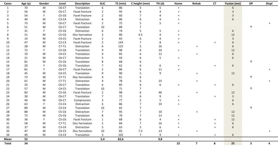
Table 2 Summary of clinicals and radiologicals findings
Cases Age (y) Gender Level Description SLIC TS (min) C height (mm) TH (d) Home Rehab CT Fusion (mo) DP Displ
1 70 M C6-C7 Translation 6 86 5 5 + 6 2 56 M C6-C7 Facet fracture 2 95 6 4 + 3 3 54 F C5-C6 Facet fracture 2 60 7 4 + 3 4 40 M C3-C4 Distraction 6 80 6 4 + 6 5 73 M C6-C7 Facet fracture 2 72 5 5 + + 6 51 M C6-C7 Translation 10 89 7 7 31 F C5-C6 Distraction 6 74 5 5 + 6 8 31 M C5-C6 Disc herniation 3 80 4.5 4 + 12 9 19 M C4-C5 Facet fracture 2 65 6 4 + 3 10 47 M C2-C3 Facet fracture 2 143 7 8 + + 6 11 28 M C7-T1 Distraction 6 125 5 16 + 6 12 72 F C5-C6 Translation 9 98 6 10 + 12 13 19 M C4-C5 Translation 9 85 6 12 + 6 14 21 M C6-C7 Distraction 5 93 6 5 + 6 15 81 M C5-C6 Translation 8 68 6 16 20 F C5-C6 Translation 7 62 6 6 + + 6 17 81 F C6-C7 Facet fracture 2 86 6 11 + + 18 45 M C4-C5 Translation 9 90 5 9 + 12 19 73 M C7-T1 Disc herniation 4 61 6 20 61 M C7-T1 Distraction 4 78 6 25 + + 21 33 M C6-C7 Translation 6 85 6 6 + + 6 22 57 M C4-C5 Translation 10 71 7 23 83 M C5-C6 Facet fracture 2 98 6 46 + 12 24 30 M C6-C7 Translation 7 72 6 9 + + 3 25 46 M C6-C7 Compression 3 77 5 5 + 6 26 63 F C3-C4 Distraction 6 86 7 10 + 6 27 80 M C3-C4 Translation 10 65 7 28 88 M C5-C6 Distraction 7 95 7 10 + 12 29 73 M C5-C6 Translation 8 70 7 13 + 12 30 36 F C5-C6 Facet fracture 1 68 6 4 + + 12 31 58 M C7-T1 Disc herniation 2 84 7 16 + + 6 32 37 M C3-C4 Distraction 5 63 5 6 + + 6 33 47 M C4-C5 Disc herniation 10 83 7.5 13 + + 34 49 M C3-C4 Translation 3 102 7 9 + + 6 Mean 52 5,4 82,6 9,8 Total 34 22 7 8 25 5 4
M Male F female SLIC Subaxial Injury Classification TS Time of Surgery C height Cage height TH Time of Hopitalization Home Patients discharged to home