HAL Id: hal-02365767
https://hal.archives-ouvertes.fr/hal-02365767
Submitted on 15 Nov 2019
HAL is a multi-disciplinary open access archive for the deposit and dissemination of sci-entific research documents, whether they are pub-lished or not. The documents may come from teaching and research institutions in France or abroad, or from public or private research centers.
L’archive ouverte pluridisciplinaire HAL, est destinée au dépôt et à la diffusion de documents scientifiques de niveau recherche, publiés ou non, émanant des établissements d’enseignement et de recherche français ou étrangers, des laboratoires publics ou privés.
LKB1 signaling is activated in CTNNB1 -mutated HCC
and positively regulates β-catenin-dependent CTNNB1
-mutated HCC
Sara Charawi, Pierre-Alexandre Just, Mathilde Savall, Shirley Abitbol,
Massiré Traore, Nolwenn Metzger, Roland Ravinger, Catherine Cavard,
Benoit Terris, Christine Perret
To cite this version:
Sara Charawi, Pierre-Alexandre Just, Mathilde Savall, Shirley Abitbol, Massiré Traore, et al.. LKB1 signaling is activated in CTNNB1 -mutated HCC and positively regulates β-catenin-dependent CTNNB1 -mutated HCC. Journal of Pathology, Wiley, 2019, 247 (4), pp.435-443. �10.1002/path.5202�. �hal-02365767�
LKB1 signaling is activated in CTNNB1-mutated HCC and
positively regulates
-catenin-dependent CTNNB1-mutated HCC
Sara Charawi1,2,3,4†, Pierre-Alexandre Just1,2,3,4,5†, Mathilde Savall1,2,3,4, Shirley Abitbol1,2,3,4, Massiré Traore1,2,3,4,
Nolwenn Metzger1,2,3,4, Roland Ravinger1,2,3,4, Catherine Cavard1,2,3,4, Benoit Terris1,2,3,4,5
and Christine Perret1,2,3,4,5*
1 Development Reproduction Cancer, INSERM, U1016, Institut Cochin, Paris, France 2 Development Reproduction Cancer, CNRS, UMR8104, Paris, France
3 Development Reproduction Cancer, Université Paris Descartes, Paris, France 4 Equipe labellisée LNCC,
5 Department of Pathology, APHP, Hôpitaux Universitaires Paris Centre, Hôpital Cochin, Paris, France
*Correspondence to: C Perret, INSERM U1016, Institut Cochin, 24 rue du Faubourg Saint-Jacques, F-75014 Paris, France. E-mail: christine.perret@inserm.fr
†These authors contributed equally to this work.
Abstract
Hepatocellular carcinomas (HCCs) are known to be highly heterogenous. Within the extensive histopathological and molecular heterogeneity of HCC, tumors with mutations in CTNNB1, encoding -catenin (CTNNB1-mutated HCC), constitute a very homogeneous group. We previously characterized a distinctive metabolic and histological phenotype for CTNNB1-mutated HCC. They were found to be well-differentiated, almost never steatotic, and often cholestatic, with a microtrabecular or acinar growth pattern. Here, we investigated whether LKB1, which controls energy metabolism, cell polarity, and cell growth, mediates the specific phenotype of CTNNB1-mutated HCC. The LKB1 protein was overexpressed in CTNNB1-mutated HCC and oncogenic activation of -catenin in human HCC cells induced the post-transcriptional accumulation of the LKB1 protein encoded by the LKB1 (STK11) gene. Hierarchical clustering, based on the expression of a murine hepatic liver Lkb1 (Stk11) signature in a human public dataset, identified a HCC cluster, composed of almost all the CTNNB1-mutated HCC, that expresses a hepatic liver LKB1 program. This was confirmed by RT-qPCR of an independent cohort of CTNNB1-mutated HCC and the suppression of the LKB1-related profile upon -catenin silencing of CTNNB1-mutated human hepatoma cell lines. Previous studies described an epistatic relationship between LKB1 and CTNNB1 in which LKB1 acts upstream of CTNNB1. Thus, we also analyzed the consequences of Lkb1 deletion on the zonation of hepatic metabolism, known to be the hallmark of -catenin signaling in the liver. Lkb1 was required for the establishment of metabolic zonation in the mouse liver by positively modulating -catenin signaling. We identified positive reciprocal cross talk between the canonical Wnt pathway and LKB1, both in normal liver physiology and during tumorigenesis that likely participates in the amplification of the -catenin signaling by LKB1 and the distinctive phenotype of the CTNNB1-mutated HCC.
Keywords: hepatocellular carcinoma; liver; CTNNB1; LKB1
Introduction
Hepatocellular carcinoma (HCC) is the fifth cause of cancer-related death worldwide (GLOBOCAN 2012 v1.0 database, International Agency for Research on Cancer, Lyon, France, 2013. Available from: http:// globocan.iarc.fr, accessed on 4 June 2018). It is a highly heterogeneous disease, with multiple risk factors and a large spectrum of histopathological features. Large-scale genomic and transcriptomic studies have classified HCC according to various molecular sub- classes, often associated with clinical and phenotypic
traits [1 – 3]. Within HCC, tumors with activating mutations of the gene encoding β-catenin (CTNNB1) (18 – 40% of cases) constitute a very homogeneous subgroup [4]. Indeed, we and others have shown that
CTNNB1-mutated HCCs are well-differentiated, often
cholestatic, chromosomally stable, and associated with a better prognosis than other HCCs [3,4]. Furthermore,
CTNNB1-mutated HCCs are almost never steatotic and
usually display microtrabecular and/or acinar growth patterns. Tumor cells in pseudoglandular or acinar struc- tures show a columnar apicobasal polarity (simple-type polarity) that differs from hepatic hepatocyte-type
polarity [4], which is required for the efficient removal of excreted bile [5]. CTNNB1-mutated HCC is also classified in the nonproliferative molecular subgroup of tumors. In contrast, non-CTNNB1-mutated HCCs dis- play heterogeneous histopathological phenotypes, usu- ally with a macrotrabecular growth pattern and poorer differentiation; steatosis is observed in approximately 30% of cases, and cholestasis is infrequent [1 – 3].
Aside from its oncogenic function in HCC when aber- rantly activated, the Wnt/β-catenin canonical pathway also plays a critical role in liver physiology as it is the master regulator of the establishment of hepatic metabolic zonation [6].
We previously sought candidate genes that could explain the distinctive phenotype of CTNNB1-mutated HCCs and identified liver kinase B1 (LKB1), encoded by STK11. STK11 encodes a serine/threonine kinase that functions as a tumor-suppressor gene. Germline mutations of STK11 are associated with Peutz – Jeghers syndrome (PJS), an autosomal-dominant disorder that is linked to an increased risk of cancer [7,8]. However, somatic STK11 mutations are not commonly found in sporadic cancers, except lung cancer [9]. The active form of LKB1 is found in a heterotrimeric complex with STRAD and MO25, which regulate its stability, kinase activity, and subcellular localization [10]. LKB1 has been called a ‘master kinase’ due to its ability to phosphorylate at least 14 downstream proteins; the AMP-activated protein kinases (AMPK); and 12 pro- teins related to AMPK, called ARK for AMP-related kinase (ARK) [11]. LKB1 regulates multiple biological pathways, including those involved in cell polarity, cell metabolism, and cell growth and proliferation [7,8]. Strikingly, LKB1 is a cell-autonomous determinant of the apical polarity of epithelial cells and can, for example, polarize single, isolated intestinal epithe- lial cells [12]. It negatively controls cell proliferation via the AMPK/mTOR, TP53, and YAP cell growth signaling pathways [7,8]. Finally, LKB1 coordinates the metabolic response to energetic stress, activating catabolic processes, such as fatty acid oxidation, and inhibiting anabolic pathways, such as lipid synthesis [7]. Therefore, the morphologically specific signature of
CTNNB1-mutated HCCs, namely, low proliferation, the
lack of steatosis, and the acquisition of a simple type of polarity, may be consistent with the activation of LKB1 in such tumors. This would confer a counterintuitive oncogenic function to STK11, which is usually con- sidered to be a tumor-suppressor gene. However, this oncogenic function of LKB1 has already been observed in other settings [13,14]. In addition, numerous studies described molecular cross talk between LKB1 and the Wnt/β-catenin signaling pathway [15 – 22].
Here, we showed that LKB1 is overexpressed in CTNNB1-mutated HCC relative to nonmutated HCC and that aberrant activation of β-catenin sig- naling induces post-transcriptional accumulation of LKB1. Using an Lkb1 transcriptomic signature that we recently established using a liver-specific deletion of Lkb1 in embryonic liver [23], we confirmed that
CTNNB1-mutated HCC express an LKB1 program. In
addition, we found that LKB1 is required for specific physiological functions of β-catenin in the liver and exerts a physiological role as a positive modulator of β-catenin-dependent metabolic zonation.
Methods
Tumor specimens
We selected 48 samples from the human HCC cohort of patients treated for liver cancer in Cochin Hospi- tal (Paris, France), previously described by Audard
et al [4], for which we had both frozen tumor and
nontumor liver samples. Half of the tumors were
CTNNB1-mutated, and half were not. All tumor sam-
ples were frozen after surgery in accordance with French laws and ethical guidelines. The frozen samples were used for western blotting analyses. We also selected a validation set of human HCCs to confirm the expression of the hepatic Lkb1 signature in CTNNB1-mutated HCC for which the RNA was already available [4]. The validation set was composed of samples of 18
CTNNB1-mutated HCCs and 22 non-CTNNB1-mutated
HCCs, together with six human normal liver samples. The mutational status of the CTNNB1 gene in these samples has been described previously [4].
Animals
Mice with a specific deletion of Lkb1 in embryonic liver, Lkb1lox,lox;Alfp-Cre mice called LKB1KOLivemb, have already been described [23]. All animal pro- cedures were carried out according to French legal regulations and approved by an ethical commit- tee (CEEA34.CP.077.12). All mice were kept in well-controlled animal housing facilities with free access to tap water and food pellets.
Cell culture and transfection assay
HuH7, HepG2, and HuH6 cells were grown in Dubel- cco’s modified Eagle’s medium (DMEM; Gibco, Life Technologies, Carlsbad, CA, USA) with 4.5 g/l glucose, supplemented with 10% heat-inactivated fetal bovine serum (FBS) and 100 U/ml penicillin/streptomycin at 37 ∘C in 5% CO2.
For siRNA transfections, cells were seeded in six-well plates (0.3 × 10⁶ cells per well) and reverse-transfected with 100 pmol siRNA in the presence of Lipofectamine RNAimax Reagent (Life Technologies) according to the procedure provided by the manufacturer. CTNNB1
Stealth Selected RNAiTM siRNAs were obtained from
Life Technologies (HSS102460, HSS102461, referred as SiRNA1 and siRNA2 in Figure 1, respectively). The StealthTM RNAi negative control duplex (referred to as Scrambled) was used as a negative control for sequence-independent effects. Cells were collected 72 h after transfection. For experiments described in
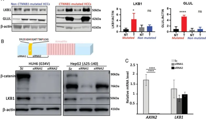
Figure 1. Oncogenic β-catenin drives LKB1 protein expression. (A) LKB1 accumulates in a large subset of CTNNB1-mutated HCCs. Western
blotting analysis of LKB1, GLUL (a β-catenin target gene in the liver), and β-actin (loading control) in HCCs (T), selected based on their CTNNB1 mutational status. NT indicates matched nontumor liver tissue for each HCC sample. Two independent representative samples are shown. Quantification of the western blots for GLUL and LKB1 is shown on the right. Statistical significance was evaluated using a two-sample unpaired Student t-test between KO and WT. ***p < 0.001. **p < 0.01. *p < 0.05. n = 24 in each group. (B) Oncogenic β-catenin drives LKB1 protein expression in HuH6 and HepG2 cells. HuH6 and HepG2 cells were treated with scrambled siRNA (Sc) or various siRNAs against CTNNB1 mRNA (siRNA1, siRNA2) and harvested 72 h after transfection. Western blotting analysis of β-catenin, LKB1, and β-actin (loading control). The diagram above the western blots depicts the localization of the siRNAs along the β-catenin coding sequence and points to the position of siRNA2 in the deleted CTNNB1 sequence in HepG2 cells. (C) β-catenin drives LKB1 expression at the post-transcriptional level. RT-qPCR analysis of AXIN2 and LKB1 gene expression of HUH6 cells transfected with siRNA against CTNNB1 or scrambled siRNA, as described in (B). Mean from three independent experiments. Error bars: standard deviations. Statistical significance was evaluated using a two-sample unpaired Student’s t-test between KO and WT. ****p < 0.001.
Figure 2, we used the SMARTpool technology of Dharmacon to inactivate CTNNB1 in CTNNB1-mutated (HUH6 and HepG2) and not mutated (HUH7) hepatoma cell lines. The siCTNNB1 (siCat) was ON-TARGET plus SMARTpool #L-003482-00-0005 and the scram- bled control (Sc) #D-001810-01-05. A total of 20 pmol siRNA was used per well in six-well plates. Cells were collected 48 h after transfection.
RNA extraction and RT-PCR
Total RNA was extracted from mouse tissues and cell lines with Trizol Reagent (Life Technologies) according to the manufacturer’s protocol. Reverse transcription was performed using 1 μg total RNA and the Tran- scriptor First Strand cDNA Synthesis Kit (Roche Diag- nostics, Life Technologies, Courtaboeuf, France) and random hexamers as primers. Quantitative PCR reac- tions were run using the Light Cycler 480 Sybr Green I Master Kit (Roche Diagnostics) and specific primers (Eurogentec, Eurogentec France SASU, Nantes, France) on a Light Cycler 480 thermocycler (Roche Diagnos- tics). Values were normalized to 18 S ribosomal RNA
levels. For the mouse data, expression of the β-catenin target genes was expressed relative to that in the livers of WT animals (n = 6 animals) as an n-fold ratio. For the human data, gene expression in the tumor samples was compared to the mean level of the corresponding gene expression in normal liver tissues (n = 6), expressed as an n-fold ratio. For human hepatoma cell lines data, gene expression in the siRNA CTNNB1-treated samples was compared to the scrambled-treated samples (n = 3). The relative amount of RNA was calculated using the 2-delta CT method. Primer sequences are shown in supplementary material, Table S1.
Immunoblotting analyses
Total protein extracts from human and mouse liver were obtained from 20 to 200 mg frozen tissue that was bead-mill homogenized in lysis buffer (50 mM
Tris – HCl pH 7.4, 150 mM NaCl, 1 mM EGTA, 1 mM
DTT, 0.1 mM 4-benzenesulfonyl fluoride, and 1% Triton X-100), supplemented with a mixture of protease and phosphatase inhibitors (Roche Diagnostics) in a 10 μl/μg ratio using a TissueLyser disruption system
Figure 2. CTNNB1-mutated HCC express a hepatic Lkb1 program. (A) LKB1 expression profile in CTNNB1-mutated HCC. RT-qPCR was
performed for the positively- (GLUL, SLC13A3, AQP9) and negatively- (SLPI, LSR, RCAN3, GSTP1, SGK1, ELOVL7) LKB1-regulated genes described in supplementary material, Figure S1 using mRNA isolated from normal human liver (N), CTNNB1-mutated HCC (M), and nonmutated HCC (NM). (B) LKB1 expression profile in human hepatoma cell line after β-catenin silencing. β-Catenin silencing was performed using siRNA against CTNNB1 (siCat) and scrambled (Sc) as controls. RT-qPCR was performed for CTNNB1 and the positive (SLC13A3) and negative (SLPI, RCAN3, ELOVL7, GSTP1) LKB1-related genes. HuH6 and HepG2 are CTNNB1-mutated hepatoma cell lines, while HuH7 is not mutated for CTNNB1. *p < 0.05; **p < 0.01; ***p < 0.001.
(Qiagen, Hilder, Germany). Samples were centrifuged at 13 000 × g for 10 min at 4 ∘C, and the supernatants were collected and stored at −80 ∘C until analysis. For human hepatoma cell lines, protein was extracted from the six-well plates using Laemmli buffer (Sigma-Aldrich,
Saint-Quentin Fallavier, France). Proteins were resolved by SDS – PAGE and then transferred to nitrocellulose, which was then blocked using 5% BSA or 5% milk. Blots were incubated with specific primary antibodies overnight at 4 ∘C, washed, incubated with the
corresponding horseradish peroxidase-conjugated secondary antibodies (Cell Signaling Technology, Ozyme, Saint-Quentin en Yvelines, France), and devel- oped by enhanced chemiluminescence (Thermo Fisher Scientific, Waltham, MA, USA). Images were recorded using a 3.2-megapixel super CCD camera driven by a LAS 4000 mini device (GE Healthcare Europe GmBH, Velizy Villacoublay, France) and quantified using Multigauge software from Fujifilm (Fujifilm France, Asnières, France). Expression was standardized to the β-actin level and normalized to a nontumor sample that was set to 1. Primary antibodies were purchased from Cell Signaling Technology [LKB1: clone D60C5, dilu- tion 1/1000; β-actin (8456, dilution 1/2000); GAPDH (97166, dilution 1/500); Gsk3β (4337, dilution 1/1000), phosphoGsk3β (9323 (S9), dilution 1/2000)] and BD Biosciences (BD, Transduction Laboratories, Rungis, France) (β-catenin (811054, dilution 1/2000) and glutamine synthase (GLUL) (610518, dilution 1/2000).
Immunohistochemistry and in situ hybridization
Mouse livers were cut into 3-mm-thick slices, fixed in 10% formalin for 12 h, and embedded in paraffin. For morphological analysis, sections were cut at 2-μm thickness, dewaxed, rehydrated, and stained with hema- toxylin and eosin. The protocols for glutamine syn- thetase immunohistochemistry and in situ hybridization have been described previously [6].Human gene expression analysis
The data for GSE62232, which includes Affymetrix U133plus v2 array (GPL570) data for 81 HCC liver tumors samples, were downloaded in an already nor- malized matrix format via Gene Expression Omnibus. The CTNNB1 mutations in this sample collection were reported in [22,23]. Hierarchical clustering was per- formed using Genesis clustering software as described in [24].
Statistical analysis
Data were analyzed using unpaired t-tests with Welch’s correction in GraphPad Prism 7.04 (GraphPad Inc., San Diego, CA, USA).
Results
Activated LKB1 signaling occurs
in CTNNB1-mutated HCC
We first analyzed LKB1 protein expression in a series of 48 human HCCs, selected based on their CTNNB1 mutational status (24 with CTNNB1 mutations and 24 without). Total proteins were extracted from cry- oconserved tumor samples and matched nontumor liver samples for each patient. The very high expres- sion of GLUL, a well-known β-catenin target in the
liver [25], confirmed activation of the β-catenin path- way in the CTNNB1-mutated tumors. LKB1 protein levels were significantly higher in CTNNB1-mutated HCCs than in nontumor liver tissue, whereas those in non-CTNNB1-mutated HCCs were no different from those in nontumor liver tissue (Figure 1A).
We then investigated whether activated β-catenin was able to stimulate LKB1 protein expression in hepatoma cell lines. HuH6 and HepG2 are two hepatoma cell lines with gain-of-function mutation alterations of
CTNNB1 (a p. G32V mutation and p. 25_140 deletion,
respectively, within the DNA sequence encoding the degradation domain of β-catenin) [26]. In HuH6, two different siRNAs against CTNNB1 mRNA, but not a scrambled siRNA, decreased LKB1 protein expression in parallel with β-catenin inactivation (Figure 1B). This was also true for HepG2 cells with siRNA1, but not siRNA2, which is targeted against a sequence deleted in the HepG2 mutated allele, preventing its inacti- vation (Figure 1B). This result shows that activation of the mutant β-catenin was required for the control of LKB1 protein expression (Figure 1B). While, as expected, RT-qPCR analyses showed that the mRNA levels of the canonical β-catenin target gene AXIN2 were strongly reduced after CTNNB1 inactivation mediated by siRNA, the LKB1 mRNA levels were not affected by β-catenin silencing, indicating that the decrease in LKB1 protein observed in Figure 1B was not associated with a decrease in LKB1 mRNA level (Figure 1C). These results show that the aberrant activa- tion of β-catenin signaling induced the accumulation of LKB1 protein in liver tumor cells, which was regulated post-transcriptionally.
We then explored whether CTNNB1-mutated HCC expressed an LKB1 program. This was performed using the liver Lkb1 signature of 253 genes that we recently obtained from the study of mice bearing a specific Lkb1 deletion in embryonic liver hepatoblasts, Lkb1lox,lox;
Alfp-Cre mice, designated LKB1KOLivemb [23]. We
studied the human HCC public dataset (GSE62232, 81 samples), for which data on the genetic status of CTNNB1 and the RNA expression profile were available (described in reference [24]). Unsupervised hierarchical clustering of the GSE62232 human HCC dataset, using the hepatic Lkb1 signature, grouped almost all CTNNB1-mutated HCCs within a single cluster, showing that most CTNNB1-mutated HCCs share a hepatic Lkb1 program (see supplementary material, Figure S1). The hepatic Lkb1 signature was composed of genes up- and downregulated in the liver of KO versus WT animals and accounted for a program suppressed by Lkb1 in mouse hepatocytes. Thus, most (19/27) CTNNB1-mutated HCCs displayed activated LKB1 signaling (see supplementary mate- rial, Figure S1). We confirmed these results using an independent cohort of human HCCs, consisting of
CTNNB1-mutated and non-CTNNB1-mutated tumors,
by RT-qPCR. The expression of several LKB1-related genes was statistically enhanced (GLUL, SLC13A3,
SGK1, ELOVL7) in CTNNB1-mutated HCCs relative
to nonmutated HCCs (Figure 2A). We validated the cross talk between β-catenin and LKB1 by analyzing the LKB1 gene expression profile in CTNNB1-mutated (HuH6, HepG2) and nonmutated (HuH7) HCC cell lines after β-catenin silencing using siRNA. Although some genes were discarded as they were not expressed in the hepatoma cell lines, the expression of the positive and negative LKB1-related genes were significantly repressed or enhanced, respectively, after β-catenin silencing in CTNNB1-mutated HCC cell lines but were not affected in the nonmutated HCC cell line (Figure 2B).
Overall, our data show that β-catenin signaling in
CTNNB1-mutated HCCs induces a post-transcriptional
accumulation of LKB1, which in turn activates an LKB1 program likely contributing to the specific histological appearance of CTNNB1-mutated HCCs.
LKB1 positively regulates
β-catenin signaling
in liver zonation
Cross talk between Wnt/β-catenin and LKB1 signal- ing has been described in several studies, with often conflicting results [15 – 22]. The first pioneering study showed LKB1 to be upstream of β-catenin signaling and to be required for full activation of β-catenin signaling in Xenopus development [15]. We explored whether LKB1 may control Wnt/β-catenin signal- ing in the mouse liver in a physiological situation in which the canonical Wnt pathway is known to have a critical function, such as metabolic zonation [6]. We thus examined liver zonation in Lkb1-deficient mice (LKB1KOLivemb) [23]. Immunohistochemistry and in
situ hybridization for Glul (glutamine synthase) and Arg1 (arginase 1), together with immunohistochemistry
for Lect2 (Leukocyte cell-derived chemotaxin 2) and Hal (histidine ammonia-lyase), showed that Lkb1 dele- tion strongly affected liver zonation in LKB1KOLivemb mice, with almost complete loss of the perivenous expression of Glul and Lect2, while the periportally expressed gene Arg1 and Hal protein were overex- pressed (Figure 3A). Accordingly, analysis of a 16-gene signature of liver β-catenin target genes confirmed decreased Wnt/β-catenin signaling in LKB1KOLivemb mice relative to controls. This decrease in β-catenin activity was confirmed by RT-qPCR using a different group of animals (Figure 3B,C). These results show that
Lkb1 is a positive modulator of Wnt/β-catenin signaling
during liver development and is required for the full establishment of liver zonation.
Previous studies also reported that LKB1 controls the Wnt/β-catenin pathway through two different kinases, GSK3β and PAR1A (MARK3) [15 – 17]. We analyzed the phosphorylation status of GSK3β (pS9-GSK3β) in the livers of LKB1KOLivemb mice by western blotting and observed higher levels of pS9-GSK3β in the mutant mice than in controls (Figure 3D). We were unable to detect phosphorylation of Par1a (Mark3) or any other Mark (data not shown). The decrease in β-catenin
signaling activity in LKBKOLivemb mice did not corre- late with increased phosphorylation of GSK3β on S9 (known to inhibit GSK3β function), normally associated with increased β-catenin activity. These results suggest that Lkb1 controls the canonical Wnt pathway in hepa- tocytes, independent of GSK3β.
Discussion
Wnt/β-catenin signaling regulates critical functions in liver physiology, and its aberrant activation is found in several liver diseases, including HCC [27]. HCCs generally display a high level of het- erogeneity [28]. However, we previously showed that
CTNNB1-mutated HCCs form a homogeneous group
of tumors, with specific cell metabolism, polarity, and growth hallmarks [4]. Here, we investigated whether the activation of LKB1 signaling could explain the specific phenotype of CTNNB1-mutated HCC as LKB1 is considered to be a master regulator of cell growth, metabolism, survival, and polarity [7,8]. We showed that
CTNNB1-mutated HCCs display significantly higher
LKB1 protein expression than non-CTNNB1-mutated HCCs. CTNNB1-mutated HCCs expressed a hepatic
LKB1 signature that we recently identified in mouse Lkb1-deficient livers [23]. Reciprocal positive control
of the canonical Wnt pathway by Lkb1 during devel- opment was previously described [15]. This led us to investigate whether Lkb1 controls liver metabolic zona- tion, a well-established function of β-catenin signaling in the liver [6]. Our data show that Lkb1 is required for the full activation of β-catenin signaling in mouse liver and is thus a positive regulator of the establishment of metabolic zonation.
Cross talk between Wnt/β-catenin and LKB1 signal- ing in both directions (β-catenin toward LKB1 and LKB1 toward β-catenin) has been described in many studies. It appears that the dialog depends on the context, developmental or oncogenic, with either a positive or a negative impact [15 – 22]. The molecular mechanisms involved are yet to be determined for all contexts. Here, we showed that oncogenic β-catenin signaling induced the accumulation of LKB1 protein in CTNNB1-mutated HCC. The regulation of LKB1 is complex and still not well characterized. It is controlled epigenetically, transcriptionally, and post-translationally through the modulation of protein stability [29]. Little is known concerning the mechanisms that regulate the stability of LKB1 protein: the ubiquitin ligase CHIP has been proposed to be involved [30], but this still needs to be confirmed. A recent study reported overexpression of
LKB1 in human HCC that predicts poor survival out-
comes. However, this was at the mRNA level and, thus, does not coincide with our study in which we showed accumulation of the LKB1 protein in CTNNB1-mutated HCC [31]. For the inverse relationship (control of β-catenin signaling by Lkb1), we showed that deletion of
Figure 3. Lkb1 positively regulates the Wnt canonical pathway and controls liver zonation. (A) Representative images for the expression of
positively regulated β-catenin target genes, Glul, by immunohistochemistry and in situ hybridization and Lect2 by immunohistochemistry
together with negatively regulated β-catenin target genes, Arg1, by in situ hybridization and Hal by immunohistochemistry. LKB1KOlivemb
mutant and control and mice were 3 weeks old. (B) Heat map of the liver β-catenin target gene signature from the gene expression profile
of LKBKOLivemb mice [23]. (C) RT-qPCR analysis of various positively (Axin2, Sp5, Glul, Lect2, Cyp2e1, Nkd1) regulated β-catenin target
genes in 14-day-old control and LKBKOlivemb mutant mice. n = 4 – 6 per group. Error bars: SEM. Statistical significance was evaluated using
a two-sample unpaired Student’s t-test between KO and WT. *p < 0.05, **p < 0.01, ***p < 0.001. (D) Western blotting analysis showing
increased pS9Gsk3β phosphorylation in mutant LKBKOLivemb mice relative to controls.
in liver zonation controlled by Wnt/β-catenin. This was associated with increased phosphorylation of Gsk3β (S9-Gsk3β), leading to its inactivation. This result does not correlate with the decreased activation of β-catenin signaling we observed in the mutant mice and suggests that Lkb1 controls β-catenin signaling independent of Gsk3β. Overall, we identified positive reciprocal cross talk between β-catenin and LKB1, in which β-catenin activity induces LKB1 expression, leading to the acti- vation of LKB1 signaling, which in turn positively regulates β-catenin signaling. Such cross talk likely results in the amplification of β-catenin signaling by
LKB1 in CTNNB1-mutated HCC.
A positive role for LKB1 signaling in CTNNB1-mutated HCC could suggest a synthetic lethal interaction between the Wnt pathway and LKB1. Although we have not been able to substantiate such an interaction (data not shown), a prosurvival role of LKB1
has already been described. Indeed, Lkb1-deficient mouse embryonic fibroblasts showed resistance to transformation by oncogenes, suggesting that the high energetic demands of malignant transformation may be incompatible with Lkb1 loss [32]. Indeed, tumor cells must sense changes in their microenvironment to support unfettered cell proliferation, and the inability to respond to such environmental cues may lead to energetic stress and ultimately cell death. LKB1 is an energetic sensor, and its function is required for the survival of tumor cells under energy stress [13]. Accord- ingly, LKB1 is upregulated in human skin cancer cells and has been shown to be required for preventing apop- tosis in several human cancer cell lines overexpressing AKT [33]. Indeed, the prosurvival role of Lkb1 is not restricted to tumor cells but has also been described for quiescent hematopoietic stem cells [34 – 36]. Overall, these data show that LKB1 may provide cancer cells with
the metabolic flexibility required to survive under condi- tions of nutrient stress, thus conferring a growth advan- tage. LKB1 is likely to play this role in CTNNB1-mutated HCC. Furthermore, we recently showed that, in
CTNNB1-mutated HCC, the tumor cells use fatty
acids for energy, corroborating the lack of steatosis of those tumors [37]. This correlates with the positive role of LKB1 on fatty acid oxidation in the liver [7].
In addition to the function of LKB1 in metabolism,
LKB1 signaling also controls cell polarity, inducing
apicobasal polarity in a cell-autonomous manner [12]. This could partially explain the cholestatic phenotype of some CTNNB1-mutated HCCs, mainly those with pseu- doglandular structures [4]. The polarity present in acinar structures differs from the hepatic polarity required for the removal of bile excreted by hepatic cells [5]. It is possible that, in CTNNB1-mutated HCC, bile is secreted at the apical pole of tumor cells and accumulates in the lumen of acinar structures and cannot be removed from the tumor, leading to cholestasis. LKB1 has been reported to be involved in the establishment of liver cell polarity [23,38]. However, the cholestasis observed in
CTNNB1-mutated HCC is also linked to the positive
effect of β-catenin signaling on bile acid metabolism that we previously described [39].
In CTNNB1-mutated HCC, LKB1 may act as a tumor-suppressor gene, conferring a better prognosis, apicobasal polarity, and a well-differentiated growth pattern, but may also be required for tumor cell survival, acting more likely as an oncogene. Increased genomic instability is one of the various mechanisms by which
LKB1 loss leads to tumor development [40]. Accord-
ingly, we recently showed that Lkb1 inhibits liver cell proliferation and favors mitotic integrity [41] that could explain why CTNNB1-mutated HCCs belong to the chromosomally stable HCC subclass [28].
We have demonstrated reciprocal cross talk between LKB1 and β-catenin, in which β-catenin enhances LKB1 protein expression, favoring expression of the hepatic Lkb1 program that enhances β-catenin signaling. In the context of CTNNB1-mutated HCCs, such cross talk results in a positive activation loop that amplifies the β-catenin signaling.
Acknowledgements
We are very thankful to Christelle Desbois-Mouthon for her help in management of human hepatoma cell lines. We also thank people of the team for their critical discussions and people at the Animal Facility of Cochin Institute who took care of the mice.
Author contributions statement
CP, PAJ, and SC conceived the study, carried out exper- iments and analyzed data. MS, SA, MT, NM, and RT carried out experiments. CC and BT characterized HCC at the genomic level. All authors were involved
in writing the paper and had final approval of the submitted version.
References
1. Boyault S, Rickman DS, de Reynies A, et al. Transcriptome classi- fication of HCC is related to gene alterations and to new therapeutic targets. Hepatology 2007; 45: 42 – 52.
2. Hoshida Y, Nijman SM, Kobayashi M, et al. Integrative transcrip- tome analysis reveals common molecular subclasses of human hepa- tocellular carcinoma. Cancer Res 2009; 69: 7385 – 7392.
3. Calderaro J, Couchy G, Imbeaud S, et al. Histological subtypes of hepatocellular carcinoma are related to gene mutations and molecular tumour classification. J Hepatol 2017; 67: 727 – 738.
4. Audard V, Grimber G, Elie C, et al. Cholestasis is a marker for hep- atocellular carcinomas displaying beta-catenin mutations. J Pathol 2007; 212: 345 – 352.
5. Treyer A, Müsch A. Hepatocyte polarity. Compr Physiol 2013; 3: 243 – 287.
6. Benhamouche S, Decaens T, Godard C, et al. Apc tumor suppressor gene is the “zonation-keeper” of mouse liver. Dev Cell 2006; 10: 759 – 770.
7. Shackelford DB, Shaw RJ. The LKB1-AMPK pathway: metabolism and growth control in tumour suppression. Nat Rev Cancer 2009; 9: 563 – 575.
8. Jansen M, Ten Klooster JP, Offerhaus GJ, et al. LKB1 and AMPK family signaling: the intimate link between cell polarity and energy metabolism. Physiol Rev 2009; 89: 777 – 798.
9. Sanchez-Cespedes M. A role for LKB1 gene in human cancer beyond the Peutz– Jeghers syndrome. Oncogene 2007; 26: 7825 – 7832. 10. Boudeau J, Baas AF, Deak M, et al. MO25alpha/beta interact with
STRADalpha/beta enhancing their ability to bind, activate and local- ize LKB1 in the cytoplasm. EMBO J 2003; 22: 5102 – 5114. 11. Alessi DR, Sakamoto K, Bayascas JR. LKB1-dependent signaling
pathways. Annu Rev Biochem 2006; 75: 137 – 163.
12. Baas AF, Kuipers J, van der Wel NN, et al. Complete polarization of single intestinal epithelial cells upon activation of LKB1 by STRAD.
Cell 2004; 116: 457 – 466.
13. Shaw RJ, Kosmatka M, Bardeesy N, et al. The tumor suppressor LKB1 kinase directly activates AMP-activated kinase and regulates apoptosis in response to energy stress. Proc Natl Acad Sci U S A 2004;
101: 3329 – 3335.
14. Hardie DG. The LKB1-AMPK pathway-friend or foe in cancer?
Cancer Cell 2013; 23: 131 – 132.
15. Ossipova O, Bardeesy N, Depinho RA, et al. LKB1 [XEEK1] regu- lates Wnt signalling in vertebrate development. Nat Cell Biol 2003;
5: 889 – 894.
16. Sun TQ, Lu B, Feng JJ, et al. PAR-1 is a dishevelled-associated kinase and a positive regulator of Wnt signalling. Nat Cell Biol 2001;
3: 628 – 636.
17. Spicer J, Rayter S, Young N, et al. Regulation of the Wnt signalling component PAR1A by the Peutz– Jeghers syndrome kinase LKB1.
Oncogene 2003; 22: 4752 – 4756.
18. Liu W, Monahan KB, Pfefferle AD, et al. LKB1/STK11 inactiva- tion leads to expansion of a prometastatic tumor subpopulation in melanoma. Cancer Cell 2012; 21: 751 – 764.
19. Ma LG, Bian SB, Cui JX, et al. LKB1 inhibits the proliferation of gastric cancer cells by suppressing the nuclear translocation of Yap and beta-catenin. Int J Mol Med 2016; 37: 1039 – 1048.
20. Pearson HB, McCarthy A, Collins CM, et al. Lkb1 deficiency causes prostate neoplasia in the mouse. Cancer Res 2008; 68: 2223 – 2232. 21. Wang J, Zhang K, Wang J, et al. Underexpression of LKB1 tumor
suppressor is associated with enhanced Wnt signaling and malig- nant characteristics of human intrahepatic cholangiocarcinoma.
22. Jacob LS, Wu X, Dodge ME, et al. Genome-wide RNAi screen reveals disease-associated genes that are common to Hedgehog and Wnt signaling. Sci Signal 2011; 4: ra4.
23. Just PA, Poncy A, Charawi S, et al. LKB1 and notch pathways interact and control biliary morphogenesis. PLoS One 2015; 10: e0145400.
24. Abitbol S, Dahmani R, Coulouarn C, et al. AXIN deficiency in human and mouse hepatocytes induces hepatocellular carcinoma in the absence of beta-catenin activation. J Hepatol 2018; 68: 1203 – 1213.
25. Cadoret A, Ovejero C, Terris B, et al. New targets of beta-catenin signaling in the liver are involved in the glutamine metabolism.
Oncogene 2002; 21: 8293 – 8301.
26. de La Coste A, Romagnolo B, Billuart P, et al. Somatic mutations of the beta-catenin gene are frequent in mouse and human hepatocellular carcinomas. Proc Natl Acad Sci U S A 1998; 95: 8847 – 8851. 27. Monga SP. Beta-catenin signaling and roles in liver homeostasis,
injury, and tumorigenesis. Gastroenterology 2015; 148: 1294 – 1310. 28. Zucman-Rossi J, Villanueva A, Nault JC, et al. Genetic landscape
and biomarkers of hepatocellular carcinoma. Gastroenterology 2015;
149: 1226 – 1239 e4.
29. Gan RY, Li HB. Recent progress on liver kinase B1 [LKB1]: expres- sion, regulation, downstream signaling and cancer suppressive func- tion. Int J Mol Sci 2014; 15: 16698 – 16718.
30. Gaude H, Aznar N, Delay A, et al. Molecular chaperone complexes with antagonizing activities regulate stability and activity of the tumor suppressor LKB1. Oncogene 2012; 31: 1582 – 1591.
31. Lee SW, Li CF, Jin G, et al. Skp2-dependent ubiquitination and activation of LKB1 is essential for cancer cell survival under energy stress. Mol Cell 2015; 68: 1203 – 1213.
32. Bardeesy N, Sinha M, Hezel AF, et al. Loss of the Lkb1 tumour suppressor provokes intestinal polyposis but resistance to transfor- mation. Nature 2002; 419: 162 – 167.
33. Zhong D, Liu X, Khuri FR, et al. LKB1 is necessary for Akt-mediated phosphorylation of proapoptotic proteins. Cancer Res 2008; 68: 7270 – 7277.
34. Gurumurthy S, Xie SZ, Alagesan B, et al. The Lkb1 metabolic sensor maintains haematopoietic stem cell survival. Nature 2010;
468: 659 – 663.
35. Gan B, Hu J, Jiang S, et al. Lkb1 regulates quiescence and metabolic homeostasis of haematopoietic stem cells. Nature 2010; 468: 701 – 704.
36. Nakada D, Saunders TL, Morrison SJ. Lkb1 regulates cell cycle and energy metabolism in haematopoietic stem cells. Nature 2010; 468: 653 – 658.
37. Senni N, Savall M, Cabrerizo Granados D, et al. β-catenin-activated hepatocellular carcinomas are addicted to fatty acids. Gut 2018. https://doi.org/10.1136/gutjnl-2017-315448.
38. Fu D, Wakabayashi Y, Ido Y, et al. Regulation of bile canalicular net- work formation and maintenance by AMP-activated protein kinase and LKB1. J Cell Sci 2010; 123: 3294 – 3302.
39. Gougelet A, Torre C, Veber P, et al. T-cell factor 4 and beta-catenin chromatin occupancies pattern zonal liver metabolism in mice.
Hepatology 2014; 59: 2344 – 2357.
40. Shorning BY, Clarke AR. Energy sensing and cancer: LKB1 function and lessons learnt from Peutz– Jeghers syndrome. Semin Cell Dev
Biol 2016; 52: 21 – 29.
41. Maillet V, Boussetta N, Leclerc J, et al. LKB1 as a gatekeeper of hep- atocyte proliferation and genomic integrity during liver regeneration.