HAL Id: inserm-01724235
https://www.hal.inserm.fr/inserm-01724235
Submitted on 6 Mar 2018
HAL is a multi-disciplinary open access
archive for the deposit and dissemination of
sci-entific research documents, whether they are
pub-lished or not. The documents may come from
teaching and research institutions in France or
abroad, or from public or private research centers.
L’archive ouverte pluridisciplinaire HAL, est
destinée au dépôt et à la diffusion de documents
scientifiques de niveau recherche, publiés ou non,
émanant des établissements d’enseignement et de
recherche français ou étrangers, des laboratoires
publics ou privés.
AMPK Re-Activation Suppresses Hepatic Steatosis but
its Downregulation Does Not Promote Fatty Liver
Development
Nadia Boudaba, Allison Marion, Camille Huet, Rémi Pierre, Benoit Viollet,
Marc Foretz
To cite this version:
Nadia Boudaba, Allison Marion, Camille Huet, Rémi Pierre, Benoit Viollet, et al.. AMPK
Re-Activation Suppresses Hepatic Steatosis but its Downregulation Does Not Promote Fatty Liver
De-velopment. EBioMedicine, Elsevier, 2018, 28, pp.194 - 209. �10.1016/j.ebiom.2018.01.008�.
�inserm-01724235�
Research Paper
AMPK Re-Activation Suppresses Hepatic Steatosis but its
Downregulation Does Not Promote Fatty Liver Development
Nadia Boudaba
a,b,c, Allison Marion
a,b,c, Camille Huet
a,b,c, Rémi Pierre
a,b,c, Benoit Viollet
a,b,c, Marc Foretz
a,b,c,⁎
aINSERM, U1016, Institut Cochin, Paris 75014, France
bCNRS, UMR8104, Paris 75014, France c
Université Paris Descartes, Sorbonne Paris Cité, Paris 75014, France
a b s t r a c t
a r t i c l e i n f o
Article history:
Received 10 November 2017 Received in revised form 5 January 2018 Accepted 5 January 2018
Available online 9 January 2018
Nonalcoholic fatty liver disease is a highly prevalent component of disorders associated with disrupted energy homeostasis. Although dysregulation of the energy sensor AMP-activated protein kinase (AMPK) is viewed as a pathogenic factor in the development of fatty liver its role has not been directly demonstrated. Unexpectedly, we show here that liver-specific AMPK KO mice display normal hepatic lipid homeostasis and are not prone to fatty liver development, indicating that the decreases in AMPK activity associated with hepatic steatosis may be a consequence, rather than a cause, of changes in hepatic metabolism. In contrast, we found that pharmaco-logical re-activation of downregulated AMPK in fatty liver is sufficient to normalize hepatic lipid content. Mech-anistically, AMPK activation reduces hepatic triglyceride content both by inhibiting lipid synthesis and by stimulating fatty acid oxidation in an LKB1-dependent manner, through a transcription-independent mecha-nism. Furthermore, the effect of the antidiabetic drug metformin on lipogenesis inhibition and fatty acid oxida-tion stimulaoxida-tion was enhanced by combinaoxida-tion treatment with small-molecule AMPK activators in primary hepatocytes from mice and humans. Overall, these results demonstrate that AMPK downregulation is not a trig-gering factor in fatty liver development but in contrast, establish the therapeutic impact of pharmacological AMPK re-activation in the treatment of fatty liver disease.
© 2018 The Authors. Published by Elsevier B.V. This is an open access article under the CC BY-NC-ND license (http://creativecommons.org/licenses/by-nc-nd/4.0/).
Keywords: AMPK Lipid metabolism
Nonalcoholic fatty liver disease (NAFLD) Fatty liver treatment
Metformin
Small-molecule AMPK activators
1. Introduction
Nonalcoholic fatty liver disease (NAFLD) is the most prevalent chronic liver disease worldwide, increasing rapidly with the expanding burden of obesity and insulin resistance (Cohen et al., 2011). However, the precise mechanisms leading to the aberrant accumulation of triglyc-erides (TG) in the liver remain poorly understood, and therapeutic ap-proaches are limited. In this context, the energy sensor AMP-activated protein kinase (AMPK) has recently garnered much attention for its ability to coordinate multiple metabolic pathways, including hepatic lipid metabolism (Viollet et al., 2009). AMPK inhibits lipogenesis by phosphorylation of acetyl-CoA carboxylase 1 (ACC1) at Ser79 and ACC2 at Ser212, key rate controlling enzymes in malonyl-CoA synthesis. Malonyl-CoA is both a critical precursor for fatty acid biosynthesis and a potent allosteric inhibitor of long-chain fatty acyl-CoA transport into mitochondria forβ-oxidation during the carnitine palmitoyltransferase 1 (CPT1) step. AMPK-mediated ACC inhibition leads to a decrease in in-tracellular malonyl-CoA levels, relieving CPT1 inhibition, resulting in an
increase in fatty acid oxidation. In addition to these short-term effects, AMPK inhibits the transcription of lipogenic genes by phosphorylating transcription factors, such as sterol regulatory element binding protein-1c (SREBP-1c) (Li et al., 2011) and carbohydrate-responsive element-binding protein (ChREBP) (Kawaguchi et al., 2002). Although AMPK has been implicated in the control of lipid partitioning between the biosynthetic and oxidative pathways in the liver in conditions of stress, its physiological relevance in normal conditions has not been for-mally investigated in vivo.
In light of the multiple effects of AMPK on lipid metabolism, it has been suggested that the impairment of hepatic AMPK activity is a key pathological event in the development of the insulin resistance and metabolic disorders associated with metabolic syndrome, including he-patic steatosis (Ruderman et al., 2013). This hypothesis is supported by the observation of low levels of hepatic AMPK activity in various rodent fatty liver models (Muse et al., 2004; Yu et al., 2004). The inhibition of AMPK may stimulate anabolic pathways, such as lipid synthesis, and at-tenuate catabolic pathways, such asβ-oxidation. Interestingly, hepatic AMPK is downregulated by hyperglycemia, the saturated fatty acid pal-mitate, branched-chain amino acids, the adipocyte-derived cytokine resistin and insulin, and this downregulation is associated with nutrient overload and the development of insulin resistance (Kraegen et al., 2006; Li et al., 2010; Mankouri et al., 2010; Muse et al., 2004; Wu
⁎ Corresponding author at: Institut Cochin, Inserm U1016, CNRS UMR8104, Université Paris Descartes, Department of Endocrinology, Metabolism and Diabetes, 24, rue du Faubourg Saint-Jacques, F-75014 Paris, France.
E-mail address:marc.foretz@inserm.fr(M. Foretz).
Contents lists available atScienceDirect
EBioMedicine
j o u r n a l h o m e p a g e :w w w . e b i o m e d i c i n e . c o m
https://doi.org/10.1016/j.ebiom.2018.01.008
et al., 2007). Furthermore, AMPK inhibition is also associated with the hepatic fat accumulation induced by chemical agents, such as ethanol, sidestream cigarette smoke and orotic acid (Jung et al., 2011; You et al., 2004; Yuan et al., 2009). Despite the recognized correlation be-tween fatty liver and AMPK downregulation, it remains unknown whether AMPK plays a causal role in the excessive accumulation of TGs in the liver or whether its downregulation is secondary to the in-crease in lipid levels or associated complications.
Conversely, by inhibiting lipid synthesis and activating fatty acid ox-idation, AMPK activation in the liver would be expected to decrease lipid accumulation. AMPK is, thus, a highly attractive target for hepatic steatosis management. Consistent with this notion, a large number of AMPK-activating compounds have been reported to have beneficial ef-fects for fatty liver treatment (Smith et al., 2016b; Viollet et al., 2009). In particular, metformin, a widely used antidiabetic drug, markedly de-creases hepatic steatosis in rodents (Lin et al., 2000), presumably by ac-tivating hepatic AMPK (Fullerton et al., 2013; Zhou et al., 2001). AMPK activation by AICAR (Bergeron et al., 2001; Yu et al., 2004), berberine (Kim et al., 2009) or the small-molecule direct AMPK activators A-769662 (Cool et al., 2006), C13 (Gomez-Galeno et al., 2010) and PF-249 and PF-739 (Cokorinos et al., 2017), has also been shown to de-crease liver fat content in obese rodents, highlighting the potential of AMPK activation for therapeutic interventions in the liver. However, most of these AMPK agonists are known to display AMPK-independent effects and may therefore interfere with various biological pathways unrelated to AMPK (Benziane et al., 2009; Foretz et al., 2010; Guigas et al., 2006; Hasenour et al., 2014; Moreno et al., 2008). The ex-tent to which specific hepatic AMPK activation can alleviate fatty liver thus remains unclear. Indeed, it cannot be excluded that at least some of the metabolic effects of AMPK-activating drugs in vivo could be me-diated independently of AMPK activation in the liver. Hence, pharmaco-logical studies should be combined with genetic studies examining the role of AMPK in mediating the observed pharmacological outcomes. To our knowledge, no study has employed genetic mouse models with spe-cific AMPK deletion in the liver to investigate the effect of direct AMPK activators on NAFLD.
In this study, we generated liver AMPK-deficient mice, to investigate the consequences of AMPK loss on the development of fatty liver dis-ease and to assess the AMPK-dependent action of indirect and direct AMPK activators in the reduction of hepatic steatosis. We found that a deficiency of AMPK in the liver was not sufficient to trigger hepatic lipid accumulation, indicating that AMPK dysfunction is not a causal fac-tor leading to the development of fatty liver disease. We also obtained genetic evidence that pharmacological AMPK activation decreased he-patic TG content through direct effects on lipogenesis andβ-oxidation rates, rather than changes in lipogenic gene expression profile. Finally, we showed that small-molecule AMPK activators efficiently inhibited lipogenic flux alone or in combination with metformin in human hepatocytes. Thus, ourfindings demonstrate the potential of pharmaco-logical AMPK activation for therapeutic interventions in fatty liver disease.
2. Materials and Methods 2.1. Study Approvals
All animal studies were approved by the Paris Descartes University ethics committee (no. CEEA34.BV.157.12) and the Direction Départementale des Services Vétérinaires of the Préfecture de Police de Paris (authorization no. 75-886).
2.2. Animals
All mice were maintained in a barrier facility under a 12-h light/12-h dark cycle (8 am-8 pm) with free access to water and standard mouse diet (in terms of energy: 65% carbohydrate, 11% fat, 24% protein).
AMPKα1 floxed mice and AMPKα1 total knockout mice were gener-ated, as described below. AMPKα2 floxed and AMPKα2 total knockout mice have been described elsewhere (Viollet et al., 2003). The genera-tion of AMPKγ1 total knockout mice has been described previously (Foretz et al., 2011). Liver double-knockout of AMPKα1 and AMPKα2 catalytic subunits was achieved by crossing AMPKα1lox/loxmice with AMPKα2lox/loxmice and then Alfp-Cre transgenic mice (kindly provided by François Tronche, Université Pierre et Marie Curie, Paris), all on the C57BL/6 background, to generate AMPKα1lox/lox
,α2lox/lox
(control) and AMPKα1lox/lox, α2lox/lox-Alfp-Cre (liver AMPK-deficient mice). Liver-specific LKB1 knockout mice were generated by crossing LKB1-lox/lox
mice (kindly provided by Ronald DePinho, Harvard University, USA) with tamoxifen-inducible albumin-Cre-ERT2 mice (kindly pro-vided by Daniel Metzger, IGBMC, France). Twelve-week-old LKB1lox/ lox-Alb-Cre-ERT2 mice were treated with vehicle (sunflower oil contain-ing 10% ethanol; control) or tamoxifen (Sigma) at a dose of 1 mg/mouse, injected intraperitoneally in afinal volume of 100 μl, over five consecu-tive days. Mice were studied or used for primary hepatocyte isolation three weeks after the start of tamoxifen administration. aP2-nSREBP-1c transgenic mice (Shimomura et al., 1998) were purchased from the Jackson Laboratory. aP2-nSREBP-1c transgenic males (C57BL/6J × SJL background) were crossed with C57BL/6J females. We studied the aP2-nSREBP-1c mice and their control littermates generated from this cross. aP2-nSREBP-1c transgenic mice were crossed with AMPKα1lox/ lox
,α2lox/lox
-Alfp-Cre mice to generate aP2-nSREBP-1ctg/wt-AMPKα1lox/ lox,α2lox/loxand aP2-nSREBP-1ctg/wt-AMPKα1lox/lox,α2lox/lox-Alfp-Cre mice. C57BL/6J mice were obtained from Harlan France. Eight-week-old control AMPKα1α2 floxed and (liver AMPK-deficient mice) were fed a high-fat diet (in terms of energy: 45% fat, 35% carbohydrate, 20% protein) (Research Diets, #D12451) for 5 months to induce a diet-induced obesity (DIO).
2.3. Gene Targeting and Generation of AMPKα1 (Prkaa1)-Knockout Mice The Prkaa1 targeting construct was generated from PCR products amplified from the DNA of 129/Sv ES cells. The 5′ and 3′ homology arms, which were 3.8 and 3.5 kb long, respectively, were inserted on ei-ther side of a PGK promoter-driven hygromycin selection cassette flanked by FRT sites and containing a 3′ loxP site, in the pL3-FRT-Hygro vector. A 2.1 kb fragment of genomic DNA bearing exons 4 and 5, partly encoding the catalytic domain, including the phosphorylation site Thr172 within the activation loop (corresponding to amino acids 113-190), with a 5′ loxP site, was introduced between the hygromycin resistance cassette and the 5′ homology arm (Supplemental Fig. S1A). Exponentially growing 129/SV CK35 embryonic stem cells were electroporated with the linearized target DNA construct and selected on plates containing hygromycin. The targeted clones were identified by PCR across both homology arms, with confirmation by Southern blot analysis. Cell populations expanded from the targeted clones were injected into C57BL/6 blastocysts, and animals displaying germline transmission were mated with C57BL/6 mice. The hygromycin resis-tance cassetteflanked by FRT sites was excised by crossing AMPKα1+/ loxmice with FLP-expressing mice. The resulting heterozygous offspring were backcrossed onto the C57BL/6J background for at least four gener-ations. These mice were crossed to generate AMPKα1lox/loxmice. Exons 4 and 5,flanked by loxP sites, were disrupted by crossing AMPKα1+/lox mice with deleter EIIa-CRE transgenic mice to produce AMPKα1+/− mice. These mice were crossed to generate wild-type (control) and AMPKα1−/−mice. Routine genotyping was carried out by multi-plex PCR with phire hot start II DNA polymerase (Thermo Scientific) on tail DNA with the P1 (5′-tattgctgccattaggctac-3′), P2 (5′-gacctgacagaataggatatgcccaacctc-3′) and P3 (5′-attaaacaccactaatt ggaaaacattccc-3′) primers, to yield amplification products of 586 bp (WT allele) and 682 bp (floxed allele) with P1/P2 and of 2365 bp (WT allele) and 348 bp (KO allele) with P3/P2 (Supplemental Fig. S1B).
2.4. Treatment of Mice with A-769662
Diet-induced obese (DIO) control AMPKα1α2 floxed and (liver AMPK-deficient mice) were treated with vehicle (0.2% hydroxypropyl methyl-cellulose, i.p., b.i.d.) or A-769662 (30 mg/kg, i.p., b.i.d.), and standard-diet-fed control AMPKα1α2 floxed and (liver AMPK-deficient mice) were treated with vehicle (i.p., b.i.d.) for 5 days. aP2-nSREBP-1c transgenic mice were treated with vehicle (i.p., b.i.d.) or A-769662 (30 mg/kg, i.p., b.i.d.), and non-transgenic littermates were treated with vehicle (i.p., b.i.d.) for 7 or 8 days, as indicated in thefigure legends. For the measurement of fatty acid synthesis in vivo, control AMPKα1α2 floxed and (liver AMPK-deficient mice) received intraperi-toneal injections of a single dose of vehicle or A-769662 (30 mg/kg), as described above.
2.5. Liver Triglyceride and Cholesterol Contents
For the extraction of total lipids from the liver, a portion of frozen tis-sue was homogenized in acetone (500μl/50 mg tissue) and incubated on a rotating wheel overnight at 4 °C. Samples were centrifuged at 4 °C for 10 min at 5000g, and the triglyceride and cholesterol concentra-tions of the supernatants were determined with enzymatic colorimetric assays (Diasys).
2.6. In Vivo Lipogenesis
The rate of hepatic lipid synthesis was quantified by determining the rate of incorporation of3H
2O into lipids in vivo. Mice were fasted for 24 h were then fed a high-carbohydrate diet (70% sucrose, Harlan, TD.08247) for 12 h. They then received a single intraperitoneal injection of vehicle or A-769662 (30 mg/kg). One hour later, the mice received an intraperitoneal injection of 0.25 ml saline containing 150μCi3H
2O. Two hours later, the mice were sacrificed by cervical dislocation. Blood was collected from the heart into heparin-containing tubes, for the determi-nation of plasma3H
2O-specific activity as an estimate of body water specific activity, and the liver was removed. Liver samples (each weighing 200 mg) were saponified and lipids were extracted with pe-troleum ether. Lipid fractions were washed with 5% acetic acid and the solvent was allowed to evaporate off to dryness. The amount of [3H] in the extracted lipids was quantified by liquid scintillation counting. Rates of fatty acid synthesis were calculated in micromoles of3H-radioactivity incorporated into lipids per gram of liver per hour.
2.7. Metabolic Parameters
All blood glucose determinations were performed on blood isolated from the tail vein with a glucometer (Roche Diagnostics). Blood was col-lected into heparin-containing tubes, and centrifuged to obtain plasma. We assessed plasma insulin levels with mouse ELISA kit (Crystal Chem). Plasma triglyceride andβ-hydroxybutyrate levels were determined en-zymatically (Diasys).
2.8. Insulin and Oral Glucose Tolerance Tests
For insulin tolerance tests, mice deprived of food for 1 h received an intraperitoneal injection of insulin (0.75 units/kg body weight; Actrapid, Novo Nordisk) and blood glucose levels were determined 0, 15, 30 and 60 min after injection. Oral glucose tolerance tests were per-formed on age-matched mice fasted for 16 h, as previously described (Foretz et al., 2010). Blood glucose levels were determined 0, 20, 40, 60, 90 and 120 min after the oral administration of glucose (3 g/kg body weight). Blood glucose concentration was determined with a glucometer (Roche Diagnostics).
2.9. Mouse Primary Hepatocytes
Mouse primary hepatocytes were isolated from 10-week-old male mice by a modified version of the collagenase method as described pre-viously (Foretz et al., 2010). The cells were plated in M199 medium con-taining Glutamax and supplemented with 100 U/ml penicillin, 100μg/ ml streptomycin, 10% (v/v) FBS, 500 nM dexamethasone (Sigma), 100 nM triiodothyronine (Sigma), and 10 nM insulin (Actrapid, Novo Nordisk), at a density of 4 × 10⁵cells/well, on six-well plates or 5 × 10⁴cells/well on 48-well plates. The hepatocytes were allowed to at-tach (3 to 4 h), and were then maintained in M199 medium with anti-biotics and 100 nM dexamethasone for 16 h. They were then stimulated with the appropriate compounds for the times indicated in thefigure legends. For the experiments using adenovirus, hepatocytes from C57BL/6J mice were infected with 2 plaque-forming units/cell of Ad-GFP or Ad-mSREBP-1c after the attachment period, in fresh medium, as detailed in thefigure legends.
2.10. Human Primary Hepatocytes
Platable cryopreserved primary human hepatocytes were purchased from Yecuris (Yecuris HepaCur, #20-0047, Donor HH05010). Frozen he-patocytes were thawed according to the manufacturer's instructions. After thawing, the viability of these human hepatocytes exceeded 70%. Hepatocytes were plated on collagen I-coated 12-well plates, at a den-sity of 1.5 × 10⁵viable cells/well, in plating medium similar to that used for mouse hepatocytes and described above. Hepatocytes were allowed to attach overnight and were then treated as described in the figure legends. Freshly plated primary human hepatocytes obtained from Biopredic International (#HEP200) were used for the experiments involving metformin.
2.11. Hepatocyte Triglyceride Content
For intracellular triglyceride content determination, hepatocytes were scraped into ice-cold PBS. After extraction with chloroform and evaporation, lipids were dissolved in acetone. Triglycerides were deter-mined with an enzymatic colorimetric assay (Diasys). Intracellular tri-glyceride content was normalized against protein content.
2.12. Measurement of Fatty Acid Oxidation in Primary Hepatocytes Primary mouse hepatocytes in 48-well plates (5 × 10⁴cells/well) were maintained in M199 medium containing antibiotics and 100 nM dexamethasone for 16 h before palmitate oxidation determinations. He-patocytes were then preincubated for 3 h in fresh M199 medium con-taining various concentrations of AMPK activators or TOFA, as described in thefigure legends. Each well was then pulse-labeled with 0.19μCi [1-14C]-palmitate (56.0 mCi/mmol) (Perkin Elmer) and 50μM palmitic acid complexed with fatty acid-free bovine serum albumin in a 6:1 molar ratio, added directly to the medium and incubated for an additional 90 min. Exogenous palmitate oxidation was monitored by measuring the production of intracellular14C-labeled acid-soluble metab-olites (ASMs), quantifying tricarboxylic acid cycle intermediates and ace-tyl esters. Reactions were terminated by aspiration of the medium and the addition of 5% HClO4for 45 min at room temperature. The ASMs were assayed in the supernatants of the acid precipitate. ASM radioactivity was determined by liquid scintillation counting. Data were normalized against protein content and expressed as nanomoles of radiolabeled ASMs produced per milligram of protein per hour.
2.13. Measurement of Lipid Synthesis in Primary Hepatocytes
Mouse primary hepatocytes in 6-well plates (4 × 10⁵cells/well) and human primary hepatocytes in 12-well plates (1.5 × 10⁴cells/well) were maintained in M199 medium containing antibiotics and 100 nM
dexamethasone for 16 h before the evaluation of fatty acid synthesis. Hepatocytes were then incubated for 3 h with 0.6μCi/ml [1-14C]-acetate (55.3 mCi/mmol) (Perkin Elmer) in fresh M199 medium containing var-ious concentrations of AMPK activators or TOFA, as described in the fig-ure legends. The cells were then scrapped into 1 ml of PBS and transferred to tubes containing 1 ml of 40% KOH. An equal volume of methanol was added and the tubes were heated at 80 °C for 1 h. Non-saponifiable and saponifiable lipids were extracted with petroleum ether before and after acidification with HCl, respectively. Lipid fractions were washed with 5% acetic acid and the solvent was allowed to evap-orate off to dryness. The samples were dissolved in liquid scintillation fluid for the determination of [14
C] incorporation. Data were normalized against protein content and are expressed in micromoles acetate incor-porated per gram of protein per hour or as a percentage of condition in-cubated in the absence of compounds.
2.14. RNA Extraction and Quantitative RT-PCR Analysis
For more details, see Supplemental Experimental Procedures. 2.15. Western Blot
Western blot protocols and antibodies used are described in Supple-mental ExperiSupple-mental Procedures.
2.16. Statistical Analysis
Results are expressed as means ± SEM. Groups were compared in unpaired two-tailed Student's t-tests or one-way ANOVA with Bonferroni post hoc test for multiple comparisons, where appropriate, performed with GraphPad Prism 5.0 (GraphPad Software Inc.). Differ-ences between groups were considered statistically significant if P b 0.05. We calculated the IC50and percentage inhibition with GraphPad Prism 5.0 software, by nonlinear regression analysis with dose-response-inhibition/Log(inhibitor) versus normalized response as the selected equation. The IC50 values for the saponifiable and non-saponifiable fractions were averaged for each compound.
Supplemental experimental procedures can be found in Supplemen-tal Data.
3. Results
3.1. Liver AMPK-Deficient Mice are not Prone to Fatty Liver Development We explored the function of AMPK in the regulation of hepatic lipid metabolism, by generating liver-specific AMPKα1α2 double-KO mice (liver AMPK-deficient mice) (Fig. S1A‐S1B). These mice had very low levels of AMPKα1 and no detectable AMPKα2 in the liver, but AMPK ex-pression was unaffected in their other organs (Fig. 1A). The presence of some AMPKα1, albeit at very low levels, can be attributed to AMPKα1 expression by non-parenchymal cells in the liver. Consistent with this conclusion, primary hepatocytes isolated from liver AMPK-deficient mice displayed no detectable expression of either AMPKα1 or AMPKα2 (Supplemental Fig. S1C). The loss of the catalytic subunits of AMPK led to a significant decrease in expression of the regulatory sub-units AMPKβ1 and AMPKγ1, whereas expression of the AMPKβ2 and AMPKγ2 subunits was unaltered (Fig. 1A and Supplemental Fig. S1C). ACC phosphorylation was abolished in the absence of AMPK, confirming that the residues Ser79 on ACC1 and Ser212 on ACC2 are AMPK-specific phosphorylation sites (Fig. 1A).
Visual examination of the livers of liver AMPK-deficient mice re-vealed an absence of steatosis,fibrosis and tumor development (data not shown). Liver AMPK-deficient mice have normal blood glucose (Fig. 1B) and plasma insulin levels (Fig. 1C) after overnight fasting or refeeding with a high-carbohydrate diet. A similar pattern of gene ex-pression for key glycolytic, lipogenic and gluconeogenic enzymes was
observed between control and liver AMPK-deficient mice in the fasted and fed states (Supplemental Fig. S2). Hepatic TG and cholesterol con-tents as well as plasma TG levels were also similar (Fig. 1D–F and Sup-plemental Fig. S5). Furthermore, there were no differences in hepatic β-oxidation, as shown by similar levels of plasma ketone bodies in liver AMPK-deficient mice compared to control mice after 24 h of fasting (Fig. 1G). Consistent with these results, de novo hepatic lipogenesis assessed by determining the incorporation of3H
2O into lipids was un-changed (Fig. 1H). Following challenge with a high-fat diet (HFD), there was no significant difference in body weight gain and fat pad weight between DIO control and liver AMPK-deficient mice (Supple-mental Fig. S3A‐C). In addition, the increases in blood glucose and insulin levels and overall glucose excursion during a 2 h oral glucose tolerance test were similar in HFD-fed mice (Fig. 1I–K). The HFD-induced accumu-lation of hepatic TG and cholesterol was not exacerbated by the absence of AMPK in the liver (Fig. 1L–M). Furthermore, the pattern of change in the expression of genes controlling glucose and lipid metabolism did not differ between DIO mice (Supplemental Fig. S4). Plasma ketone body levels were also similar (Fig. 1N). Because HFD-induced fatty liver results mainly from an increase in lipid uptake from the diet (Donnelly et al., 2005; Duarte et al., 2014), we evaluated the effect of AMPK ablation in aP2-nSREBP-1c transgenic mice, which displays exaggerated de novo lipogenesis and ectopic lipid accumulation in the liver (Shimomura et al., 1998). Hepatic TG accumulation increased by about 4-fold in aP2-nSREBP-1c transgenic mice compared to control mice but this was not enhanced by hepatic AMPK deletion (Supplemental Fig. S5). Together, these results demonstrate a clear dissociation between AMPK inactivation and the development of fatty liver disease.
3.2. AMPK Inhibits Hepatic Lipogenic Flux without Affecting Lipogenic Gene Expression
We investigated the effect of AMPK activation on hepatic lipid syn-thesis, by stimulating lipogenesis in control and AMPKα1α2-null hepa-tocytes with 25 mM glucose plus insulin in the presence of various direct (A-769662) and indirect (metformin, AICAR) AMPK activators. Treatment with metformin, AICAR or A-769662 decreased intracellular TG accumulation in a dose-dependent manner in control hepatocytes, whereas this was completely abolished in AMPKα1α2-null hepatocytes (Fig. 2A). In parallel, a dose-dependent increase in ACC phosphorylation was observed in control hepatocytes, whereas this increase was completely blunted in hepatocytes lacking AMPK (Fig. 2B). Unexpect-edly, we found that metformin inhibited the high glucose/insulin-stimulated expression of key hepatic lipogenic genes in both control and AMPKα1α2-null hepatocytes, whereas A-769662 had no effect on lipogenic gene expression (Fig. 2C). Intriguingly, AICAR inhibited Chrebp, Acly and Acaca gene expression, but simultaneously enhanced the expression of Fasn, S14 and Srebp1c genes in both control and AMPKα1α2-null hepatocytes (Fig. 2C).
It has been suggested that AMPK decreases hepatic lipid synthesis through transcriptional control, by phosphorylating and inhibiting ChREBP activity and SREBP-1c precursor maturation (Kawaguchi et al., 2002; Li et al., 2011), two major transcriptional regulators inducing lipogenic enzymes gene expression in the liver. Given the lack of corre-lation between lipogenic gene expression and intracellular TG accumu-lation in response to various AMPK activators, we then investigated whether the forced expression of lipogenic genes could bypass the inhi-bition of lipogenesis by AMPK. For this purpose, we overwhelmed the system by overexpressing the mature form of SREBP-1c (mSREBP-1c), which directly translocates into the nucleus, and we measured the ef-fects of AICAR- and A-769662-induced AMPK activation on lipogenesis in hepatocytes. As expected, mSREBP-1c expression led to the upregula-tion of lipogenic gene expression (Fig. 3A) and resulted in an increase in ACC and FAS protein levels (Fig. 3B) as well as lipid synthesis rate (Fig. 3C). In this context, AICAR and A-769662 were unable to repress lipogenic gene expression (Fig. 3A), but still inhibited lipogenesis
(Fig. 3C), indicating that AMPK suppresses lipogenesis through a transcription-independent mechanism.
Next, we assessed the impact of AMPK activation on hepatic lipid synthesis, by measuring the action of direct (salicylate, A-769662, 991, C13) and indirect (metformin, berberine, AICAR) AMPK activators on the incorporation of [14C]-acetate into saponi
fiable (fatty acids) and non-saponifiable (sterols) lipids. Importantly, basal lipid synthesis was similar in control and AMPKα1α2-null hepatocytes (5.55 ± 0.80 versus 4.83 ± 0.71 for saponifiable lipids and 0.587 ± 0.045 versus 0.565 ± 0.097 for non-saponifiable lipids, expressed in nmoles of acetate incor-porated/mg of protein/h in control and AMPKα1α2-null hepatocytes, respectively, n = 5, n.s.). When hepatocytes were treated with TOFA, a competitive inhibitor of ACC, lipid synthesis was decreased to the same extent in both control and AMPKα1α2-null hepatocytes, demon-strating that lipogenesis can be downregulated in the absence of AMPK (Fig. 4B-C). Metformin, AICAR, A-769662, 991, C13, berberine and salic-ylate dose-dependently inhibited the synthesis of saponifiable and non-saponifiable lipids in control hepatocytes, consistent with increased ACC phosphorylation observed for all these compounds, and increased AMPK phosphorylation observed for all except A-769662 and 991 (Fig. 4A–C and Supplemental Figs. S6A‐C and S7A-C). Conversely, inhibi-tion of lipid synthesis was blunted in AMPKα1α2-null hepatocytes, consistent with the lack of ACC phosphorylation at Ser79 (Fig. 4A–C and Supplemental Fig. S6A‐C), except for salicylate, which inhibited lipid synthesis similarly in both genotypes (Supplemental Fig. S7B-C). Of note, the decrease in lipogenesis observed at high concentrations of A-769662, 991, C13 and AICAR in AMPKα1α2-null hepatocytes proba-bly resulted from off-target effects. Interestingly, we observed that met-formin, AICAR and A-769662-induced inhibition of lipid synthesis is partially blunted in AMPKγ1-deficient hepatocytes (Supplemental Fig. S8). In addition, unlike metformin and AICAR, A-769662 displayed a preference for AMPKα1-containing complexes to inhibit lipogenesis (Supplemental Figs. S9 and S10). These results indicate the importance of specific AMPK heterotrimers in the control of lipogenesis and may guide the design of future therapeutic application.
3.3. AMPK-Dependent Regulation of Fatty Acid Oxidation
Measure of palmitate oxidation, as assessed by the production of 14C–labeled acid-soluble metabolites from [1-14C]-palmitate, unveiled similar rates in control and AMPKα1α2-null hepatocytes (Fig. 4D). The treatment with TOFA approximately doubled fatty acid oxidation in both genotypes (Fig. 4D and Supplemental Figs. S6D and S7D). How-ever, this activation was significantly weaker in AMPKα1α2-null hepa-tocytes. While in control hepatocytes, metformin, berberine, AICAR or C13 robustly stimulated palmitate oxidation (Fig. 4D and Supplemental Fig. S6D), A-769662, 991 and salicylate only induced a modest increase in fatty acid oxidation (Fig. 4D and Supplemental Fig. S7D). In contrast, all these compounds failed to induce fatty acid oxidation in AMPKα1α2-null hepatocytes, with the exception of salicylate (Supple-mental Fig. S7D). We noticed nonspecific effects for berberine, salicylate and 991 at the highest concentrations used, in both control and AMPKα1α2-null hepatocytes, whereas metformin had a rather inhibi-tory effect at high concentrations only in AMPKα1α2-null hepatocytes (Fig. 4D, Supplemental Figs. S6D and S7D). Surprisingly, C13 continued to induce fatty acid oxidation significantly in AMPKα1α2-null hepato-cytes at concentrations beyond 10μM (Fig. 4D). Together, these results
demonstrate that AMPK activation by metformin, berberine, AICAR, C13, A-769662 or 991 decreases hepatic lipid accumulation principally by modulating theflux of fatty acid synthesis and oxidation in an AMPK-dependent manner.
3.4. The Inhibition of Hepatic Lipogenesis by AMPK Activation is Dependent on LKB1
To investigated the importance of LKB1, a major AMPK upstream ki-nase, in the inhibition of hepatic lipogenesis by drug-activated AMPK, we generated inducible liver-specific LKB1 knockout mice. Three weeks after tamoxifen injection, no LKB1 was present in the liver and no phos-phorylation of AMPK at Thr172 or ACC1 at Ser79 and ACC2 at Ser212 was detected, although AMPK and ACC protein levels remained un-changed (Supplemental Fig. S11A). As expected, liver LKB1-null mice had high blood glucose levels, but their liver TG content was similar to that of control mice (Supplemental Fig. S11B-C). Consistent with these observations, these mice displayed increased gluconeogenic gene expres-sion (Pparg1c, Pck1, G6Pase) concomitant to dephosphorylation of the CREB-regulated transcription co-activator-2 (CRTC2), whereas lipogenic gene expression was unaffected (Acaca, Fasn) (Supplemental Fig. S11A-D). Metformin, AICAR and A-769662 failed to induce the phosphorylation of AMPK and ACC in LKB1-null hepatocytes (Supplemental Fig. S11A), but small amounts of phosphorylated ACC remained detectable at higher A-769662 concentrations. Strikingly, the inhibition of lipogenesis in re-sponse to metformin and AICAR was completely abolished in LKB1-null hepatocytes (Supplemental Fig. S11F-G). By contrast, the inhibition of li-pogenesis by A-769662 partially persisted in LKB1-null hepatocytes, highlighting the possibility that A-769662 activates AMPK allosterically in the absence of upstream kinase signaling (Supplemental Fig. S11F-G). 3.5. AMPK-Dependent Enhancement of Lipid Synthesis Inhibition and Fatty Acid Oxidation Stimulation by Combination of Small-Molecule AMPK Acti-vators with AICAR and Metformin
The combination of AICAR and A-769662 induced a synergic activa-tion of AMPK, enhancing phosphorylaactiva-tion of the downstream targets ACC, Raptor and ULK1 (Fig. 5A–B). To investigate whether this combina-tion could potentiate the AMPK-dependent inhibicombina-tion of lipogenesis, we treated control hepatocytes with submaximal doses of AICAR (75μM) or A-769662 (1μM) leading to a decrease of saponifiable and non-saponifiable lipid synthesis by ~30%. When used in combination, inhibi-tion of saponifiable and non-saponifiable lipid synthesis was further de-creased to ~ 70% and ~60%, respectively, whereas it was unaffected in hepatocytes lacking AMPK (Fig. 5C). To evaluate the impact on fatty acid oxidation, we used doses of AICAR (200μM) and A-769662 (10 μM) stimulating palmitate oxidation by ~35% and less 10%, respectively. Strikingly, combined treatment stimulated fatty acid oxidation by ~70% in control hepatocytes, whereas no such effect was observed in AMPK-deficient hepatocytes (Fig. 5D). Similarly, treatment with either A-769662 or C13 in combination with metformin inhibited lipogenesis and stimulated fatty acid oxidation in an additive manner in control but not in AMPK-null hepatocytes (Fig. 5E–H). These data demonstrate that the combination of direct small-molecule AMPK activators and indirect AMPK activators, such as AICAR or metformin enhances the inhibition of lipogenesis and the stimulation of fatty acid oxidation in an AMPK-dependent manner.
Fig. 1. Metabolic characteristics of liver AMPK-deficient mice fasted, fasted-refed a high-carbohydrate diet and fed a high-fat diet. (A) Western-blot analysis of various tissus from WT, AMPKα1lox/lox
,α2lox/lox(Lox) and AMPKα1lox/lox
,α2lox/lox-Alfp-CRE (CRE) mice, with the indicated antibodies. (B‐G) Control AMPKα1α2 floxed (WT) and liver-specific AMPKα1α2
KO (Liver AMPK KO) mice were fasted for 24 h (Fasted) or fasted for 24 h then refed a high-carbohydrate diet (Refed) for 12 h. (B) Blood glucose levels, (C) plasma insulin levels, (D) hepatic triglyceride content and (E) hepatic cholesterol content, and (F) plasma triglyceride levels and (G) plasmaβ-hydroxybutyrate levels. Data are means ± SEM. n = 6-8 per group. ⁎Pb 0.05, ⁎⁎P b 0.01, ⁎⁎⁎P b 0.001 versus fasted WT or liver AMPK KO mice by ANOVA. (H) In vivo synthesis rates of lipids in the livers of WT and liver AMPK KO mice. These data are basal values (vehicle) of experiments from theFig. 7H. (I‐N) Control AMPKα1α2 floxed (WT) and liver-specific AMPKα1α2 KO (Liver AMPK KO) mice were fed either a standard diet (SD) or a high-fat diet (HFD) for 5 months. (I) Blood glucose levels, (J) Blood glucose levels during oral glucose tolerance test (OGTT), (K) Plasma insulin levels at time 0 and at 20 min in the OGTT, (L) Hepatic triglyceride content and (M) hepatic cholesterol content and (N) plasmaβ-hydroxybutyrate levels. Data are means ± SEM. n = 6 per group. ⁎P b 0.05, ⁎⁎P b 0.01 versus SD-fed WT or liver AMPK KO mice;#
3.6. Metformin, AICAR, A-769662 and C13 Activate AMPK and Inhibit Lipid Synthesis in Human Primary Hepatocytes
We explored whether the control of hepatic lipid metabolism by drug-activated AMPK reported in primary mouse hepatocytes could
be translated in human hepatocytes. A-769662 and C13 have been re-ported to activate in a selective manner AMPK complexes containing theβ1 subunit and α1 subunit, respectively (Hunter et al., 2014; Scott et al., 2008). We therefore assessed the expression of AMPK catalytic and regulatory subunits in human primary hepatocytes. Human
Fig. 2. Effect of various AMPK activators on triglyceride content and lipogenic gene expression in control and AMPK-deficient hepatocytes. control AMPKα1α2 floxed (WT) and AMPKα1α2 KO (AMPK KO) primary hepatocytes were cultured for 16 h in medium containing 5 or 25 mM glucose (G5 or G25), with or without 100 nM insulin, in the presence or absence of various concentrations of AICAR, metformin or A-769662. (A) Intracellular triglyceride content. (B) Immunoblots were performed with the antibodies indicated. (C) Lipogenic gene expression. Data are means ± SEM from 5 independent experiments. ⁎Pb 0.05, ⁎⁎P b 0.01, ⁎⁎⁎P b 0.001 versus WT or AMPK KO hepatocytes incubated with G25 + insulin;#Pb 0.05,##Pb 0.01,###Pb 0.001 versus WT hepatocytes incubated under the same conditions; by ANOVA.
hepatocytes expressed predominantly the catalyticα1 and β2 regula-tory subunits. By contrast, mouse hepatocytes express bothα1 and α2 subunits, but predominantly theβ1 subunit. The expression of the γ1 andγ2 regulatory subunits was similar in human and mouse hepato-cytes. Although, the relative expression levels for theβ1 subunit ap-peared to be very much lower than for theβ2 subunit in human hepatocytes, they were lower than those of theβ1 subunit in mouse he-patocytes by a factor of only ~2.5 (Fig. 6A). Metformin, AICAR, and C13 induced the phosphorylation of AMPK and ACC in a concentration-dependent manner in human hepatocytes, whereas A-769662 in-creased the phosphorylation of ACC, but not of AMPK, as in mouse hepa-tocytes (Figs. 6B and4A). Althoughβ1 subunit levels were slightly lower in humans than in mice, A-769662 inhibited lipogenesis to the same extent in human and mouse hepatocytes, with IC50values of ~ 8 μM and ~9 μM, respectively (Figs. 6C–D and4B–C). Consistent with the higher levels of theα1 subunit in human hepatocytes and a prefer-ence for the activation ofα1-containing complexes, C13 decreased lipo-genesis more strongly in human hepatocytes than in mouse hepatocytes, with IC50values of ~ 115 nM and ~ 2.4μM, respectively. Likewise, human hepatocytes were slightly more sensitive to metformin-induced lipogenesis inhibition than mouse hepatocytes (IC50values of ~0.7 mM in human and ~1 mM in mouse hepatocytes). By contrast, higher concentrations of AICAR were required to reduce li-pogenesis in human hepatocytes than in mouse hepatocytes (IC50 values of ~ 199μM in human and ~61 μM in mouse hepatocytes) (Figs. 6C–D and4B–C). Importantly, the combined use of AICAR or met-formin with A-769662 and/or C13 had additive inhibitory effects on li-pogenesis (Fig. 6G–J). These data demonstrate that AMPK activation
by direct and indirect activators, either alone or in combination, ro-bustly inhibits lipid synthesis in human hepatocytes.
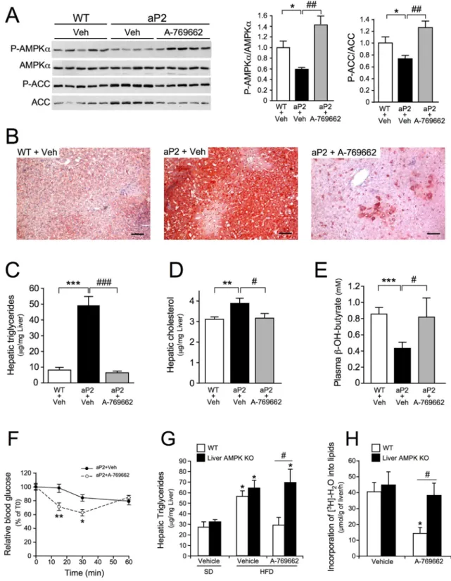
3.7. Pharmacological Re-Activation of Hepatic AMPK Abolishes Fatty Liver Disease in Mice
Lastly, we evaluated the pharmacological potential of AMPK activa-tion by the small-molecule A-769662, which acts on hepatic metabo-lism independently of an effect on food intake (Cool et al., 2006), in a fatty liver mouse model. aP2-nSREBP-1c transgenic mice which display massive fat deposition in the liver due to high levels of de novo lipogen-esis (Shimomura et al., 1998), show decreased hepatic P-Thr172-AMPK/ AMPK and P-Ser79-ACC/ACC ratios by ~ 40% and ~ 30%, respectively, compared to control livers (Fig. 7A). After chronic dosing, A-769662 sig-nificantly increased P-Thr172-AMPK/AMPK and P-Ser79-ACC/ACC ra-tios (Fig. 7A), resulting in a substantial decrease in hepatic TG content, as assessed by Oil Red O lipid staining (Fig. 7B). Hepatic triglyceride and cholesterol determinations revealed that A-769662 completely abolished hepatic lipid accumulation in these mice (Fig. 7C-D). Note that body weight was not affected by A-769662 treatment (data not shown). In addition, A-769662 restored hepatic fatty acid oxidation, as shown by the increase in plasmaβ-hydroxybutyrate levels (Fig. 7E). The decrease in liver lipid content was accompanied by an improve-ment in insulin action (Fig. 7F), suggesting that the lower fat content of the liver contributed to the increase in insulin sensitivity. In addition, to demonstrate whether the effects of A-769662 on hepatic steatosis are dependent on AMPK, DIO control and liver AMPK-deficient mice were chronically dosed with A-769662 and hepatic lipid content was
Fig. 3. The forced expression of lipogenic genes does not prevent the AMPK-induced inhibition of hepatic lipogenesis. WT primary hepatocytes were infected with Ad-GFP or Ad-mSREBP-1c adenovirus and cultured for 16 h in the presence 25 mM glucose. Hepatocytes were then incubated in fresh medium containing 25 mM glucose with or without 100μM AICAR or 30 μM A-769662 for 3 h. (A) Lipogenic gene expression. (B) Immunoblots performed with the antibodies indicated and quantification of immunoblot images for the ACC and FAS proteins normalized to GAPDH. (C) The rate of lipid synthesis was estimated from the incorporation of [1-14C]-acetate into total lipids. Data are means ± SEM from 3 independent
experiments. ⁎Pb 0.05, ⁎⁎P b 0.01, ⁎⁎⁎P b 0.001, versus Ad-GFP-infected hepatocytes;###
measured. A-769662 normalized liver TG content in control mice, but DIO liver AMPK-deficient mice were resistant to beneficial A-769662 ac-tion (Fig. 7G). A reduction of ~60% of de novo hepatic lipogenesis was observed in A-769662-treated control mice, whereas no such decrease was observed in liver AMPK-deficient mice (Fig. 7H). Thus, A-769662 attenuates fatty liver, at least partly by inhibiting de novo lipogenesis in an AMPK-dependent manner.
4. Discussion
AMPK was initially identified on the basis of its ability to phosphor-ylate and inactivate ACC, which is known to direct lipid partitioning be-tween esterification and oxidation. Thus, modulating AMPK activity in the liver may contribute to the regulation of hepatic lipid metabolism in both physiological and pathophysiological settings. Indeed, the
involvement of AMPK in the pathogenesis of hepatic lipid disorders has been suggested by the observation that decreases in AMPK activity are correlated with lipid accumulation in rodent liver (Muse et al., 2004; Yu et al., 2004). The regulation of ACC-dependent malonyl-CoA produc-tion is crucial for theflexibility of lipid metabolism in hepatocytes. Malonyl-CoA is used by fatty acid synthase to synthesize palmitic acid but is also serving to allosterically inhibit CPT1, the protein responsible for transporting long-chain fatty acids into mitochondria for β-oxidation. The role of AMPK in inhibiting ACC activity to promote the partitioning of free fatty acids for oxidation rather than TG storage is well documented (Viollet et al., 2009; Smith et al., 2016b). One of the keyfindings of our study is that, despite the lost of phosphorylation of ACC1 on Ser79 and ACC2 on Ser212 in the liver in the absence of both the AMPKα1 and α2 catalytic subunits, liver AMPK-deficient mice have unexpectedly normal hepatic TG contents and de novo lipogenesis,
Fig. 4. Effects of various AMPK activators on lipid synthesis and fatty acid oxidation rates in control and AMPK-deficient hepatocytes. control AMPKα1α2 floxed (WT) and AMPKα1α2 KO (AMPK KO) primary hepatocytes were incubated in the presence or absence of various concentrations of AICAR, metformin, A-769662, C13, 991 or TOFA for 3 h. (A) Immunoblots were performed with the indicated antibodies. (B, C) Fatty acid and sterol synthesis were assessed from the incorporation of [1-14C]-acetate into saponifiable and non-saponifiable lipids,
respectively. (D) Palmitate oxidation rates were determined by measuring the production of14C–labeled acid-soluble metabolites from [1-14
C]-palmitic acid. Data are means ± SEM from 3 to 5 independent experiments. ⁎Pb 0.05, ⁎⁎P b 0.01, ⁎⁎⁎P b 0.001 versus WT or AMPK KO hepatocytes incubated in the absence of compounds;#
Pb 0.05,##
Pb 0.01,###
Pb 0.001 WT versus AMPK KO hepatocytes incubated under the same conditions; by ANOVA.
indicating that liver AMPK is not required for the adequate metabolic regulation of hepatic lipid homeostasis. Thus, the downregulation of AMPK signaling in the liver does not necessarily lead to adverse meta-bolic consequences (e.g., development of fatty liver). The decreases in AMPK activity associated with hepatic steatosis may therefore be a con-sequence, rather than a cause, of changes in hepatic metabolism. It is possible that the decrease in AMPK activity in fatty liver results from in-hibition of its upstream kinase, LKB1, by acetylation due to a liver
steatosis-associated decrease in SIRT1 activity (Ruderman et al., 2013). In the absence of AMPK, additional regulatory mechanisms may com-pensate,fine-tuning the regulation of fatty acid metabolism. Indeed, many mechanisms are involved in the maintenance of lipid homeostasis during feeding and fasting, and in conditions of dietary lipid overload. For instance, circulating hormones, such as insulin and glucagon, play a key role in the fed/fasting responses, acting at multiple steps to control the synthesis and catabolism of fatty acids. In particular, the actions of
Fig. 5. Small-molecule AMPK activators enhance the action of metformin and AICAR on AMPK activation, lipid synthesis and fatty acid oxidation in primary hepatocytes. Control AMPKα1α2 floxed (WT) and AMPKα1α2 KO (AMPK KO) primary hepatocytes were incubated with or without various concentrations of AICAR or metformin in the absence or presence of A-769662 (1 or 10μM) or C13 (1 μM) for 3 h. (A) Immunoblots were performed with the antibodies indicated. (B) P-AMPKα/AMPKα and P-ACC/ACC ratios from the quantification of immunoblot images. Data are means ± SEM. ⁎P b 0.05, ⁎⁎P b 0.01 versus WT hepatocytes incubated in the absence of A-769662 by ANOVA. (C, E, G) Fatty acid and sterol synthesis were assessed from the incorporation of [1-14
C]-acetate into saponifiable and non-saponifiable lipids, respectively. Results are presented as a percentage of acetate incorporated into WT or AMPK KO hepatocytes incubated in the absence of compounds. (D, F, H) Palmitate oxidation rates were determined by measuring the production of14C–
labeled acid-soluble metabolites from [1-14
C]-palmitic acid. Data are means ± SEM from 3 independent experiments. ⁎Pb 0.05, ⁎⁎P b 0.01, ⁎⁎P b 0.001 versus WT hepatocytes incubated in the absence of compounds;##
Pb 0.01,###
both insulin and glucagon are unaltered by AMPK deletion in hepato-cytes (Foretz et al., 2010), and presumably overcome the lack of AMPK through additional processes maintaining hepatic lipid homeostasis. Thus, the regulation of enzymes/factors involved in the control of he-patic lipid metabolism by redundant signaling pathways may provide a simple explanation for the lack of hepatic lipid disorders in the ab-sence of AMPK activity. Supporting this notion, we have recently shown that deletion of AMPK in the liver led to an adaptation of hepatic metabolism resulting in increased secretion offibroblast growth factor 21 (FGF21) (Chalvon-Demersay et al., 2017; Hughey et al., 2017). How-ever, we cannot exclude that under particular conditions (e.g., genetic background, age, sex, diet, environment) this adjustment could be disrupted leading to an alteration of lipid metabolism.
Fullerton et al. reported that mice with alanine knock-in (KI) muta-tions in both ACC1 at Ser79 and ACC2 at Ser212 (ACC-DKI) had elevated hepatic TG content (Fullerton et al., 2013). In contrast, although the phosphorylation of ACC1 at Ser79 and ACC2 at Ser212 in the liver is lost in (liver AMPK-deficient mice), we show that hepatic TG content were unaffected compared to control mice. It is important to note that ACC-DKI mouse model is a global KI where ACC1-Ser79 and ACC2-Ser212 are mutated in all cells of the body, whereas in liver AMPK-deficient mice AMPK KO mouse model AMPK is only deleted in hepato-cytes. This may account for the different phenotype between the two mouse models. It is also important to note another important difference in the metabolic phenotype between ACC-DKI and liver-specific AMPK KO mouse models. Fullerton et al. showed that ACC-DKI mice were hy-perglycemic and also glucose intolerant, caused by the storage of excess lipid in the liver (Fullerton et al., 2013). In contrast, liver-specific AMPK KO mice are normoglycemic and display similar glucose tolerance com-pared to control mice. No change in blood glucose levels was also re-ported after hepatic AMPK deletion achieved through infection of AMPKα1/α2 floxed mice with adeno-associated virus expressing the Cre recombinase driven by a liver-specific promoter (Cokorinos et al., 2017). These results show that two different mouse models with he-patic AMPK deletion did not recapitulate the altered glucose homeosta-sis observed in ACC-DKI mice, which is conhomeosta-sistent with a normal hepatic TG content in liver-specific AMPK KO mouse models.
The pharmacological activation of AMPK has recently emerged as a new strategy for achieving a healthy balance between altered hepatic lipid metabolism and fatty liver disease (Smith et al., 2016b). However, the demonstration of AMPK-independent effects of AMPK activators, such as AICAR and metformin, in the liver (Foretz et al., 2010) raise ques-tions about the true role of AMPK activation in the regulation of hepatic lipid metabolism. AMPK-deficient hepatocytes provide a useful cell model for investigating whether the effects of AMPK activators result from an AMPK-dependent or AMPK-independent mechanism (Foretz et al., 2010). We show here that direct and indirect AMPK activators, in-cluding A-769662, 991, C13, AICAR, metformin and berberine, inhibit lipid synthesis in a dose- and AMPK-dependent manner, indicating that AMPK is a bonafide therapeutic target for limiting uncontrolled he-patic lipogenesis. Surprisingly, we found here that salicylate, which has been reported to activate AMPK through binding at a novel allosteric binding site described as the ADaM (allosteric drug and metabolite) site (Hawley et al., 2012) strongly inhibited de novo lipogenesis in the absence of AMPK (Supplemental Fig. S7). Salicylate may exert its inhibi-tory effects on fatty acid synthesis during the conversion of acetate into fatty acids (Beynen et al., 1982). The formation of salicyl-CoA from salic-ylate may inhibit fatty acid synthesis through the sequestration of coen-zyme A (Killenberg et al., 1971). It has also been suggested that salicylate decreases liver lipid levels independently of AMPKβ1, through
mitochondrial uncoupling (Smith et al., 2016a). Thesefindings suggest that salicylate operates principally via an AMPK-independent mechanism.
As previously reported, AMPK phosphorylation at Thr172 (the resi-due phosphorylated by LKB1) as well as ACC1/2 phosphorylation at Ser79/Ser212 are lost in the liver of hepatic LKB1 KO mice (Foretz et al., 2010; Shaw et al., 2005). We show here that the inhibition of lipo-genesis in response to AMPK activation is largely dependent on its up-stream kinase LKB1 in hepatocytes. However, higher concentrations of the small-molecule A-769662 are associated with low levels of ACC phosphorylation and a significant inhibition of lipogenesis in the ab-sence of LKB1. This probably reflects the ability of A-769662 to activate AMPK partially independently ofα-Thr172 phosphorylation through an allosteric mechanism and to induce the phosphorylation of its target, ACC (Foretz et al., 2010; Scott et al., 2014).
Although the action of AMPK is achieved by rapid and direct phos-phorylation of key metabolic enzymes, such as ACC (Fullerton et al., 2013), long-term effects have been suggested via the regulation of the expression of a number of gene sets. It has been suggested that AMPK promotes the inhibition of lipogenesis gene expression by direct phos-phorylation of transcription factors including ChREBP and SREBP-1c (Kawaguchi et al., 2002; Li et al., 2011). However, we found that the ef-fects of metformin and AICAR on the expression of lipogenic genes were AMPK-independent, contrasting with the resistance of hepatocytes lacking AMPK to the inhibitory action of these compounds on TG accu-mulation. We also showed that AMPK activation by A-769662 had no ef-fect on lipogenic gene expression, whereas it robustly decreased the synthesis of TGs. This is reminiscent of the absence of significant change in the expression of genes involved in lipogenesis in a genetic model with specific AMPK activation in the liver (Woods et al., 2017). Alto-gether, these results indicate that the direct inhibition of lipogenic en-zyme activity, rather than of lipogenic gene expression, may be responsible for the metabolic action of AMPK on de novo lipogenesis in hepatocytes. Interestingly, the ability of drug-activated AMPK to in-hibit hepatic lipogenesis was preserved in hepatocytes in which lipogenic gene expression was forced (Fig. 3), supporting the notion that AMPK activators directly disrupt lipogenicflux rather than the ex-pression of key lipogenic enzymes.
The hepatic ablation of AMPK did not affect fatty acid oxidation in the liver, regardless of the nutritional conditions. In the same way, fatty acid oxidation in basal conditions was similar in control and AMPKα1α2-null primary hepatocytes. The treatment of hepatocytes with TOFA, an allosteric inhibitor of ACC, increased fatty acid oxidation approximately by two-fold in both control and AMPKα1α2-null hepa-tocytes. This result demonstrates that the stimulation of fatty acid oxi-dation dependent of ACC-malonyl-CoA-CPT1 pathway is functional in these primary mouse hepatocytes. However, this activation was signi fi-cantly weaker in AMPKα1α2-null hepatocytes (~10%) (Fig. 4D and Sup-plemental Figs. S6D and S7D), indicating that fatty acid oxidative capacity is slightly altered in the absence of AMPK. This effect may re-flect the role of AMPK in mitochondrial biogenesis and dynamics (Guigas et al., 2007; Hasenour et al., 2014; Toyama et al., 2016).
Our results suggest that drug-induced AMPK activation decreases hepatic lipid accumulation both by inhibiting lipid synthesis and by stimulating fatty acid oxidation. Thesefindings conflict with a recent study from Woods et al. reporting that hepatic AMPK activation via the expression of a constitutive active form ofγ1 subunit (D316A mu-tant) prevents lipid accumulation in liver in mice fed a high-fructose diet through only de novo lipogenesis inhibition with no effect on fatty acid oxidation (Woods et al., 2017). This discrepancy may result
Fig. 6. Indirect and small-molecule AMPK activators inhibit lipid synthesis in primary human hepatocytes. (A) Assessment of AMPK subunit levels in primary human and mouse hepatocytes by western blotting with the indicated antibodies. (B-J) Human primary hepatocytes were cultured in the absence or presence of various concentrations of A-769662, C13, AICAR or metformin, alone or in combination, at the indicated concentrations for 3 h. (B) Immunoblots were performed with the indicated antibodies. (C-J) Fatty acid and sterol synthesis were assessed from the incorporation of [1-14
C]-acetate into saponifiable and non-saponifiable lipids, respectively. Data are means ± SEM from 3 independent experiments. ⁎P b 0.05, ⁎⁎P b 0.01, ⁎⁎⁎P b 0.001 versus hepatocytes incubated in the absence of activators (C\\F); for comparisons between the conditions indicated (G-J); by ANOVA.
Fig. 7. Small-molecule-mediated activation of AMPK in the liver restores fatty acid oxidation, abolishes hepatic steatosis and improves insulin sensitivity in aP2-nSREBP-1c transgenic mice. (A-E) aP2-nSREBP-1c (aP2) and non-transgenic (WT) littermate mice were treated for 8 days with vehicle or A-769662 (30 mg/kg, i.p., b.i.d.). Mice were sacrificed after a 24-h fast. (A) Western-blot analysis of liver lysates with the antibodies indicated. Each lane represents an individual mouse. The lower panels show P-AMPKα/AMPKα and P-ACC/ACC ratios from the quantification of immunoblot images (n = 5 per group). (B) Oil Red O staining of representative liver sections. Scale bars: 50 μm. (C) Liver triglyceride content, (D) liver cholesterol content and (E) plasmaβ-hydroxybutyrate levels (n = 5-6 per group). (F) Insulin tolerance test in aP2-nSREBP-1c transgenic mice (n = 5-6 per group) treated for 7 days with vehicle or A-769662 (30 mg/kg, i.p., b.i.d.). Data are means ± SEM. ⁎Pb 0.05, ⁎⁎P b 0.01, ⁎⁎⁎P b 0.001 versus WT + Veh;##
Pb 0.01,###
Pb 0.001 versus aP2 + Veh; by ANOVA. (G) Control AMPKα1α2 floxed (WT) and liver-specific AMPKα1α2 KO (Liver AMPK KO) mice fed either a standard diet (SD) or a high-fat diet (HFD) for 5 months were treated with vehicle or A-769662 (30 mg/kg, i.p., b.i.d.). After 5 days of treatment, mice were sacrificed in the fed state, and their livers were collected for hepatic triglyceride content determination. Data are means ± SEM. (n = 6 per group). ⁎Pb 0.05 HFD versus SD,#
Pb 0.05 WT versus liver AMPK KO by ANOVA. (H) In vivo synthesis rate of lipids in the livers of AMPKα1α2 floxed (WT) and liver-specific AMPKα1α2 KO mice (Liver AMPK KO) mice treated with vehicle or A-769662 were assessed by determining the incorporation of3
from the mode of AMPK activation (pharmacological versus genetic) or the method ofβ-oxidation assessment.
Importantly, we found that all the direct and indirect AMPK activa-tors tested stimulated fatty acid oxidation in an AMPK-dependent fash-ion, with the exception of C13 at high concentrations, which stimulated β-oxidation in a manner that was partly independent of AMPK. Despite metformin is well known to hamper complex I of the mitochondrial re-spiratory chain (El-Mir et al., 2000; Owen et al., 2000), we found that metformin was one of the AMPK-activating drugs most strongly activat-ing fatty acid oxidation (Fig. 4D). The simultaneous inhibition of mito-chondrial respiration and stimulation of fatty acid oxidation by metformin may appear paradoxical. In theory, the inhibition of respira-tion by metformin should impede mitochondrialβ-oxidation. A key point is that mitochondrialβ-oxidation generates abundant reduced co-enzyme FADH2, whereas carbohydrate oxidation provides mostly re-duced coenzyme NADH. This NADH is oxidized by complex I, whereas FADH2 is oxidized by complex II. The partial inhibition of respiration by metformin is therefore rescued by AMPK-induced fatty acid oxida-tion, which bypasses complex I by supplying reduced equivalents for complex II. Berberine, and, to a lesser extent, AICAR through ZMP, prob-ably act in a similar manner to metformin, because they are known to inhibit complex I of the mitochondrial respiratory chain (Guigas et al., 2007; Turner et al., 2008).
Intriguingly, all the AMPK activators tested inhibited lipid synthesis and induced ACC phosphorylation in a robust manner in control hepato-cytes, but they stimulated fatty acid oxidation to various extents. Here, we show that metformin, berberine, AICAR and C13 increased fatty acid oxidation to levels similar to those achieved with the ACC inhibitor TOFA, whereas the small-molecules A-769662 and 991 had a weaker ef-fect, even with accurate matching for lipogenesis inhibition capacity. For example, 1 mM metformin inhibited lipogenesis and induced fatty acid oxidation by ~50% in both cases, whereas the indirect activator 991, at a concentration of 1μM, reduced lipogenesis by ~80% and stimulated fatty acid oxidation by only ~30% (Fig. 4). This discrepancy raises ques-tions about the possible selective action of AMPK activators on specific AMPK heterotrimeric complexes for the differential modulation of fatty acid synthesis and oxidation. Although A-769662 activates prefer-entiallyβ1-containing complexes (Hunter et al., 2014; Scott et al., 2008), this hypothesis seems unlikely because 991 is a panβ-agonist and can activate virtually all AMPK complexes (Xiao et al., 2013). Alter-natively, indirect activators (metformin, berberine and AICAR) may en-hance β-oxidation because they rightly act on complex I of the mitochondrial respiratory chain. Partial inhibition of complex I by these indirect activators may promote FADH2 oxidation through com-plex II to increase ATP synthesis. Thus, the increase in FADH2 oxidation may enhance the fatty acid oxidationflux stimulated by AMPK activa-tion, compensating for the inhibition of complex I. Lastly, it is disturbing to observe that metformin, berberine, AICAR and C13 all increase intra-cellular AMP or generate AMP-mimetics, which, in addition to activating AMPK, may possibly modulate other AMP-sensitive enzymes involved in fatty acid catabolism. Further investigations are required to clarify this disparity between the degree of inhibition of fatty acid synthesis and the stimulation of fatty acid oxidation by AMPK activators. In partic-ular, it will be interesting to determine the relationship between the level of fatty acid oxidation and intracellular malonyl-CoA content in re-sponse to treatment with different types of AMPK activators.
The therapeutic potential of our in vitrofindings is highlighted by the AMPK-dependent action of A-769662 demonstrated in vivo, with improvements in liver steatosis in diet-induced obese (DIO) mice fol-lowing chronic treatment. We also showed that A-769662 treatment re-versed hepatic AMPK activity suppression, decreasing lipid accumulation and restoring fatty acid oxidation in the livers of insulin-resistant aP2-nSREBP-1c transgenic mice (Fig. 7). Thus despite the de-crease in AMPK activity observed in hepatic steatosis, possibly due to the inhibition of LKB1 by acetylation (Ruderman et al., 2013), AMPK can still be activated by small-molecule activators. This observation
demonstrates that, in the context of metabolic disorders, the activa-tion of AMPK by small-molecules is not subject to resistance, unlike other regulatory pathways, such as those involving insulin and lep-tin. This is probably because A-769662 partly bypasses LKB1 to acti-vate AMPK allosterically for subsequent phosphorylation of its downstream target, ACC (Scott et al., 2014). Indeed we previously re-ported that A-769662 induced the phosphorylation of ACC without affecting AMPKα-Thr172 phosphorylation in hepatocytes (Foretz et al., 2010) and, to a lesser extent, in LKB1-deficient hepatocytes (Supplemental Fig. S11A). Furthermore, small-molecule-induced AMPK re-activation in liver steatosis suggests that AMPK undergoes no intrinsic inhibition such as inhibitory post-translational modifications.
Some clinical effects in humans have reported a beneficial effect of metformin on hepatic steatosis, but the efficacy of this drug for attenu-ated fatty liver remains a matter of debate (Mazza et al., 2012). By con-trast, metformin clearly improves fatty liver in mice (Lin et al., 2000), presumably by activating hepatic AMPK (Fullerton et al., 2013). Poly-morphism of organic cation transporter 1 (OCT1), the main hepatic metformin transporter, may contribute in part to the variable clinical re-sponse to metformin in fatty liver amelioration (Shu et al., 2007). The lower metformin concentrations used in humans (20 mg/kg) than in mice (250 mg/kg) may account for this poorer efficacy. Metformin con-centrations are about 40–70 μM in the portal vein, and b250 μM in the liver (Wilcock and Bailey, 1994), whereas we show here that metformin inhibits lipogenesis and induces fatty acid oxidation in primary hepato-cytes only from concentrations of 250–500 μM. Given the risk of lactic acidosis, it is not feasible to increase metformin doses sufficiently in pa-tients for the treatment of fatty liver. As indirect and direct AMPK acti-vators activate AMPK via different mechanisms, we hypothesize that combination treatments might increase the effects of metformin on he-patic lipid metabolism by increasing the activation of AMPK, thereby significantly decreasing hepatic lipid content. We show that metformin or AICAR combined with a direct AMPK activator (A-769662 or C13) have additive effects on lipogenesis inhibition and fatty acid oxidation stimulation when used together to treat mouse and human hepato-cytes. The use of a small-molecule AMPK activator in combination treat-ments may, therefore, enhance the effects of metformin, reducing the accumulation of lipids in the livers of patients with type 2 diabetes displaying fatty liver disease.
In conclusion, we demonstrate that impaired AMPK activation is not a triggering factor in the pathogenesis of hepatic steatosis. By contrast, AMPK activation or re-activation in pathophysiological conditions has beneficial effects for the treatment of fatty liver dis-ease (Supplemental Fig. S12). Thesefindings have important impli-cations for the development of treatments for type 2 diabetes and metabolic syndrome. Indeed, by decreasing hepatic lipid content, AMPK activation may improve insulin sensitivity and, thus, increase the efficacy of insulin in the liver, thereby indirectly improving the control of hepatic glucose production and lowering blood glucose levels. Moreover, the inhibition of de novo lipogenesis by drug-induced AMPK activation may help to reduce tumor development for hepatocellular carcinomas, as reported for prostate and lung can-cers (Svensson et al., 2016; Zadra et al., 2014).
Funding Sources
This work was supported by grants from Inserm, CNRS, Université Paris Descartes, Région Ile-de-France (CORDDIM) and Agence Nationale de la Recherche (2010 BLAN 1123 01). This work was performed within the Département Hospitalo-Universitaire (DHU) AUToimmune and HORmonal diseaseS (AUTHORS). N.B. holds a doctoral fellowship from the French Government (Ministère de la Recherche et des Enseignements Supérieurs). A.M. holds a postdoctoral fellowship from the Région Ile-de-France (CORDDIM).