HAL Id: inserm-00668472
https://www.hal.inserm.fr/inserm-00668472
Submitted on 9 Feb 2012
HAL is a multi-disciplinary open access
archive for the deposit and dissemination of
sci-entific research documents, whether they are
pub-lished or not. The documents may come from
teaching and research institutions in France or
abroad, or from public or private research centers.
L’archive ouverte pluridisciplinaire HAL, est
destinée au dépôt et à la diffusion de documents
scientifiques de niveau recherche, publiés ou non,
émanant des établissements d’enseignement et de
recherche français ou étrangers, des laboratoires
publics ou privés.
Rapid determination of tricarboxylic acid cycle enzyme
activities in biological samples.
Sergio Goncalves, Vincent Paupe, Emmanuel Dassa, Jean-Jacques Brière,
Judith Favier, Anne-Paule Gimenez-Roqueplo, Paule Bénit, Pierre Rustin
To cite this version:
Sergio Goncalves, Vincent Paupe, Emmanuel Dassa, Jean-Jacques Brière, Judith Favier, et al.. Rapid
determination of tricarboxylic acid cycle enzyme activities in biological samples.. BMC Biochemistry,
BioMed Central, 2010, 11 (1), pp.5. �10.1186/1471-2091-11-5�. �inserm-00668472�
M E T H O D O L O G Y A R T I C L E
Open Access
Rapid determination of tricarboxylic acid cycle
enzyme activities in biological samples
Sergio Goncalves
1,2, Vincent Paupe
1,2, Emmanuel P Dassa
1,2, Jean-Jacques Brière
1,2, Judith Favier
3,4,
Anne-Paule Gimenez-Roqueplo
3,4,5, Paule Bénit
1,2, Pierre Rustin
1,2*Abstract
Background: In the last ten years, deficiencies in tricarboxylic acid cycle (TCAC) enzymes have been shown to cause a wide spectrum of human diseases, including malignancies and neurological and cardiac diseases. A prerequisite to the identification of disease-causing TCAC enzyme deficiencies is the availability of effective enzyme assays.
Results: We developed three assays that measure the full set of TCAC enzymes. One assay relies on the sequential addition of reagents to measure succinyl-CoA ligase activity, followed by succinate dehydrogenase, fumarase and, finally, malate dehydrogenase. Another assay measures the activity ofa-ketoglutarate dehydrogenase followed by aconitase and isocitrate dehydrogenase. The remaining assay measures citrate synthase activity using a standard procedure. We used these assays successfully on extracts of small numbers of human cells displaying various severe or partial TCAC deficiencies and on frozen heart homogenates from heterozygous mice harboring an SDHB gene deletion.
Conclusion: This set of assays is rapid and simple to use and can immediately detect even partial defects, as the activity of each enzyme can be readily compared with one or more other activities measured in the same sample.
Background
Interest in assaying tricarboxylic acid cycle (TCAC) enzyme activities has been rekindled by evidence that deficiencies in these enzymes cause a variety of human diseases [1,2], in contradiction to the long-held belief that any TCAC enzyme deficiency is lethal [3]. Several acquired conditions are characterized not only by post-translational alterations in electron transport respiratory chain proteins and impairments in mitochondrial cal-cium handling, but also by abnormalities in TCAC enzymes. Examples include heart failure in humans [4] and stress-related heart dysfunction induced in rats by chronic restraint [5]. Several inherited diseases have been ascribed to primary TCAC enzyme deficiencies (Table 1). For instance, primary succinate dehydrogen-ase (SDH; EC.1.3.99.1) deficiency results either in tissue degeneration with devastating early-onset encephalo-myopathy or in tissue proliferation with formation of paragangliomas or other tumors [2]. Similarly, a
mutation in the gene encoding fumarase (EC. 4.2.1.2) is a rare cause of encephalomyopathy and a far more com-mon cause of leiomyomas of the skin and uterus and of renal cancer [6]. TCAC dysfunction may also result from concurrent impairments in several steps of the cycle. For instance, combined deficiencies in SDH and aconitase (EC. 4.2.1.3) is observed in Friedreich’s ataxia [7].
Residual activities associated with TCAC impairments in humans vary widely and may determine the magni-tude of organic acid accumulation [8]. Organic acid accumulation has been proven instrumental in initiating tumor formation related to SDH or fumarase deficiency [9].
The ratios between TCAC enzymes are consistent for each mammalian tissues presumably reflecting their metabolic demand, as shown three decades ago in the seminal study by Pette and Hofer [10]. This echoes the occurrence of metabolons in the mitochondrial matrix [11-13], allowing for efficient channeling of substrates and co-factors through the Krebs cycle and related enzymes such as transaminase [14]. Consequently, in
* Correspondence: pierre.rustin@inserm.fr
1
Inserm, U676, Paris, 75019 France
© 2010 Goncalves et al; licensee BioMed Central Ltd. This is an Open Access article distributed under the terms of the Creative Commons Attribution License (http://creativecommons.org/licenses/by/2.0), which permits unrestricted use, distribution, and reproduction in any medium, provided the original work is properly cited.
addition to the determination of residual absolute activ-ities, estimation of ratios between enzyme activities is an effective means of detecting partial but potentially harm-ful deficiencies. When used to assess respiratory chain activities, this approach enabled the identification of sev-eral gene mutations, even in patients with partial respiratory chain deficiencies [15,16].
At present, TCAC enzyme activities are measured using a series of independent assays that are both labor-ious and time consuming. We therefore developed a limited set of assays allowing both measurement of all TCAC enzyme activities and detection of abnormalities in enzyme activity ratios. We used these assays success-fully to detect severe and partial isolated deficiencies in several TCAC enzymes.
Results
Given that TCAC enzyme activity ratios, because of their consistency [17], are important in comparing data between samples, we devised a method for measuring the activities of all eight TCAC enzymes using only three assays, which allows rapid determination of enzyme activity ratios. To define appropriate assay con-ditions, we first used mouse heart samples and assessed various parameters (detergent, pH, ionic force) that are known to independently stimulate each activity, but which might interfere with the measurement of other activities. We found that two media were sufficient for assaying all TCAC activities. The difference between these two media lies in the presence of phosphate required by some of the enzymes and in the use of elec-tron acceptors to cope with the various reduced equivalents.
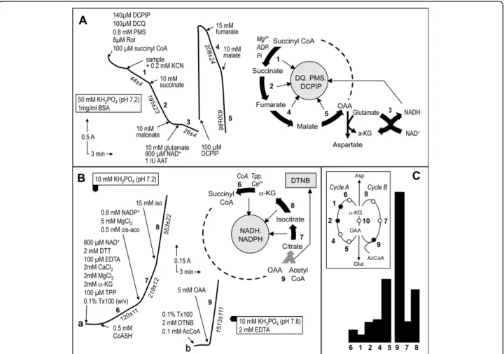
The first assay measures five enzymes sequentially in an individual sample (Fig. 1A). Importantly, while four of these enzymes catalyze steps of the TCAC, one, GDH, is measured as a consequence of the required presence of glutamate for the assay of MDH. Glutamate is required for the added aspartate amino transferase
reaction in order to transaminate the oxaloacetate pro-duced by MDH, which otherwise would rapidly block this last enzyme (Fig. 1B). The biological sample is first added to a detergent-containing medium allowing sub-strates and electron acceptors free access to their respective binding sites on the proteins. However, we found that succinyl-CoA batches variably contained reducing agents capable of interacting with the electron acceptor mixture used in the assay. Therefore, the assay is started only after most of this non-enzymatic reaction is completed. Then, biological sample is added to enable measurement of the first enzyme, GTP- and/or ATP-forming succinyl-CoA ligase, based on the amount of succinate formed by the enzyme. The succinate is then readily oxidized to fumarate by SDH concomitantly with ultimate reduction of DCPIP. In this assay, electrons from succinate are transferred by SDH to either phena-zine methosulfate or decylubiquinone, both capable of reducing DCPIP. Maximal SDH activity is then mea-sured by adding a large amount of succinate. Adding malonate, a competitive SDH inhibitor, essentially abolishes DCPIP reduction. Subsequent addition of glu-tamate, because of the presence of added NAD+, allows estimation of NAD+-dependent GDH activity. Depend-ing on the enzyme activity levels in the sample, it may be necessary at this point to add more DCPIP before performing the next assays (fumarase and MDH activ-ity). Fumarase is assayed by adding a large fumarate excess, which is readily converted to malate by fumar-ase, this latter acid being used up by MDH to produce NADH and oxaloacetate. Owing to the presence of added aspartate aminotransferase and glutamate, oxaloa-cetate does not accumulate and, therefore, does not slow the MDH reaction. The last enzyme of the assay, MDH, is then measured by adding 10 mM malate.
The second assay starts with measurement of the reduction of pyridine nucleotides (NAD+/NADP+) by KDH. This enzyme, one of the limiting steps of the TCAC, requires the presence of Ca++ ions, thiamine
Table 1 Primary deficiencies in tricarboxylic acid cycle enzymes in humans.
Enzyme1 Clinical presentation References Fumarase 1. Progressive encephalopathy
2. Hereditary leiomyomatosis and renal cell cancer
1. [19] 2. [27] Malate dehydrogenase No disease identified so far
Citrate synthase No disease identified so far Aconitase No disease identified so far
Isocitrate dehydrogenase Low-grade gliomas [32] a-ketoglutarate dehydrogenase Congenital lactic acidosis [28] Succinyl CoA ligase Encephalomyopathy with mtDNA depletion [33] Succinate dehydrogenase 1. Encephalopathy (Leigh syndrome)
2. Pheochromocytoma and paraganglioma
1. [20] 2. [34] 1
Only primary enzyme deficiencies caused by known gene mutations are indicated, excluding secondary deficiencies that may be from genetic (e.g., Friedreich’s ataxia with aconitase and succinate dehydrogenase deficiencies) or non genetic origin.
Goncalveset al. BMC Biochemistry 2010, 11:5 http://www.biomedcentral.com/1471-2091/11/5
pyrophosphate, and coenzyme A to catalyze the oxida-tion of a-ketoglutarate. After KDH measurement, cis-aconitate is added for measurement of aconitase activity based on the formation of isocitrate, which, in the pre-sence of IDH, is readily used up to reduce NAD+/NADP+. Finally, the maximal activity rate of IDH is determined after addition of a large isocitrate excess. Citrate synthase, the last TCAC enzyme to be measured, con-denses acetyl-CoA and oxaloacetate into citrate while concomitantly releasing coenzyme A, whose thiol residue readily reacts with Ellman’s reagent
(dithionitrobenzene). It is measured using the standard procedure which, in the case of cultured skin fibroblasts, concomitantly allows the detection of mycoplasma [18].
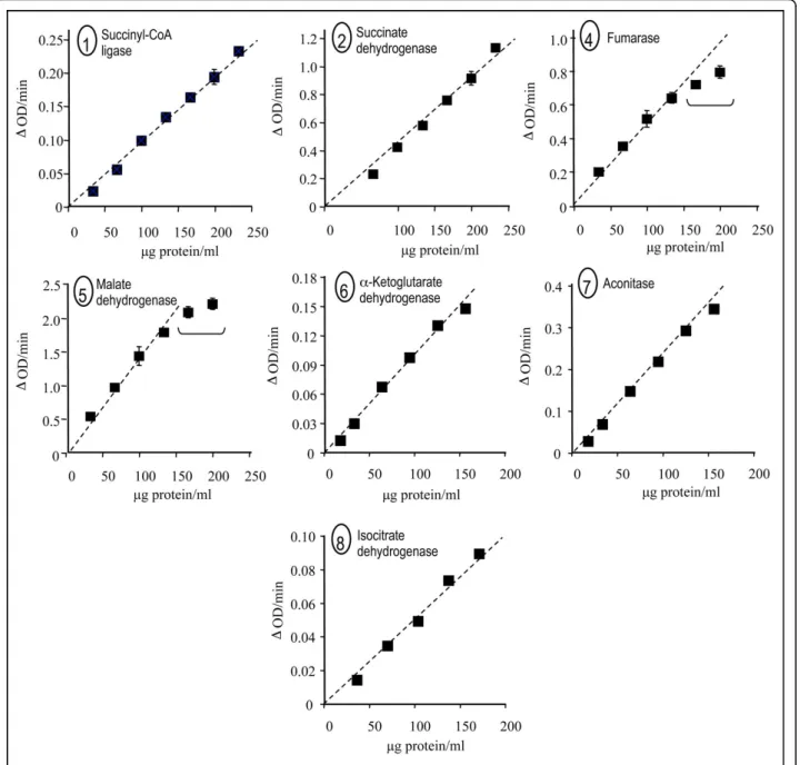
Since part of these assays relies on coupling between several successive enzymes, e.g., aconitase and IDH, we evaluated the proportionality/linearity of these assays as a function of protein concentration in heart sample homogenate (Fig. 2). For protein concentrations of up to 150μg per ml, each assay exhibited a linear response. Given that the protein concentration presumably depends on the extent of mitochondria enrichment in
Figure 1 Tricarboxylic acid cycle enzyme assays. A, The first segment of the tricarboxylic acid cycle can be conveniently evaluated using a single assay to measure five enzymes by spectrophotometrically recording the reduction of DCPIP. The sequential assay begins by measurement of succinyl-CoA ligase activity based on oxidation of the produced succinate by succinate dehydrogenase, which forwards the electrons to the electron acceptors (DQ, DCPIP, and PMS). Coupling of these two activities to estimate succinyl-CoA ligase activity is permitted by the much higher activity of SDH than of succinyl-CoA ligase. After SDH inhibition by malonate, simultaneous addition of glutamate and aspartate aminotransferase ensures elimination of any oxaloacetate in the assay medium, thereby allowing further measurement of MDH activity. Incidentally, the required presence of NAD+ permits the measurement of glutamate dehydrogenase activity. Adding more DCPIP allows subsequent measurement of fumarase and MDH activity. Again, the coupling assay to estimate fumarase activity using MDH activity is permitted by the much higher activity of MDH. B, A second spectrophotometric assay subsequently measures pyridine dinucleotide reduction by three additional enzymes starting witha-ketoglutarate dehydrogenase. The next enzyme to be measured is aconitase, whose product, isocitrate, is readily oxidized by isocitrate dehydrogenase, producing NADPH. A saturating isocitrate concentration is finally added to enable measurement of isocitrate dehydrogenase activity. C, respective proportions of TCAC enzyme activities in mouse heart. The inset shows the TCAC depicted as two interacting enzyme cycles, A and B. Cycle A:a-ketoglutarate dehydrogenase (6), succinyl CoA ligase (1), succinate dehydrogenase (2), fumarase (4), and malate dehydrogenase (5); Cycle B: citrate synthase (9), aconitase (7), and isocitrate dehydrogenase (8). The two cycles interact via the activity of aspartate aminotransferase (10).
the tissue/cell under study, linearity should be evaluated before running quantitative assays on any tissue/cell.
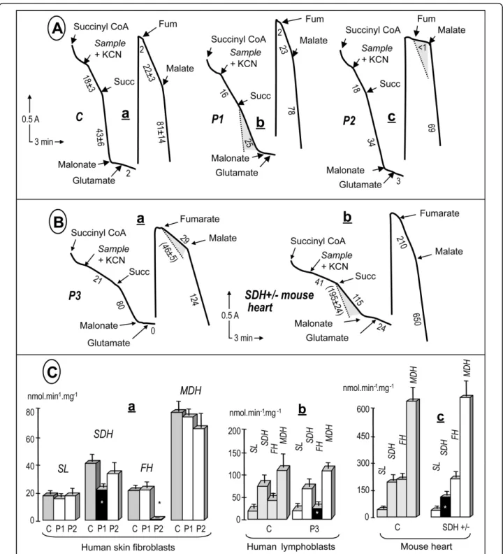
Finally, to evaluate the ability of our assays to detect deficiencies in specific TCAC enzymes, we investigated an array of samples with previously identified genetic defects resulting in deficiencies in various TCAC enzyme activities (Fig. 3). We first studied cultured human fibroblasts harboring mutations in either the SDHA or the fumarase gene (patients P1, P2; Fig. 3b, c).
In agreement with our previous studies [19,20], we found that the SDHA mutation caused an about 60% decrease, whereas the fumarase gene mutation resulted in nearly total loss of fumarase activity. Interestingly, the loss of SDH activity did not hamper our ability to mea-sure succinyl-CoA ligase activity, which was roughly similar to the control value. Then, we evaluated whether our TCAC assay was able to detect partial loss (about 50%) of fumarase activity. We studied a lymphoblastoid
Figure 2 Proportionality between TCAC enzyme activities and protein concentration in mouse heart homogenate. 1-2, 4-8: Enzyme activities plotted as a function of protein concentration: succinyl-CoA ligase (1), succinate dehydrogenase (2), fumarase (4), malate
dehydrogenase (5),a-ketoglutarate dehydrogenase (6), aconitase (7), and NADP+/NAD+-isocitrate dehydrogenase (8). Experimental conditions
were as described in Figure 1. Enzyme numbering according to Figure 1. Goncalveset al. BMC Biochemistry 2010, 11:5
http://www.biomedcentral.com/1471-2091/11/5
cell line from a human patient (P3) harboring a hetero-zygous mutation in the fumarase gene, previously shown to result in a nearly complete loss of activity when associated with a loss of the corresponding allele in tumors. Again, our assay proved capable of detecting the predictable partial (~40%) loss of fumarase activity in these cells, in terms of both the absolute activity and the activity relative to the other TCAC enzymes in the sample (Fig, 3d). Finally, heart samples from a mouse heterozygous for a deleterious mutation in the SDHB gene (exon 2 deletion) were investigated (Fig. 3e). We observed a consistent 40% decrease in SDH activity, as predicted by the heterozygous status of the animal.
Discussion
The renewed interest in measuring TCAC enzyme activity, shown to be sensitive targets under various pathological conditions, prompted us to devise a rapid assay method for detecting TCAC deficiencies in biological samples. Our previous work on the mitochondrial respiratory chain established that, in addition to absolute residual activities, relative ratios of enzyme activities in a metabolic pathway are effective in detecting even partial deficiencies in a given enzyme. We therefore developed a set of three assays that conveniently estimate all TCAC enzyme activ-ities in tissue homogenates and permeabilized cells. Although the experimental conditions had to be adapted to allow for the measurement of many enzymes using a small number of assays, they were largely based on the pioneer work done in the 1940s by Krebs and colleagues [21]. In particular, the concentrations of substrates and cofactors and the metal requirements for each enzyme were as determined by these authors.
As a first result of this work, we fully confirmed that TCAC enzyme activity ratios in each of the different tis-sues or cell investigated are consistent under basal con-ditions, as previously observed by Pette and colleagues as early as 1960[10,22].
To date there has been a lot of efforts to provide con-venient assay procedures for respiratory chain enzymes [16,23-25]. In contrast, to our knowledge, there is no report on any convenient enzymatic procedure to mea-sure the overall activity of TCAC enzymes in the con-text of screening procedures. Although our assays are rapid and sensitive, they have intrinsic limitations. First, three of the enzymes (succinyl-CoA ligase, fumarase, and aconitase) are measured via coupled assays invol-ving the next enzyme in the cycle (SDH, MDH, and IDH, respectively). Obviously, a severe deficiency in the next enzyme would impair the ability of the assay to measure the first enzyme. Therefore, deficiencies in two consecutive enzymes should be evaluated by assaying each enzyme activity separately via standard methods. Second, although our assays are sufficiently sensitive to
detect even partial deficiencies in one TCAC enzyme, measuring the slower enzymes via coupled assays (e.g., aconitase) requires a sample that is large enough to avoid problems with product dilution (e.g., isocitrate in the case of aconitase), which would impair the activity of the coupled enzyme (e.g., IDH in the case of aconi-tase). Despite these limitations, our set of assays enabled us to detect all TCAC enzyme deficiencies. Even a 40% decrease in fumarase activity in lymphoblastoid cell lines was readily detected.
So far there has been only a limited number of diseases which have been associated with primary isolated or mul-tiple defect of the TCAC [7,19,20,26-28]. Beside primary defects of the TCAC genes, as some of the TCAC proteins harbor oxygen-sensitive iron-sulfur cluster, i.e. aconitase, or require a full set of co-factors, i.e.a-ketoglutarate dehy-drogenase, a loss of activity - secondary but yet possibly instrumental in the pathophysiological process - might well be observed in a number of conditions such as aging, Parkinson’s disease or heart failure.
Methods
Biological samples
Fibroblasts derived from forearm biopsies taken with informed consent from healthy controls and patients with TCAC enzyme deficiencies were grown under stan-dard conditions as described elsewhere [29] and frozen (-80°C). Before use, cells were resuspended in 1 ml of medium (A) composed of 0.25 M sucrose, 20 mM Tris (pH 7.2), 40 mM KCl, 2 mM ethylene glycol tetra acetic acid (EGTA), 1 mg/ml bovine serum albumin (BSA), 0.01% digitonin (w/v), and 10% Percoll (v/v). After 10 min incubation at ice-melting temperature, the cells were centrifuged (5 min × 2,300 g), the supernatant dis-carded, and the pellet washed (5 min × 6000 g) with 1 ml of medium A devoid of digitonin and Percoll [30]. Lymphoblasts from patients harboring a deleterious het-erozygous fumarate hydratase gene mutation (N64T) were processed similarly to the cultured fibroblasts. Mouse colony was maintained in accordance with national and institutional guidelines. Animal procedures were approved by the ethical review panel of the Robert Debré Institut, Paris, France. Hearts were obtained from mice, snap frozen in liquid nitrogen and stored at -80°C. Frozen tissues were homogenized at ice-melting tem-perature by hand using a glass-glass potter in medium (1/10-1/20 v/v) composed of 20 mM Tris (pH 7.2), 0.8 M sucrose, 40 mM KCl, 2 mM EGTA, and 1 mg/ml BSA. Large cell debris was removed by low-speed centri-fugation (1,500 g for 5 min).
Spectrophotometry
The first assay measures succinyl-CoA ligase, SDH, glu-tamate dehydrogenase (GDH), fumarase, and malate dehydrogenase (MDH) (see below; Fig. 1). This assay is
Figure 3 Detection of severe and partial TCAC enzyme deficiencies in various biological samples. A, severe enzyme deficiencies. (a) control fibroblasts; (b) SDHA-mutant fibroblasts homozygous for a deleterious R554W mutation, and (c) fumarase-mutant fibroblasts homozygous for a deleterious E319Q mutation. Note that only the addition of organic acids and inhibitors is indicated, although the experiments also involved additions of cations, cofactors, etc, similar to those in Figure 1. B, partial enzyme deficiencies. (a) fumarase-mutant lymphoblasts heterozygous for an N64T mutation; and (b) heart homogenate from a mutant mouse heterozygous for a deleterious SDHB deletion of exon 2. Numbers along the traces are nmol/min/mg protein. The shaded areas show the decreases compared to control values. C, Graphical
representation of the values obtained for the various samples investigated. Dark symbols indicated statistically significant deficiencies. Values are means ± 1 SD. A minimal number of three independent assays (up to ten) were performed to calculate the mean values. Experimental conditions were as in Figure 1. Abbreviations: P1, patient harboring a homozygous SDHA mutation; P2, patient harboring a homozygous fumarase mutation; P3 patient harboring a heterozygous mutation; succ, succinate; fum, fumarate.
Goncalveset al. BMC Biochemistry 2010, 11:5 http://www.biomedcentral.com/1471-2091/11/5
performed in 400 μl of medium A containing 50 mM KH2PO4 (pH 7.2) and 1 mg/ml BSA. The reduction of
dichlorophenol indophenol (DCPIP) is measured using two wavelengths (600 nm and 750 nm) with various substrates and the electron acceptors decylubiquinone and phenazine methosulfate. The second assay measures a-ketoglutarate dehydrogenase (KDH), aconitase, and isocitrate dehydrogenase (IDG) activities. The same volume of the same medium is used, and pyridine nucleotide (NAD+/NADP+) reduction is measured with various substrates using wavelengths of 340 nm and 380 nm. In the third assay, citrate synthase is measured by monitoring dithionitrobenzene (DTNB; Ellman’s reagent) reduction at wavelengths of 412 nm and 600 nm as previously described[19]. For this study, all mea-surements were carried out using a Cary 50 spectro-photometer (Varian Inc., Palo Alto, CA) equipped with an 18-cell holder maintained at 37°C. Protein was mea-sured according to Bradford [31]. All chemicals were of the highest grade from Sigma Chemical Company (St Louis, MO).
List of abbreviations
AAT: Aspartate Aminotransferase; AcCoA: AcetylCoA; Asp: aspartate; BSA: Bovine Serum Albumin; aco: cis-aconitate; DCPIP: Dichlorophenol Indophenol; DQ: Duro-quinone; DTNB: Dithionitrobenzene; DTT: Dithiothreitol; EDTA: Ethylene Diamine Tetraacetic Acid; Glut: gluta-mate; GDH: Glutamate Dehydrogenase; IDH: Isocitrate Dehydrogenase; iso: isocitrate; KDH:a-Ketoglutarate Dehydrogenase;a-KG: a-ketoglutarate; MDH: Malate Dehydrogenase; OAA: oxaloacetate; PMS: Phenazine Methosulfate; Rot: rotenone; SDH: Succinate Dehydrogen-ase; TCAC: Tricarboxylic Acid Cycle (Krebs cycle); TPP: thiamine pyrophosphate; Tx100: Triton X100.
Acknowledgements
This work was supported by grants from the AFM (Association Française contre les Myopathies), the AMMi (Association contre les Maladies Mitochondriales), the Leducq foundation, the ANR Genopath MitOxy, and European Fp6 integrated project Eumitocombat
Author details
1Inserm, U676, Paris, 75019 France.2Université Paris 7, Faculté de médecine
Denis Diderot, IFR02, Paris, France.3Inserm, U970, Paris, 75015 France. 4
Université Paris Descartes, Faculté de Médecine, Paris, France.5AP-HP, Hôpital Européen Georges Pompidou, Département de Génétique, Paris, France.
Authors’ contributions
SG, JJB and PR designed the concept and experiments of the study. VP, ED and PB provided and studied the various cell types utilized, while JF and APGR provided and studied the SDH-mutant mouse. SG and PR analyzed the data. PR drafted the manuscript, helped by the other authors. All authors approved the final manuscript.
Received: 21 August 2009
Accepted: 28 January 2010 Published: 28 January 2010
References
1. Rustin P, Bourgeron T, Parfait B, Chretien D, Munnich A, Rotig A: Inborn errors of the Krebs cycle: a group of unusual mitochondrial diseases in human. Biochim Biophys Acta 1997, 1361(2):185-197.
2. Briere JJ, Favier J, Gimenez-Roqueplo AP, Rustin P: Tricarboxylic acid cycle dysfunction as a cause of human diseases and tumor formation. Am J Physiol Cell Physiol 2006, 291(6):C1114-1120.
3. Tyler D: The Mitochondrion in Health and Diseases. New York: VCH Publishers, Inc 1992.
4. Sheeran FL, Pepe S: Energy deficiency in the failing heart: linking increased reactive oxygen species and disruption of oxidative phosphorylation rate. Biochim Biophys Acta 2006, 1757(5-6):543-552. 5. Liu XH, Qian LJ, Gong JB, Shen J, Zhang XM, Qian XH: Proteomic analysis
of mitochondrial proteins in cardiomyocytes from chronic stressed rat. Proteomics 2004, 4(10):3167-3176.
6. Rustin P: Mitochondria, from cell death to proliferation. Nat Genet 2002, 30(4):352-353.
7. Rotig A, de Lonlay P, Chretien D, Foury F, Koenig M, Sidi D, Munnich A, Rustin P: Aconitase and mitochondrial iron-sulphur protein deficiency in Friedreich ataxia. Nat Genet 1997, 17(2):215-217.
8. Briere JJ, Chretien D, Benit P, Rustin P: Respiratory chain defects: what do we know for sure about their consequences in vivo?. Biochim Biophys Acta 2004, 1659(2-3):172-177.
9. Pollard PJ, Briere JJ, Alam NA, Barwell J, Barclay E, Wortham NC, Hunt T, Mitchell M, Olpin S, Moat SJ, et al: Accumulation of Krebs cycle intermediates and over-expression of HIF1alpha in tumours which result from germline FH and SDH mutations. Hum Mol Genet 2005,
14(15):2231-2239.
10. Pette D, Hofer HW: The constant proportion enzyme group concept in the selection of reference enzymes in metabolism. Ciba Found Symp 1979, , 73: 231-244.
11. Robinson JB Jr, Inman L, Sumegi B, Srere PA: Further characterization of the Krebs tricarboxylic acid cycle metabolon. J Biol Chem 1987, 262(4):1786-1790.
12. Lyubarev AE, Kurganov BI: Supramolecular organization of tricarboxylic acid cycle enzymes. Biosystems 1989, 22(2):91-102.
13. Islam MM, Wallin R, Wynn RM, Conway M, Fujii H, Mobley JA, Chuang DT, Hutson SM: A novel branched-chain amino acid metabolon. Protein-protein interactions in a supramolecular complex. J Biol Chem 2007, 282(16):11893-11903.
14. McKenna MC, Hopkins IB, Lindauer SL, Bamford P: Aspartate
aminotransferase in synaptic and nonsynaptic mitochondria: differential effect of compounds that influence transient hetero-enzyme complex (metabolon) formation. Neurochem Int 2006, 48(6-7):629-636. 15. Rustin P, Chretien D, Bourgeron T, Wucher A, Saudubray JM, Rotig A,
Munnich A: Assessment of the mitochondrial respiratory chain. Lancet 1991, 338(8758):60.
16. Rustin P, Chretien D, Bourgeron T, Gerard B, Rotig A, Saudubray JM, Munnich A: Biochemical and molecular investigations in respiratory chain deficiencies. Clin Chim Acta 1994, 228(1):35-51.
17. Pette D: Regulation of Metabolic Processes in Mitochondria. Amsterdam -London - New York: Elsevier Publishing Company 1966, 7.
18. Darin N, Kadhom N, Briere JJ, Chretien D, Bebear CM, Rotig A, Munnich A, Rustin P: Mitochondrial activities in human cultured skin fibroblasts contaminated by Mycoplasma hyorhinis. BMC Biochem 2003, 4(1):15. 19. Bourgeron T, Chretien D, Poggi-Bach J, Doonan S, Rabier D, Letouze P,
Munnich A, Rotig A, Landrieu P, Rustin P: Mutation of the fumarase gene in two siblings with progressive encephalopathy and fumarase deficiency. J Clin Invest 1994, 93(6):2514-2518.
20. Bourgeron T, Rustin P, Chretien D, Birch-Machin M, Bourgeois M, Viegas-Pequignot E, Munnich A, Rotig A: Mutation of a nuclear succinate dehydrogenase gene results in mitochondrial respiratory chain deficiency. Nat Genet 1995, 11(2):144-149.
21. Krebs HA: The citric acid cycle and the Szent-Gyorgyi cycle in pigeon breast muscle. Biochem J 1940, 34(5):775-779.
22. Pette D, Klingenberg M, Buecher T: Comparable and specific proportions in the mitochondrial enzyme activity pattern. Biochem Biophys Res Commun 1962, 7:425-429.
23. Henrickson KJ, Hall CB: Diagnostic assays for respiratory syncytial virus disease. Pediatr Infect Dis J 2007, 26(11 Suppl):S36-40.
24. Kirby DM, Thorburn DR, Turnbull DM, Taylor RW: Biochemical assays of respiratory chain complex activity. Methods Cell Biol 2007, 80:93-119. 25. Niers L, Heuvel van den L, Trijbels F, Sengers R, Smeitink J: Prerequisites
and strategies for prenatal diagnosis of respiratory chain deficiency in chorionic villi. J Inherit Metab Dis 2003, 26(7):647-658.
26. Baysal BE, Ferrell RE, Willett-Brozick JE, Lawrence EC, Myssiorek D, Bosch A, Mey van der A, Taschner PE, Rubinstein WS, Myers EN, et al: Mutations in SDHD, a mitochondrial complex II gene, in hereditary paraganglioma. Science 2000, 287(5454):848-851.
27. Tomlinson IP, Alam NA, Rowan AJ, Barclay E, Jaeger EE, Kelsell D, Leigh I, Gorman P, Lamlum H, Rahman S, et al: Germline mutations in FH predispose to dominantly inherited uterine fibroids, skin leiomyomata and papillary renal cell cancer. Nat Genet 2002, 30(4):406-410. 28. Odievre MH, Chretien D, Munnich A, Robinson BH, Dumoulin R,
Masmoudi S, Kadhom N, Rotig A, Rustin P, Bonnefont JP: A novel mutation in the dihydrolipoamide dehydrogenase E3 subunit gene (DLD) resulting in an atypical form of alpha-ketoglutarate dehydrogenase deficiency. Hum Mutat 2005, 25(3):323-324.
29. Bourgeron T, Chretien D, Rotig A, Munnich A, Rustin P: Fate and expression of the deleted mitochondrial DNA differ between human heteroplasmic skin fibroblast and Epstein-Barr virus-transformed lymphocyte cultures. J Biol Chem 1993, 268(26):19369-19376.
30. Chretien D, Benit P, Chol M, Lebon S, Rotig A, Munnich A, Rustin P: Assay of mitochondrial respiratory chain complex I in human lymphocytes and cultured skin fibroblasts. Biochem Biophys Res Commun 2003,
301(1):222-224.
31. Bradford MM: A rapid and sensitive method for the quantitation of microgram quantities of protein utilizing the principle of protein-dye binding. Anal Biochem 1976, 72:248-254.
32. Schiff D, Purow BW: Neuro-oncology: Isocitrate dehydrogenase mutations in low-grade gliomas. Nat Rev Neurol 2009, 5(6):303-304.
33. Elpeleg O, Miller C, Hershkovitz E, Bitner-Glindzicz M, Bondi-Rubinstein G, Rahman S, Pagnamenta A, Eshhar S, Saada A: Deficiency of the ADP-forming succinyl-CoA synthase activity is associated with
encephalomyopathy and mitochondrial DNA depletion. Am J Hum Genet 2005, 76(6):1081-1086.
34. Selak MA, Armour SM, MacKenzie ED, Boulahbel H, Watson DG, Mansfield KD, Pan Y, Simon MC, Thompson CB, Gottlieb E: Succinate links TCA cycle dysfunction to oncogenesis by inhibiting HIF-alpha prolyl hydroxylase. Cancer Cell 2005, 7(1):77-85.
doi:10.1186/1471-2091-11-5
Cite this article as: Goncalves et al.: Rapid determination of tricarboxylic acid cycle enzyme activities in biological samples. BMC Biochemistry 2010 11:5.
Publish with BioMed Central and every scientist can read your work free of charge
"BioMed Central will be the most significant development for disseminating the results of biomedical researc h in our lifetime."
Sir Paul Nurse, Cancer Research UK
Your research papers will be:
available free of charge to the entire biomedical community peer reviewed and published immediately upon acceptance cited in PubMed and archived on PubMed Central yours — you keep the copyright
Submit your manuscript here:
http://www.biomedcentral.com/info/publishing_adv.asp
BioMedcentral
Goncalveset al. BMC Biochemistry 2010, 11:5 http://www.biomedcentral.com/1471-2091/11/5


