Carbonic Anhydrase in the Marine Diatom Thalassiosira weissflogii
by
Samantha B. Roberts
B.S. , Biological Sciences Stanford University, 1992
Submitted to the Department of Civil and Environmental Engineering in Partial
Fulfillment of the Requirements for the Degree of
Master of Science in
Civil and Environmental Engineering
at the
Massachusetts Institute of Technology
August 1995
© 1995 Massachusetts Institute of Technology
All rights reserved
Signature of Author ...
...
Department of Civil and Environmental Engineering
_ ...
Certified by...
Professor Franqois M. M. Morel Thesis Advisor
Accepted by ...
...
...
... .
...
...
Professor Joseph M. Sussman -,OAF AGUSETTSE ISHlOGj Chairman, Departmental Committee on Graduate Studies
Carbonic Anhydrase in the Marine Diatom Thalassiosira weissflogii
by
Samantha B. Roberts
Submitted to the Department of Civil and Environmental Engineering in Partial
Fulfillment of the Requirements for the Degree of
Master of Science in
Civil and Environmental Engineering
ABSTRACT
Oceanic phytoplankton play an important role in the global carbon cycle. Some species of microalgae apparently use the enzyme carbonic anhydrase to accumulate inorganic carbon. This enzyme has been found in the marine diatom Thalassiosira
weissflogii. Zinc, an essential cofactor of carbonic anhydrase, has been shown to limit T. weissflogii growth at low carbon dioxide concentrations. The regulation of carbonic anhydrase activity by CO2 concentration implies that the enzyme is important for the
acquisition of inorganic carbon (Morel et al. 1994). In order to gain insight into the mechanism of this carbon-zinc co-limitation, the carbonic anhydrase protein was examined in greater detail. The enzyme was purified and sequenced, and the
corresponding gene cloned. The carbonic anhydrase gene sequence was different from other algal carbonic anhydrase genes, and encoded a protein of roughly 32 kilodaltons. The amino terminal amino acids sequenced from the purified T. weissflogii carbonic
anhydrase are 72 residues downstream of the putative starting methionine predicted by
the CA44 cDNA. This difference may be due to the presence of a short-lived signal
sequence designed to guide the enzyme to the correct cellular location. This work opens
the door for additional experiments to examine the mechanism of T. weissflogii inorganic carbon acquisition.
Thesis Supervisor: Dr. Francois M. M. Morel
Table of Contents
Acknowledgements 5
1. Introduction
1.1 The Zinc Hypothesis 6
1.2 The Biological Pump 6
1.3 The Redfield Ratio and HNLC Regions 8
1.4 The Rain Ratio 9
1.5 The Iron Hypothesis 9
1.6 The Zinc Hypothesis Extended 10
1.7 Carbonic Anhydrase
1.7 a Carbonic Anhydrase in Microalgae 10
1.7 b T. weissflogii Carbonic Anhydrase 12
2. Materials and Methods
2.1 T. weissflogii Growth Conditions 13
2.2 T. weissflogii Harvest and Protein Extraction 13 2.3 Bromcresol Purple Assay
2.3 a Materials 13
2.3 b The Assay 14
2.4 Carbonic Anhydrase/ C02 Experiment 14
2.5 Protein Purification
2.5 a Materials 14
2.5 b Affinity Resin Preparation 15
2.5 c Purification Protocol 15
2.6 Protein Purification
2.6 a Materials 16
2.6 b Concentration of Purified Protein 16
2.7 Protein Sequencing
2.7 a N-terminal Protein Sequencing 16
2.7 b Internal Protein Sequencing 17
2.8 Generation of T. weissflogii cDNA Library
2.8 a Total RNA Extraction
17
2.8 b mRNA Extraction 18
2.8 c cDNA Synthesis 18
2.8 d Ligation into the Lambda Zap II Vector 18
2.9 PCR on Library and Probe Generation
2.9 a Obtaining DNA from Amplified Library 19
2.9 b PCR on Library DNA 19
2.9 c Search for Sequence Upstream of the CAN2 Primer Region 20
2.9 d Generation of a Probe for Screening the Library 21
2.10 Probing the T. weissflogii cDNA library
2.10 a Solutions Used
21
2.10 b Probe Labeling 22
2.10 c Filter Lifts
22
2.10 d Probing the Filters 22
2.10 e PCR Screening to Identify Full Length Clones 23 2.10 f Isolation and Retesting of Full Length Clones 23
2.10 g Sequencing 24
3. Results
3.1 Carbonic Anhydrase/ CO2 Experiment 25
3.2 Protein Purification 25
3.3 N-terminal Protein Sequence 26
3.4 Internal Protein Sequence 26
3.5 Total RNA Extraction
27
3.6 The Library 27
3.7 PCR Using Degenerate Primers from Protein Sequence 27
3.8 PCR Product from Nested Reaction
27
3.9 Library Screening 28
3.10 Clone CA44 Vital Statistics 28
4. Conclusions
32
Appendices
One: Restriction Map of Clone CA44 34
Two: Summary of Restriction Sites 39
Three: Sequences of Primers and Locations in the Gene 41
Four: Kits Used in Materials and Methods
42
Acknowledgements
I would like to thank Franqois Morel and the Morel lab for all of their help and support. In particular, John Reinfelder provided valuable advice on aquatic chemistry, algal culture and experimental design. Jennifer Lee, Beth Ahner, Judy Sheldon and Jennifer Jay were cheerful and willing sources of information, encouragement, and friendship. Finally, I could not have even begun this project without the advice and encouragement of my husband, Radclyffe Roberts.
1 Introduction
1.1 The Zinc Hypothesis
Microalgae use a variety of mechanisms for the accumulation of inorganic carbon
from the waters in which they live. Some species apparently use the enzyme carbonic anhydrase to concentrate dissolved inorganic carbon. Algal carbonic anhydrase has been most extensively studied in freshwater species, but has also been found in marine
microalgae such as Dunaliella tertiolecta (Aizawa and Miyachi 1984) and the diatoms
Phaeodactylum tricornutum (Patel and Merrett 1986) and Thalassiosira weissflogii
(Morel at al. 1994). Since zinc is an essential cofactor of carbonic anhydrase, these diatoms, when growing under conditions of low CO02, require more zinc than cells living
in CO2 sufficient conditions. However, many areas of the ocean have extremely low zinc
concentrations. For example, in the north Pacific surface waters the zinc concentration
is about 2 picomolar (Bruland 1989). Similar to Martin's (1988) theory regarding iron
limitation of primary production in high nutrient-low chlorophyll regions of the ocean,
the zinc hypothesis states that surface water zinc levels can, through modulation of
carbonic anhydrase activity, affect phytoplankton growth, species composition, and CO2
consumption in many areas of the ocean.
1.2 The Biological Pump
The carbon cycle in the ocean is largely driven by the biological pump, which
involves the complementary reactions of photosynthesis and respiration. Photosynthesis
is the creation of biomass from CO
2and H
20 through the reaction CO
2+ H
20
->(CH
20)n + 02. Respiration is the subsequent breakdown of this biomass by the reaction
(CH
20)n + 02 -- CO
2+ H
20. Algae are the primary producers, and the biomass they
create by photosynthesis is utilized up the food chain by grazers and other
non-photosynthetic organisms. Primary productivity in the ocean is thought to be limited by nitrate or phosphate. Once one of these nutrients is exhausted, no more biomass can be produced until it is recycled during respiration. Thus respiration and photosynthesis are
The biological pump has two loops. One is the alternating cycle of
photosynthesis and respiration that occurs in the surface waters of the ocean. The
second is the cycling of organic carbon between the surface waters and the deep ocean.
Some of the biomass drops out of the surface water loop and enters the deep sea loop
through particles sinking and the downwelling of dissolved organic matter. This amount is estimated to be about 20% of the surface biomass (Sarmiento 1993), although it can vary greatly. The organic matter that sinks to the deep ocean is eventually respired back into its original components (mineralization). This causes the deep waters to become enriched in CO2. During upwelling, dissolved nutrients and CO2 are brought back to the
surface waters and re-enter the surface pool, completing the second loop of the
biological pump.
Although upwelled seawater is supersaturated with CO
2with respect to the
atmosphere, the abundant nutrients that this water contains create a fertile area for the algae. If an algal bloom occurs in an area of upwelling, it can consume much or all of the CO2 (until the nutrients are depleted) and therefore reduce the amount of CO2 that isreleased to the atmosphere at that location. Thus, the biota can play a significant role in controlling whether a region of the ocean is a local source or a sink of atmospheric CO2.
Siegenthaler and Sarmiento (1993) estimated that if the biological pump were fully effective, and surface nitrate and phosphate levels were drawn down to zero everywhere, the atmospheric CO2 level would drop from 280 ppm (pre-industrial value) to
approximately 160 ppm, whereas if the biological pump were turned off the level would
rise to 450 ppm.
The effectiveness of the biological pump is regulated to a large extent by phytoplankton physiology and ecology. In particular, it is controlled by the efficiency with which phytoplankton communities can fix carbon. This efficiency is manifested in
the ratio of carbon to nitrogen or phosphorus, or the ratio of inorganic carbon to organic
carbon in sinking particulate matter and may be controlled by the abundance of iron or zinc.
1.3 The Redfield Ratio and HNLC Regions
The Redfield Ratio describes the relationship between nitrogen, phosphorus, and
carbon in organic matter. This ratio was defined by the observation that the ratio of
carbon to nitrogen to phosphorus is the same in seawater as it is in living organisms: 106 C: 16 N: 1 P (Redfield 1963). The Redfeld Ratio can be combined with the
photosynthesis equation to write the following equation for the formation of biomass:
106 CO2 + 127 H20 + H3PO4 + 16 NO3- + 16 H+ <->(CH20)10 6(NH3)1 6(H3PO4) + 138 02
Thus, the nitrate and phosphate present in a parcel of water can be used to
calculate the maximum amount of biomass that could be produced in that parcel. In most areas of the ocean, nitrate and phosphate are depleted in the surface waters, indicating that the biological pump is operating at its maximum level. There are,
however, three well known exceptions. The Southern Ocean, the equatorial Pacific, and the northern Pacific have been classified as high nutrient-low chlorophyll (HNLC) regions (Murray et al. 1994). Although there is a large amount of biomass present in
these regions, the nitrate and phosphate are high enough to support additional biomass.
It is not known exactly why this occurs, although several theories have been proposed. One of these theories, the iron hypothesis, will be discussed in section 1.5.
The Redfield Ratio has been taken as constant in almost all ocean models. Slight
variations in this ratio, however, could affect the biological pump. If some
phytoplankton were able to fix carbon more efficiently than others and therefore use less
nitrate per CO2fixed, then more CO2 could be consumed before the limitation imposed
by nitrate depletion was reached. Sambrotto et al. (1993) have provided some evidence for this in the ocean. They measured the carbon and nitrogen content of sinking
particulate matter in the north Atlantic during an algal bloom, and estimated that using the Redfield Ratio underestimates the actual primary productivity by as much as fifty percent. This disparity could be explained by zinc-carbon colimitation of some species
1.4 The Rain Ratio
Coccolithophores are a large group of algae which produce a calcium carbonate shell by the following reaction: Ca+ 2 + 2HCO3--+ CaCO3+ H20 + CO2 (Nimer and
Merrett 1992). A portion of this calcium carbonate sinks to the deep ocean along with
the sinking particulate organic matter in the deep loop of the biological pump. The Rain Ratio is defined as the ratio of calcium carbonate to organic carbon in particulate
material falling from the surface waters to the deep ocean (Berger and Keir 1984). The
ratio depends upon the relative abundance of calcifying phytoplankton in the surface
waters, their growth rates, and the exact proportions of carbonate and organic carbon
produced by each species present. Because calcium carbonate production decreases
alkalinity, it actually causes a concurrent production of CO
2to the water and potentially
to the atmosphere (Sundquist 1993). Therefore, the percentage of a local phytoplankton
population that precipitates calcium carbonate (the total biomass is still limited by nitrate and phosphate) can affect how much CO2 is released or consumed in that particular area.
1.5 The Iron Hypothesis
One way to increase the net productivity of the biological pump is fertilization of
the HNLC regions. Although nitrate and phosphate are traditionally considered to be
limiting, in these regions they are still plentiful and therefore not limiting growth. Martin
et al. (1988) suggested that iron was the element limiting productivity in the Southern
Ocean. Large organisms, with a smaller surface area to volume ratio, would be expected to be more limited than smaller organisms (Morel et al. 1991). Although addition of iron to sea water samples caused an enhancement in biomass and growth rates (Martin et al.
1988), it is difficult to extrapolate these bottle experiments to what occurs in situ. The
algae in this area are probably iron limited, but it is uncertain whether this has a large effect on primary productivity. Phenomena such as grazing and iron complexation might limit the total biomass level that could be reached. This hypothesis was tested in the equatorial Pacific Ocean, approximately 500 kilometers south of the Galapagos Islands. Nearly 7,800 moles of iron were added to an ocean patch approximately 8 km by 8 km.
other factors. The results of this first experiment were not clear cut, however (Watson
et al. 1994, Martin et al. 1994), so a second experiment was performed. It has not yet
been analyzed, but appears to have resulted in a much larger algal bloom than was
observed in the first experiment.
1.6 The Zinc Hypothesis Extended
Similar to the theory that iron limits primary production is the theory of zinc
limitation. Experiments on the marine diatom Thalassiosira weissflogii show that if
there is sufficient available zinc (15 picomolar) in the culture medium, C02 stress caused
by diffusion limitation is relieved through the action of carbonic anhydrase (Morel et al. 1994). Coccolithophores, which either lack carbonic anhydrase, as in the case of
Emiliana huxleyi, or have very little, as in the case of Pleurochrysis carterae (Sikes and Wheeler 1982) do not require carbonic anhydrase to acquire inorganic carbon.
Therefore, in areas of the ocean with low zinc concentrations, at low C02, growth of a
population of coccolithophores might be favored over growth of a population of diatoms
or a mixture of the two. If there is abundant zinc, it is unclear which organisms will be favored. The consequence of changing this species composition is a change in the
amount of CO
2drawn down per nutrients used. If coccolithophores predominate, less
CO2 will be drawn into the biological pump than the amount a mixed population willdraw down, which in turn will be less than the amount a population composed entirely of
diatoms will sequester.
1.7 Carbonic Anhydrase
1.7 a Carbonic Anhydrase in Microalgae
Carbonic anhydrase, which requires one or more zinc ions for function, reversibly catalyzes the following reaction: HC0 3 -+ H+ -* CO2 + H20. In this way, the cell can
use the bicarbonate ion to obtain the required amount of CO
2. In plants, this enzyme
functions to supply the enzyme ribulose bisphosphate carboxylase-oxygenase
(RUBISCO) with CO
2during photosynthesis. RUBISCO then fixes the CO
2into
3-phosphoglyceric acid (PGA). In some algae, however, carbonic anhydrase has beenfound to have an additional role in inorganic carbon acquisition. Two freshwater
species, Chlamydomonas reinhardtii and Chlorella saccharophila, have been shown to
have extracellular as well as intracellular carbonic anhydrase isoforms (Husic et al. 1989, Williams and Coleman 1993).
Among algal carbonic anhydrases, the most work has been done on the green
alga Chlamydomonas reinhardtii. In addition to a chloroplastic carbonic anhydrase
isoform, two periplasmic isoforms have been found (Husic et al. 1989). The first
periplasmic form, the product of the gene CAH1, is regulated at the transcriptional level
(Ishida et al. 1993). The large and small subunits are translated as a single protein which is then cleaved. The functional enzyme is a heterotetramer composed of two 37 kD
subunits bound to two 4 kD subunits by disulfide bonds. Two zinc atoms are present per holoenzyme (Kamo et al. 1990). Activity of this isoform is induced by low CO2 levels
and repressed by high CO2 levels.
The second Chlamydomonas periplasmic carbonic anhydrase is the product of the gene CAH2, which is 91.2 % identical to the CAH1 gene sequence (Rawat and Moroney 1991). The protein consists of two 39 kD and two 4.5 kD subunits bound together in the same manner as the other isoform. This isoform is transcribed at high CO2 levels, but not at low CO2 levels (Ishida et al. 1993).
Chlorella saccharophila also has two distinct isoforms of carbonic anhydrase (Williams and Coleman 1993). The periplasmic isoform cross-reacts with antibodies raised against the Chlamydomonas periplasmic carbonic anhydrases. This isoform is present at low CO2and absent at high CO2. The chloroplastic isoform does not cross
react with the Chlamydomonas periplasmic antibody, and is present at a roughly constant
level regardless of CO2 concentration.
In both cases, the multiple isoforms of carbonic anhydrase are proposed to
function as parts of a carbon uptake and concentration system. This would involve
carbon uptake at the external cell surface in the form of bicarbonate, transport across the cell membrane, and provision as CO2 to RUBISCO in the chloroplast. The details of
exactly where and how carbonic anhydrase is involved in this system have not been worked out yet. There are at least two possible scenarios. Carbonic anhydrase may
conversion to C02 or conversion to CO2 at a later point by an additional carbonic
anhydrase. Another possibility is that bicarbonate is converted to CO2 at the cell surface
by an extracellular or periplasmic carbonic anhydrase. This creates a local area of high CO2 which then diffuses into the cell. The goal of the work in T. weissflogii is to
determine which mechanism is operating and how it is regulated.
1.7 b T. weissflogii Carbonic Anhydrase
Preliminary experiments in T. weissflogii have shown that there is at least one isoform of carbonic anhydrase present, and that there may be more. Most of this diatom's cellular zinc appears to be present in carbonic anhydrase. Zinc limited growth
curves show faster growth at high CO
2than at low CO
2, and bromcresol purple assay
experiments show a direct correlation at low CO
2between the amount of zinc present
and carbonic anhydrase activity (Morel et al. 1994). The experiments described in this thesis were designed to test the presence of carbonic anhydrase activity at different C02concentrations. It was determined that more carbonic anhydrase was present in cells
grown at low CO2 than in cells grown at high C02. Additionally, carbonic anhydrase
activity was activated or inactivated within eighteen hours of exposure to a new gas
level.
In order to learn more about the properties of the T. weissflogii carbonic
anhydrase, the protein was purified and the gene cloned. Sequence analysis revealed a
predicted protein size of 32 kD, longer than expected from the size of the purified
protein. This size disparity might be due to the presence of signal sequences designed to
guide the protein to the correct location in the cell. The protein sequence did not
resemble published sequences of other algal carbonic anhydrase genes. This work provides a foundation for many more experiments that will help elucidate the mechanism
2. Materials and Methods
2.1 T. weissflogii Growth Conditions
T. weissflogii (clone Actin) cultures were grown in modified (lacking cobalt) Aquil growth medium following the recipe in Price et al. (1988). The final metal
concentrations in media with 100 ptM EDTA were: Fe = 8.32 x 10⁻⁶ M, Cu = 1.96 x 10⁻⁸ M, Mn = 1.21 x 10- 7M, Zn = 7.97 x 10⁻⁸ M, and Co = 0. Cell concentrations were followed using a Coulter Channelyzer 256. Gas mixes were prepared in a standard air
N2/ 02 background with the designated concentration of CO
2.
2.2 T. weissflogii Harvest and Protein Extraction
Cells were harvested during late stationary phase, at densities of 90,000 to
100,000 cells per ml. They were then filtered through a 0.45 micron polycarbonate filter, resuspended in approximately 10 ml filtered sea water, and pelleted by centrifugation for 10 minutes at 800 x g in a Beckman TJ-6 swinging bucket rotor. The resultant pellet was resuspended in an appropriate volume of sea water (500 tll to 1.5 ml) and sonicated
using a Branson Sonifier 250 equipped with a microtip. Sonication was performed at
80% duty cycle, output five, for 45 seconds on ice. Unbroken cells and cellular debris were pelleted in an Eppendorfmodel 5415C microfuge at 13,800 x g for 10 minutes at 4 o
C. The supernatant was then removed to a fresh tube.
2.3 Bromcresol Purple Assay
2.3 a Materials
10 X Running Buffer (IL): 15.1 g tris base, 94 g glycine
2.3 b The Assay
Carbonic anhydrase was detected using a modification of the method of
Patterson et al. (1971). Equal amounts of protein were run on a 10% or 12%
non-dentauring polyacrylamide gel using the Laemmli method (Sambrook et al. 1989). The
gel was soaked in bromcresol purple solution and blotted dry with kimwipes. When
saturated CO2 gas was blown over the gel, red or yellow carbonic anhydrase bands
appeared against a purple gel background. The gel was then frozen on dry ice and
photographed under longwave ultraviolet light using a Wratten 74 green filter and
Polaroid 667 black and white film.
2.4 Carbonic Anhydrase/ CO
2Experiment
1 L T. weissflogii cultures were grown in Aquil media lacking cobalt (see section 2.1). The cultures were bubbled with either 100, 300, or 1000 ppm CO2, until 27920,
58192, and 34823 cells per ml respectively. For the final 18 hours, the cultures were split into two 500 ml cultures and 8 tCi 6 5Zn was added to each. One culture remained at the same gas concentration, and one was switched in the following scheme: bottle a:
1000 ppm-1000 ppm, bottle b: 1000 pm-+300 ppm, bottle c: 300 ppm-+300 ppm, bottle d: 300 ppm-> 00 ppm, bottle e: 100 ppm- 100 ppm, and bottle f: 100 ppm->300 ppm. Cells were harvested at bottle a = 141,000 cells per ml, bottle b = 96,300 cells per ml, bottle c = 168,700 cells per ml, bottle d = 164,000 cells per ml, bottle e = 61,800 cells per ml, and bottle f= 46,800 cells per ml. Harvested cells were resuspended in sterile sea water volumes which resulted in samples of equal concentrations cells per volume. The bromcresol purple assay was performed as already described, and the gel
was dried and exposed to film for two weeks.
2.5 Protein Purification
2.5 a Materials
Carbonate Buffer: 0.1 M Na2CO3, 20% v/v dioxane, pH 11
Wash I: 25 mM Tris, 22 mM Na2SO4, pH 8.2
Wash II: 25 mM Tris, 300 mM NaClO4, pH 8.7
Elution Buffer: 100 mM NaOAc, 500 mM NaC104, pH 5.6
2.5 b Affinity Resin Preparation
A carbonic anhydrase affinity resin was prepared according to the protocol of
Yang et al. (1985) as follows:
6 g epoxy-activated Sepharose 6B (Pharmacia) was dissolved in milli-Q H20, then washed over a scintered glass filter with 1 L milli-Q H20 followed by 800 ml carbonate buffer. 0.93 g p-aminomethylbenzene sulfonamide in carbonate buffer was
added to the sepharose, the volume brought to 80 ml, and the pH was returned to 11.
The solution was shaken gently for 40 hours at 50 C, then filtered on a scintered glass filter and washed with 300 ml carbonate buffer, 300 ml milli-Q H20, 300 ml 0.1 M Na2CO3 pH8, and 300 ml 0.1 M NaOAc pH 4. This was procedure was repeated two times, and then the resin was resuspended in 200 ml 1M ethanolamine pH 8 and left at
room temperature for 18 hours. Finally, the resin was filtered and washed with 1 L dialysis buffer then resuspended in 80 ml dialysis buffer and stored at 40 C until used.
2.5 c Purification Protocol
The purification protocol was a slight modification of the procedure described by
Rawat and Moroney (1991). Four liters of T. weissflogii were grown and harvested as
explained in sections 2.1 and 2.2. A 35% ammonium sulfate precipitation was
performed on the supernatant after centrifugation according to the method of Sambrook
et al. (1989). The supernatant was removed and precipitated by 70% ammonium sulfate.
The pellet obtained at this stage was resuspended in 5 ml dialysis buffer and dialysed in 15,000 dalton cutoff dialysis tubing for 1 week at 40 C. The sample was spun for 15 minutes at 40 C and 13,800 x g, and the supernatant loaded onto a 1 ml
p-aminomethylbenzene sulfonamide affinity resin column at a flow rate of approximately
12 to 15 drips per minute. The column was washed with 10 ml wash I followed by 8 ml wash II, and finally eluted with 2 x 600 jil elution buffer. To examine the results of the
procedure, 30 ptl of each step was run on a 10 % denaturing gel according to the method
of Sambrook et al. (1989).
2.6 Protein Sequencing
2.6 a Materials
Transfer buffer I: 10 mM CAPS, 20% methanol, pH 11
Transfer buffer II: (4L) 12.5% methanol, 25 mM tris, 0.19 M glycine, pH 8.3
Coomassie stain: 50% methanol, 10% acetic acid, 0.5% coomassie brilliant blue G250 Destain: 10% acetic acid, 10% isopropanol
2.6 b Concentration of Purified Protein
A 14 L culture was purified using the method described in section 2.5. The
eluate was concentrated using a Centricon 10,000 mw cutoff concentration column. To
block the column, it was filled with a solution of 1 mg per ml bovine serum albumin (Sigma A0281) in elution buffer and spun for 10 minutes at 4,000 x g in a JA-17 rotor.
The column was then rinsed by filling the loading reservoir with elution buffer then
flicking out the liquid. This was repeated 20 times, then the column was filled with
elution buffer and spun for 5 minutes at 4,000 x g in a JA-17 rotor. Next, all remaining
elution buffer was flicked out of the loading reservoir and the sample applied. The
column was not allowed to dry before the sample was loaded. After the sample was
applied, the column was spun for 2 hours at 3,400 x g in the JA-17 rotor. The final
volume was approximately 60 p1l.
2.7 Protein Sequencing
2.7 a Preparation of Sample for N-Terminal Protein Sequencing
The sample was run on a 12% denaturing polyacrylamide gel, then transferred to PVDF membrane in transfer buffer I for 1 hour at 0.47 amps. The membrane was
stained with fresh coomassie stain and the protein band cut out. N-Terminal protein
sequencing was performed by Dr. Paul Matsudaira at the Whitehead Institute for
Biomedical Research, Cambridge, MA.
2.7 b Internal Protein Sequencing
Concentrated protein was run on a 12% denaturing polyacrylamide gel and transferred onto a nitrocellulose membrane in transfer buffer II for 1 hour at 0.47 amps.
The sample was sequenced by Dr. Richard Cook, MIT Biopolymers Laboratory,
Cambridge, MA. Protein was digested by the endoproteinase Lys-C and the resultant
fragments analysed using an Applied Biosystems model 477A.
2.8 Generation of T. weissflogii cDNA library
Two liters of T weissfiogii were grown to a density of 150,000 cells per ml, then
bubbled with 300 ppm CO
2for 1 hour prior to harvesting. Cells were harvested as
previously described, total RNA was extracted, mRNA was isolated, and then the
mRNA was reverse transcribed into cDNA. Next, EcoRI linkers were ligated onto the cDNA and then cloned into Stratagene's Lambda Zap® II vector. Finally, the library was amplified.
2.8 a Total RNA Extraction
All equipment and solutions used in this section and sections 2.8 b,c were
treated with diethyl pyrocarbonate (DEPC) to remove any RNAses that might otherwise
degrade the RNA. Two aliquots of 667 ml each were harvested (10 8 cells) and
resuspended in 25 ml ice cold milli-Q H20. Total RNA was extracted by the guanidine
thiocyanate method according to the instructions in Promega's RNAgents
TMTotal RNA
2.8 b mRNA Isolation
Messenger RNA was separated from total RNA using Promega's PolyATract®
mRNA Isolation System. This technique utilizes a biotinylated poly-T oligonucleotide
probe, which hybridizes to the 3' poly-A tail and then, when reacted with streptavidin
coupled to paramagnetic particles, is separated magnetically from the rest of the RNA.
The pellet obtained after following the instructions in the kit was resuspended in 100 dtl
RNAse free water and stored at -70
°C.
2.8 c cDNA Synthesis
cDNA was created from the mRNA using Promega's Riboclone® cDNA
Synthesis System. 0.5 tpg Xba I primer was annealed to 1 tg mRNA and the first strand
synthesized using AMV reverse transcriptase according to the kit instructions. The
second strand was synthesized using DNA Polymerase I, and the product resuspended in 30 tl TE (also provided in the kit.)
2.8 d Ligation into the Lambda Zap II Vector
First, the cDNA was fractionated by separation on a 1.2 % TAE agarose gel
according to Sambrook et al. (1989). All cDNA larger than 450 base pairs was cut from
the gel and extracted using the Geneclean®II Kit. This size-fractionated cDNA was
resuspended in 5 tl sterile milli-Q H20. EcoRI linkers were ligated to 0.2 CIg
size-fractionated cDNA using Promega's Riboclone® EcoRI Linker Ligation System. This
product was next digested with EcoRI (New England Biolabs), and then ethanol
precipitated and resuspended in 2.5 ,ul sterile TE from the Linker Ligation Kit. The
following reaction was performed to ligate the cDNA into the Lambda Zap II vector: 2
pl1 digested cDNA was mixed with 1 l (1 ptg) Lambda Zap II arms, 0.5 tl 10 X Ligase Buffer and 0.5 ptl T4 DNA Ligase (New England Biolabs). This reaction was incubated
2.8 e Packaging and Library Titration
The Lambda Zap II vector with the cDNA inserts was packaged into phage
particles using Stratagene's Gigapack®II Packaging Extract Kit. Three [tl of the ligation
reaction described in the previous section were packaged. Next, the library was titered
on NZY plates following the instructions in this kit. Finally, the entire packaging
reaction was amplified according to directions in this kit and the amplified library
re-titered.
2.9 PCR on Library and Probe Generation
2.9 a Obtaining DNA from Amplified Library
Stratagene's Mass Excision Protocol was used to generate library plasmid DNA
in the vector pBluescript II sk-. 108 plaque forming units (pfu) were excised using this technique. Finally, the DNA was extracted using a Qiagen maxi prep.
2.9 b PCR on Library DNA
PCR primers were designed to the N-terminal and internal carbonic anhydase
protein sequence. Degenerate symbols used are according to the IUPAC/IUB
nomenclature system.
CANterm: 5' ggI ttY MgI MgI caY catY taY gaY 3'
CAN2:
5' gaR gtI caR gaY ggI ttY 3'
RevMQR: 5' cat Rtc Rtc IcK Ytg cat 3'
ForMQR: 5' atg caR MgI gaY gaY atg 3'
RevDLY: 5' ccN ggI gcR taI aRR tc 3'
PCR was performed using the 1 ng each of the CAN2 and RevDLY primers in a 50 tl reaction volume with 25 ng cDNA, 0.2 mM dNTP's, 1 X Taq Polymerase buffer, 2.5 mM MgC12, and 5 units Taq DNA Polymerase (Promega). Thirty three cycles were
performed with a denaturing step of 94
°C for 30 seconds, annealing at 48
°C for 30
seconds, and extension at 72 °C for 45 seconds. The PCR reaction was run on a 2% TBE agarose gel and the band at approximately 500 bp cut out. The DNA was thenextracted from agarose using the Geneclean® II Kit, ligated into the TA cloning vector
using Invitrogen's TA Cloning® Kit, and plated on LB/ ampicillin/ X-Gal plates
according to instructions in the kit. White colonies were picked into 3 ml LB/ ampicillin and grown overnight. Minipreps were performed according to Sambrook et al. (1989),
and sequenced to check for homology to the protein sequence. DNA sequencing was
performed using Amersham's Sequenase® 2.0 DNA Sequencing Kit with 3 5S dATP.
2.9 c Searching for Sequence Upstream of the CAN2 Primer Region
To determine if there was more gene sequence upstream of the 5' end of the PCR
product, nested PCR was performed using primers specific to the pBluescript vector in
conjunction with primers made from DNA sequence obtained in part 2.9 b. The
Bluescript-specific primers used were T3 and SK (available from Stratagene). The DNA sequence obtained in section 2.9 b was used to design the following primers:
BestHind: 5' gtg cgt aca agt cat cac tca tgt c 3' CA4r 5' ctt gcg atc aac ttg cca gg 3'
First, PCR was performed using I ng each of T3 and BestHind in a 50 Cil
reaction. Reaction components were otherwise identical to those described in section
2.9 b. The reaction conditions were: 23 cycles of denaturing at 950 C for 30 seconds, annealing at 550 C for 30 seconds, followed by extension at 720 C for 39 seconds. This
reaction was then diluted 1:100, and PCR performed on 1 1l of this dilution using the
primers SK and CA4r, with the rest of the components and conditions identical to those
of the T3/BestHind reaction.
This PCR reaction was then run on a TBE agarose gel, and a band of
approximately 1.1 Kb cut out of the gel. DNA was extracted from the agarose using the
Geneclean® II Kit. This was then cloned into the TA cloning vector as described in section 2.9 b. Clones TA2 and TA3 were partially sequenced and found to match the
2.9 d Generation of a Probe for Screening Library
Clone TA3 was digested with EcoRI and HindIII (New England Biolabs) to generate a DNA fragment approximately 900 bp long. The fragment was run on an agarose gel, cut and genecleaned as previously described. The fragment was
resuspended in sterile milli-Q H20 at a concentration of 2.1 ng per tl.
2.10 Probing the T.weissflogii cDNA library
2.10 a Solutions Used
Church Buffer (500 ml)
5 g BSA
1 ml 0.5 MEDTA 9.66 g NaH2PO4 25.55 g Na2HPO4 35 g SDSDenaturant Buffer
1.5 M NaCl
0.5 N NaOH
Neutralization Buffer
1.5 M NaCl
0.5 M Tris pH 8.0Rinse Buffer
0.2 M Tris
2X SSC
pH 7.5
Wash I2X SSC
0.5% SDS Wash II0.2X SSC
0.5% SDS SM Buffer100 mM NaCl
50 mM tris pH 7.5 10 mM MgSO4 Southern Neutral (500 ml) NaC1 43.85 gTris HCl 66.1 g
Trizma base 9.7 g 20 X SSC (1L)175.3 g NaCl
88.2 g NaCitrate
pH 7.0 Southern Base (500 ml) NaCl 17.5 gNaOH 4 g
NZY Media (1L)
5 g NaCIMgS047 H2
0
5 g yeast extract 10 g NZ amine pH 7.5(Note: for NZY agar, add 15 g/L bacto-Agar, and for 2 g NZY agarose, add 7 g agarose per liter.)
2.10 b Probe Labeling
The probe was created using Stratagene's Prime-it II Random Primer Labeling
Kit. In each labeling reaction, 25 ng DNA was labeled with a32P dCTP according to kit
instructions.
2.10 c Filter Lifts
Eighty 150 mm plates were plated at approximately 40,000 pfu each, for a total
of 3.2 million pfu screened. The library was plated on NZY plates according to the
instructions in Stratagene's Lambda Zap® II vector kit. Plates were then chilled for 2
hours at 40 C, and overlayed with Hybond N filters (Amersham) for 2 minutes. Filters were immersed for 2 minutes in denaturant buffer, then 5 minutes in neutralization buffer, followed by 30 seconds in rinse buffer. Finally, they were baked at 800 C for 2
hours. The plates were stored at 40 C until they were needed again.
2.10 d Probing the Filters
Probing was carried out in Stratagene's PersonalhybTM hybridization oven in 240 x 80 mm bottles. Ten 150 mm filters were screened per bottle. They were prehybridized in 30 ml Church buffer at 65 C for 30 minutes to 2 hours. The probe
was denatured by boiling for 5 minutes, then added directly to the prehybridization
I at 65° C for 15 minutes, followed by 30 ml wash I at 650 C for 30 minutes, and finally 30 ml wash II at 650 C for 30 minutes. Filters were wrapped in Saran Wrap and exposed to film with an intensifying screen at -70° C overnight.
Cores in the area of potential positives were picked into 1 ml SM buffer + 20
[ld chloroform, vortexed, and stored at 40 C. Each contained a mixture of plaques,
including the potential positive.
2.10 e PCR Screening to Identify Full Length Clones
Stratagene's Rapid Excision Kit protocol was used to generate plasmid DNA
from each of the picks. This DNA was used as template in a PCR reaction using primers
to the 5' end of the gene. The primers used were NtForw and BestHind. The sequence
of primer NtForw is: 5' gaa aca ctg cat tgg gat gg 3'.
PCR was performed using 1 l DNA and component concentrations as
previously described. The reaction consisted of 23 cycles, denaturing at 950 C for 30 seconds, annealing at 550 C for 30 seconds, and extension at 720 C for 39 seconds. The PCR reactions were run on an agarose gel, and clones with a band at approximately 500
bp were selected for further analysis.
2.10 f Isolation and Retesting of Full Length Clones
Bacteria containing the excised picks from step 2.10 e were plated onto 100
mm LB/ampicillin plates and grown at 370 C overnight. Colony lifts were then
performed. Plates were chilled at 4 C for 30 minutes, and then overlayed with Hybond N filters for 2 minutes. Next, the filters were soaked in southern base for 2 minutes, southern neutral for 2 minutes, and finally 2 X SSC for 2 minutes. Filters were then baked and probed exactly as described previously. Positive colonies were picked into 3 ml LB/ ampicillin for minipreps and sequencing.
2.10 g Sequencing
Clones were sequenced using Amersham's Sequenase ® 2.0 DNA Sequencing Kit with a thio 35 S dATP at 12.5 ptCi per tl. The primer Revcheck was used to
confirm that the clones matched carbonic anhydrase sequence. Revcheck sequence: 5'
tgt acg ctt cgt cgt cag g 3'. Two clones were selected for further analysis. They were
grown up in 100 ml LB/ampicillin and prepped using the Qiagen Plasmid Midi Kit. The
DNA was resuspended in 330 tld
sterile milli-Q H
20, extracted with phenol-chloroform
and precipitated with ethanol (Sambrook et al. 1989), and finally resuspended in 340 ll
sterile milli-Q H
20. Clone CA44 was sent to the W.M. Keck Foundation Biotechnology
Resource Laboratory at Yale University for complete sequencing. These sequencingreactions were performed on an Applied Biosystems 373A DNA Sequencer using
3. Results
3.1 Carbonic Anhydrase/ CO
2Experiment
(Carbnic alnhyvdras (Znl
I121314 516 I 1 2 1 3 4 16
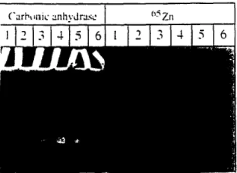
Figure 1. Bromcresol Purple Assay and Autorad. The
left panel is a photo of the bromcresol purple assay, the right is an autorad of the same gel. The CO2
concentrations were as follows: Lane 1: 1000 ppm, Lane 2: 1000 ppm switched to 300 ppm, Lane 3: 300 ppm, Lane 4: 300 ppm switched to 100 ppm, Lane 5: 100 ppm, and Lane 6: 100 ppm switched to 300 ppm.
Figure 1 is a photo of the bromcresol purple assay and corresponding
autoradiograph of the gel. Almost all of the cellular zinc was present in the same region
of the gel as the carbonic anhydrase activity, indicating that cellular zinc levels may be involved in regulating the amount of carbonic anhydrase and therefore CO2 uptake capacity. Cells grown at low CO2 levels had more carbonic anhydrase activity than cells
grown at higher C02, and adaptation to new CO2 levels occurred, at least partially,
within 18 hours of the switch to a different gas concentration.
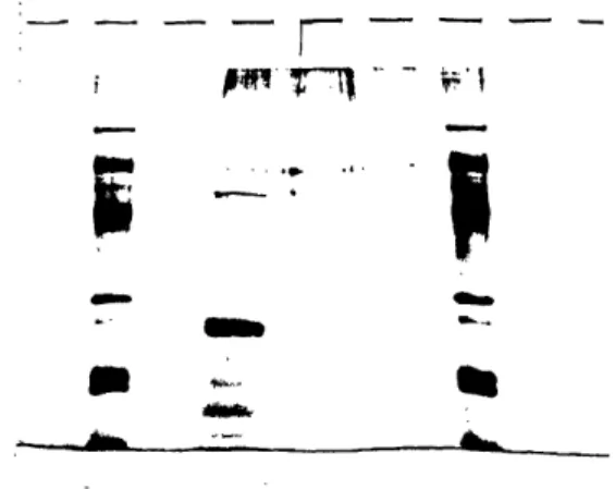
3.2 Protein Purification
The gel of the protein purification shows two proteins of very similar size at approximately 27 kilodaltons as well as several fainter bands of smaller sizes (Figure 2). The lower bands may be isoforms or degradation products of the 27 kD proteins. The doublet at 27 kD was confirmed to have carbonic anhydrase activity using the
activity, although there might not have been enough protein present for activity visible using this assay. Note that the bands present on the gel at approximately 66 kD are artifacts from the sample loading buffer.
--
r-*tiF"Z 'n
-m
Figure 2. Denaturing Polyacrylamide Gel of Protein
Purification. Bio-Rad low range SDS-PAGE standards,
found in the far left and far right lanes, are (from the top): 97.4 kD, 66 kD, 45 kD, 31 kD, 21.5 kD, and 14.5 kD. The high molecular weight bands (seen in empty lanes as well as sample lane) are artifacts from the sample loading buffer.
3.3 N-terminal Protein Sequence
N-terminal protein sequencing identified the first 15 amino acids of the protein as: 1. ala, 2. unknown, 3. glu or gly, 4. glu, 5. val, 6. gin, 7. asp, 8. gly, 9. phe, 10. arg,
11. arg or tyr, 12. his, 13. his, 14. tyr, 15. asp.
3.4 Internal Protein Sequences
Two fragments of the protein were sequenced. Eight amino acids of fragment 1
were identified as: 1. ile, 2. ser, 3. ala, 4. ser, 5. ser, 6. phe, 7. asp, 8. lys.
;-.
r,
Eighteen amino acids of fragment 2 were identified as: .lys, 2. met, 3. gin, 4. arg, 5. asp, 6. asp, 7. met, 8. ser, 9. asp, 10. asp, 11. leu, 12. tyr, 13. ala, 14. pro, 15. gly, 16. unknown, 17. arg, 18. gin.
3.5 Total RNA Extraction
An estimate of cellular RNA concentration as approximately 8 pg per cell was
obtained based on very rough calculations. The total yield of mRNA was 1.6 X 10⁻⁶ g
mRNA obtained from 2 X 10⁸ cells, or 8 X 10- 1 5g mRNA per cell. Assuming that
mRNA is about 1% of total RNA, and 10% efficiency in the harvest protocol, the
cellular RNA concentration is therefore approximately 8 pg per cell.
3.6 The Library
The titer of the T. weissflogii Lambda Zap II library on January 9, 1994 was
determined to be 1010 pfu per ml. The ratio of empty vector to vector with insert was
approximately 4 to 1.
3.7 PCR Using Degenerate Primers from Protein Sequence
From the PCR reaction using primers CAN2 and RevDLY, a PCR fragment
clone 11.3 was isolated. Clone 11.3 contained sequence coding for both the N-terminal protein fragment as well as both internal protein fragments. The location of this clone on the carbonic anhydrase gene map in figure 3 is from base 543 through base 1072.
3.8 PCR product from Nested PCR Reaction
A larger segment of the carbonic anhydrase gene sequence was obtained by performing PCR with first the T3 and BestHind primers, followed by PCR with the SK
and CA4r primers. Two clones, TA2 and TA3 were sequenced and found to partially match the sequence of clone 11.3. The position of the TA2 and TA3 sequence on the
carbonic anhydrase map in figure 3 is from base 01 to base 1076. This sequence also indicated that the carbonic anhydrase gene had more 5' terminal sequence located upstream of the N-terminal protein sequence described in section 3.3. Therefore, this
carbonic anhydrase might have a cleaved signal sequence used for cellular localization, or alternatively the protein used for N-terminal sequencing had been partially degraded by
proteases prior to the sequencing reaction.
3.9 Library Screening
Of the 3.2 million pfu screened with the probe created from clone TA3, 70 clones
were isolated that hybridized to the probe during both the primary and secondary
screening. PCR on these 70 clones using the NtForw and BestHind primers identified 11
clones that were long enough that they might contain the 5' end of the probe. Clones
CA1, 2, 3, 6, 9, 13, 27, 44, 56, 58, and CA60 were partially sequenced and found to be identical. Clone CA44 was selected for complete sequencing.
3.10 Clone CA44 Vital Statistics
Clone CA44 was 1.4 kb long and encoded a 32 kD protein (Figure 3). The coding region began with a methionine (Met 106) at position 315 and ended with a stop codon at position 1210. The sequence encoding the amino acids predicted to be at the N-terminus was present, but it was 72 amino acids into the coding region. A
comprehensive restriction map of the carbonic anhydrase gene is provided in appendices
one and two.
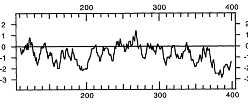
This protein had some slightly hydrophobic regions (figure 4), including one of
roughly 20 amino acids at the very beginning of the translated protein sequence (in the
putative signal sequence). The remainder of the protein was hydrophilic, with the
exception of a slightly hydrophobic stretch near the middle.
Figure 3. CA44 DNA Sequence and Corresponding Amino Acid Sequence
CAC GCC AAA GAC GAA AAC TCC AAG
his ala lys asp glu asn ser lys
GTC GGA CTT CTT GCT TCC ACC ATC val gly leu leu ala ser thr ile
GCC GCT GCA
ala ala ala
CCG GCT ACT GCC GAG pro ala thr ala glu
GAC AAC TTC CTT GTT CCC ATC GAC asp asn phe leu val pro ile asp
ATC TTC GAC GAG GGC ACC AAC GTA ile phe asp glu gly thr asn val
TGC ATC CAC ATG CCT GGT CCT CAA cys ile his met pro gly pro gln
ATG GAA GTC GAC GTA GTC CCC AAC met glu val asp val val pro asn
AAT GTT CAC TGG CAT CTT GGA ACC
asn val his trp his leu gly thr
AGT GGT CCG AAC GGA AAC GTT GGC
ser gly pro asn gly asn val gly
CAG GAT GGA TTT CGC TGC CAT CAC gln asp gly phe arg cys his his
GAA TGG
glu trp
TCT GGA
ser gly
AAA CAC TGC ATT GGG ATG
lys his cys ile gly met
GCC GGC GCA TGT GGA ACC ala gly ala cys gly thr TTC TGC AAC CTC GAT ATG GAG ACT phe cys asn leu asp met glu thr
GTT GGA GTT CAA GGA CAA ATC TTC
val gly val gln gly gln ile phe
TTG ATT CGA GGA TGG ATT GTC GAT
leu ile arg gly trp ile val asp
ACC GGA TCC ACC ACT GGG GAG AGT thr gly ser thr thr gly glu ser
ACC TGG CAA GTT GAT CGC AAG TGC
thr trp gln val asp arg lys cys
1
TAC GAT ATG AAG ATG CAA CGT GAT
tyr asp met lys met gln arg asp
1
GAA CTG GTT ACA CCC GAG TAT GTA glu leu val thr pro glu tyr val
31/11 TTG AAA leu lys 91/31 GCG CTT ala leu 151/51 GAT AAG asp lys 211/71 ATC GTC ile val 271/91 TGT GCC cys ala 331/111 GCT GGC ala gly 391/131 ACC AAG thr lys 451/151 GAA CAC glu his 511/171 GTT CCT val pro 571/191 TAC GAC tyr asp 631/211 GAA GTT glu val 691/231 ACC TAT thr tyr 751/251 CTT CAA leu gln 811/271 ACC ATT thr ile 871/291 GAA GAA glu glu 931/311 CGC AGC arg ser 991/331 CAC AAG his lys 1051/351 GAC ATG asp met 1111/371 GCT AAC ala asn GCC GCT GTG TGC GTG CTT GGA CTC ala ala val cys val leu gly leu GCC GTT CAG AAC AAC AGT TCC AGC ala val gln asn asn ser ser ser
ACC GTT GCG ACC CTC GAA GCC AGT
thr val ala thr leu glu ala ser
CCC GAA AGG GCC ACT GCT GAG ATC
pro glu arg ala thr ala glu ile
GAG AAG GCG ATC AAG CTG GAC AAC
glu lys ala ile lys leu asp asn
GCT AAC GTC ACC AAG GGA TTC AAG ala asn val thr lys gly phe lys AAT TAC TGG CAA AGC TCC ATG TGC asn tyr trp gln ser ser met cys TAC TCT GTC GGC GAG TAT GAC GAA tyr ser val gly glu tyr asp glu
TAC CGC CGT ACC CTT GCC GAG GGA tyr arg arg thr leu ala glu gly
CCT GAC GAC GAA GCG TAC ACC AGG pro asp asp glu ala tyr thr arg
GGA GAG ACA TAT GAA GTT CAT TGG
gly glu thr tyr glu val his trp CAG TAC CAA ACA CCT TTC TAC GAT gln tyr gln thr pro phe tyr asp ACT
thr
CTT GCG CCC CAG GAC ATT GCG leu ala pro gln asp ile ala
GTC AAT GAC GAC ACA TAC TAC TAC val asn asp asp thr tyr tyr tyr ATG GGA ATG GGT CAA GAC ATC GCC met gly met gly gln asp ile ala AAT GAA ATT TGT TCA TCC TAC TCC asn glu ile cys ser ser tyr ser
ATC AGT GCT TCC TCC TTC ile ser ala ser ser phe
AGT GAT GAC TTG TAC GCA ser asp asp leu tyr ala
AAC asn GAT AAG asp lys CAT GGA his gly
CAG CAA ACC CGT CGT CTC ACT gln gln thr arg arg leu thr 1/1 CAA CAA gln gin 61/21 TCC ACT ser thr 121/41 AAC GCA asn ala 181/61 GGA TCT gly ser 241/81 GCC ACC ala thr 301/101 GTT GAC val asp 361/121 GGA TTG gly leu 421/141 CCC GTC pro val 481/161 AAT GGC asn gly 541/181 GAA GTG glu val 601/201 CCC TAT pro tyr 661/221 CCT CAC pro his 721/241 GGT GTA gly val 781/261 AAC GCA asn ala 841/281 CCT GAT pro asp 901/301 ATG TAC met tyr 961/321 CCC ATT pro ile 1021/341 CTT TGC leu cys 1081/361 TCC AGG ser arg
1141/381 1171/391
GAG AAG CAT GAA CAC AAT CAC AGC CAT GGT CAC AGC CAT GTA CGT GGT CAC CAG CAC CAC glu lys his glu his asn his ser his gly his ser his val arg gly his gln his his
1201/401 1231/411
CAA TGG TTT TAG GTT GTC GAT GAG TGT ATG GAT GAT GCT CTT TAG TTT TGT ACG TCT CAC
gln trp phe AMB val val asp glu cys met asp asp ala leu AMB phe cys thr ser his
1261/421
GAA TAT GTT TAT TAC AGA TTT CCG
glu tyr val tyr tyr arg phe pro 1321/441
AAT TAT TAG TTT CCT TAA AAA AAA
asn tyr AMB phe pro OCH lys lys 1381/461
AAA AAA AAA AAA AAA AAA AAA AAT
lys lys lys lys lys lys lys asn
1291/431
GAG CCA ATA TTA ATT TCA ATT AGT TAA TTC TAA ACA
glu pro ile leu ile ser ile ser OCH phe OCH thr 1351/451
AAA AAA AAA AAA AAA AAA AAA AAA AAA AAA AAA AAA
Figure 4. Kyte Doolittle Hydrophobicity Plot of the CA44 Protein
Sequence. Translated protein sequence begining at Met 108. Amino acidposition is on the horizontal axis and hydrophobicity on the vertical axis.
200
300
400
2 1 0 -1 -2 -3200
300
400
2 1 0 -1 -2 -34. Conclusions
The T. weissflogii carbonic anhydrase gene described in this thesis does not appear to have homology to any published algal chloroplastic or periplasmic carbonic anhydrase isoforms. The Chlamydomonas reinhardtii periplasmic genes exhibit some homology to mammalian carbonic anhydrases (Fukuzawa et al. 1991), and many of the algal chloroplastic genes are homologous to the chloroplastic carbonic anhydrases of higher plants such as pea (Fukuzawa et al. 1992). Since the T. weissflogii protein is in the same size range as the other carbonic anhydrases and has the carbonic anhydrase
enzyme activity, the lack of homology might simply reflect the fact that T. weissflogii is
evolutionarily quite distant from these other organisms. Alternatively, the sequence
differences might suggest slightly different properties of the T. weissfilogii enzyme. For
example, it might be associated with a transporter, a proton pump, or some other protein utilized in the carbon accumulation mechanism.
The protein predicted to be translated from clone CA44 was 32 kD,
approximately 8 kD longer than expected. The amino terminal amino acids sequenced
from the purified carbonic anhydrase are 72 residues downstream of the putative start
methionine predicted by the CA44 cDNA. It has not been determined whether the
proteolytic events that generated the sequenced polypeptide were a result of proteolysis
during protein purification or a physiological protein processing event. If, however, the
N-terminus of the sequenced protein was not degraded, then the roughly 8 kD protein
encoded upstream of it is probably a signal sequence responsible for sending the protein
to the correct cellular location. Many of the amino acids at the beginning of this stretch
of 72 residues are slightly hydrophobic, so it is possible that this portion of the protein might serve as a signal sequence and be cleaved after insertion into the endoplasmic reticulum.
The apparent size of the purified carbonic anhydrase on the denaturing
polyacrylamide gel was 27 kD. This is different from the size of the predicted protein beginning at Met 106 (-32 kD) as well as the predicted protein beginning at Ala 178 (-24 kD). There are at least two possible explanations for these discrepancies. The simplest explanation is that salts from the purification protocol might have caused the sample to run differently from the standards in the gel. Additionally, one or more post-translational modifications such as glycosylation and signal sequence cleavage might
cause the final protein size to be larger or smaller than that predicted by the gene
sequence. There are three potential N-linked glycosylation sites present in the protein
which would act to make the protein larger than predicted. The consensus sequence for
this glycosylation event, Asn-X-Ser/Thr, is present at Asn 114, Asn 161, and Asn 386.
Therefore, if the preprotein that is initially translated is glycosylated at one or more sites,
it would be larger than 32 kD, and if a signal sequence were then cleaved, the protein
would be smaller again. This could result in a final protein of 32 kD.
This carbonic anhydrase gene is a valuable tool that can be used to characterize the inorganic carbon acquisition mechanism utilized by T. weissflogii. The cloned gene can now be manipulated to create recombinant protein which can be synthesized and
purified in large quantities. This recombinant protein can be used to perform in vitro
experiments designed to ascertain the structure and function of the enzyme. For
example, the enzyme kinetics can be examined at different CO2 concentrations. Also,
the recombinant protein can be used to raise antibodies. These antibodies can then be used to visualize carbonic anhydrase cellular localization, as well as in field studies to examine the occurrence of this enzyme in the environment. Also, the gene sequence can
be used to generate probes to examine regulation of the T. weissflogii carbonic
anhydrase in response to different environmental stimuli. Specifically, the molecular mechanisms by which zinc and CO2 modulate carbonic anhydrase enzyme levels can be
studied. The probes can be used to examine mRNA levels due to changes in
transcription induced by different zinc and CO2 concentrations. Finally, the purification
technique can be modified to try to detect additional carbonic anhydrase isoforms. All of
the information obtained by these experiments should lead to a better understanding of
Appendix One
Restriction Map of Carbonic Anhydrase clone CA44
Ple I
NspB II Hinf I
Fnu4H I Tthlll II Mme I
II I I I
CAACAACACGCCAAAGACGAAAACTCCAAGrGA-aAGCC .GCTGTGTCGTGCTTGGACTC CACTGTCGGACTTCTGC 80
GG
± ±)Gl±C-GT
GGTTTCTCC
ACGCACGA
C1A ACAGCC
I
'
11
'
I .
37 50 67 38 56 56 Fnu4H I BspW I Bbv I NspB II Fnu4H I BceF I AlwN I Msp IHinP I Fnu4H I Hpa II
Hha I BspW I CfrlO I Mnl I
BstU I Bbv I Bsg I BsaJ I
II I II 1111 II II
TTCCACCATCGCGCTTGCC GTTCAGAACACCG CTGCA C C TAACCG 160
AAGGTG;GTAGCGCGA~cGCaGtclFGTCAAGcG¶IvrGCIcG rGGCl~CGATGACGGCTCCTATTCTGGC II . II II i i I 1- 90 124 131 146 91 124 135 148 91 124 136 97 125 136 127 128 130 130 130 Sau3A I Mbo I Sau3A I Dpn II Mbo I Dpn I Dpn II BstY I BspW I
Taq I Alw I Tthlll I Hae III Dpn I
Mnl I Bsr I Taq I BsiY I Sau96 I Dde I
I I I I I I I I II I I TTGCGACCCTCGAAGCCAGTGGATCTGACAACTTCCTTGTTCCCATCGACATCGTCCCCGAAASGGCCACTGCTGAGATC 240 AACGCTGGGAGCTTCGGTCACCTAGACTGTTGAAGGAACAAGGGTAGCTGTAGCAGGGGCTTTCCCGGTGACGACTCTAG 168 176 206 216 224 233 170 181 208 225 237 181 232 182 237 182 237 182 237 182
ScrF I EcoR II Dsa V Nla III NspC I Alu I Nsp7524 I Nla IV Sau3A I Nsp I
Ban I Mbo I Fok I BstN I
Taq I Bsp1286 I BspW I Dpn II Hinc II BstX I
Mbo II Mnl I Mae II Bal I Dpn I Mae II SfaN I BstK I
I I III I I I I II II I II I
GCC ACCTTTCGACGACC AAGCGGACAA CG T TGACTGCATCCAATGCC 320
CGGTGGTAGAAGCTGCTCCCGTGGTTGCATACACGGCTCTTC
1I*1 1II. I I I I I1I 11- 111 I
248 256 266 274 285 299 308 319 251 258 274 285 301 312 259 285 309 319 259 285 314 290 314 314 315 319 319 319 Mae II Sty I Tthlll I
Mnl I HinP I BsaJ I Taq I
BsiY I Hha I Hph I Sal I
Sau96 I Hae II Mae III Tfi I Hinc II
Ava II Alu I Mae II Hinf I Acc I
I I I 11 I I I II II
¶IGMTCCTrcAAGCTGrr-rW AACGTACCAAGGGNmAAGGGATGAAGTCGACGTAGTCCCCAACACCAAGAATT 400
ACCAGGAGTTCGACCGCGA CAGTGGTTCCCTAAGTTCCCTAACTACCTTCACTGGTTCAGGGGTTGTGG TTAA
I I I II I · I II II 322 330 341 353 373 322 334 343 353 373 325 335 344 373 325 335 347 374 347 376 377 Nla III BspW I SfaN I Bsr I Alu I Bsp1286 I Bsr I Nla IV 11I I I I
ACTG GCTCCATGTGCCCCGTCAATGTCCTACTCTG AGTATGACGAA 480
TGACCGTTTCGAGGTACACGCCAGTGACAAGTGAGACAGCCGCTCATACTGCTT
II I I I .I .. 401 409 417 434 445 410 438 414 EcoN I BsiY I Rsa ISau96 I Csp6 I Mnl I Fok I Fnu4H I
Ava II Mae II BceF I BsaJ I Bsg I Bbv I
I I I I I I I I I I AATGGCAGTG GTCCGAACGGAAACGTTGGCGTTCCTTACCGCCGTACCCTTGCCGAGGGAAAGTGCAGATGGA'ITTCG 560 TTACCGTCACCAGGCTTGcTTTGCAACCGCAAGGAATGGCGGCATGGGAACGGCTCCCTCTTCACGTCCTACCTAAAGC I . I -I I I I I · I I I 490 503 521 533 544 560 490 524 535 549 560 524 528 528
BsiY I Sau96 I ScrF I EcoR II Dsa V BstN I BstK I
Rsa I EcoO109 I BsmA I
Csp6 I Hae III Fok I Mme I
I IlI 11II I
CTGCCATCACTACGACCCTGACGACGAAGCGTACACCAGGCCA
G 640
GACGGTAGTGAGCTGCTGTCCGGGATACTACCGTGACGTAACCCTACCTCAACCTC
· 591 599 626 634 591 598 640 596 596 596 596 596 599 602 NspC I Nsp7524 I HinP I Msp I Hpa IIMnl I Nae I Nla III
BsiY I CfrlO I Tthlll II
Hae III Nla IV Nsp I Rsa I
Nde I Hae I Gsu I Hha I Nla IV Csp6 I
I 111 I I II I 11 I I I
AACATATGAAGTTCATTGGC CACTC AGCCGGCGCATGTGGAACCACCTATCAGTACCAAACACCTTTCTACGAT 720
TCTGTATAC TTCAAGTAACCGGAGTGAGACCTCACACGGTGGATAGTCA TGCTA
I 11-1 I I 11 I II I I' 644 658 668 677 685 699 659 670 679 699 661 673 703 661 673 680 674 674 677 679 679 ScrF I EcoR II Dsa V Mbo II BstN I Ear I BstK I
Taq I Ple I BsaJ I
Mnl I Hinf I HinP I
BsiY I BsmA I Hha I Mme I
I I I I I I 1
GGTGTATT CTGCAAC CTCGATATGAGACTTTCAAACTCTGCGCCCCAGGACATTGCGAACGCAGTTGGAGTTCAAGG 800 CCACATAAGACGTTGGAGCTATACCTCTGAGAAGTTTGAGAACGCGGGGTCCTGTAACGCTTGCGTCAACCTCAAGTTCC I I . I I II · II I 735 745 763 787 735 747 763 737 747 767 749 768 750 768 768 768 768 Mnl I Taq I
Hph I Tfi I Fok I Mbo II
Mbo II Hinf I Taq I
I I I I I I I
ACAAATc TTCACCATTGTCAATGACcGCACA TACTACTACCCTGATTTGATTCGAGGATATGTCGATGAAGAATGG 880 TGTTTAGAAGTGGTAACAGTTACTGCTTGTGTATGATGATGGGACTAAACTAAGCTCCTACCTAACAGCTACTTCTTTACC I. . 1. -I I · · 806 849 866 809 849 856 871 852 854
BsiY I Sau3A I Mbo I Dpn II Dpn I Nla IV BstY I Msp I Hpa II Fnu4H I Rsa I BamH I Bbv I Csp6 I Alw I Ple I
Nla III Alw I Bsr I Hinf I Fok I
I I I I l I I I I I GAATGGGTCAAGACATCGCCATGACACCCCGGATCCACCACTGGGAGAGTCGCAGCAATGAAATTTGTTCATCCTACTCC 960 CTTACCCAGTTCTGTAGCGGTACATGTGGCCTAGGTGGTGACCCCTCTCAGCGTCGTTACTTTAAACAAGTAGGATGAGG I I I II I I I . I 900 910 919 927 950 903 911 927 903 910 932 908 932 908 910 910 911 911 911 911 914 ScrF I EcoR II Dsa V BstN I Sau3A I Sau3A I
BstK I Mbo I Mbo I Alu I
Pf1M I Dpn II Dpn II Taq I SfaN I
BsiY I Dpn I Dpn I Mnl I Hind III Mbo II
I I I I I I I I I
CCCATTACCGCAGT TT CAGATC GCTCCTTCGAACT CAACGATATGAAGAT 1040
GGGTAA TGGACCGG TTCAAC GTAGAA CG ATGCTATACTTCTA
II- I. I. I I I -962 979 996 1007 1018 1035 962 979 996 1013 1038 968 979 996 1019 968 979 996 968 968 ScrF I EcoR II Dsa V BstX I BstN I BstK I BsaJ I Sau3A I Mbo I Dpn II Dpn I Alw I Nla IV BstY I BamH I
Rsa I Nla III Mae III
Mae II Nla III Csp6 I Alw I Bsr I Ava I Alu I
I I I i 11 I I I I
GCAACGTGATGACATGAGTGATGACTTGTACATGGATCCAGGGAACTGGTTACACCCGAGTA TGTA GCTAACAACC 1120
CGTTGCACTACTGTACTCACTACTGAACATGCGTGTACCTAGGTCCCTTGACCAATGTACATCGATTGTT II II· I' · 1 I* 044 1053 1068 1078 1089 1099 1110 1068 1075 1093 1078 1078
Mae III Mae II
Nla III BsaA I
Sty I Rsa I
Nco I Csp6 I Hph I
Dde I Dsa I Nla III Mae III
BsmA I Nla III BsaJ I BstX I BstE II
I I I II I 11 III III
AGCAAACCCGTCGTCTCACTIACGCA GAAQCAATCACAGCCATGGTCACAGCCA GT&ACGGWACA CACCAC 1200
TCGIITnGVIKW>K K O3NCTCTTCGTACTTGTGTTAGTG CAGATTCGGTACATGCACCAGTGGTCGTGGTG
.to I 1 I II 11 111 111 1133 1147 1164 1176 1186 1139 1164 1177 1187 1164 1180 1188 1164 1180 1165 1181 1169 1182 BsmA I Mae II SfaN I Rsa I Taq I Fok I Csp6 I I I I I I I
CAATCGTTTTAGGTTGCATGAGTGTATGGATATCTCTTTAGT T TTGTACG TCTCACGAATATGrTTTATTACAGATT 1280 GTTACCAAAATCCAACAGCTACTCACATACCTACTACGAGAAATCAAAACATGCAGAGTGCTTATACAAATAATGTCTAA · I · III 1217 1230 1250 1234 1250 1252 1254 Nla IV Msp I Mse I Hpa II Ase I
BsDE I Ss I Mse I Mse I
II I I II I I