Publisher’s version / Version de l'éditeur:
Chemical Communications, 31, pp. 3924-3926, 2005
READ THESE TERMS AND CONDITIONS CAREFULLY BEFORE USING THIS WEBSITE. https://nrc-publications.canada.ca/eng/copyright
Vous avez des questions? Nous pouvons vous aider. Pour communiquer directement avec un auteur, consultez la
première page de la revue dans laquelle son article a été publié afin de trouver ses coordonnées. Si vous n’arrivez pas à les repérer, communiquez avec nous à PublicationsArchive-ArchivesPublications@nrc-cnrc.gc.ca.
Questions? Contact the NRC Publications Archive team at
PublicationsArchive-ArchivesPublications@nrc-cnrc.gc.ca. If you wish to email the authors directly, please see the first page of the publication for their contact information.
NRC Publications Archive
Archives des publications du CNRC
This publication could be one of several versions: author’s original, accepted manuscript or the publisher’s version. / La version de cette publication peut être l’une des suivantes : la version prépublication de l’auteur, la version acceptée du manuscrit ou la version de l’éditeur.
For the publisher’s version, please access the DOI link below./ Pour consulter la version de l’éditeur, utilisez le lien DOI ci-dessous.
https://doi.org/10.1039/b504186d
Access and use of this website and the material on it are subject to the Terms and Conditions set forth at
Fluorescence properties of gold nanorods and their application for
DNA biosensing
Li, Chen-Zhong; Male, Keith B.; Hrapovic, Sabahudin; Luong, John H. T.
https://publications-cnrc.canada.ca/fra/droits
L’accès à ce site Web et l’utilisation de son contenu sont assujettis aux conditions présentées dans le site LISEZ CES CONDITIONS ATTENTIVEMENT AVANT D’UTILISER CE SITE WEB.
NRC Publications Record / Notice d'Archives des publications de CNRC:
https://nrc-publications.canada.ca/eng/view/object/?id=ccde1120-19d1-4195-96f2-cd47f4cb29f6
https://publications-cnrc.canada.ca/fra/voir/objet/?id=ccde1120-19d1-4195-96f2-cd47f4cb29f6
Fluorescence properties of gold nanorods and their application for DNA
biosensing{
Chen-Zhong Li, Keith B. Male, Sabahudin Hrapovic and John H. T. Luong*
Received (in Cambridge, UK) 23rd March 2005, Accepted 27th May 2005 First published as an Advance Article on the web 4th July 2005
DOI: 10.1039/b504186d
This communication reveals new and unique optical properties with respect to enhanced fluorescence of gold nanorods as they elongate; a novel strategy for DNA hybridization studies based on monitoring the fluorescence intensity of gold nanorods has been demonstrated.
The optical and electrical properties of colloidal gold nanoparticles are known to be dramatically affected by their size, shape, and surrounding surface environments.1 The effect of rodlike gold metal structures on the optical properties has been extensively studied.2 For example, rodlike gold nanoparticles exhibit a red shift in the longitudinal plasma resonance with increasing aspect ratio while the transverse resonance remains the same.2Since size-dependent photoluminescence of nanosized semiconductor materi-als was first reported by Mooradian,3the visible photolumines-cence of sufficiently small gold nanoclusters (,5 nm) has also been observed.3 In the past years, some experimental work and theoretical calculations have been reported concerning the fluorescent properties of gold nanorods. Importantly, an enhanced (compared to bulk) detectable fluorescence emission with a low quantum efficiency for short nanorods (or the rods with a low aspect ratio) has been demonstrated by two fluorescence techniques (fluorescence emission and two-photon-induced photo-luminescence spectroscopy),4however, the quantum efficiency of such nanorods is very low, limiting the widespread use of fluorescence properties in biosensing.
The unique properties of nanoscale colloidal particles are studied increasingly for their potential in various biosensor developments.5 The optical properties of three-dimensional aggregations of gold nanoparticles have been used to detect hybridization of specific DNA sequences in solution and on surfaces6 as an alternative to fluorescent labeling of DNA. Nevertheless an analytical strategy based on gold nanorods relying on the specific fluorescent properties has not been attempted.
In this paper, an outstanding fluorescence feature of longer gold nanorods (.200 nm) excited at 690 nm was observed in terms of a strong fluorescence emission at 743 nm and a relatively weak fluorescence at 793 nm, which is remarkably different from previous reports.4 This unique fluorescence feature of gold nanorods was also imaged in the solid state with a microarray fluorescence scanner. Additionally, the decreasing fluorescence of
oligo-functionalized gold nanorods was extended to monitor DNA hybridization.
The longer gold nanorods were prepared chemically in solution by a seed-mediated method according to Murphy and co-workers,7 however short gold nanorods synthesized by an electrochemical method (ESI{, Fig. S1)7were used as seeds rather than gold nanoparticles. Optical absorption spectra and electron microscopy were used to assess the quality and polydispersity of the rods. As expected, the nanorods showed both a transverse plasmon band at 530 nm, and a longitudinal one at 1600 nm in the near-IR region (ESI{, Fig. S2), which corresponded to the high aspect ratio of the synthesized gold nanorods.7The gold nanorods have an average diameter of 17 ¡ 5 nm and an average length of about 230 ¡ 30 nm (corresponding aspect ratio 13) as verified by the analysis of TEM images (ESI{, Fig. S2).
The plasmon resonance for nanorods is known to split into two bands.1The high-energy band corresponds to the oscillation of the electron perpendicular to the rod axis (transverse surface plasmon absorption). The other absorption band, which is red-shifted to lower energies, is caused by the oscillation of electrons along the rod axis (longitudinal surface plasmon absorption). As the aspect ratio (length over width of the rod) increases, the energy separation between the resonance frequencies of the two plasmon bands increase. Fluorescence spectra were measured with a Gilford Fluoro IV Spectrofluorometer. The gold nanorods exhibit two emission bands at 741 nm and 793 nm (Fig. 1), attributed to the transverse and longitudinal surface plasmon resonances according to the theoretical calculation of the photo-induced luminescence.4,8 To be certain that impurities are not the source of visible fluorescence, the synthesized gold nanorods were redispersed in distilled deionized water and intensely washed by centrifugation at least 5 times until most of the surfactant had been removed.
Compared to shorter rods with lengths 30–120 nm prepared by the methods of Nikoobakht and El-Sayed,7 the longer rods showed very strong fluorescence bands that are red-shifted in the emission spectrum. The fluorescence intensity of gold nanorods decreased linearly with decreasing concentration of the nanorod solution (Fig. 2). The synthesized CTAB-capped (positively charged) gold nanorods agglomerate irreversibly even in very low ionic strength solution, an effect which has been well known for gold nanoparticles.9As a control experiment, all the individual chemical reagents used in the synthesis were measured and no detectable fluorescence was observed. The theoretical and experi-mental results of Mohamed et al.4also show a drastic increase in the fluorescence of gold nanoparticles as they elongate. The inset photographs of Fig. 1B shows that the longer rods (230 nm) with aspect ratio (AR) 13 are strongly fluorescent, while the inset for the
Nanobiotechnology & Biosensor Group, Biotechnology Research Institute, Canada National Research Council, 6100 Royalmount Ave., Montreal, Canada H4P 2R2. E-mail: john.luong@cnrc-nrc.gc.ca; Fax: +1 514 496 6265; Tel: +1 514 496 6270
{Electronic supplementary information (ESI) available: Experimental details, TEM characterization and UV and near-IR spectra. See http:// dx.doi.org/10.1039/b504186d
COMMUNICATION www.rsc.org/chemcomm | ChemComm
shorter one (30 nm, AR 5 2.5) is virtually nonfluorescent. It has been shown that the fluorescence quantum efficiency of gold nanorods is observed to increase with increasing the nanorod length,4 including a rationale behind such a phenomenon. The CTAB itself did not exhibit any appreciable fluorescence at 690 nm excitation.
Fluorescent labels commonly used in DNA microarray analysis, such as Cy3 and Cy5, are expensive, photosensitive and special care must be taken to minimize their exposure to light during the labeling, hybridization, washing and scanning processes.10 In contrast, gold nanorods are not photobleached and can be readily incorporated with thiol derivatized DNA targets through S–Au bonds. Therefore, gold nanorods could possibly be considered as an alternative fluorescent label for heterogeneous DNA analysis. Sequence-specific DNA detection techniques have been developed
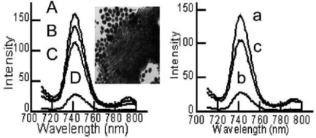
which rely upon target hybridization with radioactive, fluorescent, chemiluminescent and other types of labeled probes.10Since the first reports by Alivisatos and Mirkin6in 1996, gold nanoparticle labeling of DNA molecules has been considered as an alternative marker for the monitoring of DNA hybridization events and even for the detection of a single base mismatch in the oligonucleotide sequence. In general, the scheme is based on the colorimetric change associated with particle aggregation caused by the formation of networks of spherical gold nanoparticles crosslinked by oligonucleotide hybridization events. Similarly, the self-assembly of oligonucleotide functionalized short gold nanorods also has been established.6Herein following the design concept of DNA-nanoparticles, a fluorescence-based method for the determi-nation of DNA sequences and monitoring reversible DNA hybridization events was attempted by using DNA functionalized gold nanorods. We designed two complementary oligonucleotide sequences (I: 59-SH–(CH3)6–GGGGGGGGATGGGGGGGG-39; II: 59-SH–(CH3)6–CCCCCCCCATCCCCCCCC-39) and one noncomplementary oligo (III: 59-SH–(CH3)6–TTTTTTTTGCT-TTTTTTT-39) with SH– linkage that could be immobilized on the surfaces of two sets of gold nanorods through SH–Au specific binding. In order to minimise irreversible and nonspecific particle aggregation, the gold nanorods were modified by adding designed oligonucleotides (1 mM) into the aqueous nanorod solution at room temperature. Same sequence oligos without a thiol linkage were used as a control to identify the non specific binding (phosphate backbone–CTAB Au binding) between DNA and the gold surface. After 24 h incubation, the ionic strength of the aqueous solution was gradually increased by adding phosphate buffer to increase the specific binding efficiency of DNA on gold surfaces.9The final solution in 100 mM phosphate buffer (pH 7.5) was further incubated for 24 h, then was centrifuged for 20 min at 10 000 rpm. The precipitate was redispersed in the same amount of phosphate buffer in the presence of 100 mM NaCl for DNA concentration measurements and for the hybridization experi-ments. The final concentration of thiolated DNA was determined to be 125 pM by UV-visible spectroscopy at 260 nm by subtraction of the initial concentration of gold nanorod–DNA mixture and the concentration of the supernatant. In contrast, the low concentra-tion (20 pM) of the remaining non thiol-linked oligos measured by the same procedure indicates the relatively weaker binding (non-specific binding) between the DNA backbone and CTAB capped gold nanorods due to the opposite charge properties. When identical amounts of suspensions of two complementary oligo (I and II) modified nanorods were mixed, the intensity of the fluorescence spectra immediately started to decrease, which can be attributed to the formation of DNA-linked three-dimensional aggregates of gold nanorods. If the solution was allowed to stand for over 2 h, the fluorescence of the solution was efficiently decreased (Fig. 3D) due to the precipitation of the aggregates, down to 15% of the initial value. In contrast, for the mixture of two non-complimentary (I and III) oligo functionalized nanorod systems, no significant decrease (20%) was achieved even if the solution was kept for over 4 h (Fig. 3C). The assembled network of gold nanorods aligned in parallel stacks upon DNA hybridization (Fig. 3 inset) was also identified by transmission electron microscopy, which is consistent with the previous report.6e Fluorescence spectroscopy also revealed the reversibility of the thermally induced DNA dissociation process (melting analysis). Fig. 1 Fluorescence spectra of longer gold nanorods (230 nm, blue line)
and shorter gold nanorods (30 nm, red line) in aqueous solution. (A) Excitation spectra emitted at 741 nm. (B) Emission spectra excited at 690 nm. The inset of (B) shows the illumination photographs of long rods (a) and short rods (b) in aqueous solution. The solution was excited at 690 nm by a 150 W xenon UV lamp. We examined the fluorescence intensity of short rods, prepared from different gold salt solutions. None of these short rod containing solutions, assuming different rod concentra-tions, displayed significant fluorescence intensity.
Fig. 2 The fluorescence intensity decreases linearly with decreasing concentration units of gold nanorods.
The intensity of the fluorescence was essentially recovered up to 70% of the initial value (Fig. 3c) by heating the solution at 60 uC due to the reversible dissociation of aggregates formed by hybridization of the two complementary DNA attached gold nanorods. Compared to the initial value of fluorescence, the apparent difference in intensity of the 745 nm band which accompanies the melting process could be attributed to irreversible aggregation due to the charge screening effects of the electrolyte. The decrease of the fluorescence signal might be related to the precipitation of the aggregates as described by Dujardin et al.6
In conclusion, we have discovered that sufficiently long gold nanorods (aspect ratio . 13) exhibit novel optical properties by means of relatively intense fluorescence emission at 743 nm and one weaker band at 793 nm. After functionalization by DNA probes, the DNA hybridization event could be effectively monitored by measuring the fluorescence intensity. The results suggest that the unique fluorescent properties of gold nanorods could potentially be exploited as a sensitive probe in fluorescence-based microarray assay and optical biosensor development. Work is in progress in our laboratories using DNA sequences of interest towards the detection of important pathogenic bacteria.
Notes and references
1 S. Link and M. A. El-Sayed, Int. Rev. Phys. Chem., 2000, 19, 409; T. J. Norman, C. D. Grant, D. Magana, J. Z. Zhang, J. Liu, D. Cao, F. Bridges and A. V. Buuren, J. Phys. Chem. B, 2002, 106, 7005; Z. Zhong, S. Patskovskyy, P. Bouvrette, J. H. T. Luong and A. Gedanken, J. Phys. Chem. B, 2004, 108, 4046.
2 C. Sonnichsen, T. Franzl, T. Wilk, G. von Plessen and J. Feldmann,
Phys. Rev. Lett., 2002, 88, 77402; A. Takami, H. Kurita and S. Koda, J. Phys. Chem. B, 1999, 103, 1226; B. Nikoobakht, J. Wang and
M. A. El-Sayed, Chem. Phys. Lett., 2002, 366, 17; M. B. Mohamed, K. Z. Ismael, S. Link and M. A. El-Sayed, J. Phys. Chem. B, 1998, 103, 9370.
3 A. Mooradian, Phys. Rev. Lett., 1969, 22, 185; J. P. Wilcoxon, J. E. Martin, F. Parsapour, B. Wiedenman and D. F. Kelley, J. Chem.
Phys., 1998, 108, 9137.
4 M. B. Mohamed, V. Volkov, S. Link and M. A. El-Sayed, Chem. Phys.
Lett., 2000, 317, 517; J. Zhu, Y. C. Wang and S. N. Yan, Chin. Phys. Lett., 2004, 21, 559; K. Imura, T. Nagahara and H. Okamoto, J. Am. Chem. Soc., 2004, 126, 12730; O. P. Varnavski, M. B. Mohamed,
M. A. El-Sayed and T. Goodson, III, J. Phys. Chem. B, 2003, 107, 3101; S. R. Nicewarner-Pena, A. J. Carado, K. E. Shale and C. D. Keating,
J. Phys. Chem. B, 2003, 107, 7360; R. M. Dickson and L. A. Lyon, J. Phys. Chem. B, 2000, 104, 6095.
5 C. A. Mirkin, Inorg. Chem., 2000, 39, 2258; G. Liu, T. M. H. Lee and J. Wang, J. Am. Chem. Soc., 2004, 127, 38; C. Z. Li, Y. Liu and J. H. T. Luong, Anal. Chem., 2005, 77, 478.
6 C. A. Mirkin, R. L. Letsinger, R. C. Mucic and J. J. Storhoff, Nature, 1996, 382, 607; R. Elghanian, J. J. Storhoff, R. C. Mucic, R. L. Letsinger and C. A. Mirkin, Science, 1997, 277, 1078; J. Reichert, A. Csaki, J. M. Kohler and W. Fritzsche, Anal. Chem., 2000, 72, 6025; A. P. Alivisatos, K. P. Johnsson, X. Peng, T. E. Wilson, C. J. Loweth, M. P. Bruchez and P. G. Schultz, Nature, 1996, 382, 609; E. Dujardin, L. B. Hsin, C. R. C. Wang and S. Mann, Chem. Commun., 2001, 14, 1264.
7 N. R. Jana, L. Gearheart and C. J. Murphy, J. Phys. Chem. B, 2001, 105, 4065; B. D. Busbee, S. O. Obare and C. J. Murphy,Adv. Mater.,
2003, 15, 414; S. S. Chang, C. W. Shih, C. D. Chen, W. C. Lai and C. R. C. Wang, Langmuir, 1999, 15, 701; B. Nikoobakht and M. A. El-Sayed, Chem. Mater., 2003, 15, 1957.
8 G. T. Boyd, Z. H. Yu and Y. R. Shen, Phys. Rev. B, 1986, 33, 7923. 9 N. R. Jana, L. A. Gearheart, S. O. Obare, C. J. Johnson, K. J. Edler,
S. Mann and C. J. Murphy, J. Mater. Chem., 2002, 12, 2909; L. M. Demers, C. A. Mirkin, R. C. Mucic, R. A. Reynold, III, R. L. Letsinger, R. Elghanian and G. Viswanadham, Anal. Chem., 2000, 72, 5535. 10 J. Malicka, I. Cryczynski, J. Fang, J. Kusba and J. R. Lakowicz,
J. Fluoresc., 2002, 12, 439; J. G. Wetmur, Crit. Rev. Biochem. Mol. Biol.,
1991, 26, 227. Fig. 3 Relative fluorescence intensities of fluorescence emission spectra:
Left. (A) Gold nanorods aqueous solution. (B) Oligo functionalized gold nanorods. (C) Mixture of non-complementary DNA functionalized gold nanorods. (D) Mixture of complementary DNA functionalized gold nanorods. Right. (a) Same as (B). (b) Same as (D). (c) After melting the hybridized DNA solution at 60 uC. The inset is a TEM image of self-assembled gold nanorods by DNA hybridization.