Cell and Nanomaterial-Based Approaches for
Diagnosis and Chemotherapy of Metastatic
Cancer Cells
By
Aditya Kohli
S.B. Massachusetts Institute of Technology
MASSACHUS ES-INSTITTE
OF TECHNOLOGY
AUG 1 6 2010
LIBRARIES
SUBMITTED TO THE DEPARTMENT OF BIOLOGICAL ENGINEERING IN
PARTIAL FULFILLMENT OF THE REQUIREMENT FOR THE DEGREE OF
MASTER OF ENGINEERING
AT
THE
ARCHIVES
MASSACHUSETTS INSTITUTE OF TECHNOLOGY
JUNE 2010
@ 2010 Massachusetts Institute of Technology.
All rights reserved.
Signature of Authar
/
Department of Biological Engineering
May 21, 2010
Certified by:A
Angela M. Belcher, Thesis Supervisor
Professor Materials Science and Engineering and Biological Engineering
/I/A
Accepted by:
Darrell
J.
Irvfne, Mast'rs <Vfrigineering Department Head
Professor Materials Science and Engineering and Biological Engineering
Cell and Nanomaterial-Based Approaches for
Diagnosis and Chemotherapy of Metastatic
Cancer Cells
By
Aditya Kohli
Submitted to the Department of Biological Engineering on May 21, 2010
in Partial Fulfillment of the Requirement for the Degree of Master of
Engineering in Biological Engineering
Abstract
Metastasis is a multistep process during which tumor cells separate from a primary tumor, penetrate the bloodstream, evade host defenses, and colonize distant organs. This final and fatal step in tumor development is the cause of more than 90% of cancer related deaths. Therapies and diagnostics can be targeted to metastasis at three points in its progression: the primary tumor, the secondary tumor, and circulating tumor cells (CTCs). While much work has focused on primary tumors, less effort has concentrated on targeted isolation, detection and therapy of deeply penetrated metastases and CTCs. Here, I discuss cell and nanomaterial-based approaches for detecting and ablating these malignant populations. The number of CTCs in the blood directly correlates with disease progression; however, the lack of
definitive markers has limited their isolation and characterization. I have
demonstrated the potential use of platelets as a cell-based marker for isolation and detection of CTCs. Using phage display technology, it was possible to identify candidate peptides specific to mesenchymal-like tumor cells that may mimic the motile and aggressive CTC population. In order to detect and ablate metastases and CTCs, M13 bacteriophage was engineered into a platform for simultaneous tumor targeting, imaging, and therapy. Single-walled carbon nanotubes (SWNTs) and doxorubicin, a chemotherapeutic agent, were loaded on phage for fluorescent infrared imaging and cytotoxicity of metastatic lesions, respectively. The near-infrared optical properties of SWNTs in the "second window" make them promising candidates for imaging nascent and deeply seeded tumors. This approach provides an 'all-in-one' platform for targeted fluorescence imaging and efficient drug delivery and may allow for real-time monitoring of tumor response to drug regimens.
Thesis Supervisor: Angela M. Belcher
Title: Germeshausen Professor of Materials Science and Engineering and Biological Engineering
Table of Contents
A b stract ... ... 2
Table of Contents ... ... 3
List of Figures...4
Chapter 1: Isolation of Circulating Tumor Cells (CTCs) with Non-Epithelial M a rk e rs ... 5
1.1 B ackgro u n d ... 5
1.1.1 Circulating Tumor Cells and Metastasis... 5
1.1.2 Circulating Tumor Cells and the Epithelial to Mesenchymal Transition ... 6
1.2 Cell-based Methods for Circulating Tumor Cell Isolation... 7
1.2.1 Background: Circulating Tumor Cell Interactions with Platelets... 7
1.2.2 Formation and Characterization of Platelet/Tumor Cell Complexes in vitro ... 8
1.2.2.1 Proof of Concept Studies... 8
1.2.2.2 Effect of Shear Flow on Platelet/CTC Complex Formation... 13
1.2.3 Discussion and Future Work...16
1.3 Phage Based Approaches to Isolate CTCs ... 17
1.3.1 Isolation of Mesenchymal like CTCs Using Phage Display... 17
1.3.2 Discussion and Future Work...19
1.4 M aterials and M ethods...23
Chapter 2: M13 Bacteriophage as a Platform for Targeted, Simultaneous Near-infrared Imaging and Efficient Chemotherapy of Prostate Cancer Cells... 28
2.1 Background: Nanomaterial-Based Therapeutics...28
2.2 Theranostic Properties of M13 Bacteriophage and Single-Walled Carbon Nanotubes (SW N T s)...3 1 2.2.1 Creation of M13-SWNT Complex (Hyunjung Yi)... 31
2.2.2 Near-infrared Imaging of Tumor Cells using M13-SWNT Complex (Hyunjung Yi and D ebadyuti G hosh) ... 3 7 2.2.3 Targeted and Efficient Cytotoxicity using M13-SWNT Platform... 40
2.2.3.1 Intercellular Proteases as a Mechanism for DOX Release ... 45
2.3 T rifu n ctional Phage ... 4 7 2.4 Discussion and Future Work ... 49
2.5 M aterials and M ethods...5 1 Acknowle dge me nts ... . . ... 57
List of Figures
FIGURE 1: CONFIRMATION OF PLATELET STAINING AND ACTIVATION ... 10
FIGURE 2: COMPLEXING OF PLATELETS AND TUMOR CELLS... 11
FIGURE 3: PLATELET-TUMOR CELL COMPLEXING USING PLATELET RICH PLASMA ISOLATED FROM W HOLE BLOOD IN HIRUDIN. ... 13
FIGURE 4: THE EFFECT OF SHEAR FLOW ON PLATELET AND TUMOR CELL PROTEIN EXPRESSION .... 14
FIGURE 5: SIALYL LEWIS X STAINING OF HT29 AND LS180...16
FIGURE 6: SCHEMATIC OF BIOPANNING APPROACH... 18
FIGURE 7: BIOPANNING RESULTS... 19
FIGURE 8:A SCHEMATIC OF THE M13 VIRUS-BASED PLATFORM ... 31
FIGURE 9: BINDING TEST RESULTS. ... 32
FIGURE 10: SWNT BINDING AND M13-SWNT COMPLEX. ... 34
FIGURE 11: COLLOIDAL STABILITY OF M13 PHAGE (P8CS#3)-SWNT COMPLEX AND ITS OPTICAL PRO PERTIES. ... 3 6 FIGURE 12: STABILITY OF SWNTS IN VARIOUS SOLUTIONS... 37
FIGURE 13: SECOND WINDOW NIR IMAGING AND ANALYSIS... 39
FIGURE 14: CONJUGATION OF DOX ONTO THE M13-SBP-SWNT COMPLEX. ... 41
FIGURE 15: DOXORUBICIN CONJUGATION TO M13-SWNT COMPLEX... 42
FIGURE 16: DOSE-RESPONSE CURVES OF M13-SBP-SWNT-DOX FOR C4-2B AND DU145... 43
FIGURE 17: TIMECOURSE OF DOXORUBICIN MEDIATED CYTOTOXICITY. ... 45
FIGURE 18: DOXORUBICIN IS RELEASED FROM PHAGE BY INTERCELLULAR PROTEASES. ... 46
FIGURE 19: MALDI-TOF TO CONFIRM CHYMOTRYPSIN MEDIATED DOX CLEAVAGE... 47
FIGURE 20: SCHEMATIC OF TRIFUNCTIONAL 983 PHAGE... 48
Chapter 1: Isolation of Circulating Tumor Cells (CTCs) with
Non-Epithelial Markers
1.1 Background
1.1.1 Circulating Tumor Cells and Metastasis
Metastasis is a multistep process in which tumor cells separate from a primary tumor, penetrate the bloodstream and lymph nodes, evade host defenses, exit circulation and colonize distant organs'. This final and fatal step in tumor development is responsible for more than 90% of cancer related deaths; as a result, detecting and characterizing migrating and invasive tumor cells, the precursors of metastasis, are key obstacles in the battle against cancer2. Circulating tumor cells (CTCs), cells that have detached from a primary tumor and circulate in the
bloodstream, may be the seeds of metastasis - CTC levels in peripheral blood are a
predictor of overall survival in patients with metastatic disease. Unfortunately, little is known about the molecular characteristics of these cells. Two hurdles stand in the way of CTC characterization. First, since these cells are extremely rare (1 in 109 cells in blood), it is difficult to enrich CTCs from a large background of blood cells. Second, a lack of non-epithelial markers for CTCs may prohibit the isolation of the cells most primed for metastasis.
The formation of metastasis is an inefficient process. Nearly 106 tumor cells
per gram of tumor tissue can be introduced into the bloodstream each day3.
Epithelial cells from primary tumor tissue are unequipped to survive in circulation, as they have a rigid phenotype and are dependant on anoikis. As such, within 5
minutes of entering circulation, over 85% of these cells disappear4. The circulating
tumor cells (CTCs) that persist may be resistant to anoikis and primed to overcome the mechanical and immunological stresses of the blood such as the shearing effects of hemodynamic forces and the cytotoxic activity of natural killer cells'. Enumeration of CTCs in the peripheral blood of cancer patients has diagnostic potential: 5 or more CTCs per 7.5ml of blood in patients before first-line
chemotherapy is a better indicator of overall survival than conventional imaging
procedures5. Beyond enumeration, molecular characterization of CTCs may provide
a non-invasive method to diagnose, treat, and monitor malignancy using blood samples. CTC based diagnostics have been limited by the ability to isolate and enrich rare CTC populations from the blood. Further, a lack of proven enrichment technologies limit genomic and proteomic analysis of CTCs, as leukocyte contamination distorts molecular analysis of rare cells. As such, the most pressing problem in CTC research is the purification and enrichment of malignant CTCs. 1.1.2 Circulating Tumor Cells and the Epithelial to Mesenchymal Transition
Previous attempts to purify CTCs have employed flow cytometry6, fibre optic
array scanning7, immunomagnetic beads8, microfluidic separation9, and high
throughput optical imaging'0' 11. Most of these technologies have relied on an
epithelial marker, epithelial-cell-adhesion-molecule (EpCAM), to distinguish tumor cells from leukocytes and erythrocytes in the blood. EpCAM is frequently over expressed in a number of epithelial carcinomas in the lung, breast, prostate, head
and neck, and liver9. However, while EpCAM has been useful in establishing
proof-of-concept of the diagnostic potential of CTCs, it may not be a comprehensive CTC marker.
While epithelial cells in a solid tumor may robustly express EpCAM, disseminated tumor cells in the bloodstream are believed to undergo an epithelial to mesenchymal transition (EMT) that induces migratory and invasive properties, upregulates stem cell properties, inhibits apoptosis and senescence, contributes to
immunosuppression, and down regulates epithelial characteristics12. The
mesenchymal state is associated with the capacity to metastasize and differentiate as well as evade chemotherapy". Previous work isolating CTCs yields cells with an
epithelial phenotype reminiscent of the primary tumor13. Gene and protein
expression studies have also relied on such epithelial markers to isolate CTCs14,1s. However, this approach may fail to screen for highly malignant disseminated cells with mesenchymal characteristics. Cells that have undergone an EMT and are poised to seed a secondary tumor may not express EpCAM and would be invisible to
current CTC detection technologies. Thus, a more comprehensive marker for post EMT CTCs must be used to isolate, enrich, and subsequently characterize cells. 1.2 Cell-based Methods for Circulating Tumor Cell Isolation
1.2.1 Background: Circulating Tumor Cell Interactions with Platelets
In addition to undergoing EMT to survive in the blood, evidence suggests that
CTCs evade the immune response and extravasate from the vasculature by interacting with cells in the blood' (Figure 2a). The most compelling evidence is the inhibition of metastasis by depletion of platelets and the restoration of
metastatic potential after platelet repletion in a mouse model'6, 17.
Platelet/fibrinogen clots surrounding the tumor cells may shield them from natural killer cell surveillance. In mice lacking functional natural killer cells, fibrinogen
deficiency was not a determinant of metastatic potential'8. Platelets also promote
vascular extravasation of CTCs by releasing vascular endothelial growth factor
(VEGF) and promoting vascular hyperpermeability'. These platelet interactions are
up regulated in the tumor microenvironment. Thrombin, a potent platelet activator, is up regulated at hypoxic sites such as the tumor. Further, tumor cells activate
platelets by secreting cysteine proteases and ADP19. This interplay between
platelets and tumor cells is vital for successful metastasis and may provide insight useful in developing new therapeutic strategies to fight metastasis. Recent work has shown that platelets can induce an EMT in tumor cells via secretion of TGF-p
(Personal Communication, Myriam Labelle - Hynes Lab, MIT). This work may show
that EMT may not be restricted to the primary tumor, and that epithelial-like cells that enter the blood may undergo an EMT after interacting with platelets.
This comprehensive body of work points to a vital role for platelets in metastasis. By co-opting platelets, tumor cells are protected from cytotoxic forces and effectively escorted to a secondary site. While little is known about the molecular characteristics of CTCs, platelets are well studied. As such, they may represent a powerful marker in CTC isolation. Here I outline an approach in enumerating CTCs using platelets as a marker. Using flow cytometry, I isolated complexes with the surface expression of a platelet but with the size and
complexion of a tumor cell. These platelet tumor cell complexes may be the seeds of metastasis.
1.2.2 Formation and Characterization of Platelet/Tumor Cell Complexes in
vitro
1.2.2.1 Proof of Concept Studies
In order to investigate platelet/tumor cell complexing, I used flow cytometry to identify cells that were the size of tumor cells (10-20 microns) but had the
surface expression of platelets. Two platelet markers were used to identify
platelets: CD41 and P-selectin. CD41 (integrin aQib) associates with CD61 (integrin Pii) to form the gpllb/IlIa (CD41/CD61) complex. This complex is expressed on platelets, megakaryoctes and early hematopoietic progenitors, and binds to fibrinogen, fibronectin, vitronectin, von Willebrand factor, and thrombospondin. It is necessary for platelet adhesion and aggregation20. Selectins (CD62) are a family of cell-surface proteins that bind carbohydrates and mediate cellular interactions with leukocytes. L-selectin is expressed on the majority of B and T lymphocytes and interacts with carbohydrates on endothelial cells. P-selectin (CD62P) is expressed on activated platelets and endothelial cells and binds to sialyl-lewis-x (sLex) on neutrophils and monocytes. Interestingly, aberrant expression of carbohydrates
such as sLex is associated with tumor formation and metastasis21.
Platelet activation is the culmination of the clotting cascade and can be induced by a number of molecules and proteins including thrombin and ADP. Acting through cell surface receptors, ADP activates platelets resulting in shape change, aggregation, and release of granule contents. ADP also causes a number of intracellular events including inhibition of adenylyl cyclase, mobilization of calcium from intracellular stores, and rapid calcium influx in platelets22, 23. ADP mediated
platelet activation triggers a positive feedback loop, as activated platelets secrete
Cell Type Size (microns) Abundance Markers (Per ml blood)
Circulating tumor 10-30 Unknown Unknown
cell (CTC)
-1-100
EpCAM Erythrocyte 6-8 4.5-6 E6 Platelet 2-4 150-450 E3 CD41, CD62P Granulocyte 12-15 3-7 E3 GR-1 (Granulocyte differentiation antigen 1) Monocyte 16-20 .22-.55 E3 F4/80, CD11.b Lymphocyte 8-10 1.45-3.3 E3 CD45Table 1: Composition of blood by cell type. CTCs make up a miniscule and poorly described portion of
patient blood. A number of markers for specific blood cells were used to identify CTCs using flow cytometry.
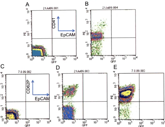
Platelet surface expression was confirmed by flow cytometry (Figure 1). Washed platelets were prepared from mouse whole blood (detailed in Materials + Methods). While unstained platelets showed no surface expression (Figure la,c), platelets stained positive for CD41 (Figure 1b) and CD62P (Figure id). As expected, platelets activated with ADP showed an up-regulation of P-selectin
(Figure le).
In order to establish proof of concept of platelets as a marker for CTCs, experiments were carried out on well-characterized mouse tumor cells lines. The
393M1 (liver to lung adenocarcinoma) and Tearly (early stage 393M1) cell lines
were used for these experiments because they represent different stages in tumor formation. While 393M1 cells are known to be metastatic, Tearly cells are isolated from an early stage primary liver adenocarcinoma and may not form metastasis (Personal Communication, Monte Winslow- Jacks Lab, MIT). Ideally, these cell lines allow for correlation of malignancy with platelet binding. The hypothesis was that those cells better primed for metastasis (393M1) would bind platelets more efficiently than earlier stage tumor cells or non-malignant cells (Tearly).
W~ F-' hJ r 7.8.09.002
Oa.
0 EpCAN OD
E
GFP GFP FFigure 1: Confirmation of platelet staining and activation. (A,c) Unstained platelets are compared to
platelets stained with (B) CD41 or (D) selectin. Addition of ADP activates platelets and up-regulates P-selectin expression (E). EpCAM staining represents a negative control, as this epithelial marker is not expressed on platelets.
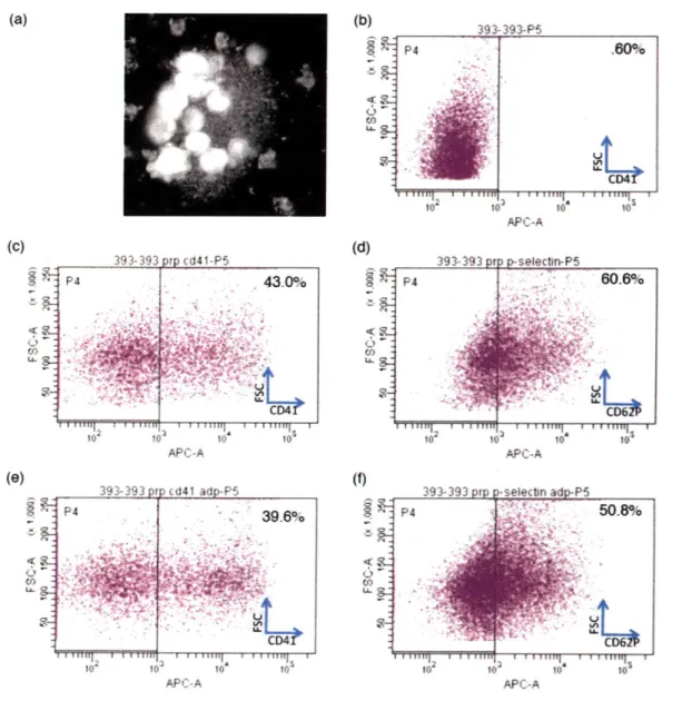
In order to recapitulate tumor cell platelet binding in vitro, 393M1 and Tearly cell lines were trypsinized and mixed with platelet rich plasma (PRP) for an hour at
37 *C (detailed in Materials and Methods). Platelet-tumor cell complexes were then
stained with platelet markers (CD-41, P-selectin), tumor cell markers (Ep-CAM) and 4', 6-diamidino-2-phenylindole (DAPI), a fluorescent dye that has a strong affinity for DNA. An outline of markers used for different cell types is shown in Table 1. Platelet-tumor cell complexes were identified by flow cytometry (Figure 2). In the absence of PRP, tumor cells did not stain for platelet markers CD41 or CD62P. When PRP was mixed with tumor cells, binding occurred and cells stained positive for both CD41 and CD62P (Figure 2c,d). Interestingly, cells stained positive for platelet marker CD62P, which is exclusively expressed on activated platelets. This result may suggest that tumor cells locally activate platelets by secreting ADP or thrombin.
W
(b) to' 10 (d) 4: APC-A APC-A <o 11 I0 toa APC-A 0 10 APC-A APC-A
Figure 2: Complexing of platelets and tumor cells. (A) Fluorescence microscopy image of platelets bound
to a tumor cell. (B) Unstained 393 tumor cells were mixed with platelet rich plasma and stained for (C) CD41 or (D) CD62P. The percentage of cells staining for platelet markers is given in the upper right corner of the plots. Cells were gated on forward scatter, DAPI negative, and GFP positive. The addition of ADP did not increase platelet-tumor cell complexing (E,F), which may show tumor cell mediated platelet activation.
To test this hypothesis, platelets were activated by ADP and incubated with tumor cells (Figure 2e,f). No increase in platelet-tumor cell binding occurred after the addition of ADP, which suggests that tumor cells are activating platelets and triggering P-selectin expression. Further, experiments carried out on the Tearly cell line yielded similar results: Tearly cells complexed with and appeared to locally
11
393-393-P5
P4 .60%
f"TfTI
I i itt i i:tun i e atu g
393-393 P p cd41 adp-PS P4CD.
... . ... . ....
activate platelets. While this data is in vitro, it may indicate that platelet binding is a universal process rather than one restricted to highly metastatic cell lines. Future work should be carried out to evaluate the mechanism of tumor-mediated platelet
activation. ELISA or western blot analysis could probe for the amount of thrombin or ADP in a particular cell type. Further, inhibitors of these molecules could be used to suppress platelet binding. This work is particularly relevant in the context of recent work carried out by Myriam Labelle of the Hynes lab at MIT. Labelle has shown that platelet binding triggers an epithelial to mesenchymal transition in tumor cells and primes them for metastasis; platelets control EMT by secreting
TGF-p
(Personal Communication - Myriam Labelle, Hynes Lab, MIT).While these initial experiments demonstrate tumor cell-platelet complexing, the extent of platelet-tumor cell binding was inconsistent. A potential reason for this inconsistency was the anti-coagulant used in whole blood isolation from mice. Initial experiments used blood drawn into sodium citrate tubes. Sodium citrate acts
by chelating calcium ions necessary for selectin binding of carbohydrate moieties24. Calcium is thus needed for both homotypic interactions between platelets and for platelet/tumor cell binding. To circumvent this issue, hirudin, an anticoagulant that
acts by inhibiting thrombin rather than chelating calcium, was used25. Platelet
binding experiments were repeated using hirudin (Figure 3). Interestingly, platelet binding to tumor cells decreased as compared to experiments done in sodium
citrate (Figure 3c,d and 3b,c). This may be because thrombin is required for
tumor cell mediated platelet activation. Residual hirudin would therefore block platelet activation and prohibit binding. Alternatively, platelet-tumor cell binding may be a selectin-independent process and binding could be integrin mediated and calcium independent.
(a) (b) 393-393-P5 393-393 prp cd4l-P5 P4 1.90% P4 383% - 4; -(c) 1o 10 10 10 APC-A 393-393 orp p-selectn-P5 P4 32.5% I I [Mfl It ill I IIIIIFt l11' 1 1112 "1 11111113 1 r1111 1 1 1 1lfl"] 1 10 10 10 10 APC-A 11 11 il3 V £ 111S I 102 10 10' 10 APC-A
Figure 3: Platelet-tumor cell complexing using platelet rich plasma isolated from whole blood in hirudin.
Calcium chelation may alter the dynamics of platelet-tumor cell complex formation. Hirudin, an anticoagulant that inhibits thrombin, was used to preserve physiological calcium levels. (A) Unstained 393 cells were compared to cells mixed with platelet rich plasma and stained for (B) CD41 or (C) P-selectin. The use of hirudin did not have a dramatic effect on complex formation.
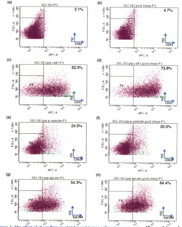
1.2.2.2 Effect of Shear Flow on Platelet/CTC Complex Formation
In order to better recapitulate in vivo conditions, platelet-tumor cell suspensions were subjected to a uniform shear field. Hydrodynamic shear induced collisions augmented platelet-tumor cell binding (Figure 4c,d). A shear rate of
100s-1 dramatically increased platelet binding to tumor cells when judged using
CD41 as a marker. However, this shear force had seemingly no effect when using
CD62P as a marker. This may be because at low shear forces (<100 s-1), platelet binding is P-selectin-independent/Arg-Gly-Asp (RGD)-dependent. At higher shear
forces (>800 s-1) this process may become P-selectin dependent26. As such, CD62P
may be up-regulated at higher shear forces because it is a stronger and more
specific interaction than RGD-based binding. In addition to platelet protein
expression, tumor cell protein expression appears to be affected by shear forces
(Figure 4gh), as EpCAM seems be upregulated under flow conditions. Thus,
platelet activation and the fluid environment of the vasculature may have an effect on platelet-tumor cell adhesive interactions, and more work should be done to examine protein expression changes in response to fluid-mechanical forces.
393-393 pr epcam-P3 54.3% PE (f) 4: () U) LL 4: CO '4-393-393 ost sheat-P3 4.7% 10 10 APC-A S 10 10 393-393 prp p-selectin post sh 3 -20.0% CD2 10o1 10' t0 APC-A
393-393 prp ep cam post shear-P3 64.4%
-1. In I # I IrUJI I, InF~
--Figure 4: The effect of shear flow on platelet and tumor cell protein expression. After applying a shear force of 100 s-1 to a suspension of platelets and tumor cells, tumor cell forward and side scatter
remained the same (AB). However, platelet expression of CD41 seemed to increase (CD), while platelet expression of P-selectin remained constant (E,F). It also appears that platelet/tumor cell complexing increased as a result of shear flow. Finally, tumor cell expression of EpCAM increased as a result of shear flow (G,H). 14 (b) APC-A (C) CA ( (0) APC-A APC-A to
-In order to evaluate flow cytometry as a tool for CTC isolation, 393 cells were spiked into whole blood and recovered via flow cytometry. Since these cells stably express GFP, their isolation can be confirmed by fluorescence. Tumor cells were spiked into mouse whole blood and incubated for 1 hr at 37 *C. After erythrocyte lysis, granulocytes, monocytes and lymphocytes were stained using markers outlined in Table 1. This counter-staining allowed for facile separation of blood cells. Using this method, I was able to resolve -10 GFP positive tumor cells per
100,000 WBCs, or 1 tumor cell per 109 total blood cells (-10 ml of total blood).
These initial experiments confirm flow cytometry as a viable method for enumerating and separating CTCs from peripheral blood.
After proof of concept studies in mice revealed platelet-tumor cell complexing to be a repeatable in vitro process, we turned to more relevant human cell lines. Human colonic carcinoma cell lines, LS180 and HT29, were tested for platelet binding. While LS180 robustly expresses sLex, an antigen for P-selectin on platelets, HT29 does not express this carbohydrate group (Figure 5). As such, I expected to see platelet binding to LS180 and negligible binding to HT29. Unfortunately, this was not the case, as neither cell line appeared to bind human platelets. Rather than a failure to bind platelets, this result may be an unfortunate experimental artifact. Antibodies to human isoforms of CD41 and CD62P on platelets appear to have been compromised during shipping from AbCAM. As a result, data from these experiments must be reproduced with a fresh batch of antibodies.
(a) (b) ( 2feb10 001 ( 2feb10 002 .8% P P sLex sLex 10( c0 1 103 10) 10 0 10 10.304 PE PE (C) 2 9b0 ar007 (B) and (D s l u w 1.51%0 Dd ste"te 103 104 102 0 104 PE PE
Figure 5: Sialyl Lewis X staining of H T29 and LS180. (A,B) While HT29 express Sialyl Lewis X, an antigen for P-selectin on platelets, at basal levels, (c,D) LS180 robustly express this carbohydrate group. (A) and
(C) show unstanined HT29 and LS180, respectively. While (B) and (D) show cells Incubated with an
antibody to CD15s.
1.2.3 Discussion and Future Work
Tumor cells bound by platelets are protected from shear forces and natural killer cell activity and may have a competitive advantage over other CTCs in seeding a secondary tumor. With the dearth of definitive CTC markers, platelets are a promising candidate for further evaluation. Here, I have delineated some early results investigating the propensity and mechanism of platelet-tumor cell binding in
vitro. From these cursory experiments, it seems that platelets may not bind some
tumor cells better than others - there is no clear correlation between metastatic
potential and platelet binding. Also, tumor cells may locally activate platelets by secreting ADP or thrombin. Further, the surface expression of both tumor cells and platelets is altered in response to shear forces, and future work investigating CTCs should take this change in protein expression into account.
Work on a human model of platelet-tumor cell binding has been hampered
by unfortunate complications with reagents. However, this work should be continued with the goal of identifying differences in platelet-tumor cell binding under varying shear forces and time courses. In addition, target proteins such as P-selectin, CD41 and carbohydrate groups such as sLex should be systematically inhibited in order to investigate the mechanism of complex formation. The final step of this work in vitro is to use FACS to sort out platelet-tumor cell complexes from whole blood and re-inject these complexes into mice in order to assay for metastatic potential. Since platelets protect from natural killer cell activity, it is imperative that these mice are not immuno-compromised, as the ability of tumor cells to overcome the immune system appears to be a critical step in metastasis mediated by platelets.
Once these in vitro experiments have elucidated the mechanism of platelet binding and its role in metastasis, patient samples should be assayed for platelet-tumor complexes. Peripheral blood samples from patients with metastatic disease should be subjected to staining and subsequent flow cytometry. If platelet-tumor cell complexes exist, they should be isolated and perpetuated in culture. These complexes should also be injected into mice in order to investigate metastatic potential. Ideally, there will be a correlation between the number of platelet-tumor complexes and the severity of a patient's disease state. If this is the case, these complexes may be the focus of future therapeutic and diagnostic development.
1.3 Phage Based Approaches to Isolate CTCs
1.3.1 Isolation of Mesenchymal like CTCs Using Phage Display
The shift from an epithelial to mesenchymal phenotype is mediated by a number of transcription factors: Snail, Zeb, E47, KLF8, FoxC2 etc. In particular, TWIST has been shown to be essential for metastasis and EMT in breast
carcinomas27. The Weinberg group has shown that up regulation of TWIST leads to
degradation of cell-cell adhesion, downregulatioon of epithelial markers (cytokeratin, E-cadherin), and upregulation of mesenchymal markers (vimentin,
line by constitutively up-regulating TWIST in mammary carcinoma cells28. Using
these two cell lines (TWIST +, TWIST -) as a proxy for mesenchymal-like and
epithelial-like tumor cells, we have used phage display of a library of random peptide 7-mers fused to minor coat protein pIll of M13 bacteriophage to select for peptides that bind to mesenchymal-like cells and not to epithelial-like cells (Figure
6).
Mesenchyrnal
M1 3 phage library Mesenchyma-ltke cells EpitheaMe cells binding peptide
(-10clones) (MBP)
amosSm#s
Collect Collect
bound unbound
phage phage
Figure 6: Schematic of biopanning approach. A library of ~1010 phage clones was incubated with
mesenchymal-ike (TWIST +) tumor cells, bound and internalized phage was collected and incubated with epithelial-like (TWIST -) tumor cells. Unbound phage was collected and this sequence was repeated three times to enrich the phage population. After enrichment, phage clones were sequenced and
mesenchymal binding peptide candidates (MBP) were Identified.
M13 is a filamentous virus that infects bacteria, consisting of five proteins that encapsulate the viral DNA. It is approximately -1 um in length and 6-7 nm in diameter. M13 phage is an attractive vector for peptide display since its genotype
relates directly to its phenotype. Consequently, peptides can be genetically
engineered into the multiple coat proteins of the M13 phage for display. Previously, others have displayed peptides and proteins on the coat proteins with affinity towards epitopes29 30, antibodies31 and mammalian cells for targeting32, 33. Our lab has engineered a phage vector for peptide display on two coat proteins, p3 and p8 and we have demonstrated multiple display of the M13 scaffold to grow and
nucleate various inorganic materials34-42. M13 filamentous bacteriophage is an
excellent biological building block due to its multiple peptide display system, controllable length, and functionality as a nanoscale scaffold for nanoparticle organization.
Initial biopaning results (detailed in Materials and Methods) yielded 12 candidate mesenchymal binding peptide (MBP) sequences (Figure 7).
Position 12345678 9 3 CPGP C 4 C rP A C Clh A'. C 76C C
8 Con
10o
C
9 C G. C 10 C P C 11 Cv ' i C 12 C§P#V#A C Consensus: C P P S P F S S C % ConservationFigure 7: Biopanning results. 12 candidate sequences for the mesenchymal binding peptide are aligned
above. The 7-mer library is book-ended by two cysteine residues, which form a disulfide bond. The consensus sequence is proline and serine rich, which is common for peptides that bind surface proteins.
While the library did not collapse to a handful of sequences, there are a number of conserved motifs between the sequences (Figure 7). The 7-mer sequences are given by positions 2-8 and are sterically constrained by two cysteine residues. The consensus sequence is proline and serine rich. Proline is a unique amino acid as its side chain is cyclized onto the backbone nitrogen. As a result, the confirmation of proline and the preceding residue are restricted. The PxxP motif is a common motif in peptides specific for proteins, as the prolines form a continuous hydrophobic strip around the surface of the helix and allows for ideal hydrogen binding sites4 3. As such, these isolated sequences are promising candidates for further investigation. 1.3.2 Discussion and Future Work
Peptides isolated via phage display should be further investigated for binding to mesenchymal-like cells. The most straightforward experiment to be carried out is a competitive ELISA in which phage specific for mesenchymal-like cells are incubated with cells and competed off with free MBP. If this competition is
successful, one can estimate the KD of peptide binding. Once a peptide has been
found using phage display, it can be used to isolate mesenchymal-like CTCs from peripheral blood.
...
In order to isolate CTCs from peripheral blood, the MBP will be biotinylated and attached to 50nm streptavidin coated Fe203 superparamagnetic bead, and magnetically labeled cells will be isolated using magnetic-activated cell sorting
(MACS) 44. MACS employs a steelwool column that is magnetized by placing it in a .6
Tesla magnetic field. The magnetic column acts as a sensitive filter for magnetically labeled cells and allows for a 5-log-fold enrichment of viable cell populations44. As proof of concept of the isolation approach, TWIST expressing mesenchymal-like cells will be spiked into whole blood to mimic a CTC population, and MBP functionalized magnetic beads will be added to the solution to label CTCs. After erythrocyte lysis, leukocytes and tumor cells will be applied to a magnetic column
(Miltenyi Biotec) - magnetically labeled tumor cells will be retained by the column
while leukocytes and plasma proteins will be discarded. Cells can be eluted from the column by removing the external magnetic field or by competing with free MBP. Several rounds of purification can be carried out in order to enrich tumor cells. If necessary, negative selection can be carried out by labeling Cd45+ leukocytes with biotinylated antibodies and running the sample through a streptavidin functionalized affinity column. This iterative approach allows for control of the purity of isolated CTCs. By closely monitoring the number of cells spiked into blood and the number of cells successfully isolated, the efficiency and resolution of this approach can be quantified. Unfortunately, this approach comes with a number of limitations. Functionalization of beads to the MBP may alter the specificity of the peptide. As such, if MACS fails to isolate tumor cells, conjugating MBP to a smaller bead with a different shape may allow for better purification. Additionally, this approach is limited by the number of MBP ligands on the surface of the tumor cells.
If these ligands are of low-abundance, cells will have few magnetic nanoparticle
bound and a low magnetic moment, which may prohibit their isolation using MACS and may require the use of an expensive multi-tesla magnet. While the MBP may show affinity for mesenchymal-like cells in vitro, this affinity may not translate well into clinical samples. Since CTCs in the blood are likely more heterogeneous than a cell line, the MBP may only isolate a subset of CTCs. In addition, this subset may not represent a particularly malignant group of cells. A number of modifications can be
made to the phage display protocol to direct the selectivity of the MBP. Increasing the number of alternating rounds of negative and positive selection during biopanning can increase its specificity. By negatively selecting against leukocytes in addition to epithelial-like cells, a MBP can be identified that does not bind to blood cells. In order to better recapitulate in vivo conditions, selection can be carried out against detached cells in media rather than cells adherent to a plate, as gene
expression may change dramatically in circulation45.
In order to generate global gene expression profiles, RNA will be extracted from isolated mesenchymal-like cells and applied to the Affymetrix Genechip Human Genome U133 Plus 2.0 Array, which allows for analysis of over 47,000
transcripts46. To benchmark expression, results will be compared to profiles from
epithelial-like cells. In order to confirm that the isolation procedure has no effect on gene expression, expression signatures from mesenchymal-like cells isolated from blood will be compared to signatures from mesenchymal-like cells isolated directly from culture. If there is a dramatic difference in gene expression attributable to the isolation procedure, a different procedure may be implemented. For example, the
MBP could be functionalized to microposts in a microfluidic device. While
leukocytes will flow through the device, mesenchymal-like CTCs will be captured. These cells can then be eluted from the posts by competition with free MBP. This
MEMS device is beneficial in that physiological flow conditions may preserve the
viability of CTCs; however, it is limited by its low throughput, as only small volumes of blood can be run through each chip at a time. Conversely, a change in gene expression of mesenchymal-like cells after incubation in blood could be due to
interactions with blood cells (personal communication, Myriam Labelle - Hynes Lab,
MIT). To test this hypothesis, cells in culture could be incubated with whole blood. After washing the cells to remove unbound blood cells, gene expression analysis would be carried out.
After these proof of concept studies have confirmed that MACS is a feasible
CTC enrichment strategy that does not alter gene expression, this approach can be.
used to characterize CTCs from patients with varying stages of metastatic disease. In collaboration with surgeons, blood and primary tumor samples will be collected
from patients that have undergone surgery to excise primary epithelial tumors. Since the cell lines from previous experiments are mammary carcinoma based, we will initially try to obtain clinical samples from patients with advanced breast cancer. Magnetic beads conjugated to the MBP will be used to purify CTCs from the blood samples. Since CTCs are so rare, multiple rounds of isolation will be carried
out to ensure minimal leukocyte contamination. Since an iterative isolation
procedure may be harmful to cells, we will have to empirically determine how many rounds of isolation are appropriate. Gene expression of isolated CTCs will be compared to expression from primary tumor cells from the same patient and to the TWIST inducible mesenchymal-like and epithelial-like cell line from the Weinberg group. When comparing patient CTC and patient tumor gene signatures, we will look for genes that are significantly up/down regulated when cells enter the bloodstream. We would expect to see up-regulation of mesenchymal markers and down regulation of epithelial markers as confirmation of an EMT. Further, we can correlate gene expression of CTCs with clinical characterization of the patient's disease stage. Cancer can be staged from 0 (carcinoma in situ), to stage IV (evidence
of metastasis)47. Using principal component analysis (PCA), we may be able to
correlate CTC gene expression with that of primary tumor cells and tumor grade.
PCA may be useful in identifying a handful of candidate genes that are responsible
for survival in the blood and metastatic potential. Future work would be carried out
by examining protein expression levels of these candidate genes, as there tends to
be no correlation between gene and protein expression levels4 8. While the utility of
gene expression data is limited, it may prove beneficial by yielding a small number
of candidate genes for further characterization49. In addition to gene and protein
expression analysis, a simpler correlation between CTC number and malignancy will be studied. Large numbers of circulating epithelial cells in peripheral blood translate into poor clinical outcomes. By isolating mesenchymal-like rather than epithelial-like tumor cells from the blood, we may be able to classify malignancy more accurately and precisely, as mesenchymal-like cells, rather than epithelial-like cells, may be the precursors of metastasis.
The paucity of CTCs isolated using MACS may prohibit gene expression analysis. Ten nanograms of total RNA from CTC and primary tumor samples is needed to prepare biotinylated hybridization targets with the Affymetrix small sample target labeling assay5o; this system is designed to reproducibly amplify 10-100ng of total RNA using T7 RNA polymerase. Assuming that each cell has
approximately 30pg of total RNA and 2% of total RNA is mRNA51, about 330 tumor
cells are needed in order to get reproducible gene expression results. If a typical blood draw is 5ml, CTC counts would have to be >60/ml in order to carry out such genetic analysis. As such, microarray analysis will only be possible if MACS can reproducibly enrich a large CTC population or if analysis is restricted to patients with high CTC counts.
An emerging alternative to microarray analysis that is well suited to rare and heterogeneous CTCs is single-cell quantitative PCR with reverse transcription (qRT-PCR) (Fluidigm Biomark system). This approach is advantageous in that it can distinguish between pooled and single cell expression, a short coming of the
microarray approach5 2, 53. Single cell resolution will be important to find
subpopulations of malignant CTCs and to understand how a single CTC can give rise
to a tumor 4. The drawback of this approach is that it will not yield a global gene
expression profile. Instead, we can examine the expression of a group of query genes. These genes would include EMT hallmark genes as well as housekeeping genes such as Actb (beta-actin) and Hprtl (hypoxanthine guanine phosphoribosyl transferase 1 in order to normalize and benchmark analysis.
1.4 Materials and Methods
Blood Collection
Blood was obtained from Research Blood Components (human blood) or Bioreclamation LLC (mouse blood). Blood was typically drawn into sodium citrate tubes. In order to avoid calcium chelation, blood was drawn into hirudin tubes (Diapharma). Mouse blood was shipped overnight on dry ice, while human blood was shipped day of on dry ice. Blood was always used within two days of receipt.
Platelet Preparation
Whole blood was centrifuged at 800g for 7 minutes in a tabletop centrifuge. Platelet rich plasma (PRP) was collected and the remaining blood cells were again centrifuged for 7 minutes at 800g. The platelet poor plasma was collected and mixed with the PRP to obtain the desired platelet concentration.
Erythrocyte Lysis
1ml of whole blood is centrifuged for 7 minutes at 800g. Remove the PRP and resuspend remaining cells in 1.5ml ACK buffer (.15M NH4Cl, 10mM KHCO3, .1mM
EDTA). Allow cells to incubate with buffer for 10 minutes. Centrifuge for 3 minutes at 3000RPM and discard supernatant. Some red blood cells will remain. Resuspend cells in ACK, incubate and centrifuge. Discard supernatant. A small white pellet should be visible (white blood cells) - resuspend in PBS to desired concentration.
Cell Lines and Culture
393 and Tearly cell lines were a gift from the Jacks Lab (MIT). HT29 and LS180 were a gift of the Hynes lab (MIT). All cells were grown in Dulbecco's Minimum Essential Medium (DMEM) supplemented with 10% fetal bovine serum (FBS) (Hyclone, Logan, UT) and 1% penicillin/streptomycin (Invitrogen, CA) at 37 *C in 5%
CO
2.
Freezing Cells
Cells were preserved by freezing at -80 *C. A confluent 225 cm2 culture plate was
trpysinized and cells were resuspended in 10% DMSO, 90% FBS. Cells were frozen in isoproponal containers to control cooling.
Cell Counting
Cells were quantified and their viability assessed using a Vi-Cell Series Cell Viability Analyzer (Beckman Coulter). This machine was used with permission from the Lauffenberger Lab (MIT). While this machine was useful in quantifying mammalian tumor cells, both human and murine platelets were too small to be reproducibly counted by this machine. Instead, these cells were counted using a hemacytometer.
Formation of Platelet Tumor Complexes
Platelet rich plasma (PRP) or washed platelets are prepared and incubated with tumor cells for 1 hour at 37 *C on a rocker. Typically 106 tumor cells were mixed with 108 platelets in PBS in a volume less than 1ml. After incubation, complexes were separated by centrifuging at 1200 RPM for 5 minutes at room temperature ( a small red ring is visible around the tumor cells at this step). If using 106 tumor cells, a pellet should be clearly visible. The supernatant was discarded and the complex
was stained for platelet markers using antibodies on ice (see above). Some
experiments included 10uM ADP during the incubation step (1 hour) for platelet activation. To form complexes with cells in whole blood, 106 tumor cells were added to a flow tube and centrifuged at 1200 RPM for 5 minutes. .5 ml of whole blood was added directly to the cells and lightly vortexed. This solution was incubated for 1 hour at 37 *C and then centrifuged at 1500 RPM for 7 minutes. The PRP and the buffy coat were carefully collected such that there was no erythrocyte contamination. These cells were then stained and run on FACS. White blood cells were counterstained with biotinylated CD45, GR-1, CD11b and F4180 incubated
with streptavidin-PE-Cy7 beads. In order to recapitulate shear forces, the
vasculature was mimicked by a simple pump apparatus. A Pump33 (Harvard
Apparatus) was connected to plastic tubing (diameter = .5mm, length 500 cm). A
shear force of 100s' was applied using a flow rate of 1.3 ul/s on the Harvard pump. PRP and tumor cells were mixed and immediately applied to the pump for about 10 minutes.
Antibody Staining
100,000 - 1,000,000 cells were incubated in 100 ul volumes of PBS. Primary
antibodies were added at 1:100 dilutions and secondary antibodies were generally added at 1:500 concentrations (see lab notebook for precise concentrations for specific experiments). Cells were incubated with antibodies on ice and protected from light for 30 minutes to 1 hour. After incubation with either primary or secondary antibodies, cells were washed 1-3x with PBS by centrifugation at 1200
RPM. In addition to surface markers, cells were incubated with DAPI immediately before analysis in order to test for cell viability
Antibody List
1. P-selectin (Psel.KO.2.7) (Santa Cruz Biotech, Inc sc-101336) (Mouse anti-human)
2. PE Goat Anti-Mouse Ig (BD Pharmingen 550589) 3. PE Goat Anti-Rat Ig (BD Pharmingen 550767)
4. Purified Rat IgG1, k Isotype Control (BD Pharmingen 553922) 5. Mouse Monoclonal (104-2) to CD45.2 PE/Cy7 (abcam 25463) 6. Ep-CAM (G8.8) (Santa Cruz Biotech, Inc sc-53532)
7. Rat Anti-mouse CD41 MWreg30 (BD Pharmingen 553847)
8. Mouse Mab CD41 (reacts with human samples) alpha-2b [sz.22] FITC (abcam 19687)
9. Mouse Mab CD62P (reacts with human samples) (AK-6) (ab 33279)
Flow Cytometry
Flow cytometry was carried out on both LSRII and FACScan cytometers. High throughput automated cytometry was carried out using the LSRII.
Flow Cytometry Data Analysis
Platelet tumor complexes were selected on the following criteria. Side and forward scatter were characteristic of the tumor cell line. If using the 393 cell line, cells were GFP positive. Cells stained positive for platelet markers CD41 or CD62P. Cells were DAPI negative, indicating viability. Cells were CD45, GR-1, CD11b and F4180 negative, indicating that they were not blood cells.
Panning
Panning was conducted with three rounds of positive selection and three rounds of negative selection. The rounds were conducted in the following order: positive, negative, negative, negative, positive, positive. Positive selection was carried out against 393 cells (Jacks Lab) while negative selection was carried out against whole blood. Phage was amplified and titered between rounds of positive selection.
Positive Selection
1. Grow epithelial-like cells such that they are over-confluent
2. Prepare phage cocktail/solution: Mix 2E11 pfu in 1 mL 1x PBS with 1% BSA and protease cocktail
3. Incubate with 3ml serum-free media for 2 h, 37 "C. This is to allow for
'clearing' of the receptors (and possibly the glycocalyx).
4. After serum-free incubation, aspirate media and add 2E11 pfu/mL phage to (target) cells.
5. Incubate phage 1 h at 37 *AC with gentle rocking to allow time for phage to
be internalized.
6. After incubation, remove supernatant (contains unbound phage) by
aspiration
7. To remove nonspecific binding phage, the cells are washed five times with 3
ml PBS supplemented with 1% BSA and 0.05% Tween-20 at room temperature in a flow tube
8. After washing add 1 ml of glycine (pH= 2.2, added by HCl) and incubate 8
mm.
9. Remove the supernatant, repeat the wash with glycine, and collect the
supernatant, transferring it into a microcentrifuge tube.
10. Add 150 uL Tris (pH -9.1) to the solution in the micro-centrifuge tube, the ACID eluted fraction. This collects weakly bound phage to the cells. Save this
fraction.
11. Recover the internalized phage by lysing the cells with 1 ml of 0.1%
triethanolamine (Sigma) in PBS (pH 7.6) for 5 min at RT
12. Neutralize the internalized phage pool with 100 pl of 0.5 M Tris-HCl (pH 7)
13. Vortex and incubate for 30 min on ice.
14. Spin down the cell lysate and collect the supernatant (-1 mL). This is the
CELL fraction. This fraction should have phage that showed affinity towards
target cell.
15. Save 10% CELL fraction for titering. Use the rest of fraction for phage
amplification. Negative Selection:
1. Clear mesenchymal cells
2. Take 1E11 pfu of amplified phage from previous round and make phage cocktail in 1 mL sterile PBS with protease inhibitor.
3. Incubate cells with phage cocktail for 1 h, 37 C.
4. Take the supernatant. This fraction will contain unbound phage that did not bind the mesenchymal-like cells and will be used for panning against metastatic liver cells.
Chapter 2: M13 Bacteriophage as a Platform for Targeted,
Simultaneous Near-infrared Imaging and Efficient Chemotherapy
of Prostate Cancer Cells
2.1 Background: Nanomaterial-Based Therapeutics
Most systematic therapeutic approaches to cancer lack specificity and are not precisely controlled after injection. The ability to control the functionality and valency of nanomaterials allows for manipulation of pharmacokinetics and dynamics and can improve safety, potency, and efficacy over traditional tumor
therapies and imaging techniques. Further, the size and shape of many
nanomaterials yield a large surface area to volume ratio and allow for the containment and functionalization of an array of targeting, therapeutic or imaging modalities.
A number of nanomaterials have shown promise in a clinical setting55-5 9 and many more candidate chemotherapy and imaging agents have been successful in murine trials. For example, iron oxide and gadolinium (III) nanoparticles can be functionalized to targeting moities in order to increase the relaxivity of water molecules in a tumor and subsequently enhance magnetic resonance images, which
may allow for more precise imaging of tumors60, 61. Gold nanorods have been
effective agents of thermolysis by acting as antennas to absorb infrared radiation
and cause tumor cell necrosis62, 63. Single-walled carbon nanotubes (SWNTs),
rolled-up tubes of graphene sheets, have also been recently studied for therapy64,65,
imaging66-68 and as fluorescent biosensors for nitric oxide69 and reactive oxygen
species70. Their quasi-one-dimensional architecture yields a large surface area for
functionalization and drug loading. Further, SWNTs are an attractive candidate for biomedical imaging because of their high absorption of near infrared (NIR) light, cross-section of Raman scattering, and NIR band-gap fluorescence ranging from 900
- 1600 nm. NIR fluorophores with emission in the "second window" (900 - 1400 nm) may have greater tissue penetration than those in the first spectral window
(650-900 nm)71. These probes are advantageous because of the low characteristic autofluorescence of biological species in the second window, which results in a
signal to noise ratio that may be 100 fold higher than that in the first window and may allow for precise and sensitive detection of nascent tumors.
Previous research has established SWNTs as viable imaging and therapeutic vectors. Several groups have shown SWNT mediated therapeutic delivery or NIR
fluorescence imaging of tumors72. Despite the recent success in utilizing SWNTs for
imaging and therapy, it remains challenging to engineer multifunctional, biocompatible, and singly-dispersed SWNT complexes that retain fluorescence. Further, no single platform has demonstrated simultaneous fluorescent second window imaging and drug delivery, as therapeutic loading may limit the stability of non-covalently functionalized SWNTs.
The next generation of nanomedicines will have encoded multifunctionality that allows for targeted and simultaneous therapy and diagnosis (so called "theranostics"). While a number of nanomedicines used in the clinic target tumors using the enhanced permeability and retention effect (EPR), targeting to specific tumor markers may improve the efficacy and safety profiles of nanomaterials by reducing nonspecific cell uptake. In addition, the ability to simultaneously image and ablate a tumor will allow for real-time monitoring of the efficacy of a drug regimen.
M13 is a promising platform for nano-scale therapeutic and diagnostic delivery because of its genetic tunability and physical characteristics. M13 has been
previously used in biomedical applications for epitope discovery73, 74, gene
delivery7s, antibody delivery76, and in vitro and in vivo ligand discovery77-79. M13 bacteriophage has five genetically modifiable proteins (proteins p3, p6, p7, p8, and
p9). Expression of material-specific peptides or targeting motifs on various coat
proteins allows for genetic control and tunability of its function40; the malleability of
the phage genome allows for directed evolution of phage proteins for biomedical applications. In addition, the highly ordered structure of the 2700 copies of the major coat protein, p8, and the filamentous shape of M13 (-6.5 nm in diameter and
-880 nm in length) enables the phage to multivalently and cooperatively interact
with one-dimensional nanomaterials such as SWNTs, and the abundance of
genetically modifiable proteins on the coat of the phage allow for encoding of multifunctionality.
Particle shape can greatly impact the cellular and tissue interactions of nanomaterials. Particles with a high aspect ratio (length to width ratio) have been shown to evade phagocytosis and can only be engulfed when cells interact with the
particle ends80. Investigators have shown that filamentous particles have
circulation times about 10-fold longer than spherical particles81. Further, surface
charge or zeta potential (electric potential at the hydrodynamic slipping plane of a particle) is an important factor influencing the behavior of nanomaterials. Particles with a negative or neutral surface charge avoid non-specific cellular uptake better than particles with a positive charge8 2. As such, the filamentous shape of M13 may help inhibit phagocytosis and also enhance circulation half-life in vivo, while the slightly negative zeta potential of the phage may reduce its nonspecific cellular uptake.
In the following section, I outline an M13 bacteriphage-based platform for manipulating single-walled carbon nanotubes (SWNTs) in biological solutions for multimodal near-infrared (NIR) fluorescent cell-specific imaging and therapy. SWNTs are stably dispersed by major coat proteins of the M13 bacteriophage
through molecular recognition and retain band-gap fluorescence in the 900 - 1350
nm wavelength range, which may allow for deep tissue imaging. Minor coat protein
p3, located at the proximal tip of M13, was genetically engineered to specifically
target SPARC matricellular protein, a marker overexpressed on metastatic breast and prostate tumors. The targeted virus-SWNT complex serves as a NIR imaging agent and allowed for sensitive and selective NIR imaging of cells with varying expression levels of SPARC. Controlling M13-SWNT stoichiometry allowed further functionalization of the viral major coat with a chemotherapeutic agent for targeted drug delivery in vitro. Doxorubicin (DOX) delivered by the M13-SWNT complex inhibited cell growth 400x more efficiently than free DOX, and was restricted to SPARC expressing cells. This approach provides an 'all-in-one' platform for targeted NIR fluorescence imaging and efficient drug delivery.
0 ON 0
(a)
SPPTGI N00ONo p3 p8 (b) DVYESALP 880 nmFigure 8:A schematic of the M13 virus-based platform. (Figure components not to scale) (A)
M13-SBP-SWNT-DOX complex. The filamentous shape of M13 bacteriophage and high aspect ratio allow for favorable physical interactions In vivo. p3 protein is engineered for targeting SPARC (SSPTGIN) and p8
protein is modified for SWNT binding and drug conjugation (DVYESALP). The secondary structure of a single p8 protein Is shown inset and an inserted 8-mer peptide sequence is displayed on the amino-terminus of the p8 protein. The tyrosine residue In red is hypothesized to interact with hydrophobic SWNTs while the free carboxylate group on aspartic acid is used for drug conjugation. . Doxorubicin is depicted with a blue sphere. (B) A helical structure of 2,700 copies of p8 protein along the M13 virus with five-fold rotational and two-fold screw symmetry, allowing for multivalent and cooperative binding of SWNTs. Rendering was based on a structural model of M13 virus, 2COW.pdb. (software: Swiss-PDB) (Courtesy of Hyunjung Yi)
2.2 Theranostic Properties of M13 Bacteriophage and Single-Walled Carbon Nanotubes (SWNTs)
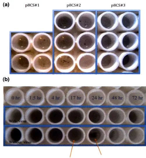
2.2.1 Creation of M13-SWNT Complex (Hyunjung Yi)
To bind and disperse SWNTs non-covalently along the length of virus wrapped by p8 major coat proteins, 8mer peptide sequences displayed on p8 that show binding affinity towards SWNTs were identified through biopanning (see Materials and Methods). After several rounds of iterative panning against SWNT film, consensus sequences were identified. The three most frequently appearing clones, DPSRLANE, DKSIEPLP, and DVYESALP (designated p8cs#1, p8cs#2, p8cs#3
respectively), were investigated for binding and dispersion of SWNTs. Well-dispersed HiPco (high-pressure CO decomposition) SWNTs in water with 2wt% sodium-cholate were dialyzed extensively against surfactant-free buffer in the
presence of each virus clone8 3. SWNT solution without virus was also dialyzed as a
negative control. After dialysis, only p8cs#3 formed a homogeneous solution with SWNTs whereas other clones and negative control formed black aggregates (Figure
9). This solubility suggests that p8cs#3 had a binding affinity strong enough to
disperse SWNTs in aqueous solution. Interestingly, only p8cs#3 had an aromatic residue, tyrosine (Y), which is thought to preferentially interact with the graphene
sidewall of carbon nanotubes (Figure 8a) 84,85.
(a) p8cS#1 p8CS#2 p8CS#3
(b)
Figure 9: Binding test results. (A) Each clone was incubated with sodium cholate-dispersed HipCo SWNT
in water and dialyzed against tris-buffered saline (TBS, Tris-HCI 100 mM, NaCI 150 mM, pH. 7.5) for three days with frequent buffer exchanges. The upper column has the highest virus concentration and each virus clone has duplicate samples. Only clone p8cs#3 shows homogeneous solutions while others
form black aggregates. (B) SWNT dialyzed with and without virus clone p8cs#3. After 24 hr, black aggregates are clearly observed for SWNTs dialyzed without virus. (Courtesy of Hyunjung Yi)
To further probe the binding mechanism of p8cs#3 to SWNTs, the
hydrophobicity of the peptide sequence was calculated and plotted (Figure 10a)86.
The peptide insert of p8cs#3 has a hydrophobic moiety sandwiched within a hydrophilic region and this moiety is believed to interact with SWNTs. Meanwhile, p8cs#1 lacked a hydrophobic region and p8cs#2 showed lower hydrophobicity than
p8cs#3. In general, t- stacking and hydrophobic interactions are considered the
primary interaction mechanisms between biological molecules and SWNTs84, 85.
Therefore, it is reasonable to conclude that the combination of iT-n stacking and the
hydrophobic properties of the p8cs#3 sequence are the driving force of the
M13-SWNT binding. While the hydrophobic moiety is small, the overall interaction
between SWNT and the virus is amplified by multivalent binding, as each M13 virus has 2,700 copies of p8 major coat protein that form well ordered structures with five-fold-symmetry and two-fold screw symmetry about the major axis (Figure 8b). Based on the crystallographic data of wild-type M13 virus (PDB ID: 2COW), there
are about 31 p8 peptides in series per 100 nm along the virus. This multivalent and
cooperative binding scheme makes detachment of SWNT off the virus unlikely and energetically unfavorable.