HAL Id: cea-01233690
https://hal-cea.archives-ouvertes.fr/cea-01233690
Submitted on 25 Nov 2015
HAL is a multi-disciplinary open access
archive for the deposit and dissemination of
sci-entific research documents, whether they are
pub-lished or not. The documents may come from
teaching and research institutions in France or
abroad, or from public or private research centers.
L’archive ouverte pluridisciplinaire HAL, est
destinée au dépôt et à la diffusion de documents
scientifiques de niveau recherche, publiés ou non,
émanant des établissements d’enseignement et de
recherche français ou étrangers, des laboratoires
publics ou privés.
Arg375 tunes tetrahydrobiopterin functions and
modulates catalysis by inducible nitric oxide synthase.
Zhi-Qiang Wang, Jesús Tejero, Chin-Chuan Wei, Mohammad Mahfuzul
Haque, J. Santolini, Mohammed Fadlalla, Ashis Biswas, Dennis J Stuehr
To cite this version:
Zhi-Qiang Wang, Jesús Tejero, Chin-Chuan Wei, Mohammad Mahfuzul Haque, J. Santolini, et al..
Arg375 tunes tetrahydrobiopterin functions and modulates catalysis by inducible nitric oxide synthase..
Journal of Inorganic Biochemistry, Elsevier, 2012, 108, pp.203-15. �10.1016/j.jinorgbio.2011.11.015�.
�cea-01233690�
Arg375 tunes tetrahydrobiopterin functions and modulates catalysis by inducible
nitric oxide synthase
Zhi-Qiang Wang
a,⁎
, Jesús Tejero
b, Chin-Chuan Wei
c, Mohammad Mahfuzul Haque
b, Jerome Santolini
d,
Mohammed Fadlalla
b, Ashis Biswas
b, Dennis J. Stuehr
b,⁎⁎
a
Department of Chemistry and Biochemistry, Kent State University at Tuscarawas, New Philadelphia, OH 44663, United States
b
Department of Pathobiology, Lerner Research Institute, Cleveland Clinic Foundation, Cleveland, OH 44195, United States
c
Department of Chemistry, Southern Illinois University Edwardsville, Edwardsville, IL 62026, United States
d
iBiTec-S; LSOD, C. E. A. Saclay; 91191 Gif-sur-Yvette Cedex, France
a b s t r a c t
a r t i c l e i n f o
Article history: Received 24 July 2011
Received in revised form 12 November 2011 Accepted 14 November 2011
Available online 23 November 2011 Keywords:
NO
Single turn over Arg375
Nitric oxide synthase Tetrahydrobiopterin Midpoint potential
NO synthase enzymes (NOS) support unique single-electron transitions of a bound H4B cofactor during
catal-ysis. Previous studies showed that both the pterin structure and surrounding protein residues impact H4B
redox function during catalysis. A conserved Arg residue (Arg375 in iNOS) forms hydrogen bonds with the H4B ring. In order to understand the role of this residue in modulating the function of H4B and overall NO
syn-thesis of the enzyme, we generated and characterized three mutants R375D, R375K and R375N of the oxyge-nase domain of inducible NOS (iNOSoxy). The mutations affected the dimer stability of iNOSoxy and its binding affinity toward substrates and H4B to varying degrees. Optical spectra of the ferric, ferrous, ferrous
dioxy, ferrous-NO, ferric-NO, and ferrous-CO forms of each mutant were similar to the wild-type. However, mutants displayed somewhat lower heme midpoint potentials and faster ferrous heme-NO complex reactiv-ity with O2. Unlike the wild-type protein, mutants could not oxidize NOHA to nitrite in a H2O2-driven
reac-tion. Mutation could potentially change the ferrous dioxy decay rate, H4B radical formation rate, and the
amount of the Arg hydroxylation during single turnover Arg hydroxylation reaction. All mutants were able to form heterodimers with the iNOS G450A full-length protein and displayed lower NO synthesis activities and uncoupled NADPH consumption. We conclude that the conserved residue Arg375 (1) regulates the tempo and extent of the electron transfer between H4B and ferrous dioxy species and (2) controls the
reac-tivity of the heme-based oxidant formed after electron transfer from H4B during steady state NO synthesis
and H2O2-driven NOHA oxidation. Thus, Arg375 modulates the redox function of H4B and is important in
con-trolling the catalytic function of NOS enzymes.
© 2011 Elsevier Inc. All rights reserved.
1. Introduction
Nitric oxide (NO), an inorganic radical, plays an important role in physiology and pathology[1–3]. In mammals NO is largely produced by nitric oxide synthase (NOS) enzymes fromL-arginine (Arg). NOSs areflavo-heme enzymes that catalyze a stepwise oxidation ofL-Arg to form nitric oxide (NO) andL-citrulline[4–6]. The overall
biosyn-thetic reaction consumes 1.5 NADPH and 2 O2 and involves two
steps: the first being Arg hydroxylation to form N-hydroxy-L-Arg (NOHA) as a bound intermediate, and the second being NOHA oxida-tion to form citrulline and NO (Scheme 1). Three mammalian NOS isozymes have been characterized: neuronal NOS (nNOS, type I), macrophage inducible NOS (iNOS, type II) and endothelial NOS
(eNOS, type III) [7–9]. Each NOS is only active as a homodimer [10,11], with each monomer being comprised of a N-terminal oxygenase domain that binds Fe-protoporphyrin IX (heme), the substrate Arg, and the cofactor 6R-tetrahydrobiopterin (H4B)[12,13], and a C-terminal
fla-voprotein domain that binds FAD, FMN, and NADPH[14–16]. A central calmodulin (CaM) binding motif links the two domains[10,15–18]. The heme is ligated to a cysteine thiolate and acts in conjunction with H4B
to catalyze a reductive activation of molecular oxygen in both steps of NO synthesis[19–21]. During catalysis, NADPH-derived electrons are transferred to FAD and FMN in the NOSflavoprotein domain. CaM bind-ing activates NO synthesis by triggerbind-ing inter-domain electron transfer between the reduced FMN and ferric heme groups in the NOS homodi-mer (Fig. 1)[22–26]. Once ferrous heme forms, it can bind O2, obtain
an electron from H4B, generate reactive heme dioxy species, and catalyze
substrate oxidation in each NOSoxy domain[22–24,27].
H4B was found to play novel structural and electronic roles in NOS
[12,20,28,29]. It promotes dimer assembly of iNOS heme-containing monomers, increases Arg binding affinity, modifies heme midpoint ⁎ Corresponding author. Tel.: +1 330 308 7564; fax: +1 330 339 5022.
⁎⁎ Corresponding author. Tel.: +1 216 445 6950; fax: +1 216 636 0104. E-mail addresses:zwang3@kent.edu(Z.-Q. Wang),stuehrd@ccf.org(D.J. Stuehr). 0162-0134/$– see front matter © 2011 Elsevier Inc. All rights reserved.
doi:10.1016/j.jinorgbio.2011.11.015
Contents lists available atSciVerse ScienceDirect
Journal of Inorganic Biochemistry
j o u r n a l h o m e p a g e : w w w . e l s e v i e r . c o m / l o c a t e / j i n o r g b i opotential and induces a high spin shift of the heme iron[30–33]. Moreover, H4B performs a critical redox function in NOS: it reduces
the heme ferrous dioxy intermediate (FeIIO
2) that is formed during
oxygen activation and becomes an enzyme-bound H4B radical
[20,21,34–40]. NOSflavoprotein domain then catalyzes reduction of the biopterin radical to H4B which remains bound in the enzyme to
be used again in the second step of NO synthesis. The one-electron redox cycling of bound H4B in NOS enzymes is unique and differs
from other enzymes like tryptophan/phenylalanine/tyrosine hydrox-ylases which also use H4B as a cofactor but only catalyze two-electron
oxidation of H4B to generate H2B, and need a steady supply of fresh
H4B during catalysis[27,41].
Crystal structures of nNOS, iNOS, and eNOS oxygenase domain di-mers (i.e., NOSoxy)[12,13,15,42]reveal that H4B binds near the heme
with its 2-amino-4-hydroxy-pyrimidine ring hydrogen bonded with a heme propionate group[43,44]. The H4B ring also makesπ-stacking
and/or hydrogen-bonding interactions with several conserved resi-dues that are provided by both subunits of the NOSoxy dimer (Fig. 2). Point mutagenesis has been utilized to probe the roles of these residues in modulating the functions of H4B and catalytic
prop-erties of NO synthesis[37,38,45–48]. For instance, the conserved res-idue Trp457 in iNOS and its corresponding amino resres-idue Trp678 in nNOS were found to regulate delivery of both electrons required for O2activation (i.e., kinetics of ferric heme reduction by the NOS
flavo-protein domain and reduction of the heme-dioxy intermediate by H4B)[37,38,45]. Here we are particularly interested in a conserved
Arg residue (Arg375 in iNOS), which is situated on the substrate bind-ing helix and forms hydrogen bonds with O4 and N5 of the H4B ring
(Fig. 2). To investigate the role of Arg375 in regulating the redox func-tion of H4B and overall NO synthesis of the enzyme, we used point
mutagenesis to generate the R375D, R375K and R375N in iNOSoxy. We then characterized each mutant with regard to their dimer con-tent, H4B and substrate Arg binding, heme midpoint potential, extent
of H4B radical formation, and a range of catalytic properties. Our
re-sults indicate that conserved residue Arg375 modulates the redox function of H4B and is important in controlling the catalytic function
of NOS enzymes. Scheme 1. Biosynthesis of NO from Arginine by NOS enzymes.
Fig. 1. Model of NOS dimer structure and electron transfer pathway. Inter-domain elec-tron transfer occurs between FMN and the heme located on the adjacent subunit.
Fig. 2. Protein environment of the H4B cofactor. A, Interactions between H4B and iNOS.
Several iNOS residues and the heme cofactor form a hydrogen bonding network with the H4B cofactor. Relevant iNOS residues are shown in green; H4B (yellow), Heme
(pink) and the substrateL-Arg (blue) are shown as sticks. Two water molecules that mediate H-bonding interactions are shown as red spheres. The hydrogen bonding in-teractions are shown as yellow dashes. Note theπ-stacking interaction between the Trp457 side chain and H4B. Thefigure was made using PyMOL (http://www.pymol.
org/) with the crystal structure of the mouse iNOSoxy dimer (PDB entry 1NOD
[12,79]). B; Hydrogen bond interactions (dashed lines) between the H4B cofactor and
iNOS. Ser112, Ile456 and Trp457 make H-bonds through the main chain carbonyl groups; Arg375 interacts through its side chain.
2. Materials and methods 2.1. Materials
All reagents and materials were obtained from Sigma, Aldrich, Alexis, or sources described previously[37,38].
2.2. Mutagenesis
Site-directed mutagenesis of Δ65iNOSoxy domain (amino acid 66–498) was performed using the QuikChange XL Mutagenesis Kit (Agilent Technologies-Stratagene, La Jolla, CA). Mutations were con-firmed by DNA sequencing at the Cleveland Clinic Genomics Core Fa-cility. Mutation bases (bold and underlined) and a silent restriction site (Italic) were introduced to the primers:
R375K_Fw: 5′ ACT GGG TGG CGA TTG TGC CTC CCA TGT CGG GAT
CCA TCA CCC CTG TCT TCC 3′
R375K_Rev: 5′ GGA AGA CAG GGG TGA TGG ATC CCG ACA TGG
GAG GCA CAA TCG CCA CCC AGT 3′
R375N_Fw: 5′ ACT GGG TGT TCA TTG TGC CTC CCA TGT CGG GAT
CCA TCA CCC CTG TCT TCC 3′
R375N_Rev: 5′ GGA AGA CAG GGG TGA TGG ATC CCG ACA TGG
GAG GCA CAA TCG ACA CCC AGT 3′
R375D_Fw: 5′ GGA AGA CAG GGG TGA TGG ATC CCG ACA TGG GAG
GCA CAA GA ACA CCC AGT 3′
R375D_Rev: 5′ GGA AGA CAG GGG TGA TGG ATC CCG ACA TGG
GAG GCA CAA TCT TCA CCC AGT 3′
DNA isolation, restriction enzyme digestion, transformation, etc. were carried out using standard protocols.
2.3. Protein expression and purification
Wild type and mutants with a six-histidine tag at C terminus were over expressed in BL21 using pCWori vector and purified as reported previously by Ni2 +-nitrilotriacetate (NTA) chromatography [46].
Concentrations of NOS proteins were determined from the 444 nm absorbance of the ferrous-CO complex, using an extinction coefficient 76 mM− 1cm− 1[49].
2.4. Dimerization assessment
Protein stock solutions were incubated overnight at 4 °C in the presence of 1 mM H4B and 10 mM Arg. The dimer contents of the
pro-teins were estimated by chromatography on an Amersham Pharmacia Biotech Superdex-200 HR size-exclusion column equilibrated with 40 mM EPPS buffer (pH 7.4), containing 10% glycerol, 250 mM NaCl and 3 mM DTT. The dimer:monomer ratios were derived from mea-suring the integrated area of the corresponding elution peaks deter-mined at 280 nm. Molecular weights of the protein peaks were estimated relative to protein molecular weight standards.
Fig. 3. Spectral properties of the R375 mutants. The spectra of proteins which were pu-rified in the presence of 0.5 mM Arg and 3 μM H4B are shown in solid lines. Dashed
lines show the spectra of mutants in the presence of 10 mM Arg and 200μM H4B
after incubation proteins with high concentration of Arg and H4B overnight at 4 °C. A
difference spectrum created by subtracting the spectrum of the ferric enzyme from the spectrum of the enzyme ferrous-CO complex is shown in dotted line in each panel.
Table 1
Soret band peak absorbance values and dimeric contents of the wild-type and mutant enzymes. UV–visible spectra were recorded at room temperature. The dimerization contents were estimated by gelfiltration chromatography at 4 °C.
WT R375K R375D R375N FeIII(−Arg, −H 4B) 418 nm 418 nm 418 nm 418 nm FeIII (+Arg, + H4B) 395 nm 395 nm 388 nm 394 nm FeII 411 nm 412 nm 413 nm 412 nm Ferrous-CO 444 nm 444 nm 445 nm 444 nm Ferrous-NO 437, 567 nm 437, 567 nm 439, 568 nm 437, 567 nm Dimer content 100% 100% 30% 38%
2.5. Peroxide assay
H2O2-dependent NOHA oxidation assays were performed as
de-scribed previously [46,50,51]. In short, enzymes were incubated at room temperature with NOHA, DTT and different concentrations of H4B in 96-well plates. Reactions were initiated by adding 30 mM H2O2
and stopped after 10 min by adding catalase. Griess reagent solution was then added to enable detection of nitrite production as determined by the absorbance change at 550 nm. Nitrite was quantitated based on NaNO2standard solutions.
2.6. Midpoint potential titration of the mutants
Midpoint potential measurements were performed as previously described with some modifications[52]. Spectroelectrochemical titra-tions were carried out at room temperature in the presence of mixed dye (40μM neutral red, 20 μM benzyl viologen, and 100 μM methyl
viologen). Proteins (~15μM), which were incubated with 10 mM
Arg and 1 mM H4B at 4 °C overnight, were added to 40 mM
phos-phate buffer (pH 7.0) including 200μM H4B, 10 mM Arg, 0.1%
Tween and the mixture of dyes described above. Protein samples and working electrode (Au) in the cell were made anaerobic by alter-nating cycles of vacuation andflushing with nitrogen. The anaerobic reference electrode (AgCl(s)/Ag(s)/Cl(aq)) and auxiliary electrode (Ag(s)/KCl(aq)) were promptly placed into the cell under N2
pres-sure. Potential titrations were performed by gradually adding small amount of current to the system using a Radiometer PGP201 tiostat/galvanostat. The spectra were then recorded when the poten-tial in the system were stabilized after each addition. The midpoint potential was determined using the absorbance changes at 400 and 645 nm, which indicate the ferric to ferrous heme transition. The midpoint potential was calculated according to the Nernst equation and is reported relative to the standard hydrogen electrode. 2.7. Single-turnover Arg hydroxylations
Arg hydroxylation single-turnover reactions were carried out in a Hi-Tech SF-61 stoppedflow apparatus equipped for anaerobic work and coupled to a Hi-Tech MG-6000 diode array detector, as reported previ-ously[20,37]. The R375 mutants were pre-incubated with Arg plus H4B or H2B on ice overnight before performing experiments because of
their lower affinity toward substrate and cofactor. An anaerobic solution that contained dithionite-reduced enzyme were transferred into the stopped-flow instrument and rapidly mixed with air-saturated EPPS buffer at 10 °C. Ninety-six spectral scans were obtained after each
mixing. Sequential spectral data werefit to different reaction models using the Specfit/32 global analysis software, Version 3.0 (Spectrum Software Associates), which could calculate the number of different en-zyme species, their spectra, and their concentrations versus time during the single turnover reactions. Data from eight to ten sequential reactions were averaged to obtain thefinal traces.
2.8. EPR spectra measurement
EPR spectra were recorded on a Bruker ESP 300 electron paramagnetic resonance spectrometer equipped with an ER 035 NMR gauss meter and a Hewlett–Packard 5352B microwave frequency counter. All spectra were obtained at 150 K using a microwave power of 2 mW, microwave quency of 9.45 GHz, modulation amplitude of 5.0 G, and modulation fre-quency of 100 kHz. 10 scans per sample were accumulated to improve the signal to noise ratio. Spin quantitations were calculated by double in-tegration. We observed some variations in the protein concentration (around 25%). Thus, in order to improve the measurement, protein sam-ples were thawed after EPR data collection and protein concentration were calculated. Double integration values were divided by thefinal pro-tein concentration. Radical concentrations versus time werefit to an A → B→ C kinetic model, where B is the radical, using the Origin 7.5 software (OriginLab, Northampton, MA).
2.9. Product stoichiometry analysis for Arg hydroxylation single-turnover reaction
NOHA production from Arg was measured using the following method. Reaction samples collected from single turnover reactions run in anaerobic vessels were derivatized with o-phthalaldehyde, and then analyzed by reverse-phase HPLC withfluorescence detec-tion as described previously [20,37]. In summary, after reactions were completed, sample solutions werefiltered through an Amicon Centricon device (10,000 MW cut-off) prior to derivatization for 2 min. An aliquot was then immediately injected into a Hewlett– Packard ODS-Hypersil column and eluted with a gradient of buffer A (5 mM ammonium acetate, pH 6.0, 20% methanol) and buffer B (100% methanol). Retention times and concentrations of amino acids were calculated based on Arg and NOHA standard solutions. 2.10. Ferrous NO oxidation
Reduced protein was generated by the method described previ-ously[53,54]. Small amount of anaerobic NO-saturated buffer were added to the reduced protein to form ferrous NO complex. Such Fig. 4. Potentiometric reductive titration of R375K iNOSoxy. Left Panel shows spectra recorded during titration of the ferric enzyme in the presence of H4B, Arg, and mediator dyes.
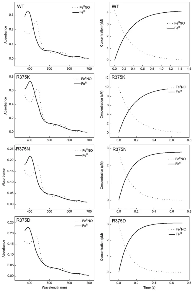
Fig. 5. Spectra and kinetics of iNOSoxy FeIINO oxidation reactions. Anaerobic ferrous iNOSoxy proteins in the presence of Arg (10 mM) and H
4B (400μM) were titrated by small
amounts of anaerobic NO-saturated buffer to form the ferrous NO complex, which was then rapid-mixed in a stoppedflow spectrophotometer with air-saturated buffer at 10 °C. Left panels contain spectra of the beginning ferrous-NO and ending ferric heme species as calculated by global analysis of the spectral data, while right panels show the con-centration of both species versus time after mixing. Data are representative of three experiments.
solution was transferred to the stopped-flow instrument by a gastight syringe and rapidly mixed with air saturated buffer. Sequential spec-tral data of 96 scans werefit to A → B reaction models using the Spec-fit global analysis software.
2.11. Heterodimer studies of Arg375 mutant monomer and G450A iNOS full length monomer
60μM iNOS wild type or mutant was incubated with urea (5 M) for 2.5 h at 15 °C to form protein monomer. For the heterodimer formation, the resulting urea-treated monomer was incubated with G450A iNOS full length protein (ratio 7:1) at 30 °C for 1 h in 40 mM EPPS, 10% glyc-erol, 250 mM NaCl buffer containing 1 mg/ml BSA, 1 mM H4B, 3 mM
DTT, 10 mM Arg and then further incubated at 4 °C overnight. Nitrite productions of heterodimer were assayed as described previously in 96 well plates[46]. In short, about 50–80 μl heterodimers were incubat-ed with cofactor FAD, FMN, and other reagents including SOD and cata-lase in each well, and NADPH was added to initiate the reaction at 37 °C. After 1 h, excess NADPH was consumed by adding lactate dehydroge-nase (LDH) and sodium pyruvate. Absorbance difference at 550 and 650 nm was measured after adding Griess reagent to each well. Nitrite productions were then calculated according to a standard curve made with nitrite solutions of known concentration.
NO synthesis of heterodimers were determined by spectrophoto-metric oxyhemoglobin assay [38,55] using extinction coefficient of 38 mM−1cm−1for methemoglobin minus oxyhemoglobin. All sample solutions contained 100μM H4B, 5 mM Arg, 0.3 mM DTT, 0.1 mg/ml
BSA, 10 U/ml SOD, 346 U/ml catalase, 1 mM oxyhemoglobin and 4μM FMN/FAD. Reactions were initiated by adding NADPH (100μM final concentration), and the absorbance changes at 401 nm were recorded at room temperature. NADPH oxidation was monitored at 340 nm using an extinction coefficient of 6.22 mM−1cm−1.
Antagonist experiments were done in a similar way except that 300 nM wild type iNOSoxy monomer was included during the het-erodimer formation of the mutants.
3. Results and discussion
3.1. Spectral property, stability, binding affinity for H4B and Arg, and
di-mer content of mutants
Mutations at Arg375 in iNOSoxy changed enzyme's binding affinity toward substrate Arg and cofactor H4B significantly. When the proteins
were purified in the absence of Arg and H4B, adding high concentration
of Arg and H4B at room temperature afterwards or incubating the
en-zyme overnight at 4 °C could not cause a complete high-spin shift in the heme iron spin state, which is a characteristic of substrate and
cofactor binding (data not shown). Mutants were also found to be less stable compared to the wild type: two mutants R375N and R375D dis-played a P420 peak instead of a P450 peak after the addition of CO to their dithionite reduced forms[56]. To maximize mutants' stability and their binding affinity for substrate and cofactor, we henceforth uti-lized mutant proteins that were purified in the presence of 1 mM Arg and 2μM H4B for our studies, unless noted otherwise.
All three mutants displayed the Soret absorbance peaks at 418 nm when purified in the presence of H4B and Arg, indicating that they
contained low-spin heme iron (Fig. 3) which is also observed in the wild type NOSoxy and other mutant like W457A iNOSoxy when puri-fied in the absence of substrate and cofactor[46,57–59]. Overnight in-cubation of proteins at 4 °C with additional H4B and Arg induced
Soret peak maxima shift to 395 nm for R375K and R375N, and 388 nm for R375D, which indicates that Arg and H4B could bind and
stabilize the heme iron in afive-coordinate high-spin state as occurs in the wild type NOS (Table 1,Fig. 3). After addition of the reducing agent dithionite and CO gas to the solution, mutants generated stable ferrous-CO complexes with maximal Soret absorbance at 444 nm (Table 1,Fig. 3). Arg375 mutants were also able to form other heme species including ferric/ferrous NO and ferrous dioxy which displayed similar Soret band maxima as that of the corresponding wild type heme species. These spectral properties demonstrate that the muta-tion does not perturb the electronic properties of the heme signi fi-cantly. Thisfinding agrees well with the crystal structures of R375 iNOSoxy mutants which showed that there are very limited structural changes around the heme pocket compared to the wild-type iNO-Soxy.1We also estimated the possible conformation changes after
the mutations and observed that Lys residue in R375K can still main-tain the interaction with the heme, as well as forming the hydrogen bonding with O4 of H4B without significant rearrangements of the
protein structure. However, the side chains of Asn and Asp are too short to make direct interactions with H4B in the absence of
notice-able structural changes (Supplementary Fig. S1).
The dimer–monomer ratio of each Arg375 mutant was estimated by gelfiltration chromatography[38,46,58]. After incubating the enzyme with sufficient amounts of substrate and cofactor, R375K mutant was approximately 100% dimeric. However, only approximately 38% and 30% dimer were obtained for R375N and R375D, respectively. Such de-creases in dimer content were also observed in R375A and Trp678 mu-tants where Trp 678 is another conserved amino residue involved in the hydrogen bonding andπ stacking with H4B ring in nNOSoxy[38].
3.2. Peroxide assay
We next examined whether the mutants could oxidize NOHA to ni-trite in a H2O2-driven reaction. Measurements of the reaction end
product-nitrite were carried out photometrically using the Griess Assay in a 96-well microplate at 37 °C as described previously[46,50,51]. For this experiment we used mutants purified in the absence of Arg and with the presence of H4B. Protein solutions werefirst incubated with
1 mM H4B overnight at 4 °C, then were diluted to the desired
concentra-tion and further incubated with various concentraconcentra-tion of NOHA (1 mM, 2 mM, 10 mM) or H4B (100μM, 500 μM, 1 mM) at 30 °C for 30 min.
H2O2solution was added to initiate the reaction, then the reaction
mix-tures were incubated for 10 min at 30 °C.
Wild type iNOSoxy protein being as a positive control displayed normal activity comparable to our previous results[46]. However, none of the mutants showed activities under these experimental con-ditions regardless of increasing the enzyme concentration from 110 nM to 1.1μM. To test whether the lower activity was attributed to the lower binding ability of substrate NOHA and H4B, we measured
the spectra of these mutants in the presence of 1 mM NOHA and
1
Z.-Q.Wang, M. Aoyagi, E. D. Getzoff and D. J. Stuehr, unpublished data. Table 2
Rates of heme and H4B redox transitions and product yield during Arg single turnover
reactions. Ferrous wild type and mutant iNOSoxy proteins containing substrates Arg and H4B were rapid-mixed at 10 °C with air-saturated buffer to start the reactions.
Sub-sequent heme transitions were followed by stopped-flow rapid-scanning spectroscopy. Rates were calculated by Specfit global analysis of diode array spectral data. H4B radical
formation and decay were monitored by EPR. NOHA productions were calculated based on HPLC results described in the Experimental section. Values are the means ± S.D. of three determinations. NOS FeIIO 2 formation (s− 1) FeIIO 2 decay (s− 1) H4B radical formation (s− 1) H4B radical decay (s− 1) NOHA produced per heme WT 67.8 ± 3.0 20.8 ± 0.5 11.1a 0.7 ± 0.5 0.55 ± 0.06 R375K 55.9 ± 3.3 29.7 ± 1.1 30.2 ± 9.8 1.6 ± 0.5 0.30 ± 0.10 R375N 72.4 ± 3.2 17.9 ± 0.5 34.8 ± 4.6 1.2 ± 0.6 0.25 ± 0.08 R375D NA 0.13 ± 0.01 8.8 ± 1.6 0.3 ± 0.1 0.11 ± 0.02 a
0.5 mM H4B. About 80–90% of heme iron of R375K and R375N were in
the high spin state suggesting normal binding of NOHA and H4B
to-ward the enzyme under the assay conditions. However, heme iron of R375D mainly existed in the low spin state.
In peroxide assay system, H2O2binds directly to the NOS ferric
heme forming reactive heme-oxy species and further react with Arg or NOHA to produce nitrite in the presence or absence of H4B[46].
Since the NOS heme does not need to acquire electrons from the re-ductase domain or H4B[50,51,58]. This method can be used to test
the reactivity of the heme pocket of the oxygenase domain for NOS enzymes. For R375K and R375N mutants, their lower activities com-pared to wild type iNOSoxy may be either due to the lower affinity of the enzyme for H2O2or the reaction generating a heme-oxy species
that is less reactive toward NOHA than wild type iNOSoxy. In the case of R375D, low binding affinity toward NOHA plus the two factors mentioned above contribute to its lower activity.
3.3. Heme midpoint potential
The NOS ferric heme midpoint potential is important in determin-ing the thermodynamic drivdetermin-ing force for electron transfer between the reductase domainflavins and the ferric heme. We next measured the heme midpoint potential for R375K and R375D. A spectro-electrochemical reductive titration was performed for the ferric
en-zyme in the presence of Arg, H4B, and mediator dyes [52].
Representative data were shown inFig. 4. The heme midpoint
poten-tial were determined to be−294±3 mV for R375K and −283±3 mV
for R375D, which are about 30 to 40 mV more negative than the wild
type iNOSoxy when measured under similar conditions (−263 mV
[52];−253 mV2).
In NOSs, the heme iron has a proximal cysteine thiolate ligand [12,13,42] (Fig. 2). Studies have shown that the strength of the heme-thiolate bond determines the heme midpoint potential of the enzyme. One conserved residue Trp409 in nNOS (Trp188 in iNOS) en-gages inπ-stacking with the heme and forms hydrogen bond to the proximal cysteine ligand. Mutation on this residue turns out to alter the heme midpoint potential greatly by either weakening or strength-ening the heme-thiolate bond[60]. Interestingly, our Arg375 mutation lowers the heme midpoint potential, indicating that such mutation couldfine tune the heme environment even though spatially Arg375 is far from the cysteine thiolate ligand. These effects are probably me-diated through the direct interaction of the Arg375 side chain with the heme propionate. The decrease in the redox potential creates unfavor-able thermodynamic change for the electron transfer from theflavins in the reductase domain and the ferric heme. In addition, the lower heme midpoint potential also implied the conceivable changes of the
2
J. Santolini and D. J. Stuehr, unpublished data.
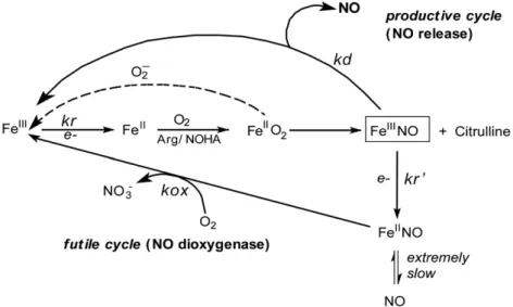
Fig. 6. Global kinetic model for NO synthesis by NOS. During steady state catalysis, the enzyme molecules engage in a productive cycle that releases free NO and in a futile cycle that releases a higher oxide of nitrogen (nitrate). Reduction of ferric enzyme to ferrous (kr) enables the heme to bind O2and initiates catalytic reactions. After NO is made, an immediate
product of catalysis is the ferric heme-NO complex (FeIII–NO), which can either release NO (k
d) or become reduced (kr′) to generate a ferrous heme-NO complex (FeII–NO). The
ferrous heme-NO complex dissociates extremely slowly and instead regenerates the active ferric enzyme by reacting with O2(kox). The FeIIO2intermediate can also undergo an
uncoupled reaction that generates ferric enzyme and superoxide.
heme reactivity which is supported by NOHA oxidation reaction de-scribed above.
3.4. Oxidation rate of the ferrous heme-NO (FeIINO) complex (kox)
The differences in kox values between the wild type and mutants often indicate subtle changes of the heme environment. We next
determined kox for the wild-type and Arg375 mutants by mixing their ferrous heme-NO complexes with an air-saturated buffer in the stopped-flow spectrophotometer, and following their subse-quent conversion to ferric enzyme[61–64]. The starting FeIINO
com-plex and ending ferric enzyme were observed for all proteins, and their conversionfit well to a monophasic transition (Fig. 5). The cal-culated kox values of WT, R375K, N and D are 3.11 ± 0.15, 6.35 ± 0.35, Fig. 7. Stopped-flow analysis of heme transitions and kinetics during single-turnover Arg hydroxylation catalyzed by R375K, R375N and wild type iNOSoxy. Anaerobic ferrous iNO-Soxy proteins (4μM) in the presence of Arg (10 mM) and H4B (400μM) were mixed in a stopped flow instrument with air-saturated buffer at 10 °C and diode array spectra were
collected. Left panels contain three heme species that were detected during each reaction as calculated by global analysis of the spectral data, while right panels show their con-centration profiles versus time after mixing. Data are representative of three similar experiments.
7.72 ± 0.33 and 8.73 ± 0.37 s–1, respectively. All mutants showed higher kox rates, which were about 2.1 to 2.8 times faster than that of the wild-type iNOSoxy. In combination with the results of the heme midpoint potential measurements, mutation at Arg375 low-ered the heme midpoint potential and increased the oxidation rate of the ferrous heme-NO complex. Such relationship was observed in other mutants as well. For example, W409F, W409Y nNOSoxy mu-tants and mesoheme-nNOSoxy also displayed lower heme midpoint potentials and high kox values relative to their wild-type enzyme [54,60,61,65–67]. For iNOSoxy, replacing Trp188 to His increases the heme midpoint potential by 80 mV due to the strengthened heme-thiolate bond, since His is able to form stronger hydrogen bonding with proximal Cys ligand, and its kox rate of W188H was found to decrease by 40% relative to wild type iNOSoxy correspondingly.3
The correlation between the kox rate and heme midpoint poten-tial provides a good indication about the heme environment and the characteristics of the heme-thiolate bond. NOS heme FeII
–NO com-plexes oxidize at much higher rates compared to any other heme pro-tein including Hb or Mb[28,54,65,68,69]. Such difference is attributed in part by the heme-thiolate bond, causing the NOS heme FeIINO complex to have a lower midpoint potential. Our results on Arg375 further indicate that besides the difference between the heme proxi-mal ligands (cysteine in NOSs versus histidine in Hb, Mb and other
heme proteins), modifications of nearby amino residue in the heme pocket could also modulate the heme midpoint potentials and the kox rate of NOS enzymes.
The rate of ferrous NO oxidation (kox) is one of the key parame-ters controlling the steady state NO synthesis in full length enzymes [63,70](Fig. 6). During NO synthesis, practically all generated NO binds to the NOS ferric heme forming FeIIINO before exiting the
en-zyme. Part of the FeIIINO is reduced by the attached reductase domain
and the resulting FeIINO complex must react with O2(kox inFig. 6)
afterwards to return to the catalytic cycle [63,70]. Unlike other heme-thiolate proteins[54,71], NOS enzyme posses relatively high oxidization rate of FeIINO complex which is crucial to avoid the for-mation of a dead-end product during catalysis. The observed faster kox rates of Arg375 mutants possibly increase the steady-state activ-ity of iNOS by speeding the return of enzyme molecules to the pro-ductive cycle (Fig. 6)[62,70,72].
3.5. Kinetics of heme transitions, H4B radical formation and decay during
Arg hydroxylation
Previous studies show that H4B performs a novel redox function in
NOS: it reduces the ferrous dioxy intermediate (FeIIO2) that forms
dur-ing oxygen activation. This step is critical because it enables NOS to form the heme-oxy species that react with Arg or NOHA (Scheme 2). Since Arg375 forms hydrogen bond with the H4B ring, we investigated
whether mutation affects the redox function of H4B during single
cata-lytic turnover Arg hydroxylation.
First, we monitored the heme transitions that occur during a sin-gle turnover Arg hydroxylation reaction run at 10 °C in the stopped-flow spectrophotometer. Air-saturated buffer was mixed with anaer-obic ferrous iNOSoxy proteins that contained H4B and Arg and the
subsequent heme transitions were followed by UV–visible scanning [20,37,73].Figs. 7 and 8contain representative data collected during reactions catalyzed by wild type, R375K, R375N and R375D iNOSoxy. InFig. 7, the left panels show the light absorbance spectra of the three enzyme species that were detected during each reaction, while the right panels show the concentration of each species changed with time during the reaction. In all mutants we could identify a beginning ferrous species, a heme-dioxy (FeIIO
2) intermediate, and an ending
ferric species. Their Soret absorbance maxima for each heme species matched very well among wild-type and mutants. The ending ferric species for all mutants displayed a 390–393 nm peak, which indicated that their heme groups were predominantly in high spin state at the end of the single turnover reactions. InFig. 8, Panel A shows the scan-ning traces collected at different time after mixing anaerobic ferrous R375D proteins with air-saturated buffer at 10 °C. Panel B contains cross section analysis of the kinetic data at 426 nm for the decay of the heme ferrous dioxy and 393 nm for the formation of the ferric en-zyme. These results showed that Arg375 mutations still permitted buildup of a FeIIO
2intermediate, and did not significantly alter its
spectral characteristics or those of the ferrous or ferric species. For-mation and disappearance of the FeIIO
2intermediate were best
de-scribed by monophasic transitions, consistent with previous reports [20,37,73]. The mutants showed similar FeIIO
2formation rate as of
the wild type iNOSoxy (Table 2). However, the transition of the FeIIO
2 intermediate to ferric enzyme was found to be different
among mutants: R375N and R375K mutants demonstrated similar or faster rate, respectively, compared to wild type, but much slower FeIIO
2decay rate was observed in the R375D mutant (Table 2). Our
previous results show that the electron transfer from H4B to ferrous
dioxy species in wild type iNOSoxy increases the ferrous dioxy decay rate by 40 fold compared to the H2B-bound protein (H2B
can-not reduce the ferrous dioxy species). To elucidate the redox role of H4B in Arg375 mutants, we performed the single turnover Arg
hy-droxylation reactions using H2B-bound R375D mutant as well. Three
heme species were observed, whose spectral features were identical
3
Z.-Q.Wang, and D. J. Stuehr, unpublished data.
Fig. 8. Heme transitions and kinetics of R375D iNOSoxy during Arg hydroxylation sin-gle turnover reactions. Panel A contains spectral scans collected at 0.0089 s (…), 0.00135 s (—) and 13.35 s (– –) after mixing anaerobic ferrous proteins with air-saturated buffer at 10 °C. Panel B contains cross section analysis of the kinetic data at 426 nm and 393 nm. Data are representative of three similar experiments.
to the ferrous, FeIIO
2, and ferric species in the H4B-bound proteins
(data not shown). However, conversion of the FeIIO
2 intermediate
to ferric enzyme was 5 times slower in the H2B-bound proteins,
sug-gesting H4B is able to transfer an electron to the heme ferrous dioxy
species of Arg375 mutants during Arg hydroxylation single turnover reactions.
We next studied H4B radical formation and decay during Arg
hy-droxylation. Rapid-freeze experiments showed that a radical with g Fig. 9. H4B radical formation and decay during Arg hydroxylation in Arg375 mutants and wild-type iNOSoxy. Anaerobic ferrous iNOSoxy proteins were rapidly mixed with an
air-saturated solution at 10 °C to start the reaction. Reactions were aged at the various times (16 ms to 5 s), and then quenched by rapid freezing. EPR traces are shown for Arg375 mutants and wild-type iNOSoxy.
Fig. 10. Calculated concentrations of the H4B radical versus time during Arg hydroxylation in Arg375 mutants and wild-type iNOSoxy. The relative concentration of H4B radical was
value of 2.0 built up in Arg375 mutants during the single turnover re-actions (Fig. 9). The spectra and saturation properties of the mutant radicals were either identical or similar to those of the H4B radical
formed in wild-type iNOSoxy [13,20,37,42,74]. Signal intensities were used to calculate the amount of H4B radical per heme that was
present at each time point. The kinetic data werefit to a two step pro-cess including the radical formation and decay (Fig. 10). The relative rates of H4B radical formation are consistent with the different
fer-rous dioxy disappearance rates we observed in the mutants, and imply that there is faster ferrous dioxy reduction by H4B in the
R375K and R375N mutants compared to the wild type. Apparently R375 mutations alter the tempo of electron transfer: in R375K and R375N mutants, H4B transfers an electron faster than in the wild
type protein, while H4B transfers an electron slower in the R375D
mutant. Our kinetic data also suggest the mutations do not greatly af-fect the stability of the H4B radical since all mutants displayed similar
H4B decay rates compared to the wild type within experimental error.
Our data analysis also shows that a normal level of H4B radical
accu-mulated in the R375K mutant reaction (about 0.78 per heme) compared to wild type iNOSoxy[37]. However, there was less H4B radical
accumu-lation (0.4 and 0.2 radical per heme, respectively) during the single turn-over reactions of R375N and R375D iNOSoxy. Increasing the H4B
concentration in the reactions from 1 to 5 mM did not alter the results in any way (data not shown), suggesting that the observed differences were not due to incomplete H4B binding. The poor radical buildup
ob-served in these mutants might be due to their incomplete dimer forma-tion, or possibly some unreacted H4B remaining in the enzyme.
We then studied the stoichiometry of Arg hydroxylation in the Arg375 mutants. NOHA was the only amino acid product in all cases and its formation depended completely on H4B (data not shown).
The R375K, R375N and R375D mutants consistently catalyzed about 0.30, 0.25 and 0.11 of Arg hydroxylation on a per heme basis during the single turnover Arg hydroxylation reactions, respectively, which are all less extensive than in the wild type (0.55,Table 2).
Previous studies suggest that a complete and well coupled electron transfer between H4B and FeIIO2occurred for wild type NOS proteins
in Arg hydroxylation single turnover reactions. Thus, one would expect that Arg hydroxylation should generate 1 NOHA/heme. However, we and others[20,73–75]typically observe substoichiometric NOHA for-mation in single turnover reactions ranging from 0.2 to 0.8 NOHA formed per heme, which implies that subsequent steps (i.e. conversion of the iron-peroxo intermediate to iron-oxo or its reaction with Arg) be-come uncoupled in the wild type iNOSoxy within this setting. We re-cently identified a heme-oxy reaction intermediate in the W188H iNOSoxy[60], further studies should be able to shed light on this issue. Here, the mutation at Arg375 even further decreased the product stoichiometry compared to the wild type protein. One possibility is that Arg375 mutations might diminish the reactivity of the heme-based
oxidant toward Arg, since this effect will only occur after H4B reduces
the FeIIO
2intermediate. Our H2O2-driven NOHA oxidation results
sup-ported this assumption since the heme-based oxidant formed when fer-ric NOS reacts with H2O2is thought to be similar or identical to the
species that hydroxylates NOHA during normal NOS catalysis[33,61]. For the R375N and D mutants, their incomplete enzyme dimerization would also diminish the product stoichiometry.
3.6. Activities of mutant heterodimers
To test whether mutation at Arg375 affect the NO synthesis activ-ity, we made heterodimers by incubating the monomer of wild type iNOSoxy (or mutants) and full-length G450A iNOS. The point muta-tion Gly450 to Ala prevents itself forming homodimer, thus the re-ductase domain from G450A is only capable of transferring electron to the iNOSoxy which enables NO production from the oxygenase do-main[46,76,77]. Wefirst converted oxygenase domains of wild type and mutants to monomer by adding urea, then incubated such mono-mer with full-length G450A protein in the presence of H4B and Arg to
form heterodimer. We next measured the rates of NO synthesis and NADPH oxidation of these heterodimers in the presence of Arg and H4B at 25 °C using the spectrophotometric oxyhemoglobin assay.
Re-sults are summarized inTable 3.
The NO synthesis activity of the heterodimer containing iNOSoxy wild type was very close to the native wild type full length iNOS en-zyme, which validates the experimental approach. All mutants showed lower NO synthesis activities with only 35%, 17% and 7% of the wild type for R375K, N and D, respectively. However, their concurrent rates of NADPH oxidation were also reduced to a lesser degree. In general, NO synthesis from Arg displays a stoichiometry of 1.5 NADPH oxidized per NO formed for wild type full length NOSs[18,78]. This minimum value was observed for our wild-type heterodimer iNOS at 25 °C, while a stoichiometry of 3.5, 7.5, and 29 NADPH oxidized per NO formed were found for R375K, R375N and R375D, respectively (Table 3). This indicates that a majority of the NADPH oxidation is uncoupled from their NO synthesis in the Arg375 mutants.
Nitrite production of heterodimer measured by Griess assay also showed the lower activity of mutants compared to the wild type, which is consistent with the results from our NO measurements (Table 3). One could argue that the lower activity might be due to the low affinity of mutant monomer toward the G450A subunit. To test this, we measured the capability of each mutant monomer to“compete” or“antagonize” wild type iNOSoxy monomer in forming a heterodimer with full length G450A iNOS. First, 300 nM wild type iNOSoxy monomer formed heterodimer with G450A iNOS. If mutant monomers could com-pete with wild type effectively, then addition of excess of mutant
mono-mer (~3μM) to the protein solution should reduce the nitrite
production to a level similar to that of the heterodimer we formed by mixing mutant monomer and G450 iNOS in the absence of wild type iNOSoxy monomer. Our results inTable 3clearly indicate that Arg375 mutants compete successfully and completely with wild-type iNOSoxy monomer in forming the heterodimer, eliminating the possibility that lower activity is associated with a lower binding affinity of the mutant monomer to full length G450A iNOS. These results are also consistent with our previous heterodimer studies of R375A mutant[46].
The catalytic profiles we observed for heterodimer are consistent with our single turnover Arg hydroxylation results. Since all the hetero-dimers have the same reductase domain subunit, lower NO synthesis observed in the mutants solely comes from the mutant oxygenase do-main itself and/or the inter-dodo-main electron transfer. Negative shift of the heme midpoint potential of Arg375 mutants disfavored the electron transfer from reductase domain to the oxygenase domain, which can decrease the NO synthesis activity. Also the less efficient electron trans-fer from H4B and/or reduced reactivity of heme-based oxidant further
lower the NO synthesis rate in Arg375 mutants. Table 3
Catalytic activities of heterodimers formed by the Arg375 mutants or wild type iNO-Soxy and G450A iNOS. NO synthesis and NADPH oxidation were measured at room temperature and NO2−production measurement was performed at 30 °C as described
under“Materials and methods.” The values are the mean±S.D. of three measurements. WT, wild type. Heterodimer Native WT iNOS R375K R375N R375D WT NO synthesis (min− 1) 17.6 ± 0.6 8.6 ± 0.3 1.7 ± 0.1 51.8 ± 0.4 50.9 ± 0.2 NADPH oxidation (min− 1) 62.5 ± 4.0 64.2 ± 3.8 50.1 ± 4.4 80.3 ± 6.2 78.2 ± 3.1 NO2−production (μM) (NO2−/μM G450A/min) 7.8 ± 0.2 6.3 ± 0.1 1.8 ± 0.1 30.3 ± 0.7 NA NO/NADPH 3.6 7.5 29.4 1.6 1.5 Antagonist Study 29% 30% 13% 100% NA
4. Conclusions
Mutation of the conserved residue Arg375, whose side chain H-bonds with heteroatoms in the ring of H4B, greatly changes iNOS properties and
catalytic behavior. The decreased binding affinity toward substrate and cofactor can be overcome by purifying the enzyme in the presence of substrates and cofactor and later incubating the enzyme with additional amount of substrate and cofactor. However, the maximum dimer forma-tion obtained in the R375N and D mutant are only 38% and 30%, respec-tively. Such a low percentage in these two mutants partially contributes to their lower Arg hydroxylation observed in the single turnover reactions and decreased NO synthesis rate. Mutation at Arg375 also perturbs the heme environment which leads to the neg-ative shift of the heme midpoint potential, faster ferrous NO oxida-tion rate and vanished peroxide activity in H2O2-drived NOHA
oxidation compared to the wild type protein. Although replacement of Arg375 by Lys (K) increases the tempo of electron transfer be-tween H4B and the ferrous dioxy species formed during O2
activa-tion, R375K still shared similar properties like other mutants, for instance, lower NOHA production in single turnover reactions and lower NO synthesis activities and uncoupled NADPH oxidation. We attribute these properties mainly to the reduced reactivity of heme-based oxidant formed after the electron transfer from H4B.
Our results suggest that the conserved residue Arg375 modulates the redox and structural functions of H4B, and is thereby important
in controlling the catalytic behavior of NOS enzymes.
Supplementary materials related to this article can be found on-line atdoi:10.1016/j.jinorgbio.2011.11.015.
Abbreviations
NOS nitric oxide synthase
iNOS inducible nitric oxide synthase
iNOSoxy the oxygenase domain of inducible nitric oxide synthase
Arg L-arginine
DTT dithiothreitol
NO nitric oxide
EPPS 4-(2-hydroxyethyl)-1-piperazinepropanesulfonic acid
NOHA Nω-hydroxy-L-arginine
H4B (6R)-5, 6, 7, 8-tetrahydro-L-biopterin
H2B 7,8-dihydro-L-biopterin
NED N-1-naphthylethylenediamine dihydrochloride
LDH lactate dehydrogenase
FeII ferrous heme species
FeIII ferric heme species
FeIIO
2 ferrous dioxy species
FeIIINO ferric NO species FeIINO ferrous NO species
Acknowledgments
We thank Manisha Sharma and John MacDonald for excellent technical support. This work was supported by the National Institutes of Health Grants GM51491 and CA53914 (D. J. S.), American Heart As-sociation Beginning Grant-in-aid 0565297B (Z-Q. W.), KSU Farris In-novation Award (Z-Q. W.) and KSU Tuscarawas Faculty Professional Development Release Time Award (Z-Q. W.).
References
[1] C. Nathan, M.U. Shiloh, Proc. Natl. Acad. Sci. U. S. A. 97 (2000) 8841–8848. [2] M. Sasaki, M. Gonzalez-Zulueta, H. Huang, W.J. Herring, S. Ahn, D.D. Ginty, V.L.
Dawson, T.M. Dawson, Proc. Natl. Acad. Sci. U. S. A. 97 (2000) 8617–8622. [3] H. Stopper, M. Moller, H.M. Bommel, H.H. Schmidt, Toxicol. Lett. 106 (1999)
59–67.
[4] D.J. Stuehr, S. Ghosh, Handbook of Experimental Pharmacol, 2000, pp. 33–70. [5] W.K. Alderton, C.E. Cooper, R.G. Knowles, Biochem. J. 357 (2001) 593–615.
[6] A.C. Gorren, B.M. List, A. Schrammel, E. Pitters, B. Hemmens, E.R. Werner, K. Schmidt, B. Mayer, Biochemistry 35 (1996) 16735–16745.
[7] D.S. Bredt, S.H. Snyder, Proc. Natl. Acad. Sci. U. S. A. 87 (1990) 682–685. [8] Q.W. Xie, H.J. Cho, J. Calaycay, R.A. Mumford, K.M. Swiderek, T.D. Lee, A. Ding, T.
Troso, C. Nathan, Science 256 (1992) 225–228.
[9] J.S. Pollock, U. Forstermann, J.A. Mitchell, T.D. Warner, H.H. Schmidt, M. Nakane, F. Murad, Proc. Natl. Acad. Sci. U. S. A. 88 (1991) 10480–10484.
[10] B.S. Masters, K. McMillan, E.A. Sheta, J.S. Nishimura, L.J. Roman, P. Martasek, FASEB J. 10 (1996) 552–558.
[11] D.J. Stuehr, Annu. Rev. Pharmacol. Toxicol. 37 (1997) 339–359.
[12] B.R. Crane, A.S. Arvai, D.K. Ghosh, C. Wu, E.D. Getzoff, D.J. Stuehr, J.A. Tainer, Sci-ence 279 (1998) 2121–2126.
[13] T.O. Fischmann, A. Hruza, X.D. Niu, J.D. Fossetta, C.A. Lunn, E. Dolphin, A.J. Prongay, P. Reichert, D.J. Lundell, S.K. Narula, P.C. Weber, Nat. Struct. Biol. 6 (1999) 233–242. [14] E.A. Sheta, K. McMillan, B.S. Masters, J. Biol. Chem. 269 (1994) 15147–15153. [15] R. Gachhui, A. Presta, D.F. Bentley, H.M. Abu-Soud, R. McArthur, G. Brudvig, D.K.
Ghosh, D.J. Stuehr, J. Biol. Chem. 271 (1996) 20594–20602.
[16] E.D. Garcin, C.M. Bruns, S.J. Lloyd, D.J. Hosfield, M. Tiso, R. Gachhui, D.J. Stuehr, J.A. Tainer, E.D. Getzoff, J. Biol. Chem. 279 (2004) 37918–37927.
[17] B. Mayer, B. Hemmens, Trends Biochem. Sci. 22 (1997) 477–481. [18] D.J. Stuehr, Biochim. Biophys. Acta 1411 (1999) 217–230. [19] H. Li, T.L. Poulos, J. Inorg. Biochem. 99 (2005) 293–305.
[20] C.C. Wei, Z.Q. Wang, Q. Wang, A.L. Meade, C. Hemann, R. Hille, D.J. Stuehr, J. Biol. Chem. 276 (2001) 315–319.
[21] C.C. Wei, Z.Q. Wang, C. Hemann, R. Hille, D.J. Stuehr, J. Biol. Chem. 278 (2003) 46668–46673.
[22] O.W. Griffith, D.J. Stuehr, Annu. Rev. Physiol. 57 (1995) 707–736.
[23] B.S. Masters, K. McMillan, E.A. Sheta, J.S. Nishimura, L.J. Roman, P. Martasek, FASEB J. 10 (1996) 552–558.
[24] M.A. Marletta, Cell 78 (1994) 927–930.
[25] H.M. Abu-Soud, L.L. Yoho, D.J. Stuehr, J. Biol. Chem. 269 (1994) 32047–32050. [26] L.J. Roman, R.T. Miller, M.A. de La Garza, J.J. Kim, B.S. Siler Masters, J. Biol. Chem.
275 (2000) 21914–21919.
[27] C.C. Wei, B.R. Crane, D.J. Stuehr, Chem. Rev. 103 (2003) 2365–2383.
[28] H.M. Abu-Soud, R. Gachhui, F.M. Raushel, D.J. Stuehr, J. Biol. Chem. 272 (1997) 17349–17353.
[29] M. Sorlie, A.C. Gorren, S. Marchal, T. Shimizu, R. Lange, K.K. Andersson, B. Mayer, J. Biol. Chem. 278 (2003) 48602–48610.
[30] N.C. Gerber, C.R. Nishida, P.R. Ortiz de Montellano, Arch. Biochem. Biophys. 343 (1997) 249–253.
[31] B. Mayer, C. Wu, A.C. Gorren, S. Pfeiffer, K. Schmidt, P. Clark, D.J. Stuehr, E.R. Wer-ner, Biochemistry 36 (1997) 8422–8427.
[32] H.M. Abu-Soud, M. Loftus, D.J. Stuehr, Biochemistry 34 (1995) 11167–11175. [33] K.M. Rusche, M.M. Spiering, M.A. Marletta, Biochemistry 37 (1998) 15503–15512. [34] P.P. Schmidt, R. Lange, A.C. Gorren, E.R. Werner, B. Mayer, K.K. Andersson, J. Biol.
Inorg. Chem. 6 (2001) 151–158.
[35] A.R. Hurshman, C. Krebs, D.E. Edmondson, M.A. Marletta, Biochemistry 42 (2003) 13287–13303.
[36] V. Berka, H.C. Yeh, D. Gao, F. Kiran, A.L. Tsai, Biochemistry 43 (2004) 13137–13148.
[37] Z.Q. Wang, C.C. Wei, S. Ghosh, A.L. Meade, C. Hemann, R. Hille, D.J. Stuehr, Bio-chemistry 40 (2001) 12819–12825.
[38] Z.Q. Wang, C.C. Wei, J. Santolini, K. Panda, Q. Wang, D.J. Stuehr, Biochemistry 44 (2005) 4676–4690.
[39] C.C. Wei, Z.Q. Wang, D. Durra, C. Hemann, R. Hille, E.D. Garcin, E.D. Getzoff, D.J. Stuehr, J. Biol. Chem. 280 (2005) 8929–8935.
[40] C.C. Wei, Z.Q. Wang, J. Tejero, Y.P. Yang, C. Hemann, R. Hille, D.J. Stuehr, J. Biol. Chem. 283 (2008) 11734–11742.
[41] T. Nagatsu, H. Ichinose, Mol. Neurobiol. 19 (1999) 79–96.
[42] C.S. Raman, H. Li, P. Martasek, V. Kral, B.S. Masters, T.L. Poulos, Cell 95 (1998) 939–950.
[43] M. Aoyagi, A.S. Arvai, S. Ghosh, D.J. Stuehr, J.A. Tainer, E.D. Getzoff, Biochemistry 40 (2001) 12826–12832.
[44] B.R. Crane, A.S. Arvai, S. Ghosh, E.D. Getzoff, D.J. Stuehr, J.A. Tainer, Biochemistry 39 (2000) 4608–4621.
[45] Z.Q. Wang, C.C. Wei, D.J. Stuehr, J. Biol. Chem. 277 (2002) 12830–12837. [46] S. Ghosh, D. Wolan, S. Adak, B.R. Crane, N.S. Kwon, J.A. Tainer, E.D. Getzoff, D.J.
Stuehr, J. Biol. Chem. 274 (1999) 24100–24112.
[47] I. Sagami, Y. Sato, S. Daff, T. Shimizu, J. Biol. Chem. 275 (2000) 26150–26157. [48] P.F. Chen, V. Berka, K.K. Wu, Arch. Biochem. Biophys. 411 (2003) 83–92. [49] D.J. Stuehr, M. Ikeda-Saito, J. Biol. Chem. 267 (1992) 20547–20550.
[50] R. Gachhui, D.K. Ghosh, C. Wu, J. Parkinson, B.R. Crane, D.J. Stuehr, Biochemistry 36 (1997) 5097–5103.
[51] R.A. Pufahl, J.S. Wishnok, M.A. Marletta, Biochemistry 34 (1995) 1930–1941. [52] A. Presta, A.M. Weber-Main, M.T. Stankovich, D.J. Stuehr, J. Am. Chem. Soc. 120
(1998) 9460–9465.
[53] Z.Q. Wang, C.C. Wei, M. Sharma, K. Pant, B.R. Crane, D.J. Stuehr, J. Biol. Chem. 279 (2004) 19018–19025.
[54] J. Tejero, J. Santolini, D.J. Stuehr, FEBS J. 276 (2009) 4505–4514. [55] S. Adak, K.S. Aulak, D.J. Stuehr, J. Biol. Chem. 276 (2001) 23246–23252. [56] L. Huang, H.M. Abu-Soud, R. Hille, D.J. Stuehr, Biochemistry 38 (1999) 1912–1920. [57] H.M. Abu-Soud, C. Wu, D.K. Ghosh, D.J. Stuehr, Biochemistry 37 (1998) 3777–3786. [58] D.K. Ghosh, C. Wu, E. Pitters, M. Moloney, E.R. Werner, B. Mayer, D.J. Stuehr,
Bio-chemistry 36 (1997) 10609–10619.
[59] H.M. Abu-Soud, J. Wang, D.L. Rousseau, D.J. Stuehr, Biochemistry 38 (1999) 12446–12451.
[60] J. Tejero, A. Biswas, Z.Q. Wang, R.C. Page, M.M. Haque, C. Hemann, J.L. Zweier, S. Misra, D.J. Stuehr, J. Biol. Chem. 283 (2008) 33498–33507.
[61] S. Adak, Q. Wang, D.J. Stuehr, J. Biol. Chem. 275 (2000) 17434–17439. [62] J. Santolini, A.L. Meade, D.J. Stuehr, J. Biol. Chem. 276 (2001) 48887–48898. [63] Z.Q. Wang, C.C. Wei, D.J. Stuehr, J. Inorg. Biochem. 104 (2010) 349–356. [64] S. Adak, Q. Wang, D.J. Stuehr, J. Biol. Chem. 275 (2000) 33554–33561. [65] J. Tejero, A. Biswas, M.M. Haque, Z.Q. Wang, C. Hemann, C.L. Varnado, Z. Novince,
R. Hille, D.C. Goodwin, D.J. Stuehr, Biochem. J. 433 (2010) 163–174.
[66] S. Adak, C. Crooks, Q. Wang, B.R. Crane, J.A. Tainer, E.D. Getzoff, D.J. Stuehr, J. Biol. Chem. 274 (1999) 26907–26911.
[67] S. Adak, D.J. Stuehr, J. Inorg. Biochem. 83 (2001) 301–308. [68] T.W. Ost, S. Daff, J. Biol. Chem. 280 (2005) 965–973. [69] C.E. Cooper, Biochim. Biophys. Acta 1411 (1999) 290–309.
[70] D.J. Stuehr, J. Santolini, Z.Q. Wang, C.C. Wei, S. Adak, J. Biol. Chem. 279 (2004) 36167–36170.
[71] L.G. Quaroni, H.E. Seward, K.J. McLean, H.M. Girvan, T.W. Ost, M.A. Noble, S.M. Kelly, N.C. Price, M.R. Cheesman, W.E. Smith, A.W. Munro, Biochemistry 43 (2004) 16416–16431.
[72] J. Santolini, S. Adak, C.M. Curran, D.J. Stuehr, J. Biol. Chem. 276 (2001) 1233–1243. [73] S. Boggs, L. Huang, D.J. Stuehr, Biochemistry 39 (2000) 2332–2339.
[74] A.R. Hurshman, C. Krebs, D.E. Edmondson, B.H. Huynh, M.A. Marletta, Biochemis-try 38 (1999) 15689–15696.
[75] N. Bec, A.C. Gorren, C. Voelker, B. Mayer, R. Lange, J. Biol. Chem. 273 (1998) 13502–13508.
[76] U. Siddhanta, C. Wu, H.M. Abu-Soud, J. Zhang, D.K. Ghosh, D.J. Stuehr, J. Biol. Chem. 271 (1996) 7309–7312.
[77] U. Siddhanta, A. Presta, B. Fan, D. Wolan, D.L. Rousseau, D.J. Stuehr, J. Biol. Chem. 273 (1998) 18950–18958.
[78] M.A. Marletta, A.R. Hurshman, K.M. Rusche, Curr. Opin. Chem. Biol. 2 (1998) 656–663.
[79] B.R. Crane, A.S. Arvai, R. Gachhui, C. Wu, D.K. Ghosh, E.D. Getzoff, D.J. Stuehr, J.A. Tainer, Science 278 (1997) 425–431.