An angiogenic role for the #5#1 integrin in promoting
endothelial cell proliferation during cerebral hypoxia
The MIT Faculty has made this article openly available.
Please share
how this access benefits you. Your story matters.
Citation
Li, Longxuan, Jennifer Welser-Alves, Arjan van der Flier,
Amin Boroujerdi, Richard O. Hynes, and Richard Milner. “An
Angiogenic Role for the Α5β1 Integrin in Promoting Endothelial Cell
Proliferation During Cerebral Hypoxia.” Experimental Neurology
237, no. 1 (September 2012): 46–54.
As Published
http://dx.doi.org/10.1016/j.expneurol.2012.06.005
Publisher
Elsevier
Version
Author's final manuscript
Citable link
http://hdl.handle.net/1721.1/101277
Terms of Use
Creative Commons Attribution-Noncommercial-NoDerivatives
An angiogenic role for the α5β1 integrin in promoting
endothelial cell proliferation during cerebral hypoxia
Longxuan Lia, Jennifer Welser-Alvesa, Arjan van der Flierb, Amin Boroujerdia, Richard O.
Hynesb, and Richard Milnera
aDepartment of Molecular and Experimental Medicine, The Scripps Research Institute, 10550
North Torrey Pines Road, La Jolla, CA 92037
bHoward Hughes Medical Institute, Koch Institute for Integrative Cancer Research at the
Massachusetts Institute of Technology, Cambridge, MA 02139
Abstract
Fibronectin is a critical regulator of vascular modelling, both in development and in the adult. In the hypoxic adult central nervous system (CNS), fibronectin is induced on angiogenic vessels, and endothelial cells show strong induction of the two fibronectin receptors α5β1 and αvβ3 integrins. In a previous study, we found that the αvβ3 integrin is dispensable for hypoxic-induced cerebral angiogenesis, but a role for the endothelial α5β1 integrin was suggested. To directly investigate the role of endothelial α5 integrin in cerebral angiogenesis, wild-type mice and mice lacking α5 integrin expression in endothelial cells (α5-EC-KO) were subject to hypoxia (8% O2) for 0, 2, 4, 7
or 14 days. Quantification of cerebral vessel density and endothelial-specific proteins claudin-5 and Glut-1 revealed that α5-EC-KO mice displayed an attenuated angiogenic response, which correlated with delayed endothelial proliferation. α5-EC-KO mice showed no defect in the ability to organize a cerebrovascular fibronectin matrix, and no compensatory increase in vascular αvβ3 integrin expression. Consistent with these findings, primary α5KO brain endothelial cells (BEC) in culture exhibited delayed growth and proliferation. Taken together, these studies demonstrate an important angiogenic role for the α5β1 integrin in promoting BEC proliferation in response to cerebral hypoxia.
Keywords
angiogenesis; cerebral; hypoxia; endothelial; proliferation; integrin; fibronectin
INTRODUCTION
The blood supply to the central nervous system (CNS) is tightly regulated to maintain optimal delivery of oxygen and other nutrients (Attwell et al. 2010). Actually, the oxygen level in intact brain tissue is much lower than the 21% oxygen present in air at sea level; more in the range of 2–4% (Ndubuizu and LaManna 2007). Cerebral hypoxia or ischemia
© 2012 Elsevier Inc. All rights reserved.
Corresponding author: Dr. Richard Milner at: Department of Molecular and Experimental Medicine, MEM-132, The Scripps Research Institute, 10550 North Torrey Pines Road, La Jolla, CA 92037. Tel: (858) 784-8569, Fax: (858) 784-2174, rmilner@scripps.edu. Longxuan Li and Jennifer Welser-Alves contributed equally to this work.
Publisher's Disclaimer: This is a PDF file of an unedited manuscript that has been accepted for publication. As a service to our
customers we are providing this early version of the manuscript. The manuscript will undergo copyediting, typesetting, and review of
NIH Public Access
Author Manuscript
Exp Neurol
. Author manuscript; available in PMC 2013 August 20. Published in final edited form as:Exp Neurol. 2012 September ; 237(1): 46–54. doi:10.1016/j.expneurol.2012.06.005.
NIH-PA Author Manuscript
NIH-PA Author Manuscript
trigger activation of several physiological adaptive responses, including increased ventilation rate, tachycardia, cerebral vasodilatation, cerebral angiogenesis, and raised hemotocrit (LaManna et al. 2004). Angiogenesis is defined as the active sprouting of new blood vessels (Folkman 1995), and within days of an ischemic event, growing capillaries sprout specifically in the area bordering the ischemic core, also called the ischemic penumbra (Chen et al. 1994; Krupinski et al. 1994; Wei et al. 2001). Interestingly, experimental mice exposed to chronic hypoxia display a widespread, robust angiogenic response throughout the CNS, which acts to reduce the average distance between capillary and neuron (Kanaan et al. 2006; LaManna et al. 1992). As animals that are hypoxic pre-conditioned are subsequently protected from ischemic insults (Dowden and Corbett 1999; Miller et al. 2001), this raises the interesting possibility that the hypoxic-induced angiogenic remodeling may account for some of this protection.
To identify the molecular mechanisms that mediate this angiogenic response, much attention has focused on the transcription factor, hypoxia-inducible factor-1α (HIF-1α) (Chavez et al. 2000), and the associated growth factor signalling pathways, which include vascular endothelial growth factor (VEGF) (Kuo et al. 1999). However, the role of the extracellular matrix (ECM) in regulating cerebral angiogenesis has only recently come under the spotlight. This seems highly relevant in light of the evidence implicating a critical angiogenic role for the ECM, both during development and in adult tissues (Astrof and Hynes 2009; del Zoppo and Milner 2006; Hynes et al. 2002; Silva et al. 2008). In particular, the ECM protein fibronectin and its integrin receptors are essential for developmental angiogenesis, as shown by failure of angiogenesis in mutant mice lacking the fibronectin gene (George et al. 1993) or the fibronectin-specific α5 integrin (Yang et al. 1993). Evidence suggests that fibronectin might also play an important role in cerebral
angiogenesis. During development, cerebral vessels are surrounded by a fibronectin-rich ECM (Risau and Lemmon 1988), and vascular cells within these vessels express high levels of fibronectin receptors, which are downregulated with vessel maturation (Milner and Campbell 2002b). This raised the question of whether this developmental expression pattern of fibronectin and its receptors is re-capitulated on angiogenic vessels in the adult CNS. By examining this question in a model of hypoxia-induced cerebral angiogenesis, we
demonstrated that angiogenic vessels in the hypoxic adult mouse CNS show parallel upregulation of fibronectin and endothelial expression of two BEC fibronectin receptors,
α5β1 and αvβ3 integrins (Li et al. 2010b; Milner et al. 2008a). To determine whether the αvβ3 integrin is functionally important in the angiogenic response, we analyzed this process
in β3 integrin-null mice. This revealed that while the overall hypoxic increase in vessel density was unaffected by absence of the αvβ3 integrin, surprisingly, endothelial proliferation in these mice was accelerated. Interestingly, this correlated closely with compensatory increase in BEC α5β1 integrin expression (Li et al. 2010b), suggesting a role for the α5β1 integrin in BEC proliferation. As the global knockout of the α5 integrin subunit has an embryonic lethal phenotype (Yang et al. 1993), it is not possible to test this hypothesis in these mice. To overcome this problem, in the current study, we used mice lacking α5 integrin expression in endothelial cells (α5-EC-KO) (van der Flier et al. 2010) to directly investigate the role of endothelial α5 integrin in hypoxic-induced cerebral
angiogenesis.
MATERIALS AND METHODS
Animals
The studies described have been reviewed and approved by The Scripps Research Institute Institutional Animal Care and Use Committee. The generation of the Tie2-Cre/α5
integrin+/− strain, and α5 integrinflox/flox strains of mice have been described previously
NIH-PA Author Manuscript
NIH-PA Author Manuscript
(Kisanuki et al. 2001; van der Flier et al. 2010; Yang et al. 1993). Both strains were backcrossed >6 times onto the C57BL/6 background and maintained under specific pathogen-free conditions in the closed breeding colony of The Scripps Research Institute (TSRI). Breeding of these two strains generated mice that lacked expression of the α5 integrin specifically in endothelial cells (referred to here as α5 endothelial cell knockout mice (α5-EC-KO) mice; i.e. α5f/−; Tie2-Cre), accounting for 25% of the offspring, as well as mice that were Tie2-Cre negative, having two copies of the α5 integrin gene (α5+/f), which were used as controls. Genotyping was performed using previously described protocols (Kisanuki et al. 2001; van der Flier et al. 2010; Yang et al. 1993).
Chronic Hypoxia Model
α5-EC-KO mice or littermate controls (α5flox/wt), 8–10 weeks of age, were housed 4 to a
cage, and placed into a hypoxic chamber (Biospherix, Redfield, NY), maintained at 8% oxygen for periods up to 14 days. Littermate controls of each strain were also kept in the same room under similar conditions except that they were kept at normal oxygen levels (normoxia, ~21% oxygen at sea level) for the duration of the experiment. Every few days, the chamber was opened for cage cleaning and food and water replacement as needed.
Immunohistochemistry and antibodies
Immunohistochemistry was performed as described previously (Milner and Campbell 2002b) on 10µm frozen sections of cold saline-perfused brains taken from mice subject to either normoxia (control, ~21% O2) or hypoxia for 2, 4, 7 or 14 days. The following
monoclonal antibodies were obtained from BD Pharmingen (La Jolla, CA): α5 (clone 5H10-27 (MFR5)), CD31 (PECAM-1, clone MEC13.3), the hamster monoclonal antibodies reactive for the β3 integrin subunit (clone 2C9.G2) and isotype control antibodies; rat anti-KLH (A110-2) and hamster anti-TNP-anti-KLH (G235-1). Rabbit polyclonal antibodies against the following proteins were used in this study: fibronectin (Sigma), Ki67 (Vector
Laboratories, Burlingame, CA), claudin-5 (Invitrogen, Carlsbad, CA), Glut-1 (Santa Cruz Technologies, Santa Cruz, CA), and beta-actin (Neo-marker, Fremont, CA). The Cy3-conjugated secondary antibodies against rat IgG, hamster IgG and rabbit IgG were all obtained from Jackson Immunoresearch (West Grove, PA). Alexafluor 488-conjugated secondary anti-rat IgG was obtained from Invitrogen. Quantification of the number of capillaries positive for the different antigens was performed as previously described (Li et al. 2010b).
Western blotting
Western blotting was used to determine levels of claudin-5 or Glut-1 in brain lysates, as previously described (Milner et al. 2007). Within each sample, levels of these proteins were normalised to the level of β-actin, and expressed as the fold-increase over the level present within the normoxic brain (n = 3 mice per condition). Statistical significance was assessed by using Student’s t test, in which p < 0.05 was defined as statistically significant.
Brain endothelial cell culture
Pure cultures of mouse brain endothelial cells (BEC) were obtained as described previously (Milner et al. 2008b), with the modification that puromycin (4 µg/ml, Alexis GmbH, Grunberg, Germany) was included in culture media between days 1–3 to remove
contaminating cell types. Endothelial cell purity was >99% as determined by CD31 in flow cytometry. BEC were used only for the first passage.
NIH-PA Author Manuscript
NIH-PA Author Manuscript
Cell adhesion assays
Adhesion assays were performed as previously described (Milner and Campbell 2002a). Adhesion was quantified by phase microscopy by counting all attached cells within 5 fields of view per condition.
Flow cytometry
Integrin expression of α5-EC-KO or littermate control BEC was examined as previously described (Milner et al. 2008b). The fluorescent intensity of labeled cells was analyzed with a Becton Dickinson FACScan machine (San Diego, CA), with 10,000 events recorded for each condition.
Proliferation assays
Freshly isolated primary BEC were cultured on fibronectin-coated (10 µg/ml fibronectin (Sigma) for two hours at 37°C) glass coverslips. After 3 days, BEC were cultured overnight in the presence of BrdU (Invitrogen, Carlsbad, CA), then fixed in acid/alcohol and analyzed for BrdU incorporation. BrdU-positive cells were expressed as the percentage of total cells (Hoechst staining), and the results presented as the mean ± SEM of four experiments.
Migration assay
10 µl drops containing 10,000 BEC were plated into the center of wells of a 24 well plate, on top of a small area coated with a 10 µl drop containing 10 µg/ml fibronectin. Cells were allowed to attach for one hour before the cell drop was surrounded by a 50 µl drop of medium containing 10 µg/ml fibronectin. Two hours later, the well was filled with 0.5 ml medium. At this time, the diameter of the cell mass (both vertical and horizontal) was measured using an eyepiece graticule. The diameter of the expanding cell mass was subsequently measured after 24 and 48 hours.
Immunofluorescence of primary BECs
Coverslips were coated with fibronectin or laminin (10 µg/ml) for two hours at 37°C. BEC from α5-EC-KO or control mice were plated onto the coverslips and cultured for 18–24 hours. After this time, cells were fixed for 5 minutes in acetone/methanol (1:1) and blocked in 5% normal goat serum in PBS. Cells were then incubated with primary antibodies against the α5 (clone 5H10-27 (MFR5)), αv (clone RMV-7) or β3 (clone 2C9.G2) integrin subunits or fibronectin (polyclonal), for one hour, washed, then incubated with Cy3-conjugated secondary antibodies, washed then mounted on glass slides. The nuclear marker Hoechst (Sigma) was included in cells stained for fibronectin.
Quantification and statistical analysis
Quantification of the number of blood vessels positive for the different antigens was performed by capturing images of the two regions of interest (brainstem and cerebral cortex) with X 20 objective on a Zeiss Axio Observer A1 microscope and Zeiss Axiocam MRC digital camera. Multiple images (> 3) were taken of each section, the number of positive events per field of view quantified in the two different regions in each brain (n = 3 mice), and the results expressed as the mean ± SEM of the number of antigen-positive events per field of view. In the in vitro assays of cell adhesion, proliferation and cell migration, all assays were performed in duplicate a minimum number of 4 times, and the results presented as the mean ± SEM. In all experiments, statistical significance was assessed by using Student’s t test, in which p < 0.05 was defined as statistically significant.
NIH-PA Author Manuscript
NIH-PA Author Manuscript
RESULTS
Confirmation of the absence of α5 integrin expression in brain endothelial cells (BEC) of Tie2-Cre/α5 integrin flox mice
To directly investigate the contribution of endothelial α5 integrin to hypoxic-induced cerebral angiogenesis, we used mice that lack α5 integrin expression in endothelial cells (α5-EC-KO), as previously described (van der Flier et al. 2010). Transgenic mice
expressing Cre recombinase under the control of the Tie2 promoter, (Tie2-Cre; α5−/+) were crossed with mice in which the α5 integrin gene was flanked by LoxP sites (α5f/f). From this breeding strategy, approximately 25% of the offspring were Tie2-Cre, α5f/− which lacked α5 integrin expression in endothelial cells (referred to as α5-EC-KO mice).
Littermate mice that had two copies of the α5 integrin gene (α5+/f; Tie2-Cre negative) were used as controls. To confirm that this genetic approach was effective at deleting α5 integrin from BEC, we conducted two different analyses. First, using immunofluorescence (IF) staining, we examined α5 integrin expression in the brains of mice, maintained under normoxic conditions or after exposure to 8% hypoxia for 4 days, a stimulus we have previously shown promotes α5 integrin expression in angiogenic BEC (Milner et al. 2008a). As shown in Figure 1A–B, cerebral blood vessels in control mice under normoxic conditions showed low levels of α5 integrin expression, and this expression was strongly increased after 4 days hypoxia. In contrast, α5 integrin was undetectable on cerebral blood vessels in
α5-EC-KO mice under any condition. This confirmed first, that the α5 integrin gene was
totally deleted from BEC within α5-EC-KO mice, and second, underlines our previous observation that BEC are the major cell type expressing α5 integrin in cerebral vessels (Milner and Campbell 2002b; Milner et al. 2008a). Second, flow cytometry was used to examine α5 integrin expression on primary cultures of BEC derived from control littermates or α5-EC-KO mice. As illustrated in Figure 1C, whereas control BEC expressed high levels of the α5 integrin (greater than 82% of control BEC were α5 integrin-positive), BEC isolated from α5-EC-KO mice showed total absence of this integrin.
α5 integrin-deficient mice show a reduced angiogenic response to cerebral hypoxia
To examine directly the role of α5β1 integrin in mediating an angiogenic response, we employed a mouse model of cerebral hypoxia, in which mice are subject to hypoxia (8% O2)
for 0, 2, 4, 7 or 14 days. This induces marked angiogenic remodelling within the CNS, culminating in increased vascular density in all areas of the CNS examined (LaManna et al. 1992; Milner et al. 2008a). The angiogenic response of α5-EC-KO mice was compared with that of littermate controls. Measuring blood vessel density using CD31 labeling, cerebral hypoxia induced a strong time-dependent increase in cerebral blood vessel density. The hypoxic-induced changes in cerebral vessel density in α5-EC-KO mice were significantly less than control mice (Figure 2A–C). Quantification of this response revealed that under normoxic conditions, there was no difference in vessel density (vessels per field of view) between α5-EC-KO and control mice, either in the brainstem (49.8 ± 2.8 compared with 50.2 ± 4.3) or cerebral cortex (43.6 ± 1.4 compared with 44.1 ± 2.8). However, following 14 days hypoxia, cerebral blood vessel density in the α5-EC-KO mice was significantly less than in controls, both in the brainstem (73.8 ± 1.2 compared with 93.0 ± 3.8, p < 0.01), and cerebral cortex (64.0 ± 2.3 compared with 81.7 ± 2.5, p < 0.01). In addition, linear
regression analysis, plotting vessel density against time in hypoxia, revealed a significantly reduced slope in α5-EC-KO mice, both in the brainstem (1.62 ± 0.37 versus 3.01 ± 0.46, p = 0.027) and cerebral cortex (1.37 ± 0.27 versus 2.71 ± 0.46, p = 0.015). To seek confirmation of this difference, we performed western blotting to quantify expression levels of two proteins specifically expressed at high levels by BEC, the tight junction protein, claudin-5 and the glucose transporter protein, Glut-1. As shown in Figure 2D and E, in control mice, 14 days hypoxia produced a strong increase in levels of claudin-5 and Glut-1 protein
NIH-PA Author Manuscript
NIH-PA Author Manuscript
expression, reflecting the increased capillary density induced by hypoxia. Whereas levels of these two proteins were not significantly different in normoxic brain lysates made from α5-EC-KO or control mice, clear differences became obvious after 14 days hypoxia. Compared to controls, 14-day hypoxic brain samples from α5-EC-KO mice contained less claudin-5 (64.5 ± 12.8% of the control level, p<0.05), and less Glut-1 (45.8 ± 13.5% of the control level, p<0.05). In summary, using two different techniques (IF and western blot) and three different endothelial cell markers (CD31, claudin-5 and Glut-1), these studies demonstrate that mice lacking endothelial cell expression of the α5 integrin showed a significantly reduced angiogenic response to cerebral hypoxia.
α5 integrin-deficient BEC show a delayed mitogenic response to cerebral hypoxia in vivo
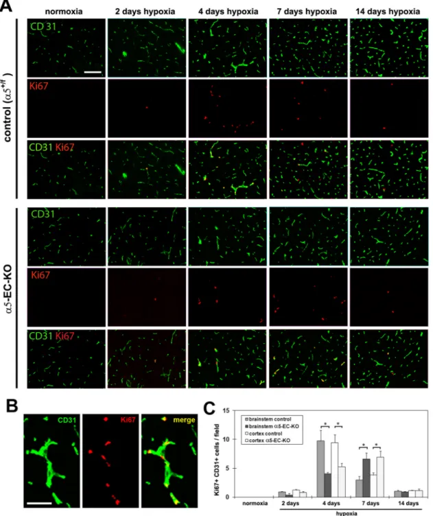
Fibronectin drives angiogenesis, in part by promoting endothelial cell proliferation (Astrof and Hynes 2009; Wang and Milner 2006). As α5-EC-KO mice displayed an attenuated angiogenic response to cerebral hypoxia, we next examined whether this might be a result of reduced endothelial proliferation in these mice, by performing dual-IF for the proliferation marker Ki67 and the endothelial marker CD31. As shown in Figure 3, after 4 days hypoxia, the number of dual-positive Ki67+/CD31+ cells per field of view in the brain of α5-EC-KO mice was significantly lower than in control mice, both in the brainstem (4.1 ± 0.3 compared with 9.8 ± 1.8, p < 0.05) and the cerebral cortex (5.3 ± 0.6 compared with 9.4 ± 1.4, p < 0.05). However, after 7 days hypoxia, this ranking had reversed, with the number of dual-positive Ki67+/CD31+ cells in the brain of α5-EC-KO mice now significantly higher than in controls, both in the brainstem (6.6 ± 1.1 compared with 3.0 ± 0.6, p < 0.05) and cerebral cortex (6.9 ± 1.0 compared with 3.8 ± 0.4, p < 0.05). After 14 days hypoxia, levels of Ki67+/CD31+ positive events in both strains of mice were reduced to much lower levels with no significant difference detected at this time-point. This demonstrates that in keeping with the attenuated angiogenic response, BEC in α5-EC-KO mice show a delayed and reduced mitogenic response to cerebral hypoxia. It also shows that the α5 integrin is not absolutely essential for BEC to mount a mitogenic response. Another possibility that might account for these findings is that lack of the α5 integrin could lead to increased BEC apoptosis during angiogenic remodeling, which would contribute to the attenuated angiogenic response we observed in α5-EC-KO mice. To investigate this, we performed TUNEL IF, which revealed a total absence of any TUNEL-positive cells during the hypoxic angiogenic response, in both control and α5-EC-KO mice (not shown), demonstrating that absence of the α5 integrin does not lead to BEC cell death during the angiogenic response.
Mice deficient of endothelial α5 integrin show no defect in vascular fibronectin deposition upon cerebral hypoxia, and no compensatory increase in the expression of αvβ3 integrin
Organization of a fibronectin matrix depends on cell surface fibronectin receptors (Wierzbicka-Patynowski and Schwartzbauer 2003). Cerebral hypoxia promotes parallel increases in vascular expression of fibronectin and BEC expression of the α5β1 integrin, which peak after 4 days hypoxia and then decline with time (Li et al. 2010a). As the α5β1 integrin is one of the main fibronectin receptors expressed by BEC (Wang and Milner 2006), and its demonstrated role in fibronectin assembly (Wierzbicka-Patynowski and
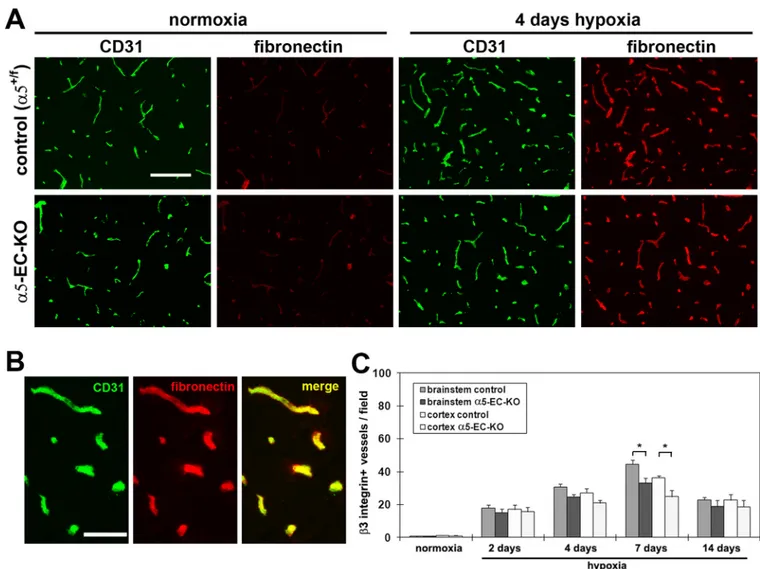
Schwartzbauer 2003), it is possible that absence of the α5 integrin might prevent fibronectin binding to BEC or result in diminished organization of a fibronectin matrix. To address this question, we used IF to examine the distribution of vascular fibronectin during the hypoxic response. This showed that in the brain of both control and α5-EC-KO mice, 4 days hypoxia induced a marked increase in the level of fibronectin expression in cerebral vessels (Figure 4A), clearly demonstrating that lack of endothelial α5 integrin expression does not affect fibronectin distribution within cerebral vessels in the hypoxic CNS.
NIH-PA Author Manuscript
NIH-PA Author Manuscript
BEC express two fibronectin integrin receptors, α5β1 and αvβ3 (Wang and Milner 2006), raising the possibility of redundancy in this system. In light of our recent finding that β3 integrin-null mice show no defect in hypoxic-induced angiogenesis, but display
compensatory upregulation of the α5β1 integrin (Li et al. 2010b), we next examined whether compensation worked both ways, by comparing the number of β3 integrin-positive vessels in α5-EC-KO and littermate control mice. As shown in Figure 4C, hypoxic brains of
α5-EC-KO mice contained marginally fewer β3 integrin-positive vessels compared to
littermate controls (which was significant after 7 days hypoxia, p < 0.05), consistent with the reduced vessel density seen in α5-EC-KO mice, clearly demonstrating the absence of any compensatory upregulation of αvβ3 integrin in α5-EC-KO mice.
α5 integrin-deficient BEC show delayed proliferation and growth in primary culture
Previous antibody-blocking studies suggest that both α5β1 and αvβ3 integrins play
functional roles in mediating BEC adhesion, survival and proliferation on fibronectin (Wang and Milner 2006). To directly examine the role of the α5 integrin in mediating interactions with fibronectin, BEC were isolated from α5-EC-KO mice, and their adhesion and growth on fibronectin compared with BEC from littermate controls. During the first few days of primary BEC culture, endothelial islands slowly disperse as BEC migrate out and proliferate. Compared with control cells, though similar numbers of α5 integrin-deficient BEC attached to fibronectin (Figure 5A), they were much slower to disperse from
endothelial islands and displayed slower growth kinetics on fibronectin (Figure 5B). BrdU incorporation studies showed that after 3 days primary culture, α5 integrin-deficient BEC displayed a significantly reduced proliferation rate (6.3 ± 3.5 cells/field compared with 22.2 ± 4.5 cells/field for control cells, p< 0.05; Figure 5C). This demonstrates that while α5 integrin-deficient BEC can adhere to fibronectin, absence of this integrin results in delayed proliferation and growth of BEC.
Re-distribution of αv integrins to focal contacts in α5 integrin-deficient BEC
In a recent study, we demonstrated that αv integrin fibronectin receptors can functionally compensate for lack of α5 integrin by re-distributing into focal contacts normally occupied by α5 integrins (van der Flier et al. 2010). To address whether this shift also occurs in BEC, we compared the cellular distribution of the αv and β3 integrin subunits on BEC derived from α5-EC-KO mice and control mice plated on fibronectin (Figure 6). In control BEC, the
α5 and αvβ3 integrins showed a clear difference in their sub-cellular localization. While the αvβ3 integrin (demonstrated by αv and β3 integrin IF) was predominantly located in focal
adhesions at the cell border, the α5 integrin was found in focal adhesions across the entire cell surface. Interestingly, in α5 integrin-deficient BEC, the αvβ3 integrin shifted its distribution to a pattern similar to the α5 integrin in control cells. Thus, consistent with our studies on embryonic endothelial cells (van der Flier et al. 2010), αv integrins on adult α5 integrin-deficient BEC show functional compensation, re-locating to the position normally occupied by α5 integrins. In light of the distinct compartmentalization of α5 and αvβ3 integrins demonstrated by our IF studies; α5 localized under the body of the cell, and αvβ3 localized specifically at the cell border, we wondered whether these two integrins might play distinct roles in promoting BEC migration. To test this idea, and specifically examine the role of α5 integrin in driving BEC migration, we compared the migration rates of α5 integrin-deficient and control BEC. This revealed no obvious deficiency in the ability of α5 integrin KO cells to migrate on fibronectin (Figure 5D). Next, we evaluated whether α5 integrin-deficient BEC could assemble a fibronectin fibrillar network. BEC were plated onto a laminin substrate, and fibronectin IF performed 24 hours later. As shown in Figure 6, BEC lacking the α5 integrin were still capable of producing a fibronectin fibrillar network.
NIH-PA Author Manuscript
NIH-PA Author Manuscript
DISCUSSION
Cerebral hypoxia stimulates a robust angiogenic response (LaManna et al. 1992), which is associated with strong upregulation of fibronectin and the fibronectin receptors α5β1 and
αvβ3 integrins on vascular endothelium (Li et al. 2010b; Milner et al. 2008a). A recent
study from our laboratory using β3 integrin-null mice, showed that the αvβ3 integrin is dispensable for the angiogenic response, but suggested a role for the α5β1 integrin (Li et al. 2010b). To address directly whether the α5β1 integrin is required for the vascular
remodeling response to cerebral hypoxia, we examined this process in mice lacking α5 integrin expression in endothelial cells. This revealed that the angiogenic response to cerebral hypoxia was markedly attenuated in α5-EC-KO mice, as these mice showed reduced hypoxia-induced increases in vessel density relative to control littermates. In keeping with the decreased angiogenic response, BEC in α5-EC-KO mice showed a delayed mitogenic response to hypoxia, which was mirrored by delayed proliferation and growth of
α5-KO BEC in primary culture. Taken together, these studies demonstrate an important
angiogenic role for the α5β1 integrin in promoting BEC proliferation in response to cerebral hypoxia.
Chronic hypoxia drives an angiogenic response in the CNS
Relative to other tissues, the brain has extremely high demands for oxygen. This fact is well illustrated by the fact that the CNS constitutes less than 2% of the body weight, but uses up to 20% of the total oxygen consumption in the body (Attwell et al. 2010). Interestingly, the oxygen levels in intact brain tissue of animals breathing 21% oxygen air at sea level are much lower than 21%, actually more in the range of 2–4% (15–30 torr) (Ndubuizu and LaManna 2007). Thus, when animals breathe 8% oxygen, the oxygen concentration in the brain falls proportionally. This decline is detected by oxygen chemoreceptors, which trigger a number of physiological adaptive responses, including increased ventilation rate,
tachycardia, cerebral vasodilatation, cerebral angiogenesis, and raised hematocrit (LaManna et al. 2004). All these mechanisms have a common aim: to raise the oxygen delivery to the brain, so as to compensate for the reduced oxygen availability in the atmospheric air.
The role of α5β1 integrin in angiogenesis
Current data suggest that the α5β1 integrin plays an important role in angiogenesis. Global deletion of the α5 integrin gene results in an embryonic lethal phenotype, with aberrant blood vessel formation in the embryo (Yang et al. 1993). Similar vascular defects are also apparent in α5 integrin-null embryoid bodies and teratoma cells (Taverna and Hynes 2001). In addition, while quiescent endothelial cells express only low levels of α5β1 integrin, angiogenic endothelial cells strongly upregulate this integrin, and functional blockade of the
α5β1 integrin in tumor-induced neovasculature inhibits angiogenesis and tumor growth in
vivo (Kim et al. 2000). In the current study, mice lacking endothelial expression of the α5 integrin displayed a blunted angiogenic response to cerebral hypoxia. α5-EC-KO mice showed reduced BEC proliferation at 4 days hypoxia, though interestingly, by 7 days hypoxia, BEC proliferation in these mice had caught up and was even slightly higher than littermate control mice. This demonstrates that in the adult CNS, α5β1 integrin plays an important role in stimulating endothelial cell proliferation at an early step in the angiogenic process. Surprisingly, developmental angiogenesis is relatively unperturbed in α5-EC-KO mice (van der Flier et al. 2010), implying that the mechanisms promoting angiogenesis in developmental and pathological situations might be different. Interestingly, the same situation has also been described for α5β1 integrin function in lymphangiogenesis, in that developmental lymphangiogenesis appears unaffected by absence of the α5 integrin (van der Flier et al. 2010), but blockade of the α5β1 integrin in the adult inhibits lymphangiogenesis (Okazaki et al. 2009). Why α5β1 should play an important role in adult remodelling
NIH-PA Author Manuscript
NIH-PA Author Manuscript
situations but not be essential during development, remains to be determined. One possibility is that a higher level of plasticity is present during development and that with maturation, a more limited number of molecular mechanisms are available. This suggests that developmental angiogenesis has considerably more redundancy and/or compensatory mechanisms compared to the adult situation.
The relative importance of α5β1 and αvβ3 integrins in cerebral angiogenesis
BEC express two fibronectin receptors, the α5β1 and αvβ3 integrins (Wang and Milner 2006). As both these integrins show strong upregulation during hypoxia-induced cerebral angiogenesis, it is important to determine the relative importance of these two integrins. In a recent study with β3 integrin-null mice, we showed that while β3 was not essential for cerebral angiogenesis, lack of β3 led to compensatory increases in BEC α5 integrin expression, and increased rate of BEC proliferation, suggesting a role for the α5β1 integrin in driving BEC mitogenic responses during angiogenesis. Our current data support this idea, by showing that in response to hypoxia, α5-EC-KO mice show attenuated angiogenesis and delayed BEC proliferation. This confirms that the α5β1 integrin is important in driving BEC proliferation during the cerebral angiogenic response to hypoxia, and lends extra weight to the idea that of the two BEC fibronectin receptors, α5β1 integrin is more important than
αvβ3. However, while the absence of BEC α5β1 led to a delayed and weaker angiogenic
response, it did not totally abrogate this response. Several explanations might account for this. First, in the absence of α5β1 integrin, αvβ3 may mediate the effect of fibronectin on BEC proliferation. In the current study, we did not observe increased vascular expression of the αvβ3 integrin in α5-EC-KO mice, though consistent with previous findings in
embryonic endothelial cells (van der Flier et al. 2010), in α5-KO BEC, αvβ3 re-localized to cell surface focal adhesions where normally only α5β1 integrin resides, suggesting a functional compensatory mechanism. It is important to note however, that while αvβ3 integrin may compensate to some degree, our current findings clearly demonstrate that the
αvβ3 integrin is not as effective in promoting the angiogenic response as α5β1 integrin.
Second, other fibronectin receptors (integrins or non-integrins) may be upregulated in a compensatory manner. Third, other cell adhesion molecules may also promote BEC proliferation and angiogenesis, thus providing other redundant or compensatory
mechanisms. For example, collagen I and its integrin receptors α1β1 and α2β1 have well described pro-angiogenic roles (Senger 1996; Senger et al. 1997), making it feasible that in the absence of fibronectin or fibronectin receptors, collagen I can still promote an
angiogenic response. In the next phase of experiments, we plan to address the first two possibilities, by examining the hypoxic response in double KO mice lacking both αvβ3 and
α5β1 in endothelial cells.
In summary, we have addressed the requirement of the α5β1 integrin in mediating cerebral angiogenesis in response to cerebral hypoxia, by using mice lacking α5 integrin expression in endothelial cells (α5-EC-KO). These mice showed a significantly attenuated angiogenic response, which was paralleled by a delayed endothelial mitogenic response. In vitro experiments confirmed the proliferation defect in α5-KO BEC. Taken together, these studies demonstrate an important angiogenic role for the α5β1 integrin in promoting BEC
proliferation in response to cerebral hypoxia.
Acknowledgments
RM was supported by a Harry Weaver Neuroscience Scholar Award (JF 2125A1/1) from the National Multiple Sclerosis Society (RM), and by the NIH RO1 grant NS060770. Additional support came from the NIH (PO1-HL66105 and the NIGMS Cell Migration Consortium, GC11451.126452, PI, A.F. Horwitz), and the Howard Hughes Medical Institute, of which ROH is an Investigator. This is manuscript number 20792 from The Scripps Research Institute.
NIH-PA Author Manuscript
NIH-PA Author Manuscript
REFERENCES
Astrof S, Hynes RO. Fibronectins in vascular development. Angiogenesis. 2009; 12:165–175. [PubMed: 19219555]
Attwell D, Buchan AM, Charpak S, Lauritzen M, MacVicar BA, Newman EA. Glial and neuronal control of brain blood flow. Nature. 2010; 468:232–243. [PubMed: 21068832]
Chavez JC, Agani F, Pichiule P, LaManna JC. Expression of hypoxic inducible factor 1α in the brain of rats during chronic hypoxia. J Appl Physiol. 2000; 89:1937–1942. [PubMed: 11053346] Chen HH, Chien CH, Liu HM. Correlation between angiogenesis and basic fibroblast growth factor
expression in experimental brain infarct. Stroke. 1994; 25:1651–1657. [PubMed: 7518971] del Zoppo GJ, Milner R. Integrin-matrix interactions in the cerebral microvasculature. Arterioscler
Thromb Vasc Biol. 2006; 26:1966–1975. [PubMed: 16778120]
Dowden J, Corbett D. Ischemic preconditioning in 18- to 20-month-old gerbils: long-term survival with functional outcome measures. Stroke. 1999; 30:1240–1246. [PubMed: 10356107]
Folkman J. Angiogenesis in cancer, vascular, rheumatoid and other disease. Nat Med. 1995; 1:27–31. [PubMed: 7584949]
George EL, Georges-Labouesse EN, Patel-King RS, Rayburn H, Hynes RO. Defects in mesoderm, neural tube and vascular development in mouse embryos lacking fibronectin. Development. 1993; 119:1079–1091. [PubMed: 8306876]
Hynes RO, Lively JC, McCarty JH, Taverna D, Francis SE, Hodivala-Dilke K, Xiao Q. The diverse roles of integrins and their ligands in angiogenesis. Cold Spring Harb Symp Quant Biol. 2002; 67:143–153. [PubMed: 12858535]
Kanaan A, Farahani R, Douglas RM, LaManna JC, Haddad GC. Effect of chronic continuous or intermittent hypoxia and reoxygenation on cerebral capillary density and myelination. Am J Physiol Regul Integr Comp Physiol. 2006; 290:R1105–R1114. [PubMed: 16322350]
Kim S, Bell K, Mousa SA, Varner JA. Regulation of angiogenesis in vivo by ligation of integrin a5b1 with the central cell-binding domain of fibronectin. Am J Pathol. 2000; 156:1345–1362. [PubMed: 10751360]
Kisanuki YY, Hammer RE, Miyazaki J, Williams SC, Richardson JA, Yanagisawa M. Tie2-Cre transgenic mice: a new model for endothelial cell-lineage analysis in vivo. Dev Biol. 2001; 230:230–242. [PubMed: 11161575]
Krupinski J, Kaluza J, Kumar P, Kumar S, Wang JM. Role of angiogenesis in patients with cerebral ischemic stroke. Stroke. 1994; 25:1794–1798. [PubMed: 7521076]
Kuo N-T, Benhayon D, Przybylski RJ, Martin RJ, LaManna JC. Prolonged hypoxia increases vascular endothelial growth factor mRNA and protein in adult mouse brain. J Appl Physiol. 1999; 86:260– 264. [PubMed: 9887138]
LaManna JC, Chavez JC, Pichiule P. Structural and functional adaptation to hypoxia in the rat brain. J Exp Biol. 2004; 207:3163–3169. [PubMed: 15299038]
LaManna JC, Vendel LM, Farrell RM. Brain adaptation to chronic hypobaric hypoxia in rats. J Appl Physiol. 1992; 72:2238–2243. [PubMed: 1629078]
Li L, Welser JV, Dore-Duffy P, Del Zoppo GJ, LaManna JC, Milner R. In the hypoxic central nervous system, endothelial cell proliferation is followed by astrocyte activation, proliferation, and increased expression of the α6β4 integrin and dystroglycan. Glia. 2010a; 58:1157–1167. [PubMed: 20544851]
Li L, Welser JV, Milner R. Absence of the αvβ3 integrin dictates the time-course of angiogenesis in the hypoxic central nervous system: accelerated endothelial proliferation correlates with
compensatory increases in α5β1 integrin expression. J Cereb Blood Flow Metab. 2010b; 30:1031– 1043. [PubMed: 20087368]
Miller BA, Perez RS, Shah AR, Gonzales ER, Park TS, Gidday JM. Cerebral protection by hypoxic preconditioning in a murine model of focal ischemia-reperfusion. Neuroreport. 2001; 12:1663– 1669. [PubMed: 11409736]
Milner R, Campbell IL. Cytokines regulate microglial adhesion to laminin and astrocyte extracellular matrix via protein kinase C-dependent activation of the α6β1 integrin. J Neurosci. 2002a; 22:1562–1572. [PubMed: 11880486]
NIH-PA Author Manuscript
NIH-PA Author Manuscript
Milner R, Campbell IL. Developmental regulation of β1 integrins during angiogenesis in the central nervous system. Mol Cell Neurosci. 2002b; 20:616–626. [PubMed: 12213443]
Milner R, Crocker SJ, Hung S, Wang X, Frausto RF, Del Zoppo GJ. Fibronectin- and Vitronectin-Induced Microglial Activation and Matrix Metalloproteinase-9 Expression Is Mediated by Integrins α5β1 and αvβ5. J Immunol. 2007; 178:8158–8167. [PubMed: 17548654]
Milner R, Hung S, Erokwu B, Dore-Duffy P, LaManna JC, del Zoppo GJ. Increased expression of fibronectin and the α5β1 integrin in angiogenic cerebral blood vessels of mice subject to hypobaric hypoxia. Mol Cell Neurosci. 2008a; 38:43–52. [PubMed: 18343155]
Milner R, Hung S, Wang X, Berg G, Spatz M, del Zoppo G. Responses of endothelial cell and astrocyte matrix-integrin receptors to ischemia mimic those observed in the neurovascular unit. Stroke. 2008b; 39:191–197. [PubMed: 18032737]
Ndubuizu O, LaManna JC. Brain tissue oxygen concentration measurements. Antioxid Redox Signal. 2007; 9:1207–1219. [PubMed: 17536959]
Okazaki T, Ni A, Aveni OA, Baluk P, Yao LC, Vossmeyer D, Zischinsky G, Zahn G, Knolle J, Christner C, et al. alpha5 beta1 integrin blockade inhibits lymphangiogenesis in airway inflammation. Am J Pathol. 2009; 174:2378–2387. [PubMed: 19443705]
Risau W, Lemmon V. Changes in the Vascular Extracellular Matrix during Embryonic Vasculogenesis and Angiogenesis. Dev Biol. 1988; 125:441–450. [PubMed: 3338622]
Senger DR. Molecular framework for angiogenesis: a complex web of interactions between
extravasated plasma proteins and endothelial cell proteins induced by angiogenic cytokines. Am J Pathol. 1996; 149:1–7. [PubMed: 8686733]
Senger DR, Claffey KP, Benes JE, Perruzzi CA, Sergiou AP, Detmar M. Angiogenesis promoted by vascular endothelial growth factor: regulation through alpha1beta1 and alpha2beta1 integrins. Proc Natl Acad Sci. 1997; 94:13612–13617. [PubMed: 9391074]
Silva R, D'Amico G, Hodivala-Dilke KM, Reynolds LE. Integrins: the keys to unlocking angiogenesis. Arterioscler Thromb Vasc Biol. 2008; 28:1703–1713. [PubMed: 18658045]
Taverna D, Hynes RO. Reduced blood vessel formation and tumor growth in alpha5-integrin-negative teratocarcinomas and embryoid bodies. Cancer Res. 2001; 61:5255–5261. [PubMed: 11431367] van der Flier A, Badu-Nkansah K, Whittaker CA, Crowley D, Roderick T, Bronson DT, Lacy-Hulbert
A, Hynes RO. Endothelial α5 and αv Integrins Cooperate in Remodeling of the Vasculature During Development. Development. 2010; 137:2439–2449. [PubMed: 20570943]
Wang J, Milner R. Fibronectin promotes brain capillary endothelial cell survival and proliferation through α5β1 and αvβ3 integrins via MAP kinase signaling. J Neurochem. 2006; 96:148–159. [PubMed: 16269008]
Wei L, Erinjeri JP, Rovainen CM, Woolsey TA. Collateral growth and angiogenesis around cortical stroke. Stroke. 2001; 32:2179–2184. [PubMed: 11546914]
Wierzbicka-Patynowski I, Schwartzbauer JE. The ins and outs of fibronectin assembly. J Cell Sci. 2003; 116:3269–3276. [PubMed: 12857786]
Yang JT, Rayburn H, Hynes RO. Embryonic mesodermal defects in α5 integrin-deficient mice. Development. 1993; 119:1093–1105. [PubMed: 7508365]
NIH-PA Author Manuscript
NIH-PA Author Manuscript
Highlights
• Angiogenic vessels upregulate fibronectin and integrin receptors, α5β1 and
αvβ3.
• Examined role of α5 in mice lacking α5 integrin in endothelial cells (α5-EC-KO)
• These mice showed less cerebral angiogenesis and reduced endothelial proliferation
• Suggests that α5β1 integrin drives endothelial proliferation and angiogenesis
NIH-PA Author Manuscript
NIH-PA Author Manuscript
Figure 1. Confirmation of the absence of α5 integrin expression in BEC of α5-EC-KO mice
A. Frozen sections of brainstem taken from mice exposed to normoxia (~21% O2) or 4 days
hypoxia were processed for dual-IF for CD31 and the α5 integrin. Scale bar = 100µm. Note that in contrast to the control CNS, where strong hypoxic-induction of endothelial α5 integrin was observed, vessels in α5-EC-KO mice showed a total lack of the α5 integrin. B. High power image of CD31/α5 integrin dual-IF of 4 day hypoxia exposed brainstem. Scale bar = 50µm. C. BEC from control or α5-EC-KO mice were cultured on fibronectin and expression of the α5 integrin subunit analyzed by flow cytometry. Note that in contrast to control BEC, α5 integrin was totally absent on BEC obtained from α5-EC-KO mice.
NIH-PA Author Manuscript
NIH-PA Author Manuscript
Figure 2. Reduced hypoxia-induced cerebral angiogenic response in α5-EC-KO mice
A. α5-EC-KO mice and control littermates (α5+/f) were maintained at normoxia (~21% O 2)
or exposed to 8% hypoxia for 2, 4, 7 or 14 days before frozen brain sections were
immunostained to assess blood vessel density (CD31) in the brainstem. Scale bar = 100µm. B. High power images of CD31 IF performed on brainstem of α5-EC-KO and littermate control mice exposed to 14 day hypoxia. Scale bar = 50µm. C. Quantification of analyses. Brain sections were examined for the number of CD31+ vessels. Experiments were performed with three different animals per condition, and the results expressed as the mean ± SEM of the number of CD31+ vessels per field of view. Note that after 14 days hypoxia, blood vessel density in α5-EC-KO mice was significantly less than in control mice, both in the brainstem and cerebral cortex, * p < 0.01. D. Western analysis of brain expression of claudin-5 and Glut-1 during normoxia (~21% O2) and after 14 days hypoxia. E.
Quantification of western analysis. Note that after 14 days hypoxia, levels of the BEC proteins, claudin-5 and Glut-1 were significantly lower in the α5-EC-KO mice relative to controls,* p < 0.05.
NIH-PA Author Manuscript
NIH-PA Author Manuscript
Figure 3. Delayed and reduced BEC proliferation during the angiogenic response to cerebral hypoxia in α5-EC-KO mice
A. Frozen sections of brainstem taken from control or α5-EC-KO mice exposed to normoxia (~21% O2) or 2, 4, 7 or 14 days hypoxia were examined for evidence of BEC proliferation,
as assessed by CD31/Ki67 dual-IF. Scale bar = 100µm. B. High power image of CD31/Ki67 dual-IF of 4 day hypoxia exposed brainstem of control mouse. Scale bar = 50µm. C. Quantification of CD31+/Ki67+ proliferating BEC, in which each experiment was
performed with three different animals per condition, and the results expressed as the mean ± SEM of the number of CD31+/Ki67+ cells per field of view. Note that hypoxia promoted
NIH-PA Author Manuscript
NIH-PA Author Manuscript
an increase in the number of proliferating BEC, though this response was delayed in α5-EC-KO mice. * p < 0.05.
NIH-PA Author Manuscript
NIH-PA Author Manuscript
Figure 4. Analysis of hypoxic-induced alterations in cerebrovascular expression of fibronectin and β3 integrin
A. Frozen sections of cerebral cortex taken from control or α5-EC-KO mice exposed to normoxia (~21% O2), or 4 days hypoxia, underwent dual-IF for CD31 and fibronectin. Scale
bar = 100µm. Note that cerebral hypoxia promoted a strong increase in fibronectin
expression in cerebral vessels of both control and α5-EC-KO mice. B. High power image of CD31/fibronectin dual-IF of 4 day hypoxia exposed brainstem. Scale bar = 50µm. C. Quantification of β3 integrin-positive vessels in control or α5-EC-KO mice exposed to hypoxia. Experiments were performed with three different animals per condition, and the results expressed as the mean ± SEM of the number of β3 integrin-positive vessels per field of view. Note that cerebral hypoxia increased the number of β3 integrin-positive vessels (that peaked after 7 days hypoxia), and that this increase was significantly lower in α5-EC-KO mice relative to controls,* p < 0.05.
NIH-PA Author Manuscript
NIH-PA Author Manuscript
Figure 5. Analysis of growth defects in primary α5-EC-KO cultures
BEC from α5-EC-KO or littermate control mice were isolated and cultured on fibronectin. A. Adhesion assays. Note that α5 integrin-deficient BEC showed no defect in adhesion to fibronectin or collagen I. B. Growth in primary culture. Note that after 2 days culture, while control BEC had dispersed from the endothelial islands, α5 integrin-deficient BEC were slow to disperse. After 5 days culture, BEC from both strains had dispersed and formed expanding colonies, though α5 integrin-deficient BEC were slower to grow, relative to control cells. Scale bar = 25µm. C. BEC proliferation in 3-day old primary cultures cultured on fibronectin was examined over 16 hours, and expressed as the % of BEC that
incorporated BrdU. All points represent the mean ± SEM of four experiments. Note that the proliferation rate of α5 integrin-deficient BEC was significantly lower than control BEC (p < 0.05). D. Migration rates compared. All points represent the mean ± SEM of four
experiments. Note that α5 integrin-deficient BEC migrated on fibronectin at equivalent rates to control cells.
NIH-PA Author Manuscript
NIH-PA Author Manuscript
Figure 6. Immunolocalization of α5 and αvβ3 integrins on BEC
BEC from α5-EC-KO and littermate control mice were cultured on fibronectin and integrin localization determined by IF. Scale bars = 25µm. Note that in control BEC, the α5 integrin was found in focal contacts throughout the body of the cell, while in contrast, the αv and β3 subunits were specifically localized at the cell border. Interestingly, in α5 integrin-deficient BEC, the αvβ3 integrin showed a re-distribution to focal contacts within the body of the cell. Fibronectin IF of cells plated onto laminin demonstrated that α5 integrin-deficient BEC were still capable of producing a fibronectin fibrillar network.