Publisher’s version / Version de l'éditeur:
Vous avez des questions? Nous pouvons vous aider. Pour communiquer directement avec un auteur, consultez la première page de la revue dans laquelle son article a été publié afin de trouver ses coordonnées. Si vous n’arrivez pas à les repérer, communiquez avec nous à PublicationsArchive-ArchivesPublications@nrc-cnrc.gc.ca.
Questions? Contact the NRC Publications Archive team at
PublicationsArchive-ArchivesPublications@nrc-cnrc.gc.ca. If you wish to email the authors directly, please see the first page of the publication for their contact information.
https://publications-cnrc.canada.ca/fra/droits
L’accès à ce site Web et l’utilisation de son contenu sont assujettis aux conditions présentées dans le site LISEZ CES CONDITIONS ATTENTIVEMENT AVANT D’UTILISER CE SITE WEB.
Journal of the American Chemical Society, 140, 48, pp. 16783-16791, 2018-11-05
READ THESE TERMS AND CONDITIONS CAREFULLY BEFORE USING THIS WEBSITE. https://nrc-publications.canada.ca/eng/copyright
NRC Publications Archive Record / Notice des Archives des publications du CNRC :
https://nrc-publications.canada.ca/eng/view/object/?id=57f37fd0-1d6f-4706-b221-59ea90dc8ea0 https://publications-cnrc.canada.ca/fra/voir/objet/?id=57f37fd0-1d6f-4706-b221-59ea90dc8ea0
NRC Publications Archive
Archives des publications du CNRC
This publication could be one of several versions: author’s original, accepted manuscript or the publisher’s version. / La version de cette publication peut être l’une des suivantes : la version prépublication de l’auteur, la version acceptée du manuscrit ou la version de l’éditeur.
For the publisher’s version, please access the DOI link below./ Pour consulter la version de l’éditeur, utilisez le lien DOI ci-dessous.
https://doi.org/10.1021/jacs.8b10017
Access and use of this website and the material on it are subject to the Terms and Conditions set forth at
Gramillin A and B: cyclic lipopeptides identified as the nonribosomal
biosynthetic products of Fusarium graminearum
Bahadoor, Adilah; Brauer, Elizabeth K.; Bosnich, Whynn; Schneiderman,
Danielle; Johnston, Anne; Aubin, Yves; Blackwell, Barbara; Melanson,
Jeremy E.; Harris, Linda J.
Gramillin A and B: Cyclic Lipopeptides Identified as the
Nonribosomal Biosynthetic Products of Fusarium graminearum
Adilah Bahadoor,
*
,†Elizabeth K. Brauer,
‡Whynn Bosnich,
‡Danielle Schneiderman,
‡Anne Johnston,
‡Yves Aubin,
§Barbara Blackwell,
‡Jeremy E. Melanson,
†and Linda J. Harris
*
,‡†
Metrology, National Research Council Canada, Ottawa, Ontario K1A 0R6, Canada ‡
Ottawa Research and Development Centre, Agriculture and Agri-Food Canada, Ottawa, Ontario K1A 0C6, Canada §
Centre for Biologics Evaluation, Biologics, and Genetic Therapies Directorate, Health Canada, Ottawa, Ontario K1A 0K9, Canada
*
S Supporting InformationABSTRACT: The virulence and broad host range of Fusarium
graminearum is associated with its ability to secrete an arsenal of
phytotoxic secondary metabolites, including the regulated mycotoxins belonging to the deoxynivalenol family. The TRI genes responsible for the biosynthesis of deoxynivalenol and related compounds are usually expressed during fungal infection. However, the F. graminearum genome harbors an array of unexplored biosynthetic gene clusters that are also co-induced with the TRI genes, including the nonribosomal peptide synthetase 8 (NRPS8) gene cluster. Here, we identify two bicyclic lipopeptides, gramillin A (1) and B (2), as the biosynthetic end products of NRPS8. Structural elucidation by high-resolution LC-MS and NMR, including 1H-15N-13C HNCO and HNCA on
isotopically enriched compounds, revealed that the gramillins
possess a fused bicyclic structure with ring closure of the main peptide macrocycle occurring via an anhydride bond. Through targeted gene disruption, we characterized the GRA1 biosynthetic gene and its transcription factor GRA2 in the NRPS8 gene cluster. Further, we show that the gramillins are produced in planta on maize silks, promoting fungal virulence on maize but have no discernible effect on wheat head infection. Leaf infiltration of the gramillins induces cell death in maize, but not in wheat. Our results show that F. graminearum deploys the gramillins as a virulence agent in maize, but not in wheat, thus displaying host-specific adaptation.
■
INTRODUCTIONMillions of years of co-evolution have shaped the diverse strategies that fungi use to overcome plant host defenses and establish infection. Perturbation of host defense is often associated with interference with pathogen recognition, suppression of the immune response, disruption of the plant cell structure or metabolism through deployment of proteins, secondary metabolites, or other small molecules.1 Fungal secondary metabolites are often biosynthesized by enzymes encoded in gene clusters transcribed under specific environ-mental or developenviron-mental conditions,2 and the hemibiotrophic or necrotrophic fungi are particularly noted for producing toxins that promote plant cell death, disabling immunity and providing the fungus with nutrients.3
Fusarium graminearum is a hemibiotrophic pathogen that
infects a wide range of plant species including cereal crops such as wheat, maize, and barley.4 The fungus is a rich source of secondary metabolites, including sesquiterpenoids, polyketides, and nonribosomal peptides. Comparative genomic studies have identified an array of biosynthetic genes encoding terpenoid synthases (TPS), polyketide synthases (PKS), and
non-ribosomal peptide synthases (NRPS) which are often within co-regulated clusters of genes in the F. graminearum genome.5−7
Delving into the interactions of F. graminearum with maize, barley, and wheat, we revealed that several genes from the
NRPS8 biosynthetic cluster are expressed during infection of
maize kernels but exhibited transient expression during barley and wheat spike infection.8 Furthermore, NRPS8 was co-expressed alongside the TRI biosynthetic genes.8−10The TRI genes participate in the biosynthesis of the regulated mycotoxin 4-deoxynivalenol, and their involvement in virulence within plant reproductive tissue is well docu-mented.11−13
To understand the role of the NRPS8 gene cluster in infection, we characterized the GRA1 biosynthetic gene and its transcription factor GRA2. We identified the biosynthetic products of GRA1 as bicyclic lipopeptides, which we named gramillin A and B. Full structure elucidation revealed
Received: September 14, 2018
Published: November 5, 2018
pubs.acs.org/JACS
Cite This:J. Am. Chem. Soc. 2018, 140, 16783−16791
nonproteinogenic amino acid building blocks, amino acid modifications, and two macrocyclizations that could not be anticipated through sequence analysis.14,15 Further, GRA1 is required for full virulence on maize silks, and gramillin A and B induce cell death in maize leaves. During wheat spike infection, the absence of GRA1 has no effect on virulence, and gramillin A and B do not induce cell death in wheat leaves. Together, this suggests that the gramillins are nonribosomal peptide (NRP) phytotoxins that promote virulence of the pathogen in specific host microenvironments.
■
RESULTSGramillin A and B Identified as the Biosynthetic Products of NRPS8. The NRPS8 gene cluster (FGSG_00036−FGSG_00049) containing GRA1 includes the transcription factor GRA2 and multiple putative peptide tailoring genes (Figure 1A).5,9 GRA1 encodes the predicted
multimodular 865 kDa NRPS8 containing seven sets of adenylation (A) and condensation (C) domains and peptidyl carrier proteins, indicating the potential incorporation of seven amino acids in the gramillins.16 GRA1 was disrupted in the Fg180378 and Fg233423 wild-type backgrounds via Agro-bacterium-mediated transformation (Figure S1).17 A similar strategy was used to disrupt GRA2 (data not shown). A number of true disruptants were produced as referred to in Figure 1B (see also Table S1). The gene expression of GRA1 was confirmed to be undetectable or severely reduced in Δgra1 and Δgra2 mutants by quantitative droplet digital PCR (ddPCR) (Table S2). The secondary metabolite profile of wild-type and Δgra1 mutants were compared (Figure 1B and Figures S2−S5). The LC-MS screen revealed a major peak at 6.6 min belonging to a co-eluting mixture of gramillin A (m/
zobs= 847.4, [M + H]+) and gramillin B (m/zobs= 861.4, [M +
H]+) (Figure 1B,C). Gramillin A, with a molecular formula of
C35H58O12S2N8, was detected as the [M + H]+ion at an m/z of
847.3695 (mcalc847.3688, Δm = −1.7 ppm). Similarly, the [M
+ H]+ion of gramillin B was detected at an m/z of 861.3853 (mcalc 847.3845, Δm = −0.9 ppm), corresponding to a
molecular formula of C36H60O12S2N8. The mass difference of
14 amu between gramillin A and B already suggested that gramillin B possessed an extra CH2 group (Figure 1C). A
5-fold reduction in the production of the gramillins was observed from the transformation control strain (TC), and disrupting the transcription factor GRA2 greatly minimized (>40 fold) the production of the gramillins. Mycelial growth and spore production of the Δgra1 (GRA1 disruption) mutants was similar to wild-type in both genetic backgrounds, while Δgra2_9 displayed impaired mycelial growth and conidiation (Supporting Information, Tables S1 and S3). All together, these results point to GRA1 as a biosynthetic gene leading to the gramillins and GRA2 as a transcription factor regulating gramillin biosynthesis.
Isolation and Structural Elucidation of Gramillin A and B. To isolate gramillin A and B for structural characterization, a Δtri1 mutant of Fg180378 was grown in liquid media.18Deletion of the Tri1 gene blocks the final steps in 15-acetyl-4-deoxynivalenol biosynthesis without affecting the production of the gramillins, thereby simplifying their purification from liquid culture (Supporting Information Figure S6). Two major challenges, namely solubility and apparent facile decomposition, stood in the way of isolating and purifying the gramillins from liquid culture. Gramillin A and B are hydrophilic peptides, so biphasic extraction of the aqueous medium with organic solvents was futile. Rather, a solid-phase extraction (SPE) of the aqueous medium provided a crude mixture of gramillin A and B, which after preparative HPLC purification provided a cleaner, but inseparable mixture of the gramillins.
In hindsight, the decomposition of the gramillins was due to the presence of the labile anhydride bond, which we were not aware of at the onset of this study. To circumvent the issue, we speculated that the gramillins must prefer an acidic medium. The pH of the production medium for the gramillins hovers around pH 2−2.5, and no apparent decomposition has been observed under such conditions. Upon switching to acidic phases for isolation and extraction procedures, no decom-position was observed, and enough gramillin material was obtained for NMR. Apart from the complexity of character-izing gramillin A and B as an inseparable mixture, these compounds were also poorly soluble or degraded in a range of solvents. In acidified D2O, a concentration of up 1 mg/mL of a
gramillin A and B mixture was achievable. Alternate NMR
Figure 1.Characterization of GRA1 and GRA2 in the NRPS8 gene cluster. (A) The predicted genes in the NRPS8 gene cluster include GRA1 and its transcription factor GRA2. GRA1 encodes the seven-modular NRPS8. (B) Gramillin A and gramillin B co-elute and their production followed by LC-MS from wildtype Fg180378 and Fg233423 and their respective GRA1, GRA2, and TC disruptants. (C) The mass spectrum of gramillin A and B.
Journal of the American Chemical Society Article
DOI:10.1021/jacs.8b10017 J. Am. Chem. Soc. 2018, 140, 16783−16791 16784
solvents, CD3OD and DMSO-d6, were unsuitable due to
cross-reactivity with the anhydride bond. Taken together, the pH sensitivity and the cross-reactions with mild nucleophilic solvents, such as methanol and DMSO, pointed toward the presence of a base-sensitive bond in the gramillins.
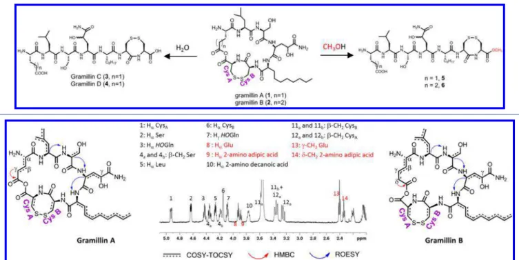
This inherent sensitivity to mild nucleophiles provided an opportunity to gather crucial structural information about the gramillins. Once enough purified gramillins were obtained, they were exposed to water and methanol, respectively, and their reactions tracked by LC-MS. Upon exposure to water, two new products appeared instantly (Scheme 1). Each contained an extra 18 amu suggesting the incorporation of water. Subsequent MS2 fragmentations suggested the new
products gramillin C (3) and gramillin D (4) were linear (Figures S7−S8). On the other hand, gramillin A (1) and B (2) exhibited significantly different MS2fragmentation patterns
than 3 and 4 (Figures S9−S10). When dissolved in methanol, gramillin A and B hydrolyzed to the new products 5 and 6 with an extra 32 amu, pointing to the incorporation of methanol. MS2 fragmentation determined that nucleophilic attack by
methanol occurred exclusively at the CysA residue with the formation of a methyl ester at the C-terminal Cys A (Figures S11−S12). Both hydrolysis reactions irrevocably rule out a typical macrocylization via an amide bond. Amide bonds are hydrolyzed under harsh acidic or basic conditions. Cyclization via an ester was also ruled out. Methanolysis of ester bonds typically requires an acid catalyst, but the gramillins reacted instantly in water or methanol without any catalyst. Moreover, the molecular formula of the gramillins precluded the presence of an ester bond. On the other hand, an anhydride bond matches the observed molecular formula and is a base-labile bond. Taken together, these observations strongly suggested the existence of an anhydride bond formed from the
condensation of the carboxylic acids moieties on the Glu or 2-amino adipic acid side chain to CysA.
Although the presence of serine as one of components of the gramillins was clear by MS2fragmentation, the other residues
were either unnatural (2-amino decanoic acid and 2-amino adipic acid), modified (HOGln), or isomeric (Leu vs Ile) and thus required NMR for structural confirmation. Furthermore, 1,4-dithiothreitol (DTT) treatment19 of the gramillins confirmed the presence of a disulfide bond (Figure S13); however, the adjacent position of the two cysteines in the structure was eventually settled by NMR.
Cognizant that the NMR characterization was performed on a gramillin A and B mixture, rather than the individual compounds, it was imperative to obtain NMR spectra with unambiguous signals that would provide clear distinction between gramillin A and B. This was achieved by performing the NMR experiments in different NMR solvent systems, with each providing optimal conditions for observing key correlations to distinguish between gramillin A and B (Figures S14−S17). The1H-1H COSY and TOCSY coupled to a
1D-proton worked best in D2O:CD3CN (4:1) + 0.1% formic acid
(Figure 2 and Figures S18−19). From these series of NMR data, the individual amino acid spin systems of two cysteine residues CysA and CysB, Leu, Ser, HOGln and 2-amino decanoic acid were readily established. Furthermore, in the 3− 5 ppm region of the spectrum, the α-protons of all the amino acids, as well as the β-CH2 groups of Ser, CysA, and CysB,
appeared as nonoverlapping distinct signals unambiguously confirming their assignments. A clear CH3-(CH2)n
COSY-TOCSY correlation indicated the presence of a lipid chain in 2-amino decanoic acid. The α-H signal for the suspected Glu residue did not overlap, producing two visibly separate triplets, thus differentiating gramillin A from gramillin B (Figure 2). The COSY and TOCSY experiments connected the α-CH, β-Scheme 1. Hydrolysis of the Gramillins in Water and Methanol
Figure 2.1H NMR of a gramillin A and B mixture recorded in D2O:CD3CN (4:1) + 0.1% formic acid differentiates between the Hαand γ-CH2of
Glu and the Hαand δ-CH2of 2-amino adipic acid. The HMBC recorded in D2O + 0.1% TFA identifies the carbonyl on the side chains of Glu and
CH2, and γ-CH2hydrogens of Glu in a three spin system for
gramillin A at 4.01, 2.10, and 2.46 ppm, respectively. On the other hand, the TOCSY revealed that the α-CH at 3.96 ppm in gramillin B belonged to a four spin system including three methylene units, whose multiplicities were clearly identified by a phase-sensitive HSQC, at 1.85 (β-CH2), 1.5−1.6 (γ-CH2),
and 2.38 (δ-CH2) ppm, respectively.
In D2O + 0.1% TFA and H2O:D2O (95:5) + 0.1% TFA,
suppression of the residual water peak eliminated the α-H signal of Ser. On the other hand, 1H-13C HSQC provided reliable proton-carbon correlations. The13C chemical shifts for
all primary, secondary, and tertiary carbon atoms in the gramillins were approximated from the best estimate of the center of the cross-peaks from the HSQC spectrum (Table 1 andFigure S20). All amino acids exhibited similar cross-peaks,
except Glu and 2-amino adipic acid. Hydroxylation at the γ-position of HOGln was confirmed by the downfield chemical shift of the γ-H at 4.13 ppm and γ-C at 68.5 ppm. However, the stereochemistry of the γ-OH remains unknown at this point. Two-bond correlations from the HMBC spectrum were used to assign the13C chemical shifts for the carbonyls in the
side-chains of Glu (γ-CH2to CO) and 2-amino adipic acid
(δ-CH2 to CO) (Figure 2 and Figure S21). The 13C
chemical shifts of the amide carbonyls were obtained from15N NMR. Partial connectivity obtained from a continuous series of ROESY correlations revealed Leu-Ser-HOGln-2-amino decanoic acid was connected in sequence (Figure 2 and Figure S22).
Full connectivity was revealed by 15N NMR experiments,
namely 15N-HSQC, HSQC-TOCSY, HNCO, and HNCA
Table 1.1H and13C Chemical Shifts of Gramillin A and B in D
2O + 0.1% TFA
aδ
N(shown in parentheses) and δHof the NH groups were obtained in H2O:D2O (95:5) + 0.1% TFA; δNwas obtained from the15N-HSQC
spectrum of15N-enriched gramillin A.
Figure 3.Key1H-15N-13C NMR correlations. (A)1H-15N HSQC-TOCSY links the individual amino acid spin system of Leu, Ser, HOGln, 2-amino
decanoic acid, Cys B, and Cys A to their respective15NH. (B)1H-15N-13C HNCO identifies the amide carbonyls of all the amino acids, except for
Glu. (C)1H-15N-13C HNCA detects the amide carbonyl of Glu and confirms the amino acid sequence of the gramillins.
Journal of the American Chemical Society Article
DOI:10.1021/jacs.8b10017 J. Am. Chem. Soc. 2018, 140, 16783−16791 16786
performed on a 15N-labeled gramillin A and B mixture
(gramillin A: gramillin B, ≥ 90:10) (Figures S23−S26). The NH signals of Glu and 2-amino adipic acid were unexpectedly missing from all NMR spectra, in spite of the fact that LC-MS showed all eight nitrogen positions to be enriched (approx-imately 80%15N enrichment,Figure S27). The protons of free
NH2 groups have been postulated to undergo fast exchange
with water resulting in a reduced signal, which could explain the missing NH signals for Glu and 2-amino adipic acid if their NH2 groups were free.20 This observation corroborated MS2
data on compounds 3 to 6 that also pointed to a free NH2
group at the Glu and 2-amino adipic acid positions (Figures S7−S8 and S11−S12). The 15N-HSQC and HSQC-TOCSY
experiments readily identified the 15NH protons of Leu, Ser, HOGln, 2-amino decanoic acid, Cys A, and Cys B in gramillin
A (Figure 3A). In turn, these were linked to their respective carbonyls from the HNCO spectrum (Figure 3B). The HNCA experiment, together with the HNCO data, correlated the inter-residue sequence in gramillin A as Glu-Leu-Ser-HOGln-2-amino decanoic acid-CysB-CysA (Figure 3C). No correla-tion between the N-terminal of Glu to the C-terminal of CysA was detected in the HNCA spectrum, further confirming that these two residues were not linked via an amide bond and bolstering the hypothesis that the terminal NH2group of Glu
was free. Rather, it was the HNCA correlation of the15NH of
Leu to the amide carbonyl of Glu that identified its presence, and hence its chemical shift. Altogether, the LC-MS and NMR data indicated the participation of the carboxylic acid group of the Glu and 2-amino adipic acid side chains in the cyclization of the macrocycle via an anhydride bond. To our knowledge, this is the first time an anhydride bond has been implicated in the cyclization of a cyclic peptide.
The absolute stereochemistry of the gramillins was confirmed by chiral LC-MS, after acid hydrolysis. The gramillins hydrolyzed to reveal the L-configurations of Glu
(gramillin A only), 2-amino adipic acid (gramillin B only), Leu, Ser, and 2-amino decanoic acid (Figure S28). With NaN3 in
the hydrolysis mixture, the cysteine residues were detected as
L-cysteic acid. The absolute stereochemistry of the HOGln
residue could not be confirmed by acid hydrolysis. The deamidation of Gln to Glu under acid hydrolysis is documented.21 A signal potentially belonging to HOGlu (m/
zobs 164.05516, Δm = −1.2 ppm) was detected from the
hydrolysate (Figure S29). However, without an appropriate standard for comparison, its absolute configuration cannot be verified. Alternatively, since no epimerization domains were identified in NRPS8 (Figure 1A), it is likely that HOGln exists in theL-configuration.
The Dynamics of GRA1 and TRI1 Co-Expression.With previous research suggesting that the NRPS8 gene cluster and
TRI genes are induced under similar conditions,8−10 we investigated the dynamics and range of gramillin production alongside 15-acetyl-4-deoxynivalenol, the TRI end-product. We monitored GRA1 (encoding NRPS8, the main biosynthetic gene of the gramillins) gene expression by quantitative ddPCR and gramillin accumulation in the liquid cultures of Fg180378, grown in two stages.22The first stage medium contains all the requirements for the fungus to undergo exponential growth. After 48 h, the fungus approaches stationary phase. Under these conditions, transient expression of the GRA1 gene, concomitant with low level detection of the gramillins, is observed. At this point, the rich medium is discarded along with any metabolites produced by the fungus and replaced
with a minimal medium for the second stage which induces secondary metabolism (day 0 in Figure 4A). Accordingly,
GRA1 gene expression rose sharply at day 1 in second stage
medium and reached maximum expression levels at day 3, and peak concentrations of gramillin A and B were detected at day 4 (Figure 4A). Expression of the TRI1 gene and the production of the trichothecene, 15-acetyl-4-deoxynivalenol, also followed the same trend (Figure S30). More importantly, gramillin production did not appear to be influenced by F.
graminearum chemotype, as a 3-acetyl-4-deoxynivalenol
producer (FgLH03) and NX-2 producer (FgA4-10-9) readily produced gramillin A and B (Figure 4B and Figure S31). In addition, Δgra1 and Δgra2 mutations did not perturb 15-acetyl-4-deoxynivalenol biosynthesis, and a Δtri1 mutation had no impact on gramillin production (Figures S6 and S32−S33). These combined observations suggest that while GRA1 and
TRI genes are co-expressed, they are involved in distinct
biosynthetic processes, thus providing F. graminearum the flexibility to exploit either independently or simultaneously depending on its needs.
Gramillins as Host-Specific Virulence Factors. Several genes, including GRA1, from the NRPS8 gene cluster are expressed throughout maize kernel infection and at the beginning of wheat spike infection.8 To determine if the gramillins modulate plant cell function, a purified inseparable mixture of gramillin A and B was infiltrated into maize and wheat leaves. After 24 h exposure, complete tissue collapse and cell death were observed in maize leaves with 10 μM of gramillin, while wheat leaves remained unaffected (Figure 5A and Figure S34). The effect was dose dependent in maize, where between 1 and 10 μM produced significant cell death and ion leakage within 4 h (Figure 5A,B andFigure S34). To
Figure 4. Dynamics and range of gramillin production among F.
graminearum isolates. (A) Gramillin A and B production, measured
relative to Day 0 in 2nd stage medium, closely tracks GRA1 gene
expression. (B) Gramillin A and gramillin B are produced from various F. graminearum strains.
determine if GRA1 contributes to virulence, wild-type
Fg180378, Δgra1_1, Δgra1_3, Δgra2_9, and TC conidia
were used to inoculate wheat spikes and maize silks (Figure 5C). In maize, both Δgra1 and Δgra2 strains produced smaller lesions relative to the wild-type and TC strains. Reduced
virulence was unrelated to infection structure formation as the mutants were able to form intracellular hyphae and infection cushions similar to the wild-type mutants (Figure S35). In wheat, no differences in infection rate were observed between strains, except for the Δgra2_9 mutant. We also analyzed for gramillins in maize silks 5 days after inoculation with Fg180378 or Δgra1_1 or silks left untreated for the same period. Gramillins (approximately 1:1 cyclic:linear) were only detected in the wild-type Fg180378-inoculated silks, but not Δgra1_1-infected or untreated silks (Figure 5D). Together, this indicates that the gramillins are phytotoxins and virulence factors produced by F. graminearum in maize and that these functions are attenuated or ineffective in wheat.
■
DISCUSSIONF. graminearum is a virulent plant pathogen infecting a wide
variety of cereal crops, thus endangering food supplies, limiting their marketability, and necessitating expensive monitoring and regulation.23−25Its versatile ability to adapt to different plant hosts and variable environmental conditions has been linked to its propensity to secrete a diverse array of enzymes and small metabolites. The bioproducts of the NRPS8 gene cluster are of interest since this gene cluster is co-expressed alongside the trichothecene genes and up-regulated during infection of cereal crops.8−10,26,27 Here, we identify gramillin A and B as two host-specific virulence factors and NRPS8/GRA1-dependent metabolic products. GRA1 is a hepta-modular NRPS and is a member of the Euascomycete-only synthetases (EAS), a diverse fungal-specific subfamily of NRPS, rendering their amino acid specificity hard to predict by bioinformatic analysis.28 Therefore, we relied on NMR and LC-MS to elucidate the full structure of the gramillins. Given the structural characterization presented here, more work is necessary to determine if other genes in the NRPS8 gene cluster (Table 2) are involved in modification of the final
product. However, from the predicted functions of some of these genes, their involvement with the biosynthesis of the gramillins is plausible.
The biosynthesis of 2-amino decanoic acid for incorporation in gramillins could be initiated by a fatty acid synthase, of which the α and β subunits29 are possibly encoded by
FGSG_00036 and FGSG_11656, respectively. FGSG_15680, Figure 5.Gramillins are host-specific toxins and virulence factors. (A)
Leaves of young maize and wheat plants were infiltrated with the indicated concentrations of the gramillins. Cell death within the infiltrated area was recorded after 24 h by visual assessment (0 indicated no necrosis, 1 = 1−25% necrosis, 2 = 26−50%, 3 = 51−75% and 4 = 76−100% necrosis within the infiltration zone) (n = 20) (Figure S34). (B) Electrolyte leakage in maize leaves (n = 20) measured after exposure to the gramillins or the control. (C) Maize silks and wheat spikelets were inoculated with Fg180378, Δgra1, Δgra2 disruptants, and transformant control (TC), and extent of disease quantified after 5 days (maize silks, left as lesion size in mm) and 14 days (wheat spikelets, right as % infected spikelets). (D) The gramillins are detected in planta on maize silks infected with
Fg180378 (n = 5). A, gramillin A; B, gramillin B; C, gramillin C;
D, gramillin D.
Table 2. NRPS8 Biosynthetic Gene Cluster5
gene predicted function
FGSG_00036 fatty acid synthase (α subunit)
FGSG_15680 cytochrome P450
FGSG_11656 fatty acid CoA synthase (β subunit) FGSG_00038 hypothetical protein
FGSG_00039 conserved hypothetical protein
FGSG_00040 conserved hypothetical protein, SET domain FGSG_11657 conserved hypothetical protein
FGSG_11658 (GRA2) helix−loop−helix transcription factor FGSG_15673 (GRA1) NRPS8
FGSG_00043 thioredoxin reductase FGSG_00044 α/β-hydrolase family FGSG_00045 α/β-hydrolase family
FGSG_00046 putative multidrug resistance protein FGSG_00047 α/β-hydrolase family
FGSG_00048 oxidoreductase
FGSG_00049 branched-chain amino acid aminotransferase
Journal of the American Chemical Society Article
DOI:10.1021/jacs.8b10017 J. Am. Chem. Soc. 2018, 140, 16783−16791 16788
encoding a cytochrome P450 enzyme related to hydroxylases, could hydroxylate the fatty acid chain. Subsequent oxidation to the ketone by an oxidoreductase (putatively encoded by
FGSG_00048) and transamination by an aminotransferase
(potentially encoded by FGSG_00049) could form 2-amino-decanoic acid. On the other hand, FGSG_15680 could also be responsible for the HO-modified glutamine at the γ-position. Whether hydroxylation occurs on the fully assembled product or on the Gln residue prior to assembly into the gramillins requires further proof. The thioredoxin gene FGSG_00043 could also be required for the disulfide-bond formation between CysA and CysB. The specific involvement of the remaining genes in the cluster is more difficult to discern, but could have broader regulatory (FGSG_00040 and
FGSG_11657) or enzymatic functions (FGSG_00044 and FGSG_00045) based on sequence comparison to orthologous
genes. The final C-domain of GRA1 did not possess the expected sequence of a termination CT domain, often
implicated in macrocyclization and release of a cyclopeptide in fungal NRPs (Figure S36).30We postulate that in the case of the gramillins, FGSG_00047, a thioesterase-encoding gene, perhaps acting in concert with the terminal C-domain of GRA1, can catalyze the formation of the macrocyclic anhydride and release of the products.
Determination of the amino acid specificity of the A domains of GRA1 by ATP-[32P]PP
i exchange assay was
beyond the scope of this study. However, with LC-MS and NMR evidence pointing to two adjacent cysteines in the gramillins, it was possible that a high degree of conservation existed between the 10 key positions of the ribosomal code of two adjacent A-domains.31 More convincingly, based on the Stachelhaus model,1 4 the binding pocket codes DVGFLGSIWK of the A-domain of module 6 and DVGFVGSIWK of the A domain of module 7 differed by only one mutation substituting L for V. It is therefore possible that CysB and CysA are encoded by the A-domains of modules 6 and 7. As such, we cautiously propose the biosynthesis of the gramillins to initiate with Glu or 2-amino adipic acid on module 1. Serial annexation of Leu, Ser, HOGln, 2-amino decanoic acid, Cys B, and Cys A then follows by means of modules 2−7, respectively.
A number of nonribosomal peptides (NRP) promote fungal virulence in plants by acting as either siderophores or toxins.32 Among the 19 predicted F. graminearum NRPSs, only NRPS1, NRPS2, NRPS6, NRPS7, and NRPS14 are associated with known products including malonichrome, ferricrocin and fusarinine, as well as fusaristatin and chrysogine.16,33−36 In addition, NRPS9 has a role in promoting F. graminearum virulence on wheat coleoptiles, however the biosynthetic end product remains unknown.26The product of NRPS4 may be involved in modifying host surface hydrophobicity but also remains unknown.37 The gramillins lack the essential hydroxamate moieties to bind Fe2+ ions, suggesting that they
do not function as siderophores. Fusaristatin A, a PKS-NRPS7 hybrid, is the only other cyclic lipopeptide identified from F.
graminearum and is a mild antifungal agent.38While it appears that all the necessary enzymes to synthesize 2-amino decanoic acid are putatively present in the NRPS8 gene cluster, for fusaristatin A, the lipid tail is assembled by a PKS gene cluster, then transferred to NRPS7 to be appended to the growing peptide chain.38
A thorough search of the literature did not reveal any cyclic peptides containing anhydride bonds, although we found a
number of small molecule natural products with maleic anhydride moieties.39 The maleic acid anhydride-containing natural products share the same pH dependence toward ring stability with the gramillins. In addition, the maleic anhydride structure and, as a result, biological activity were maintained at low pH.40,41Alkaline conditions, on the other hand, promote ring opening, leading to biologically inactive compounds. The biological activity of the linear gramillins is currently under investigation. The gramillins share some structural similarity to the malformins, isolated from Aspergillus niger, which are cyclic pentapeptides that also possess two cysteines joined in an amide and disulfide bond but lack the lipid tail.42Malformins have been reported to inhibit cell elongation to cause root curvature and growth abnormalities in select plants and possess antibacterial activity.43−45
We also investigated the relationship between gramillin and trichothecene biosynthesis. In our experiments, 15-acetyl-4-deoxynivalenol production coincided with gramillin production over time, but was unaffected by GRA1 or GRA2 expression (Figures S21−22). This suggests that the NRPS8 and TRI gene clusters may be regulated in a similar manner but that their products are not cross-regulating. Numerous transcriptomic studies have profiled F. graminearum gene expression during the first few days after plant inoculation. In vivo, the NRPS8 gene cluster is expressed throughout maize kernel infection, but is only expressed transiently in wheat and barley spikes, while TRI genes are expressed in all three hosts.8Both the TRI and NRPS8 gene clusters are not induced in wheat coleoptiles, while the NRPS8 cluster is induced at 18 and 48 h post-inoculation during maize stalk infection, whereas the TRI genes are not induced.26,46The increased expression of NRPS8 cluster genes in maize relative to other hosts is interesting when considering that gramillin is a virulence factor and phytotoxin in maize but not in wheat. GRA1 disruption did not impair growth in vitro, as expected for a gene involved in secondary metabolism, but did inhibit maize silk infection. Disruption of GRA2 appeared to have more pleiotropic effects and probably influences genes outside of the NRPS8 gene cluster, reflected in reduced growth on solid media and consequently reduced virulence on both maize and wheat.
These observations support the idea that F. graminearum responds to host-specific cues to deploy only the virulence factors that are effective within a host microenvironment. However, the mechanisms underlying fungal perception of the host environment remain unknown, and the identity and host specificity of virulence factors remain masked by functional redundancy.47In addition, our observation that wheat remains insensitive to gramillin may provide avenues for developing resistance in maize. For example, wheat cells may be detoxifying or sequestering gramillin, or wheat may lack a host susceptibility factor. Manipulation of similar genes and processes in maize could have an impact on pathogen virulence and disease progression. Overall, the discovery of the gramillins as disease-promoting metabolites furthers our understanding of the biosynthetic capabilities and attack strategies of the F.
graminearum fungal pathogen.
■
ASSOCIATED CONTENT*
S Supporting InformationThe Supporting Information is available free of charge on the ACS Publications websiteat DOI:10.1021/jacs.8b10017.
All supplementary figures (Figures S1 to S36) and tables (Tables S1−S5) mentioned in this report and full experimental details (PDF)
■
AUTHOR INFORMATION Corresponding Authors *Adilah.Bahadoor@nrc-cnrc.gc.ca *Linda.Harris@canada.ca ORCID Adilah Bahadoor:0000-0002-8130-5070 NotesThe authors declare no competing financial interest.
■
ACKNOWLEDGMENTSWe thank Dr. Juris Meija of the National Research Council Canada, developer of the 15N calculator, for assistance in
calculating15N incorporation in the gramillins.
■
REFERENCES(1) Rodriguez-Moreno, L.; Ebert, M. K.; Bolton, M. D.; Thomma, B. P. Tools of the Crook-Infection Strategies of Fungal Plant Pathogens.
Plant J. 2018, 93, 664−674.
(2) Brakhage, A. A.; Schroeckh, V. Fungal Secondary Metabolites -Strategies to Activate Silent Gene Clusters. Fungal Genet. Biol. 2011,
48, 15−22.
(3) Scharf, D. H.; Heinekamp, T.; Brakhage, A. A. Human and Plant Fungal Pathogens: The Role of Secondary Metabolites. PLoS Pathog. 2014, 10, No. e1003859.
(4) Kazan, K.; Gardiner, D. M.; Manners, J. M. On the Trail of a Cereal Killer: Recent Advances in Fusarium graminearum Pathoge-nomics and Host Resistance. Mol. Plant Pathol. 2012, 13, 399−413.
(5) Sieber, C. M. K.; Lee, W.; Wong, P.; Münsterkötter, M.; Mewes, H.-W.; Schmeitzl, C.; Varga, E.; Berthiller, F.; Adam, G.; Güldener, U. The Fusarium graminearum Genome Reveals More Secondary Metabolite Gene Clusters and Hints of Horizontal Gene Transfer.
PLoS One 2014, 9, No. e110311.
(6) Ma, L.-J.; Geiser, D. M.; Proctor, R. H.; Rooney, A. P.; O’Donnell, K.; Trail, F.; Gardiner, D. M.; Manners, J. M.; Kazan, K.
Fusarium Pathogenomics. Annu. Rev. Microbiol. 2013, 67, 399−416.
(7) Ma, L.-J; Van der Does, H. C.; Borkovich, K. A.; Coleman, J. J.; Daboussi, M.-J.; Di Pietro, A.; Dufresne, M.; Freitag, M.; Grabherr, M.; Henrissat, B.; Houterman, P. M.; Kang, S.; Shim, W.-B.; Woloshuk, C.; Xie, X.; Xu, J.-R.; Antoniw, J.; Baker, S. E.; Bluhm, B. H.; Breakspear, A.; Brown, D. W.; Butchko, R. A. E.; Chapman, S.; Coulson, R.; Coutinho, P. M.; Danchin, E. G.; Diener, A.; Gale, L. R.; Gardiner, D. M.; Goff, S.; Hammond-Kosack, K. E.; Hilburn, K.; Hua-Van, A.; Jonkers, W.; Kazan, K.; Kodira, C. D.; Koehrsen, M.; Kumar, L.; Lee, Y.-H.; Li, L.; Manners, J. M.; Miranda-Saavedra, D.; Mukherjee, M.; Park, G.; Park, J.; Park, S.-Y.; Proctor, R. H.; Regev, A.; Ruiz-Roldan, M. C.; Sain, D.; Sakthikumar, S.; Sykes, S.; Schwartz, D. C.; Turgeon, B. G.; Wapinski, I.; Yoder, O.; Young, S.; Zeng, Q.; Zhou, S.; Galagan, J.; Cuomo, C. A.; Kistler, H. C.; Rep, M. Comparative Genomics Reveals Mobile Pathogenicity Chromosomes in Fusarium. Nature 2010, 464, 367−373.
(8) Harris, L. J.; Balcerzak, M.; Johnston, A.; Schneiderman, D.; Ouellet, T. Host-preferential Fusarium graminearum Gene Expression During Infection of Wheat, Barley, and Maize. Fungal Biol. 2016, 120, 111−123.
(9) Gardiner, D. M.; Kazan, K.; Manners, J. M. Novel Genes of
Fusarium graminearum That Negatively Regulate Deoxynivalenol
Production and Virulence. Mol. Plant-Microbe Interact. 2009, 22, 1588−1600.
(10) Jonkers, W.; Dong, Y.; Broz, K.; Kistler, H. C. The Wor1-like Protein Fgp1 Regulates Pathogenicity, Toxin Synthesis and Reproduction in the Phytopathogenic Fungus Fusarium graminearum.
PLoS Pathog. 2012, 8, No. e1002724.
(11) Proctor, R. H.; Hohn, T. M.; McCormick, S. P. Reduced Virulence of Gibberella zeae Caused by Disruption of a Trichothecene Toxin Biosynthetic Gene. Mol. Plant-Microbe Interact. 1995, 8, 593− 601.
(12) Harris, L. J.; Desjardins, A. E.; Plattner, R. D.; Nicholson, P.; Butler, G.; Young, J. C.; Weston, G.; Proctor, R. H.; Hohn, T. M. Possible Role of Trichothecene Mycotoxins in Virulence of Fusarium
graminearum on Maize. Plant Dis. 1999, 83, 954−960.
(13) Langevin, F.; Eudes, F.; Comeau, A. Effect of Trichothecenes Produced by Fusarium graminearum During Fusarium Head Blight Development in Six Cereal Species. Eur. J. Plant Pathol. 2004, 110, 735−746.
(14) Stachelhaus, T.; Mootz, H. D.; Marahiel, M. A. The Specificity-Conferring Code of Adenylation Domains in Nonribosomal Peptide Synthases. Chem. Biol. 1999, 6, 493−505.
(15) Challis, G. L.; Ravel, J.; Townsend, C. A. Predictive, Structure-Based Model of Amino Acid Recognition by Nonribosomal Peptide Synthetase Adenylation Domains. Chem. Biol. 2000, 7, 211−224.
(16) Hansen, F. T.; Gardiner, D. M.; Lysøe, E.; Fuertes, P. R.; Tudzynski, B.; Wiemann, P.; Sondergaard, T. E.; Giese, H.; Brodersen, D. E.; Sørensen, J. L. An Update to Polyketide Synthase and Non-Ribosomal Synthetase genes and Nomenclature in Fusarium.
Fungal Genet. Biol. 2015, 75, 20−29.
(17) Frandsen, R. J. N.; Frandsen, M.; Giese, H. In Plant Fungal
Pathogens: Methods and Protocols; Bolton, D., Thomma, B. P. H. J.,
Eds.; Springer Science+Business Media: New York, 2012; Vol. 835, p 17−45.
(18) McCormick, S. P.; Harris, L. J.; Alexander, N. J.; Ouellet, T.; Saparno, A.; Allard, S.; Desjardins, A. E. Tri1 in Fusarium
graminearum Encodes a P450 Oxygenase. Appl. Environ. Microbiol.
2004, 70, 2044−2051.
(19) Scigelova, M.; Green, P. S.; Giannakopulos, A. E.; Rodger, A.; Crout, D. H. G.; Derrick, P. J. A Practical Protocol for the Reduction of Disulfide Bonds in Proteins Prior to Analysis by Mass Spectrometry. Eur. J. Mass Spectrom. 2001, 7, 29−34.
(20) Schmidt, J. M.; Thuring, H.; Werner, A.; Ruterjans, H.; Quaas, R.; Hahn, U. Two-dimensional 1H, 15N-NMR Investigation of
Uniformly15N-labeled Ribonuclease T1. Eur. J. Biochem. 1991, 197,
643−653.
(21) Li, X.; Lin, C.; O’Connor, P. B. Glutamine Deamidation: Differentiation of Glutamic Acid and gamma-Glutamic Acid in Peptides by Electron Capture Dissociation. Anal. Chem. 2010, 82, 3606−3615.
(22) Miller, J. D.; Blackwell, B. A. Biosynthesis of 3-acetyldeox-ynivalenol and Other Metabolites by Fusarium culmorum HLX 1503 in a Stirred Jar Fermentor. Can. J. Bot. 1986, 64, 1−5.
(23) Marin, S.; Ramos, A.; Cano-Sancho, G.; Sanchis, V. Mycotoxins: Occurrence, Toxicology, and Exposure Assessment.
Food Chem. Toxicol. 2013, 60, 218−237.
(24) Wilson, W.; Dahl, B.; Nganje, W. Economic Costs of Fusarium Head Blight, Scab and Deoxynivalenol. World Mycotoxin J. 2018, 11, 291−302.
(25) Ferrigo, D.; Raiola, A.; Causin, R. Fusarium Toxins in Cereals: Occurrence, Legislation, Factors Promoting the Appearance and Their Management. Molecules 2016, 21, 627.
(26) Zhang, X.; Jia, L.; Zhang, Y.; Jiang, G.; Li, X.; Zhang, D.; Tang, W. In planta Stage-Specific Fungal Gene Profiling Elucidates the Molecular Strategies of Fusarium graminearum Growing Inside Wheat Coleoptiles. Plant Cell 2012, 24, 5159−5176.
(27) Stephens, A. E.; Gardiner, D. M.; White, R. G.; Munn, A. L.; Manners, J. M. Phases of Infection and Gene Expression of Fusarium
graminearum During Crown Rot Disease of Wheat. Mol. Plant-Microbe Interact. 2008, 21, 1571−1581.
(28) Bushley, K. E.; Turgeon, B. G. Phylogenomics Reveals Subfamilies of Fungal Nonribosomal Peptide Synthetases and Their Evolutionary Relationships. BMC Evol. Biol. 2010, 10, 26.
(29) Singh, N.; Wakil, S. J.; Stoops, J. K. Yeast Fatty Acid Synthase: Structure to Function Relationship. Biochemistry 1985, 24, 6598− 6602.
Journal of the American Chemical Society Article
DOI:10.1021/jacs.8b10017 J. Am. Chem. Soc. 2018, 140, 16783−16791 16790
(30) Gao, X.; Haynes, S. W.; Ames, B. D.; Wang, P.; Vien, L. P.; Walsh, C. T.; Tang, Y. Cyclization of Fungal Nonribosomal Peptides by a Terminal Condensation-like Domain. Nat. Chem. Biol. 2012, 8, 823−830.
(31) Kalb, D.; Lackner, G.; Hoffmeister, D. Fungal Peptide Synthetases: An Update on Functions and Specificity Signatures.
Fung. Biol. Rev. 2013, 27, 43−50.
(32) Hoffmeister, D.; Keller, N. P. Natural Products of Filamentous Fungi: Enzymes, Genes, and Their Regulation. Nat. Prod. Rep. 2007,
24, 393−416.
(33) Oide, S.; Berthiller, F.; Wiesenberger, G.; Adam, G.; Turgeon, B. G. Individual and Combined roles of Malonichrome, Ferricrocin, and TAFC Siderophores in Fusarium graminearum Pathogenic and Sexual Development. Front. Microbiol. 2015, 5, 759.
(34) Tobiasen, C.; Aahman, J.; Ravnholt, K. S.; Bjerrum, M. J.; Grell, M. N.; Giese, H. Nonribosomal Peptide Synthetase (NPS) genes in
Fusarium graminearum, F. culmorum and F. pseudograminearium and
Identification of NPS2 as the Producer of Ferricrocin. Curr. Genet. 2007, 51, 43−58.
(35) Varga, J.; Kocsube, S.; Toth, B.; Mesterhazy, A. Nonribosomal Peptide Synthetase Genes in the Genome of Fusarium graminearum, Causative Agent of Wheat Head Blight. Acta Biol. Hung. 2005, 56, 375−388.
(36) Wollenberg, R. D.; Saei, W.; Westphal, K. R.; Klitgaard, C. S.; Nielsen, K. L.; Lysøe, E.; Gardiner, D. M.; Wimmer, R.; Sondergaard, T. E.; Sørensen, J. L. Chrysogine Biosynthesis is Mediated by a Two-module Nonribosomal Peptide Synthetase. J. Nat. Prod. 2017, 80, 2131−2135.
(37) Hansen, F. T.; Droce, A.; Sorensen, J. L.; Fojan, P.; Giese, H.; Sondergaard, T. E. Overexpression of NRPS4 Leads to Increased Surface Hydrophobicity in Fusarium graminearum. Fungal Biol. 2012,
116, 855−862.
(38) Sorensen, J. L.; Sondergaard, T. E.; Covarelli, L.; Fuertes, P. R.; Hansen, F. T.; Frandsen, R. J. N.; Saei, W.; Lukassen, M. B.; Wimmer, R.; Nielsen, K. F.; Gardiner, D. M.; Giese, H. Identification of the Biosynthetic Gene Clusters for the Lipopeptides Fusaristatin A and W493B in Fusarium graminearum and F. pseudograminearum. J. Nat.
Prod. 2014, 77, 2619−2625.
(39) Chen, X.; Zheng, Y.; Shen, Y. Natural Products with Maleic Anhydride Structure: Nonadrides, Tautomycin, Chaetomellic Anhy-dride, and Other Compounds. Chem. Rev. 2007, 107, 1777−1830.
(40) Singh, S. B.; Zink, D. L.; Liesch, J. M.; Goetz, M. A.; Jenkins, R. G.; Nallin-Omstead, M.; Silverman, K. C.; Bills, G. F.; Mosley, R. T.; Gibbs, J. B.; Albers-Schonberg, G.; Lingham, R. B. Isolation and Structure of Chaetomellic Acid A and B from Chaetomella acutiseta: Farnesyl Pyrophosphate Mimic Inhibitors of Ras Farnesyl-Protein Transferase. Tetrahedron 1993, 49, 5917−5926.
(41) Futagawa, M.; Rimando, A. M.; Tellez, M. R.; Wedge, D. E. pH Modulation of Zopfiellin Antifungal Activity to Colletotrichum and
Botrytis. J. Agric. Food Chem. 2002, 50, 7007−7012.
(42) Cutler, H. G. Microbial Natural Products that Affect Plants, Phytopathogens, and Certain Other Microorganisms. Crit. Rev. Plant
Sci. 1995, 14, 413−444.
(43) Suda, S.; Curtis, R. W. Antibiotic Properties of Malformin.
Appl. Microbiol. 1966, 14, 475−476.
(44) Izhar, S.; Bevington, J. M.; Curtis, R. W. Effect of Malformin on Root Growth. Plant Cell Phys. 1969, 10, 687−698.
(45) Curtis, R. W. Studies on Response of Bean Seedlings and Corn Roots to Malformin. Plant Physiol. 1961, 36, 37−43.
(46) Zhang, Y.; He, J.; Jia, L.-J.; Yuan, T.-L.; Zhang, D.; Guo, Y.; Wang, Y.; Tang, W.-H. Cellular Tracking and Gene Profiling of
Fusarium graminearum During Maize Stalk Rot Disease Development
Elucidates Its Strategies in Confronting Phosphorus Limitation in the Host Apoplast. PLoS Pathog. 2016, 12, No. e1005485.
(47) Adam, G.; Wiesenberger, G.; Güldener, U. In Biosynthesis and
Molecular Genetics of Fungal Secondary Metabolites; Springer: New