HAL Id: ird-01563113
https://hal.ird.fr/ird-01563113v3
Submitted on 7 Apr 2018HAL is a multi-disciplinary open access
archive for the deposit and dissemination of sci-entific research documents, whether they are pub-lished or not. The documents may come from teaching and research institutions in France or abroad, or from public or private research centers.
L’archive ouverte pluridisciplinaire HAL, est destinée au dépôt et à la diffusion de documents scientifiques de niveau recherche, publiés ou non, émanant des établissements d’enseignement et de recherche français ou étrangers, des laboratoires publics ou privés.
Transfer of Pseudomonas pictorum Gray and Thornton
1928 to genus Stenotrophomonas as Stenotrophomonas
pictorum comb. nov., and emended description of the
genus Stenotrophomonas
Aboubakar Sidiki Ouattara, Jean Le Mer, Manon Joseph, Hervé Macarie
To cite this version:
Aboubakar Sidiki Ouattara, Jean Le Mer, Manon Joseph, Hervé Macarie. Transfer of Pseu-domonas pictorum Gray and Thornton 1928 to genus Stenotrophomonas as Stenotrophomonas pic-torum comb. nov., and emended description of the genus Stenotrophomonas. International Journal of Systematic and Evolutionary Microbiology, Microbiology Society, 2017, 67 (6), pp.1894 - 1900. �10.1099/ijsem.0.001880�. �ird-01563113v3�
Transfer of Pseudomonas pictorum Gray & Thornton 1928 to
genus Stenotrophomonas as Stenotrophomonas pictorum
comb. nov. and emended description of the genus
Stenotrophomonas.
Aboubakar S. Ouattara1, Jean Le Mer2, Manon Joseph2, Hervé Macarie3.
1
Laboratoire de Microbiologie et de Biotechnologie Microbienne, Ecole Doctorale Sciences et Technologies, Université Ouaga 1 Pr Joseph KI ZERBO, 03 BP 7021, Ouagadougou 03, Burkina Faso.
2 Aix Marseille Univ, Univ Toulon, CNRS, IRD, MIO, Marseille, France.
3 Aix Marseille Univ, Univ Avignon, CNRS, IRD, IMBE, Marseille, France.
Running title: Stenotrophomonas pictorum, comb. nov.
Subject category for the Contents list: New Taxa, Proteobacteria
Keywords: polyphasic taxonomy, reclassification, cellular fatty acids, 16S rRNA
sequences, DNA-DNA hybridization, Stenotrophomonas pictorum comb. nov. Accession number of 16S rRNA gene sequence of the type strain: AJ131116 (GenBank/EMBL/DDBJ)
Abstract
A polyphasic taxonomic approach including analysis of phenotypic, physiologic and genotypic characteristics, 16S rRNA sequence and DNA-DNA hybridization analysis was used to determine the most consistent affiliation of Pseudomonas
pictorum. Pseudomonas pictorum ATCC 23328T exhibited phenotypic traits of
members of genus Stenotrophomonas including cellular fatty acid composition, quinone and limited range of substrate that could be used. Antibiotic susceptibility and physiological characteristics were determined. The DNA base composition was 65.7 mol % G+C. Phylogenetic analysis revealed that type strains of Stenotrophomonas terrae, S. humi, S. nitritireducens and S. acidaminiphila were the nearest relatives (16S rRNA similarity of 98.0 to 98.8 %). All the other type strains of Stenotrophomonas species showed high 16S rRNA sequence similarities (96.8 to 97.2 %). DNA-DNA hybridizations revealed 31.0, 32.0, 43.3 and 43.6 % reassociation between Pseudomonas pictorum
ATCC 23328T and type strains of Stenotrophomonas terrae, Stenotrophomonas
humi, Stenotrophomonas nitritireducens and Stenotrophomonas acidaminiphila, respectively. Our overall results indicate that Pseudomonas pictorum should be transferred in genus Stenotrophomonas as a new species of this genus, Stenotrophomonas pictorum comb. nov. Since the original description of the genus Stenotrophomonas with only one species (S. maltophilia), an emendation of the genus description is proposed in order to match better with the characteristics of the eleven new species assigned to this genus since then.
Author’s preprint version of the article published after peer review in Int J Syst Evol Microbiol 67:1894–1900, 2017 and available at Microbiology Society via http://dx.doi.org/10.1099/ijsem.0.001880
The first description of Pseudomonas pictorum proposed by Gray & Thornton (1928) was a report of the phenotypic characteristics of cells and morphology of colonies. Among the years, phenotypic studies (cellular fatty acid composition, quinone type content of cells, polyamine pattern and esterase polymorphism) and phylogenetic analysis (16S rRNA sequences, gyrB sequence, full genome sequence) clearly pointed out that Pseudomonas pictorum should rather be reclassified within the Xanthomonadaceae lineage and that its closest relatives were Stenotrophomonas species (Oyaizu & Komataga, 1983, Van den Mooter and Swings, 1990; Yang et al., 1993a: Singer et al., 1994; Anzai et al., 2000; Assih et al., 2002; Kaparullina et al., 2009; Kim et al., 2010; Ramos et al., 2011; Svensson-Stadler et al., 2012; Patil et al., 2016). As a consequence, the type strain of Pseudomonas pictorum was included in several studies about the genomic and phenotypic diversity of the Stenotrophomonas genus in which this type strain was even sometimes presented as a member of S. maltophilia with which however it shows only 30% DNA-DNA reassociation (Hauben et al., 1999; Yang et al., 1993a; Coenye et al., 2004). Despite of this body of evidence and wide acceptance, Pseudomonas pictorum has still not been formerly assigned to the genus Stenotrophomonas. The aim of the present paper is to determine the most consistent taxonomic position of Pseudomonas pictorum by a polyphasic taxonomic approach including analysis of phenotypic, physiologic and genotypic (16S rRNA sequence similarities and DNA-DNA hybridization) properties and characteristics of this species. Since no other P. pictorum strain than the type strain is presently available in public culture collections and all the accessible cultures of the type strain are derived from the strain deposited in the
Czech collection of microorganisms under the number CCM 284T, our study
was limited to the type strain.
Cultures of all the type strains were done using nutrient broth media (meat
extract 3 g/l, peptone 5 g/l, yeast extract 5 g/l). Unless otherwise indicated,
cultures of microorganisms to perform phenotypic tests were done under aerobic conditions at 35 °C, pH 7. All analyses were performed at least in duplicate. For phenotypic and biochemical characteristic determination as well as DNA-DNA hybridization experiments, we worked with Pseudomonas
pictorum ATCC 23328T. The 16S rRNA sequence used for the phylogenetic
analysis corresponds to that of Pseudomonas pictorum LMG 981T (GenBank
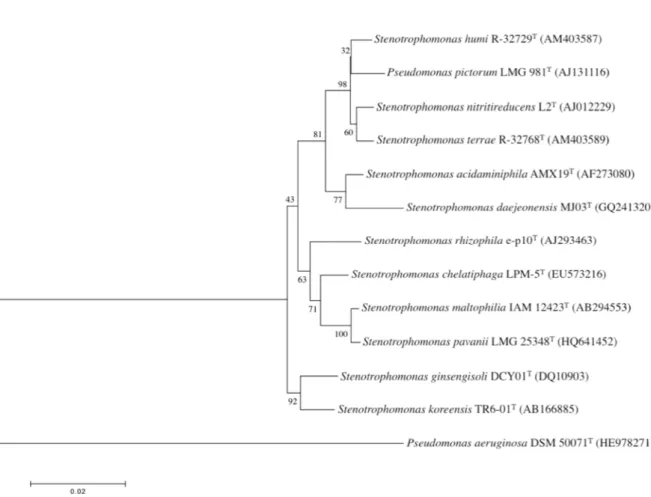
accession number AJ131116; 1,502 positions used). All the other type strains of Stenotrophomonas species used during this study were obtained from public culture collections. The culture collection references of these type strains are listed in Table S1. The phylogenetic analysis was done as described by Assih et al. (2002) except that this time the MEGA software (version 5.2; http://www.megasoftware.net) and the Muscle algorithm were used to align the 16S rRNA sequences. This analysis revealed that the closest relatives were the type strains of Stenotrophomonas humi, S. terrae, S. nitritireducens and S. acidaminiphila with similarity levels of 98.8 %, 98.8 %, 98.6 % and 98.0 % respectively. All the other type strains of Stenotrophomonas species showed high 16S rRNA sequence similarities ranging from 96.8 to 97.2 % (Fig 1). Pseudomonas pictorum cannot be assigned to Stenotrophomonas daejeonensis, S. koreensis, S. maltophilia and S. pavanii since they share less
Fig. 1. 16S rRNA phylogenetic dendrogram showing the position of
Pseudomonas pictorum LMG 981T among the representative members of the
genus Stenotrophomonas. The scale bar indicates 2 nucleotides substitution per 100 nucleotides. Numbers (percentages) at the node corresponds to bootstrap values based on 1000 resampling. Sequence accession numbers are indicated.
than 97 % 16S rRNA similarity (Stackebrandt & Goebel 1994, Wayne et al., 1987). Recently, a 16S rRNA similarity of 98.2 % has been proposed as the new cut off point above which DNA-DNA reassociation experiments should be necessary for testing the genomic uniqueness of a novel isolate (Stackebrandt & Ebers, 2006; Meier-Kolthoff et al., 2013; Kim et al., 2014). Based on this criteria, P. pictorum could not be assigned to Stenotrophomonas chelatiphaga, S. rhizophila or S. ginsengisoli with which it shares 97.1 to 97.2 % 16S rRNA sequence similarity. Therefore, DNA-DNA tests were performed only with the four type strains sharing at least 98% 16S rRNA sequence similarities. The
DNA-DNA hybridizations with S. acidaminiphila CIP 106456T and S.
nitritireducens DSM 12575T were done by DSMZ using the spectroscopic
method as described elsewhere (De Ley et al., 1970, Escara & Hutton 1980, Huss et al., 1983, Jahnke & Bahnweg, 1986; Jahnke, 1992) while the DNA-DNA
hybridizations with S. humi CCUG 54881T and S. terrae CCUG 54880T were
determined by BCCM/LMG using the microplate method developed by Ezaki et al. (1989) with the modifications implemented by Goris et al. (1998) and
Cleenwerck et al. (2002). These tests revealed that P. pictorum ATCC 23328T
hybridized at 31.0 ± 3 % with S. humi CCUG 54881T, at 32.0 ± 10 % with S.
terrae CCUG 54880T and at 43.6 % with S. acidaminiphila CIP 106456T and
43.3 % with S. nitritireducens DSM 12575T, all values significantly below the 70
% cut off limit for species delineation. The 16S rRNA analysis coupled to the DNA-DNA hybridization with the closest relatives showed that Pseudomonas pictorum belongs to Stenotrophomonas genus but that it cannot be assigned to any of the Stenotrophomonas species with valid standing in nomenclature described so far. As a consequence, the most consistent alternative seems to be the affiliation of Pseudomonas pictorum as a distinct new species of genus Stenotrophomonas. Such conclusion is in line with results pointed out by
several authors (Anzai et al., 2000, Svensson-Stadler et al., 2012) including one
based on the analysis of the full genome of Pseudomonas pictorum (Patil et al., 2016).
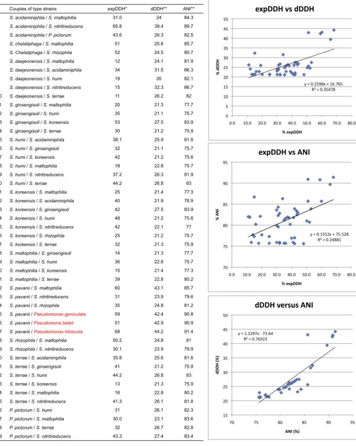
On this last point it is interesting to note that contrarily to what was observed by Goris et al. (2007), no correlation could be found between the ANI (Average Nucleotide Identity) or dDDH (digital DNA-DNA hybridization) derived from the analysis of the full genome sequences of all Stenotrophomonas species by Patil et al. (2016) and the 46 experimental DNA-DNA hybridization values (expDDH)
published in the descriptions of the different Stenotrophomonas species (Tables
S4 and S5). It is important however not to forget that the 46 experimental DNA-DNA hybridization values available for these species are all well below 70 % since they were determined to confirm that close strains belonged to different species and that this may have created a statistical bias for such comparison. Although dDDH and experimental DNA-DNA hybridization did not gave identical values for the Stenotrophomonas species they were always in phase with respect to the 70% cutoff limit between species.
Additional biochemical characteristics provided by our study were the cellular
fatty acid (CFA) composition and GC mole % of P. pictorum ATCC 23328T. The
CFA composition of the type strains of all the validly published species assigned presently to the Stenotrophomonas genus was redetermined at DSMZ using the Sherlock Microbial Identification systems (MIDI) and MIDI standard procedures
for strain cultivation (24h at 28°C in trypticase soy broth supplemented with 15 g
agar l-1). The CFA profiles obtained were compared by unweighted arithmetic
average clustering to those of all the Pseudomonas, Pseudoxanthomonas and Xanthomonas species with valid standing in nomenclature present in MIDI TSBA6 library using MIDI proprietary software that disclose the results in the form of dendrogram. The CFA Profiles for each species of the MIDI TSBA6 library correspond to the average profile of several strains including the type
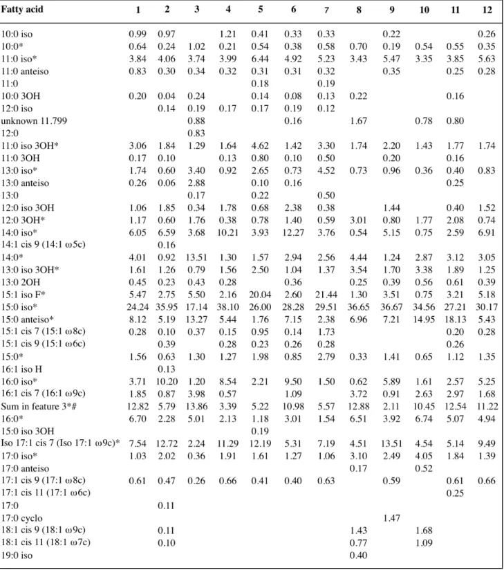
strain ones. The major fatty acids of Pseudomonas pictorum ATCC 23328T
were iso-C15:0 (24.2 %), anteiso-C15:0 (8.1 %), iso-C17:1ω9c (7.5 %), C16:0 (6.7 %),
iso-C14:0 (6.1 %) and iso-C15:1 F (5.5 %). Up to fifteen CFA were always present
in the CFA profile determined for the type strains of all the Stenotrophomonas species and in the cell fatty acid pattern of Pseudomonas pictorum ATCC
23328T (Table S2 and genus description). The unresolved CFA mixture iso-C15:0
2OH/C16:1ω7c was also present in the CFA composition determined for all the
strains included in our study (Table S2). The three CFA (iso-C11:0, iso-C11:0-3OH
and iso-C13:0-3OH) identified by Yang et al. (1993b) as characteristic of genus
Stenotrophomonas and Xanthomonas were detected in the cell fatty acid
pattern of Pseudomonas pictorum ATCC 23328T (Table S2). Other fatty acids
present at a level of more than 1 % in most of the type strains of
Stenotrophomonas species tested were: iso-C11:0 (3.4-6.4 %), iso-C11:0 3OH
(1.3-3.3 %), C14:0 (1.2-13.5 %), iso-C15:1 F (1.1-20.0 %), iso-C15:0 (17.1-38.1 %),
anteiso-C15:0 (1.8-18.1 %), iso-C16:0 (1.2-11.9 %), C16:0 (1.2-6.7 %) iso-C17:1ω9c
(2.2-13.5 %). The comparison by unweighted arithmetic average clustering of
the CFA profile of Pseudomonas pictorum ATCC 23328T showed that
Stenotrophomonas terrae CCUG 54880T and S. humi CCUG 54881T were the
two type strains with the closest CFA profiles (Fig. S1) which is in line with the 16S rRNA phylogeny (Fig. 1) and gyrB sequence analysis (Svensson-Stadler et al., 2012). The CFA average clustering clearly showed also that Pseudomonas
pictorum ATCC 23328T does not cluster with the Pseudomonas species but
rather with those belonging to the Xanthomonadaceae lineage (Fig. S2). Within this lineage, the Stenotrophomonas species did not form however a homogenous separate cluster from the Xanthomonas and other related species (e.g. Pseudoxanthomonas spp). This indicates that CFA is not an adequate discrimination tool among the different genus of this lineage.
The guanine-plus-cytosine content (G+C%) of the bacterial DNA of
Pseudomonas pictorum ATCC 23328T was determined by BCCM/LMG using
the HPLC technique (Mesbah et al., 1989). The value reported is the mean value of three independent analyses of the same DNA sample. The G+C mole
% of P. pictorum ATCC 23328T found during this study was 65.7 which is within
the range of values (64.0 – 69.2) reported for the other Stenotrophomonas species (Lee et al., 2011, Table 1) and is identical to the value reported earlier by De Vos et al., (1989). The two previous experimental values are in agreement with the G+C % (66.00 %) calculated from the full sequence of
Pseudomonas pictorum JCM 9942T by Patil et al. (2016).
Procedures for determination of general phenotypic characteristics for P. pictorum were as described elsewhere (Assih et al., 2002, Ouattara et al., 2003, Thierry et al., 2004). Our results of phenotypic characterization were partially
consistent with those reported by Gray & Thornton (1928) and the general
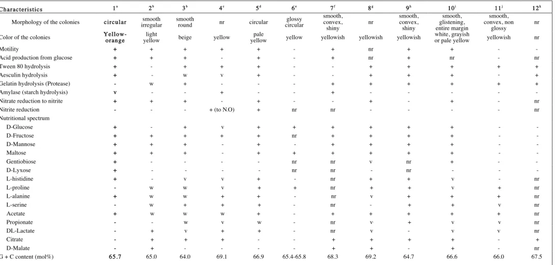
Table 1. General phenotypic characters and G+C% of the type strain of Pseudomonas pictorum and that of type strains of Stenotrophomonas species with standing in nomenclature
1 = Pseudomonas pictorum ATCC 23328T, 2 = Stenotrophomonas humi, 3 = Stenotrophomonas terrae, 4 = Stenotrophomonas nitritireducens, 5 = Stenotrophomonas acidaminiphila, 6 = Stenotrophomonas
ginsengisoli, 7 = Stenotrophomonas chelatiphaga, 8 = Stenotrophomonas rhizophila, 9 = Stenotrophomonas daejeonensis, 10 = Stenotrophomonas maltophilia, 11 = Stenotrophomonas koreensis, 12 = Stenotrophomonas pavanii.. + : positive, - : negative, w : weak, v : variable, nr: not reported,. All the species : gram negative, oxidase positive, catalase positive. The following substrates were not utilized by Pseudomonas pictorum : D-galactose, D-trehalose, L-sorbose, D-melibiose, sucrose, D-raffinose, lactose, lactulose, 1-O-Methyl-β-galactoside, 1-O-Methyl-α-galactoside, D-cellobiose,
1-O-Methyl-β-glucoside, ribose, L-arabinose, xylose, palatinose, L-rhamnose, L-fucose, melizitose, arabitol, L-arabitol, xylitol, dulcitol, tagatose, glycerol, myo-inositol, mannitol, maltitol, turanose, D-sorbitol, adonitol, HQ-β-glucuronide, i-erythritol, 1-O-Methyl-α-D-glucoside, 3-O-methyl-D-glucose, D-saccharate, mucate, L-tartrate, D-tartrate, meso-tartrate, D-malate, L-malate, cis-aconitate, trans-aconitate, tricarballylate, citrate, D-glucuronate, D-galacturonate, 2-ketogluconate, 5-ketogluconate, tryptophan, D-gluconate, phenylacetate, protocatechuate, 4-hydroxybenzoate, quinate, gentisate, 3-hydroxybenzoate, benzoate, 3-phenylpropionate, m-coumarate, trigonelline, betaine, putrescine, 4-aminobutyrate, histamine, DL-lactate, caprate, caprylate, succinate, fumarate, glutarate, DL-glycerate, 5-aminovalerate, ethanolamine, tryptamine, D-glucosamine, Itaconate, 3-hydroxybutyrate, L-aspartate, L-glutamate, L-proline, D-alanine, L-serine, malonate, propionate, L-tyrosine, 2-ketoglutarate
Characteristics 1a 2b 3b 4c 5d 6e 7f 8g 9h 10i 11j 12k
Morphology of the colonies circular irregular smooth smooth round nr circular circular glossy smooth, convex,
shiny nr smooth, convex, shiny smooth, glistening, entire margin smooth, convex, non glossy nr
Color of the colonies Yellow-orange yellow light beige yellow yellow pale yellow yellowish yellowish yellowish white, grayish or pale yellow yellowish nr
Motility + + + + + - + nr + + - -
Acid production from glucose + + + - + - + nr + nr - nr
Tween 80 hydrolysis + - + + + - - + + + + +
Aesculin hydrolysis + - w v + - - + + + - +
Gelatin hydrolysis (Protease) - w + - - - + + + + + +
Amylase (starch hydrolysis) v - - + - - + - - - - -
Nitrate reduction to nitrite + + + - + - - + - + - nr
Nitrite reduction - - - + (to N2O) + nr nr - - - - nr
Nutritional spectrum D-Glucose + - + v + + + + + + - - D-Fructose + + + + + nr + + + + - - D-Mannose + + + - + - + + + + - - Maltose + + + - + + + + + + - - Gentiobiose + - - - - nr nr v nr + - - D-Lyxose + - - - - nr nr - nr - - - L-histidine + - v v + - nr + + v - nr L-proline - w w v + + nr + + v + nr L-alanine + w w + + - nr v + + + nr L-serine - w + + + - nr - + + v nr Acetate + w w w + - + + + + + nr Propionate - - w v w - nr vj + v v nr DL-Lactate - + v + + - nr v - v v nr Citrate - + + + - - + + + + - + D-Malate - + - - - - + + - + - nr G + C content (mol%) 65.7 65.0 64.0 69.1 66.9 65.4-65.8 68.3 69.2 64.7 66.6 66.0 67.5
a data from this study, Gray and Thornton, 1928 and Lipski et al., 1992; b data from Heylen et al., 2007, Kim et al., 2010, Kapparulina et al., 2009 and Lee et al., 2011; c data from Finkmann et al., 2000, Heylen et al., 2007, Kim et al., 2010, Kapparulina et al., 2009, Lee et al., 2011, Palleroni 2005, Yang et al., 2006 and Lipsky & Altendorf 1997; d data from Assih et al., 2002 and Kim et al., 2010; e data from Kim et al., 2010, f data from Kapparulina et al., 2009, g data from Wolf et al., 2002, Heylen et al., 2007, Kapparulina et al., 2009, Wolf et al. 2002, Kim et al., 2010 and Yang et al. 2006; h data from Lee et al., 2011, i data from Palleroni 2005, Stanier et al., 1966, Heylen et al., 2007, Kim et al., 2010, Yang et al. 2006, Lipsky & Altendorf 1997 and Lee et al., 2011; j data from Yang et al., 2006, Heylen et al., 2007, Yang et al. 2006 and Kim et al., 2010; k data from Ramos et al., 2011.
characteristics given by Palleroni & Bradbury (1993) and Palleroni (2005) for the Stenotrophomonas genus.
The overall results of phenotypic characterization are given in the genus and species description and in Table 1. P. pictorum could be easily distinguished from the other Stenotrophomonas species by the color of its colonies on nutrient agar (data not shown) and from its closest relatives S. humi, S. terrae, S. nitritireducens or S. acidaminiphila, by its ability to assimilate gentiobiose, D-lyxose but not L-proline and L-serine. Motility and fructose utilization were the common properties shared by P. pictorum and its closest relatives pertaining to genus Stenotrophomonas (Table 1).
The disk diffusion technique as described by Thierry et al. (2004) was used to evaluate the susceptibility of the type strain of Pseudomonas pictorum towards
a set of medical antibiotics. Our data showed that P. pictorum ATCC 23328T
presented low MICs (Minimum Inhibitory Concentrations) to 14 of the 16 antibiotics tested and was only apparently resistant to cephalothin, a first generation cephem, and amoxillin that both act on peptidoglycan synthesis. P.
pictorum ATCC 23328T was however susceptible to the 6 other antibiotics
tested working with the same mechanism (Table S3). These results suggest that P. pictorum is highly susceptible to antibiotics which is logical since it was isolated from soil in 1928 much before the start of the massive use of antibiotics that has resulted in an increase of the abundance of antibiotic resistance gene in soils since then (Knapp et al., 2010). This observation is also in line with the analysis of the full genome of P. pictorum that has shown the absence of chromosomally encoded β-lactamases contrarily to S. maltophilia, S. pavanii and other related strains (Patil et al., 2016).
Phylogenetic differences between the type strain of Pseudomonas pictorum and any of the type strains of genus Stenotrophomonas species are supported by phenotypic and biochemical differences. The overall present results together with past analyses (Anzai et al., 2000, Assih et al., 2002, De Vos et al., 1989, Kersters et al., 1996, Oyaizu & Komagata 1983, Patil et al., 2016, Singer et al., 1994, Svensson-Stadler et al., 2012, Van den Mooter & Swings 1990, Yang et al., 1993a) support that Pseudomonas pictorum should be transferred in genus Stenotrophomonas as a new species of this genus, Stenotrophomonas pictorum comb. nov. An emended description of the genus Stenotrophomonas is therefore proposed in order to take in account new taxonomic data available through the description of several new species assigned to this genus since its creation by Palleroni and Bradbury (1993).
Emended description of genus Stenotrophomonas.
The etymology, morphology and biochemical properties are as indicated in the genus description (Palleroni and Bradbury 1993, Palleroni 2005). Additional or modified properties are: reduction of nitrate to nitrite is variable, oxidase reaction is variable, tween 80, gelatin and starch hydrolysis variable, species are non-motile or motile by means of a single polar flagellum or several polar flagella, some species may grow anoxically using nitrate as alternate electron acceptor, colonies are white, beige, grayish, yellowish, pale yellow, yellow light, orange-yellow or yellow on common solid media, colonies are smooth,
glistening and often circular, growth is not accompanied by odour on common solid media but odour could develop on some media. The cellular fatty acids are
of the iso/anteiso type with iso-C15:0 normally clearly predominating. The other
predominating fatty acids present are iso-C11:0, anteiso-C15:0 and iso-C17:1ω9c.
Other CFA usually or always present in cells are: C10:0, iso-C11:0 3OH, iso-C13:0,
C12:0 3OH, iso-C14:0, C14:0, iso-C13:0 3OH, iso-C15:1 F, ,C15:0, iso-C16:0, C16:0, and
iso-C17:0. DNA G+C content is 64.0–69.2 mole %. Members of the genus are
widely distributed in nature. The type species is Stenotrophomonas maltophilia.
Description of Stenotrophomonas pictorum comb. nov.
Stenotrophomonas pictorum (pic.to’rum). L. gen. pl. n. pictorum, of painters; here, intended to mean of the Picts, named after the Picts, a Scottish tribe. Exhibits all of the characteristics of the members of the genus. Cells size: 0.5-0.8 x 1.5-3 µm. Colonies are yellow and circular on Trypticase soy agar. On nutrient agar, colonies are orange-yellow or yellow. Cells are positive for catalase, aesculin, tween 80 esterase but negative for urease, indole, ONPG, Simmons citrate, lysine and ornithine decarboxylase, arginine dihydrolase, DNAse and proteolysis. Oxidase and starch hydrolysis variable. Nitrate is reduced but not nitrite. Polyamines: spermidine (major), cadaverine and spermine (minor). Quinone type: Q8. A limited range of substrate can be utilized (11 on 99 tested) including D-glucose, D-fructose, D-mannose, maltotriose, maltose, gentiobiose, D-lyxose, N-Acetyl-D-glucosamine, L-histidine, L-alanine and phenol. Acid is produced from D-glucose (dextrose) and maltose. Cholesterol is depleted when grown on bovine calf serum but not used as sole carbon source in mineral medium. Substrates not used are listed in Table 1. No growth was observed at 4 °C or/and 41 °C. Antibiotics susceptibility: susceptible to Ticarcillin, Piperacillin, Piperacillin + Tazobactam, Imipenem, Cefotaxime, Ceftazidime, Tobramycin, Amikacin, Gentamicin, Netilmicin, Colistin, trimethoprim + Sulfamethoxazole, Ofloxacin and Ciprofloxacin; resistant to Amoxicillin and Cephalothin. All the CFA characteristic of genus Stenotrophomonas are present. Predominant fatty acids are by decreasing
order of abundance iso-C15:0, anteiso-C15:0, iso-C17:1ω9c, C16:0, iso-C14:0 and
iso-C15:1 F. The DNA G + C content is 65.7 mole %. Habitat: originally isolated from
soil. Type strain : ATCC 23328T, CCM 284T, CCUG 1823T, CCUG 3368T, CIP
103273T, DSM 19282T, JCM 9942T, LMG 981T, NCIMB 9152T, VKM 1240T. The
GenBank/EMBL/DDBJ accession number for the 16S rRNA gene sequence of the type strain is AJ131116.
Funding information
The authors state that this work received no specific grant from any funding agency.
Acknowledgements
The authors thank Bernard Ollivier and Jean-Luc Cayol for fruitful discussions.
Conflicts of interest
The authors declare that there is no conflict of interest.
References
Anzai, Y., Kim, H., Park, J. Y., Wakabayashi, H., & Oyaizu, H. (2000).
Phylogenetic affiliation of the pseudomonads based on 16S rRNA sequence. Int J Syst Evol Microbiol 50, 1563–1589.
Assih, E. A., Ouattara, A. S., Thierry, S., Cayol, J. L., Labat, M., & Macarie, H. (2002). Stenotrophomonas acidaminiphila sp. nov., a strictly aerobic
bacterium isolated from an upflow anaerobic sludge blanket (UASB) reactor. Int J Syst Evol Microbiol 52, 559–568.
Cleenwerck, I., Vandemeulebroecke, K., Janssens, D. & Swings J. (2002).
Re-examination of the genus Acetobacter, with description of Acetobacter cerevisiae sp. nov. and Acetobacter malorum sp. nov. Int J Syst Evol Microbiol
52, 1551–1558.
Coenye, T., Vanlaere, E., LiPuma, J. J., & Vandamme, P. (2004).
Identification of genomic groups in the genus Stenotrophomonas using gyrB RFLP analysis. FEMS Immunol Medical Microbiol 40, 181-185.
De Ley, J., Cattoir, H. & Reynaerts, A. (1970). The quantitative measurement
of DNA hybridization from renaturation rates. Eur J Biochem 12, 133-142.
De Vos, P., Van Landschoot, A., Segers, P., Tytgat, R., Gillis, M., Bauwens, M., Rossau, R., Goor, M., Pot, B., Kersters, K, Lizzaraga, P., & De Ley, J. (1989). Genotypic relationships and taxonomic localization of unclassified
Pseudomonas and Pseudomonas-like strains by desoxyribonucleic acid: ribosomal nucleic acid hybridizations. Int J Syst Bacteriol 39, 35–49.
Escara, J. F. & Hutton, J. R. (1980). Thermal stability and renaturation of DNA
in dimethylsulphoxide solutions: acceleration of renaturation rate. Biopolymers
19, 1315-1327.
Ezaki, T., Hashimoto, Y. & Yabuuchi E. (1989). Fluorimetric deoxyribonucleic
acid-deoxyribonucleic acid hybridization in microdilution wells as an alternative to membrane filter hybridization in which radioisotopes are used to determine genetic relatedness among bacterial strains. Int J Syst Evol Microbiol 39, 224– 229.
Finkmann, W., Altendorf, K., Stackebrandt, E., & Lipski, A. (2000).
Characterization of N2O-producing Xanthomonas-like isolates from biofilters as
Stenotrophomonas nitritireducens sp. nov., Luteimonas mephitis gen. nov., sp. nov. and Pseudoxanthomonas broegbernensis gen. nov., sp. nov. Int J Syst Evol Microbiol 50, 273–282.
Goris, J., Konstantinidis, K. T., Klappenbach, J. A., Coenye, T., Vandamme, P., & Tiedje, J. M. (2007). DNA–DNA hybridization values and 9
their relationship to whole-genome sequence similarities. Int J Syst Evol Microbiol 57, 81-91.
Goris, J., Suzuki, K., De Vos, P., Nakase, T. & Kersters, K. (1998).
Evaluation of a microplate DNA-DNA hybridization method compared with the initial renaturation method. Can J Microbiol 44, 1148-1153.
Gray, P. H. H. & Thornton, H. G. (1928). Soil bacteria that decompose certain aromatic compounds. Zentralblatt fur Bakteriologie, Parasitenkunde,
Infektionskrankheiten und Hygiene. Abteilung II, 73, 74-96.
Hauben, L., Vauterin, L., Moore, E. R. B., Hoste, B. & Swings, J. (1999).
Genomic diversity of the genus Stenotrophomonas. Int J Syst Bacteriol 49, 1749–1760.
Heylen, K., Vanparys, B., Peirsegaele, F., Lebbe, L., & De Vos, P. (2007).
Stenotrophomonas terrae sp. nov. and Stenotrophomonas humi sp. nov., two nitrate-reducing bacteria isolated from soil. Int J Syst Evol Microbiol 57, 2056– 2061.
Huss, V. A. R., Festel, H. & Scheilfer, K. H. (1983). Studies on the
spectrometric determination of DNA hybridization from renaturation rates. Syst Appl Microbiol 4, 184-192.
Jahnke, K.-D. (1992). Basic computer program for evaluation of spectroscopic
DNA renaturation data from GILFORD system 2600 spectrometer on a PC/XT/AT type personal computer. J Microbiol Methods 15, 61-73.
Jahnke, K.-D. & Bahnweg, G. (1986). Assessing natural relationships in the
basidiomycetes by DNA analysis. Trans Br Mycol Soc 87, 175-191.
Kaparullina, E., Doronina, N., Chistyakova, T., & Trotsenko, Y. (2009).
Stenotrophomonas chelatiphaga sp. nov., a new aerobic EDTA-degrading bacterium. Syst Appl Microbiol 32, 157–162.
Kersters, K., Ludwig, W., Vancanneyt, M., De Vos, P., Gillis, M., & Schleifer, K.-H. (1996). Recent changes in the classification of the
pseudomonads: an overview. Syst Appl Microbiol 19, 465–477.
Kim, H. B., Srinivasan, S., Sathiyaraj, G., Quan, L. H., Kim, S.H., Bui, T. P. N., Liang, Z., Kim, Y. J., & Yang, D. C. (2010). Stenotrophomonas ginsengisoli
sp. nov., isolated from a ginseng field. Int J Syst Evol Microbiol 60, 1522–1526.
Kim, M., Oh, H.-S., Park, S.-C., & Chun J. (2014). Towards a taxonomic
coherence between average nucleotide identity and 16S rRNA gene sequence similarity for species demarcation of prokaryotes. Int J Syst Evol Microbiol 64, 346–351.
Knapp, C. W., Dolfing, J., Ehlert, P. A., & Graham, D. W. (2010). Evidence of
increasing antibiotic resistance gene abundances in archived soils since 1940. Environ Sci Technol 44, 580-587.
Lee, M., Woo, S. G., Chae, M., Shin, M. C., Jung, H. M., & Ten, L. N. (2011).
Stenotrophomonas daejeonensis sp. nov., isolated from sewage. Int J Syst Evol Microbiol 61, 598–604.
Lipski, A. & Altendorf K. (1997). Identification of heterotrophic bacteria
isolated from ammonia-supplied experimental biofilters. System Appl Microbiol
20, 448-457.
Lipski, A., Klatte, S., Bendinger, B., & Altendorf, K. (1992). Differentiation of
Gram-negative, nonfermentative bacteria isolated from biofilters on the basis of fatty acid composition, quinone system, and physiological reaction profiles. Appl Environ Microbiol 58, 2053-2065.
Meier-Kolthoff, J. P., Auch, A. F., Klenk, H. P. & Göker, M. (2013). Genome
sequence-based species delimitation with confidence intervals and improved distance functions. BMC Bioinformatics 14, 60.
Mesbah, M., Premachandran, U. & Whitman, W. B. (1989). Precise
measurement of the G+C content of deoxyribonucleic acid by high-performance liquid chromatography. Int J Syst Bacteriol 39, 159-167.
Moore, E. R. B., Tindall, B. J., Martins dos Santos, V. A. P., Pieper, D. H., Ramos J. L., Palleroni, N. J. (2006). Nonmedical: Pseudomonas. In The
Prokaryotes, a Handbook on the Biology of Bacteria, 3rd Edition, Vol 6 Proteobacteria Gamma Subclass, pp. 646-703. Edited by M. Dworkin (Editor-in-Chief), S. Falkow, E. Rosenberg, K-.H. Schleifer, E. Stackebrandt. New York : Springer Science+Business Media, LLC.
Ouattara, A. S., Assih, E. A., Thierry, S., Cayol, J.-L., Labat, M., Monroy, O. & Macarie, H. (2003). Bosea minatitlanensis sp. nov., a strictly aerobic
bacterium isolated from an anaerobic digester. Int J Syst Evol Microbiol 53, 1247–1251.
Oyaizu H., & Komataga, K. (1983). Grouping of Pseudomonas species on the
basis of cellular fatty acid composition and the quinone system with the special reference to the existence of 3-hydroxy fatty acids. J Gen Appl Microbiol 29, 17-40.
Palleroni, N. J. (2005). Genus IX. Stenotrophomonas Palleroni and Bradbury
1993, 608VP In Bergey’s Manual of Systematic Bacteriology, 2nd edition, vol 2,
part B The Gammaproteobacteria, pp. 107-115. Edited by D. J. Brenner, N. R. Krieg, J. T. Staley (volume editors) & G. M. Garity (editor-in-chief). New York : Springer Science+Business Media, LLC
Palleroni, N. J., & Bradbury, J. F. (1993). Stenotrophomonas, a new bacterial
genus for Xanthomonas maltophilia (Hugh 1980) Swings et al. 1983. Int J Syst Bacteriol 43, 606–609.
Patil, P. P., Midha, S., Kumar, S., & Patil, P. B. (2016). Genome sequence of
type strains of genus Stenotrophomonas. Front Microbiol 7, 1-6.
Ramos, P. L., Trappen, S. V., Thompson, F. L., Rocha, R. C. S., Barbosa, H. R., Vos, P. D., & Moreira-Filho, C. A. (2011). Screening for endophytic
nitrogen-fixing bacteria in Brazilian sugar cane varieties used in organic farming and description of Stenotrophomonas pavanii sp. nov. Int J Syst Evol Microbiol
61, 926–931.
Singer, E., Debette, J., Lepretre, A., & Swings, J. (1994). Comparative
esterase electrophoretic polymorphism and phenotypic analysis of Xanthomonas maltophilia and related strains. System Appl Microbiol 17, 387-394.
Stackebrandt, E. & Ebers, J. (2006). Taxonomic parameters revisited:
tarnished gold standards. Microbiol Today 33, 152-155.
Stackebrandt, E. & Goebel, B. M. (1994). Taxonomic note: a place for
DNA-DNA reassociation and 16S rRNA sequence analysis in the present species definition in bacteriology. Int J Syst Bacteriol 44, 846-849.
Stanier, R.Y., Palleroni, N.J. Doudoroff, M. (1966). The aerobic
pseudomonads: a taxonomic study. J. Gen. Microbiol., 43, 159-271.
Svensson-Stadler, L. A., Mihaylova, S. A., & Moore, E. R. B. (2012).
Stenotrophomonas interspecies differentiation and identification by gyrB sequence analysis. FEMS Microbiol Lett 327, 15-24.
Thierry, S., Macarie, H., Iizuka, T., Geiβdörfer, W., Assih, E. A., Spanevello, M., Verhe, F., Thomas, P., Fudou, R., Monroy, O., Labat, M. & Ouattara, A. S. (2004) Pseudoxanthomonas mexicana sp. nov. and Pseudoxanthomonas
japonensis sp. nov., isolated from diverse environments, and emended descriptions of the genus Pseudoxanthomonas Finkmann et al., 2000 and of its type species. Int J Syst Evol Microbiol 54, 2245–2255.
Van den Mooter, M., & Swings, J. (1990). Numerical analysis of 295
phenotypic features of 266 Xanthomonas strains and related strains and an improved taxonomy of the genus. Int J Syst Bacteriol 40, 348–369.
Wayne, L. G., Brenner, D. J., Colwell, R. R., Grimont, P. A. D., Kandler, O., Krichevsky, M. I., Moore, L. H., Moore, W. E. C., Murray, R. E. G. & 3 other authors (1987). International Committee on Systematic Bacteriology. Report of
the ad hoc committee on reconciliation of approaches to bacterial systematics. Int J Syst Bacteriol 37, 463-464.
Wolf, A., Fritze, A., Hagemann, M., & Berg, G. (2002). Stenotrophomonas
rhizophila sp. nov., a novel plant-associated bacterium with antifungal properties. Int J Syst Evol Microbiol 52, 1937–1944.
Yang, H. C., Im, W. T., Kang, M. S., Shin, D. Y., & Lee, S. T. (2006).
Stenotrophomonas koreensis sp. nov., isolated from compost in South Korea. Int J Syst Evol Microbiol 56, 81–84.
Yang, P., De Vos, P., Kersters, K., & Swings, J. (1993a). Polyamine patterns
as chemotaxonomic markers for the genus Xanthomonas. Int J Syst Evol
Microbiol43, 709-714.
Yang, P., Vauterin, L., Vancanneyt, M., Swings, J. & Kersters, K. (1993b).
Application of fatty acid methyl esters for the taxonomic analysis of the genus Xanthomonas. Syst Appl Microbiol 16, 47-71.
SUPPLEMENTARY MATERIAL
Transfer of Pseudomonas pictorum Gray & Thornton 1928 to genus
Stenotrophomonas as Stenotrophomonas pictorum comb. nov. and
emended description of the genus Stenotrophomonas
Aboubakar S. Ouattara1, Jean Le Mer2, Manon Joseph2, Hervé Macarie3
1
Laboratoire de Microbiologie et de Biotechnologie Microbienne, Ecole Doctorale Sciences et Technologies, Université Ouaga 1 Pr Joseph KI ZERBO, 03 BP 7021, Ouagadougou 03, Burkina Faso.
2 Aix Marseille Univ, Univ Toulon, CNRS, IRD, MIO, Marseille, France.
3 Aix Marseille Univ, Univ Avignon, CNRS, IRD, IMBE, Marseille, France.
*Corresponding author: as.ouattara@yahoo.fr
CONTENTS:
Fig. S1. Dendrogram generated from CFA compositions showing the relationship of Pseudomonas
pictorum ATCC 23328T with the type strains of all Stenotrophomonas species as well as to the type
species of Pseudomonas (P. aeruginosa), Pseudoxanthomonas (P. broegbernensis) and Xanthomonas (X. campestris pv. campestris) genus but also to Pseudoxanthomonas mexicana. The CFA of the species associated with strain code in microbial collections were determined in this study. The CFA of all other species corresponds to those present in MIDI TSBA6 library.
Fig. S2. Dendrogram generated from CFA compositions showing the relationship of Pseudomonas
pictorum ATCC 23328T with the type strains of all Stenotrophomonas species (determined in this study) as well as all the Pseudomonas, Pseudoxanthomonas and Xanthomonas species with valid standing in nomenclature present in MIDI TSBA6 library. The references including two species signify that their CFA profile is indistinguishable by the MIDI system.
Table S1. Accession number in public culture collections of the type strains of the Stenotrophomonas
species with standing in nomenclature used during the study.
Table S2. Fatty acid profile of Pseudomonas pictorum type strain compared to those of all type
strains of Stenotrophomonas species with standing in nomenclature.
Table S3. Susceptibility of Pseudomomas pictorum ATCC 23328T towards selected antibiotics covering all mechanism of action.
Table S4. Correlation between expDDH, dDDH (experimental and digital DNA-DNA hybridization
respectively) and ANI (Average Nucleotide Identity) reported in literature for the type strains of the Stenotrophomonas species and related strains.
Table S5. Experimental DNA/DNA hybridization (expDDH) reported in literature between the type
strains of the Stenotrophomonas species and related strains.
!
Fig. S1. Dendrogram generated from CFA compositions showing the relationship of
Pseudomonas pictorum ATCC 23328T with the type strains of all Stenotrophomonas species
as well as to the type species of Pseudomonas (P. aeruginosa), Pseudoxanthomonas (P. broegbernensis) and Xanthomonas (X. campestris pv. campestris) genus but also to Pseudoxanthomonas mexicana. The CFA of the species associated with strain code in microbial collections were determined in this study. The CFA of all other species corresponds to those present in MIDI TSBA6 library.!
!
! ! ! ! ! ! ! ! ! ! ! ! ! ! ! ! ! ! ! ! ! ! ! ! !
Fig. S2. Dendrogram generated from CFA compositions showing the relationship of
Pseudomonas pictorum ATCC 23328T with the type strains of all Stenotrophomonas species
(determined in this study) as well as all the Pseudomonas, Pseudoxanthomonas and Xanthomonas species with valid standing in nomenclature present in MIDI TSBA6 library. The references including two species signify that their CFA profile is indistinguishable by the MIDI system.
Pseudomonas aeruginosa GC subgroup A……….! Stenotrophomonas ginsengisoli DSM 24757T……….! Stenotrophomonas koreensis CCUG 53887T…………..! Stenotrophomonas chelatiphaga DSM 21508T………..!
Pseudoxanthomonas mexicana………..! Stenotrophomonas daejeonensis CCUG 59871T………! Stenotrophomonas acidaminiphila……….! Stenotrophomonas humi CCUG 54881T……….!
Pseudomonas pictorum ATCC 23328T………!
Stenotrophomonas terrae CCUG 54880T………...! Stenotrophomonas pavanii DSM 25135T………! Stenotrophomonas maltophilia………...!
Pseudoxanthomonas broegbernensis……….! Stenotrophomonas rhizophila DSM 14405T…………..! Xanthomonas campestris pv. campestris……….!
Euclidian distance! 0! 10! 20! 30! 40! 50! 60!
Xanthomonas hortorum pv. hederae……….! Xanthomonas arboricola pv. juglandis………! Xanthomonas pisi………!
Xanthomonas cucurbitae………..!
Xanthomonas translucens pv. translucens………..! Xanthomonas fragariae (48 h)………! Xanthomonas axonopodis pv. axonopodis………..!
Stenotrophomonas nitritireducens DSM 12575T…….! Xanthomonas hyacinthi………..!
Xanthomonas albilineans (48 h)………!
Pseudomonas chlororaphis………! Pseudomonas savastanoi pv. fraxinus………! Pseudomonas veronii……….! Pseudomonas putida biotype A……….! Pseudomonas synxantha………! Pseudomonas corrugata………! Pseudomonas mucidolens………..! Pseudomonas cichorii / Pseudomonas viridiflava...! Pseudomonas fluorescens biotype A………..! Pseudomonas fuscovaginae………! Pseudomonas lundensis……….!
Pseudomonas agarici……….! Xanthomonas oryzae pv. oryzae……….! Pseudomonas oleovorans………! Pseudomonas mendocina / Pseudomonas straminea….!
Pseudomonas stuzeri………..! Pseudomonas resinovorans……….! Pseudomonas balearica………..! Pseudomonas alcaligenes………...! Pseudomonas pertucinogena………..! Pseudomonas syringae pv. syringae………..!
Table S1. Accession number in public culture collections of the type strains of the Stenotrophomonas species with standing in
nomenclature used during the study.
Species Strain accession number
in culture collections
Analysis performed
Stenotrophomonas acidaminiphila CIP 106456T DNA-DNA hybridization, phenotypic characterization, CFA
Stenotrophomonas chelatiphaga DSM 21508T CFA
Stenotrophomonas daejeonensis CCUG 59871T CFA
Stenotrophomonas ginsengisoli DSM 24757T CFA
Stenotrophomonas humi CCUG 54881T DNA-DNA hybridization, CFA
Stenotrophomonas koreensis CCUG 53887T CFA
Stenotrophomonas maltophilia CIP 60.77T CFA
Stenotrophomonas nitritireducens DSM 12575T DNA-DNA hybridization, phenotypic characterization, CFA
Stenotrophomonas pavanii DSM 25135T CFA
Stenotrophomonas rhizophila DSM 14405T CFA
Stenotrophomonas terrae CCUG 54880T DNA-DNA hybridization, CFA
Table S2. Fatty acid profile of Pseudomonas pictorum type strain compared to those of all type
strains of Stenotrophomonas species with standing in nomenclature.
Species: 1, P. pictorum; 2, S. acidaminiphila; 3, S. chelatiphaga; 4, S. daejeonensis; 5, S. ginsengisoli; 6, S. humi; 7, S. koreensis; 8, S. maltophilia; 9, S. nitritireducens; 10, S. pavanii; 11, S. rhizophila; 12, S. terrae.
Values are percentages of total fatty acids. The fatty acids are listed in the order of elution obtained during analysis by gas chromatography (GC). The fatty acid names are given according to the carbon numbering from the carboxyl group (!-end). When required, equivalence to the methyl ("-end) nomenclature numbering is given within parenthesis to easier the conversion between the two nomenclatures. *Fatty acid is present in all tested strains. # Unresolved 15:0 iso 2OH / 16:1 cis 9, i.e. the two CFA 15:0 iso 2OH and 16:1 cis 9 (16:1 "7c) elute with the same retention time upon GC analysis.
Table S3. Susceptibility of Pseudomomas pictorum ATCC 23328T towards selected antibiotics covering all mechanism of action.
Antibiotics are listed by mechanism of action and class. The values obtained in the present study correspond to MICs (Minimum inhibitory concentration) in mg per litre. The values within parenthesis after the name of the antibiotics correspond to the disk charge. S and R within parenthesis after the MIC values mean that the strains are respectively susceptible or resistant to the antibiotics. The S & R categories are based on the guidelines of the antibiogram committee of the French Society of Microbiology (CASFM, 2002).
Antibiotic MIC mg/l (sensitivity)
Inhibition of protein synthesis
Aminoglycosides
Amikacin (30 !g) 0.01 (S)
Gentamicin (15 !g) 0.03 (S)
Netilmicin (30 !g) 0.01 (S)
Tobramycin (10 !g) 0.06 (S)
Inhibition of nucleic acid synthesis or DNA damage
Fluoroquinolones
Ciprofloxacin (5 !g) 0.04 (S)
Ofloxacin (5 !g) 0.04 (S)
Disruption of cell membrane
Polypeptides
Colistin (50 !g) < 2 (S)
Inhibition of folate synthesis
Sulfonamides + di-aminopyrimidines (1.25 + 23.75 !g)
Trimethoprim – Sulfamethoxazole 0.02 (S)
Inhibition of peptidoglycan synthesis
Carbapenems Imipenem (10 !g) 0.57 (S) Cephems Cefotaxime (30 !g) 0.71 (S) Ceftazidime (30 !g) 0.02 (S) Cephalothin (30 !g) 128 (R) Penams Amoxicillin (25 !g) > 16 (R) Piperacillin (75 !g) 0.25 (S) Piperacillin (75 !g) – Tazobactam (10 !g) 0.14 (S) Ticarcillin (75 !g) 0.12 (S) Reference.
CASFM (2002). Communiqué 2002. Special number SFM, pp. 1–47. Paris: Comité de l’Antibiogramme
de la Société Française de Microbiologie (in French). http://www.sfm.asso.fr
Couples of type strains expDDH* dDDH** ANI** 1 S. acidaminiphila / S. maltophilia 31.0 24 84.3 2 S. acidaminiphila / S. nitritireducens 65.8 39.4 89.7 3 S. acidaminiphila / P. pictorum 43.6 26.3 82.5 4 S. chelatiphaga / S. maltophilia 51 25.6 85.7 5 S. Chelatiphaga / S. rhizophila 52 24.5 80.7 6 S. daejeonensis / S. maltophilia 12 24.1 81.9 7 S. daejeonensis / S. acidaminiphila 34 31.5 86.3 8 S. daejeonensis / S. humi 19 26 82.1 9 S. daejeonensis / S. nitritireducens 15 32.3 86.7 10 S. daejeonensis / S. terrae 11 26.2 82 11 S. ginsengisoli / S. maltophilia 20 21.3 77.7 12 S. ginsengisoli / S. humi 35 21.1 75.7 13 S. ginsengisoli / S. koreensis 53 27.5 83.9 14 S. ginsengisoli / S. terrae 30 21.2 75.9 15 S. humi / S. acidaminiphila 38.1 25.9 81.8 16 S. humi / S. ginsengisoli 32 21.1 75.7 17 S. humi / S. koreensis 42 21.2 75.6 18 S. humi / S. maltophilia 18 22.8 75.7 19 S. humi / S. nitritireducens 37.2 26.3 81.9 20 S. humi / S. terrae 44.2 26.8 83 21 S. koreensis / S. maltophilia 25 21.4 77.3 22 S. koreensis / S. acidaminiphila 40 21.9 76.9 23 S. koreensis / S. ginsengisoli 42 27.5 83.9 24 S. koreensis / S. humi 48 21.2 75.6 25 S. koreensis / S. nitritireducens 42 22.1 77 26 S. koreensis / S. rhizophila 25 21.2 75.7 27 S. koreensis / S. terrae 32 21.3 75.9 28 S. maltophilia / S. ginsengisoli 14 21.3 77.7 29 S. maltophilia / S. humi 36 22.8 75.7 30 S. maltophilia / S. koreensis 15 21.4 77.3 31 S. maltophilia / S. terrae 39 22.8 80.2 32 S. pavanii / S. maltophilia 60 43.1 85.7 33 S. pavanii / S. nitritireducens 31 23.9 79.6 34 S. pavanii / S. rhizophila 35 24.8 81.2
35 S. pavanii / Pseudomonas geniculata 59 42.4 90.8
36 S. pavanii / Pseudomona beteli 51 42.9 90.9
37 S. pavanii / Pseudomonas hibiscola 68 44.2 91.4
38 S. rhizophila / S. maltophilia 50.2 24.8 81 39 S. rhizophila / S. nitritireducens 30.1 23.9 79.9 40 S. terrae / S. acidaminiphila 35.8 25.6 81.6 41 S. terrae / S. ginsengisoli 41 21.2 75.9 42 S. terrae / S. humi 44.2 26.8 83 43 S. terrae / S. koreensis 13 21.3 75.9 44 S. terrae / S. maltophilia 16 22.8 80.2 45 S. terrae / S. nitritireducens 41.3 26.1 81.8 46 P. pictorum / S. humi 31 26.1 82.3 47 P. pictorum / S. maltophilia 30.0 23.1 83.6 48 P. pictorum / S. terrae 32 26.7 82.8 49 P. pictorum / S. nitritireducens 43.3 27.4 83.4 *see Table S5
** Patil, P. P., Midha, S., Kumar, S., & Patil, P. B. (2016). Genome sequence of type strains of genus Stenotrophomonas. Front Microbiol 7, 1-6.
Table S4. Correlation between expDDH, dDDH (experimental and digital DNA-DNA hybridization respectively) and ANI (Average Nucleotide Identity) reported in literature for the type strains of the Stenotrophomonas species and related strains y"="0.2598x"+"16.765" R²"="0.35478" 0" 5" 10" 15" 20" 25" 30" 35" 40" 45" 50" 0.0" 10.0" 20.0" 30.0" 40.0" 50.0" 60.0" 70.0" 80.0" %" dD D H " %"expDDH" expDDH"vs"dDDH" y"="0.1552x"+"75.528" R²"="0.24881" 70" 75" 80" 85" 90" 95" 0.0" 10.0" 20.0" 30.0" 40.0" 50.0" 60.0" 70.0" 80.0" %" AN I" %"expDDH" expDDH"vs"ANI" y"="1.2297x"3"73.64" R²"="0.76923" 15" 20" 25" 30" 35" 40" 45" 50" 70" 75" 80" 85" 90" 95" dD D H "(%) " ANI"(%)" dDDH"versus"ANI" 20
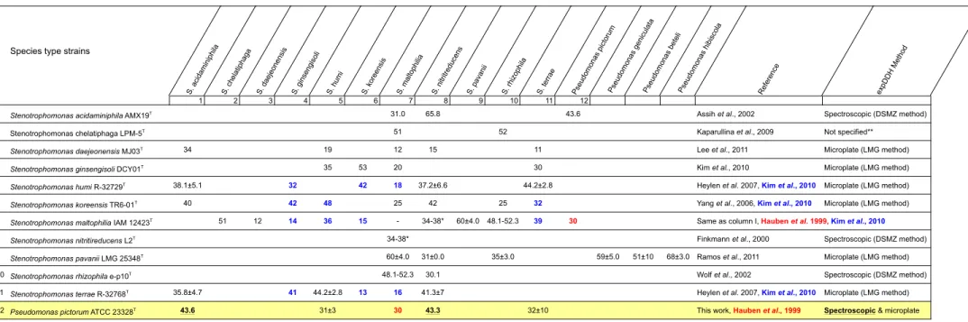
Table S5. Experimental DNA/DNA hybridization (expDDH) reported in literature between the type strains of the Stenotrophomonas species and related strains
Species type strains
S. a cida mini phila S. ch elat ipha ga S. d aeje onen sis S. g inse ngiso li S. h umi S. ko reen sis S. ma ltoph ilia S. n itritre duce ns S. p ava nii S. rh izo phila S. te rrae Pse udomo nas pict oru m Pse udomo nas geni cula ta Pse udomo nas bete li Pse udomo nas hibi sco la Ref ere nce exp DD H Me thod 1 2 3 4 5 6 7 8 9 10 11 12
1 Stenotrophomonas acidaminiphila AMX19T 31.0 65.8 43.6 Assih et al., 2002 Spectroscopic (DSMZ method)
2 Stenotrophomonas chelatiphaga LPM-5T 51 52 Kaparullina et al., 2009 Not specified**
3 Stenotrophomonas daejeonensis MJ03T 34 19 12 15 11 Lee et al., 2011 Microplate (LMG method)
4 Stenotrophomonas ginsengisoli DCY01T 35 53 20 30 Kim et al., 2010 Microplate (LMG method)
5 Stenotrophomonas humi R-32729T 38.1±5.1 32 42 18 37.2±6.6 44.2±2.8 Heylen et al. 2007, Kim et al., 2010 Microplate (LMG method)
6 Stenotrophomonas koreensis TR6-01T 40 42 48 25 42 25 32 Yang et al., 2006, Kim et al., 2010 Microplate (LMG method)
7 Stenotrophomonas maltophilia IAM 12423T 51 12 14 36 15 - 34-38* 60±4.0 48.1-52.3 39 30
8 Stenotrophomonas nitritireducens L2T 34-38* Finkmann et al., 2000 Spectroscopic (DSMZ method)
9 Stenotrophomonas pavanii LMG 25348T 60±4.0 31±0.0 35±3.0 59±5.0 51±10 68±3.0 Ramos et al., 2011 Microplate (LMG method)
10 Stenotrophomonas rhizophila e-p10T 48.1-52.3 30.1 Wolf et al., 2002 Spectroscopic (DSMZ method) 11 Stenotrophomonas terrae R-32768T 35.8±4.7 41 44.2±2.8 13 16 41.3±7 Heylen et al. 2007, Kim et al., 2010 Microplate (LMG method) 12 Pseudomonas pictorum ATCC 23328T 43.6 31±3 30 43.3 32±10 This work, Hauben et al., 1999 Spectroscopic & microplate
*S. maltophilia LMG11114 not the type strain, **Kaparullina et al. (2009) make reference to a general article describing all the methods that can be used for DNA-DNA reassociation experiments without specifying which of them they used References
Assih, E. A., Ouattara, A. S., Thierry, S., Cayol, J. L., Labat, M., & Macarie, H. (2002). Stenotrophomonas acidaminiphila sp. nov., a strictly aerobic bacterium isolated from an upflow anaerobic sludge blanket (UASB) reactor. Int J Syst Evol Microbiol 52, 559–568.
Hauben, L., Vauterin, L., Moore, E. R. B., Hoste, B. & Swings, J. (1999). Genomic diversity of the genus Stenotrophomonas. Int J Syst Bacteriol 49, 1749–1760.
Heylen, K., Vanparys, B., Peirsegaele, F., Lebbe, L., & De Vos, P. (2007). Stenotrophomonas terrae sp. nov. and Stenotrophomonas humi sp. nov., two nitrate-reducing bacteria isolated from soil. Int J Syst Evol Microbiol 57, 2056–2061. Kaparullina, E., Doronina, N., Chistyakova, T., & Trotsenko, Y. (2009). Stenotrophomonas chelatiphaga sp. nov., a new aerobic EDTA-degrading bacterium. Syst Appl Microbiol 32, 157–162.
Kim, H. B., Srinivasan, S., Sathiyaraj, G., Quan, L. H., Kim, S.H., Bui, T. P. N., Liang, Z., Kim, Y. J., & Yang, D. C. (2010). Stenotrophomonas ginsengisoli sp. nov., isolated from a ginseng field. Int J Syst Evol Microbiol 60, 1522–1526. Lee, M., Woo, S. G., Chae, M., Shin, M. C., Jung, H. M., & Ten, L. N. (2011). Stenotrophomonas daejeonensis sp. nov., isolated from sewage. Int J Syst Evol Microbiol 61, 598–604.
Wolf, A., Fritze, A., Hagemann, M., & Berg, G. (2002). Stenotrophomonas rhizophila sp. nov., a novel plant-associated bacterium with antifungal properties. Int J Syst Evol Microbiol 52, 1937–1944. Yang, H. C., Im, W. T., Kang, M. S., Shin, D. Y., & Lee, S. T. (2006). Stenotrophomonas koreensis sp. nov., isolated from compost in South Korea. Int J Syst Evol Microbiol 56, 81–84.
Same as column I, Hauben et al. 1999, Kim et al., 2010
Finkmann, W., Altendorf, K., Stackebrandt, E., & Lipski, A. (2000). Characterization of N2O-producing Xanthomonas-like isolates from biofilters as Stenotrophomonas nitritireducens sp. nov., Luteimonas mephitis gen. nov., sp. nov. and Pseudoxanthomonas broegbernensis gen.
nov., sp. nov. Int J Syst Evol Microbiol 50, 273–282.
Ramos, P. L., Trappen, S. V., Thompson, F. L., Rocha, R. C. S., Barbosa, H. R., Vos, P. D., & Moreira-Filho, C. A. (2011). Screening for endophytic nitrogen-fixing bacteria in Brazilian sugar cane varieties used in organic farming and description
of Stenotrophomonas pavanii sp. nov. Int J Syst Evol Microbiol 61, 926–931.