HAL Id: hal-01685637
https://hal.archives-ouvertes.fr/hal-01685637
Submitted on 18 Jan 2018HAL is a multi-disciplinary open access archive for the deposit and dissemination of sci-entific research documents, whether they are pub-lished or not. The documents may come from teaching and research institutions in France or
L’archive ouverte pluridisciplinaire HAL, est destinée au dépôt et à la diffusion de documents scientifiques de niveau recherche, publiés ou non, émanant des établissements d’enseignement et de recherche français ou étrangers, des laboratoires
A metallo pro-drug to target Cu(II) in the context of
Alzheimer’s disease
Amandine Conte-Daban, Vinita Ambike, Régis Guillot, Nicolas Delsuc,
Clotilde Policar, Christelle Hureau
To cite this version:
Amandine Conte-Daban, Vinita Ambike, Régis Guillot, Nicolas Delsuc, Clotilde Policar, et al.. A metallo pro-drug to target Cu(II) in the context of Alzheimer’s disease. Chemistry - A European Journal, Wiley-VCH Verlag, 2018, 24 (20), pp.5095-5099. �10.1002/chem.201706049�. �hal-01685637�
A metallo pro-drug to target Cu(II) in the context of Alzheimer’s
disease
Amandine Conte-Daban,[a,b] Vinita Ambike,[e] Régis Guillot,[e] Nicolas Delsuc,[c,d] Clotilde Policar* [c,d] and Christelle Hureau*[a,b]
[a] CNRS, LCC (Laboratoire de Chimie de Coordination), 205 route de Narbonne, BP 44099 31077 Toulouse Cedex 4, France
[b] Université de Toulouse, UPS, INPT, 31077 Toulouse Cedex 4, France
[c] Laboratoire des Biomolécules, Département de chimie, École normale supérieure, UPMC Univ. Paris 06, CNRS, PSL Research University, 24 rue Lhomond, 75005 Paris, France
[d] Sorbonne Universités, UPMC Univ. Paris 06, École normale supérieure, CNRS, Laboratoire des Biomolécules (LBM), 75005 Paris, France
[e] Institut de Chimie Moléculaire et des Matériaux d'Orsay, UMR CNRS 8182, Bâtiments 420, Université Paris-Sud 11, Université Paris-Saclay, Rue du doyen Georges Poitou, 91405 Orsay cedex, France.
E-mail : christelle.hureau@lcc-toulouse.fr; clotilde.policar@ens.fr.
Abstract. Alzheimer’s disease and oxidative stress are connected. In the present communication, we report the use of a Mn(II)-based superoxide dismutase mimic ([MnII(L)]+, 1+) as a pro-drug candidate
to target Cu(II) associated events, i.e. Cu(II)-induced formation of reactive oxygen species (ROS) and modulation of the amyloid- (A) peptide aggregation. Complex 1+ is able to remove Cu(II) from A,
stop ROS and prevent alteration of A aggregation as would do the corresponding free ligand LH. Using 1+ instead of LH in further biological applications would have the double advantage to avoid the
cell toxicity of LH and to benefit from its proved SOD-like activity.
Most of neurodegenerative diseases are characterized by the aggregation of intrinsically disordered peptides/proteins (IDP).[1] Such a process can be altered by metal ions, mainly copper and zinc.[2] Another common central element in the pathogenesis of neurodegenerative diseases is the production of Reactive Oxygen Species (ROS) by redox-active metal centres bound to the corresponding IDP.[2a, 3] In Alzheimer's disease (AD), copper ions bound to extracellular amyloid- (A) peptide produce ROS and contribute to the oxidative stress observed in the disease.[3] It has indeed been shown that the Cu(A) species can catalyse the incomplete reduction of dioxygen leading to O2•-, H2O2 and HO•.[4] In living organisms, efficient pathways to finely tune the concentrations in O2°- and H2O2 have evolved to protect endogenous components against oxidative damage and to control the ROS concentrations to appropriate levels. They can involve stoichiometric antioxidants (vitamins E and C, glutathione
(GSH), etc.) or redox enzymes, such as catalase or GSH peroxidase and also superoxide dismutases (SODs), which act as the primary protection system.[5] Oxidative stress arises in AD when these protective pathways are overwhelmed;[6] a link between SOD levels and A aggregation has also been proposed.[7] Oxidative stress is also observed in many others physiopathological processes including, aging, arthritis, stroke, cancer, or inflammation.[5b, 8] Anti-oxidants in general, including recombinant SODs, are now quite well documented for their beneficial effects in oxidative stress situations.[5b, 9] Nonetheless, major drawbacks such as the immunogenicity, the poor penetration into cells and the cost of purified enzymes limit their applications in therapeutics. These limitations can be overcome with low molecular-weight complexes reproducing the catalytic activity of SODs, also called SOD mimics.[5b, 9b-d] The use of inorganic complexes as therapeutics in the AD context has been previously reported,[10] and transmetallation reactions have been recently exemplified as therapeutic approaches against cancer[11] or ALS.[12] In line with such previous works, a therapeutic strategy relying on the use of a SOD mimic is reported here. The [MnII(L)]+ complex (1+) (see Figure 1 and S1 for the structure of
LH, N-(2-hydroxybenzyl)-N,N’-bis[2-(N-methylimidazolyl)methyl]ethane-1,2-diamine and of the
corresponding complexes), is known to dismutate O2•- out of any cellular context with a kcat of 7x10⁶ M-1.s-1.[13] Recently, some of us have set up an approach in inorganic cellular and biological chemistry to characterize the antioxidant bio-activity of SOD-mimics directly in cellular models of oxidative stress. 1+ was shown to efficiently limit superoxide flow in activated macrophages[14] and to limit inflammation with a potential intracellular SOD-activity, while the ligand alone (LH) showed a cellular toxicity.[15] In the present communication, we used complex 1+ instead of following a classical
chelating strategy[16] and using a free ligand that might be toxic. Furthermore in the context of AD, the importance of oxidative stress is widely acknowledged,[3] proving relevant the use of a SOD mimict. We will thus describe here how compound 1+, which has an interesting intrinsic SOD-like ability,
prevents Cu-induced processes by removing Cu(II) from A, that is to say how it prevents ROS production and alteration of A aggregation.
Cu(A) + Mn(L) (1+) → Mn(A) + Cu(L) (2+) (1)*
Figure 1. Scheme of the ligand LH (left) and X-Ray structure of [(Cu)LH]2+ (2H2+, right). Selected
bond distances and angles: Cu-N1 = 1.963(2), Cu-N2 = 1.973 (2), Cu-N3 = 2.011(3), Cu-N4 = 2.046(2) and Cu-O1 = 2.378(2) Å. N1-Cu-N3 = 82.68(11), N3-Cu-N4 = 84.32(10), N4-Cu-N2 = 84.09(9)°.
The ability to remove Cu(II) from Aβ, or metal-swap ability, is described by reaction (1) and the constant K. Reaction (1), has been probed under our working conditions (i.e. buffered solution at pH 7.1): (i) by direct measurements of Cu(II)/Mn(II) swap between the A peptide and the LH ligand and (ii) by evaluation of the affinity constants of each metallic species involved in (1), i.e. the constant of each individual reaction M + L → M(L), (M = Cu(II), Mn(II), L = LH, A). Because all the four species involved in the reaction are paramagnetic, EPR technique is a method of choice to monitor it. The partial dissociation of Mn(A) leads to an intense six-line spectrum corresponding to loosely bound Mn(II) species near the g = 2 region, which precludes the straight-forward observation of the metal exchange (see Figure S2). However, careful inspection of the EPR spectra on the edge of the 6-lines signal show that the signature of a mixture of Cu(A) and 1+ perfectly matches the hyperfine
features of [Cu(L)]+ (complex noted 2+) recorded under the very same conditions (Figure 2). Note (i)
that the 9-GHz EPR signature of 1+ gives a broad signal with no hyperfine structure in the g = 2
region, as expected for highly distorted Mn(II) species[13, 15] (Figure S2); (ii) as a parallel experiment, the removal of Cu from the A peptide by the ligand LH has been recorded by EPR (Figures S3) and (iii) the metal swap experiment has also been followed by UV-Vis (Figure S4), the data obtained matching the EPR results. Affinity of Mn(II) for LH has been evaluated by direct titration monitored by UV-Vis (Figure S5) leading to an apparent affinity constant of 1.3x10⁶ M-1 (pH 7.1, 100 mM HEPES) in line with previous measurement by calorimetry.[17] Affinity of Cu(II) for LH has been determined by competition with ligands of known affinity (EPR, Figure S6) and an apparent value in the range of 1016 M-1 was obtained. The affinity of Cu(II) for A at the same pH (i.e. pH 7.1) is 109 M -1.[18] We were not able to determine precisely the affinity of Mn(II) for A but the value should be (i) below 105 M-1 since 10 equivalents of A is not able to remove Mn(II) from LH (Figure S7) and (ii) above 103 M-1 if we consider the presence of three histidine and four carboxylate amino-acid residues
in the A peptide sequence and affinity values of multidentate imidazole and carboxylate-based synthetic ligands as tabulated in ref. [19] (see Table S3), in line with the recent evaluation of the affinity
value of Aβ by NMR.[20] This indicates that the constant K of reaction (1) ( ) is in the 104
-106 range, consistent with the metal swap observed by the previously described direct exchange experiments. Structure of complex 2H2+ was characterized by X-ray crystallography (see experimental
data for details, Tables S1-S2). Briefly, 2H2+ is a dicationic Cu(II) complex where the metal centre lies
in a square-planar pyramidal environment, made of the four nitrogen atoms from the ligand in the equatorial plane with an averaged Cu-Nequatorial distance of 1.998(25) Å and the oxygen atom form its phenol moiety in the apical position. The apical distance is significantly long (2.378(2) Å) in line with the protonated nature of the phenol group, in line with previous report on a series of similar [N4O] complexes where the apical distance has been shown to increase from about 2.15 to 2.5 Å upon protonation of the phenolate moiety bound to the central Cu(II) ion.[21] The integrity of the environment of the Cu centre in 2H2+ and in particular the presence of the protonated phenol moiety in
the apical position in solution was assayed by determination of the pH at which the pendant phenol group is deprotonated in the complex and in the free ligand. pKa-values of 6.4 (2+) (to be compared to
6.5 for 1+) and 11.3 were determined, respectively (Figure S8). The pKa values in presence of metal
ions indicates that the phenol arm is mainly deprotonated at neutral pH and at pH 7.1 the repartition between 2H2+ and 2+ is about 20:80 to 15:85. Phenolate coordination to the Cu(II) centre is also
proved by the UV-Vis band detected at 410 nm characteristic of a phenolate to Cu(II) charge transfer transition (Figure S8).[22]. In addition, the EPR spectrum and parameters (g//= 2.23 and A//(65Cu) = 200x10-4 cm-1) of 2+ are consistent with a square-based pyramidal geometry with four nitrogen atoms
in the equatorial plane and a monocationic species.[23] The cyclic voltammogram of 2+ was also
recorded (Figure S9). When scanning to the cathodic potential, a quasi-reversible wave was observed at E1/2 = -0.42 V vs. SCE (See Supporting information for details) and the phenolate oxidation was seen on the reverse scan at Epa = 0.80 V vs. SCE. When scanning first towards anodic potential, the irreversible oxidation of the phenolate was observed at the same potential value and on the reverse scan the reduction of the Cu(II) centre is almost fully abolished: the phenolate binding ability being removed by the oxidation (see red trace in Figure S9) This is consistent with the fact that the phenolate is a ligand for Cu(II) in 2+. Altogether these data show that the phenol moiety is mainly as a Cu(II)
coordinated phenolato at pH 7.1 whereas LH is bound to Cu(II) in its protonated form in the solid state, both phenolate (solution) or phenol (solid-state) groups lying in the apical position (see Supporting Information for details).
Figure 2. 65Cu EPR signatures of a) Cu(Aβ16), b) 2+, c) Cu(Aβ16) + 1+. [65Cu] = [Mn(II)] = 180 µM, [Aβ16] = [LH] = [1+] = 200 µM, [HEPES] = 50 mM, pH = 7.1. 10% of glycerol was used as a
cryoprotectant. T= 110 K, 0.5 mT of modulation amplitude, under non saturating conditions.
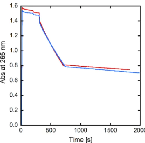
Based on the value of its redox potential, it was anticipated that 2+ should be resistant to the reduction
by ascorbate that shows an anodic potential under the very same recording conditions at 0.l V vs. SCE,[24] which is the very first step of ROS production by Cu(II) complexes. We confirmed this by measuring the catalytic consumption of Asc under different conditions. Either Cu(A) and 1+ (or
Cu(A) and LH as control) were first pre-mixed and then Asc was added (Figure S10) or Cu(A) was reacted with Asc and then 1+ (or LH as control) was added (Figure 3), according to a previously
published procedure.[24-25] The absence of Asc consumption or its preclusion was observed, respectively. This indicates that 1+ is able to remove Cu(II) from A leading to 2+, which is not
competent for Asc oxidation. Asc oxidation is directly associated to the production of ROS, since it is the (physiological) reductant required to fuel the reduction of dioxygen to O2•-, H2O2 and HO•.[3] In addition, release of HO• by the Cu(A)/Asc/O2 catalytic system has been monitored using the 3-CCA fluorescence assay (Figure S11).[26] Results parallel those obtained in the Asc consumption test indicating the prevention of HO• release when 1+ is added to the mixture This important observation
indicates that both 1+ and LH are thermodynamically and kinetically competent for the swapping and
removal of Cu(II) bound to A. This awakens hope of possible usage of 1+ or LH for controlling
Figure 3. Kinetics of ascorbate consumption, followed by UV-visible spectroscopy at 265 nm with subtraction of the background signal at 800 nm. Asc + Aβ40 + Cu(II) + LH (red curve), Asc + Aβ40 + Cu(II) + 1+ (blue curve). LH or 1+ are added at t = 600 s. [Aβ40] = [LH] = [1+] = 12 µM, [Cu(II)] = 10
µM, [HEPES] = 100 mM, T = 25°C, pH = 7.1 (For source and preparation of Aβ40, see Supporting Information). For negative (Asc + Aβ40 + Cu(II)) and positive (Asc) controls, see Figure S10.
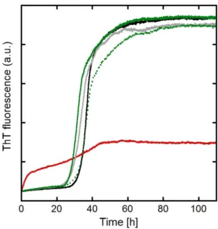
The aggregation of the disordered A peptide into fibrillary structures present in the senile plaques of AD brains is also a key event in the aetiology of AD and Cu(II) has been shown to impact A aggregation.[2b, 27] Hence we probe the activity of 1+ and LH on the Cu(II)-mediated formation of small
A aggregates. The kinetic monitoring of A aggregation has been performed using the classical ThT fluorescence assay, where ThT is a dye that acquires a strong fluorescence upon binding to fibrillary -sheet structure (Figure 4).[28] Mn(II) has no significant impact on the aggregation of A (within the error bar of the experiment). In contrast, Cu(II) strongly modifies the aggregation kinetic profile. The sigmoid-shape curve obtained in case of apo-A is no more observed. Instead, an ill-defined process leading to a weak maximum fluorescence value -about 1/3 of the maximum fluorescence obtained for the apo peptide- is detected. This indicates that Cu(II) strongly alters the aggregation process of A, in line with previous reports.[27e] Upon addition of either 1+ or LH along with Cu(II), the Cu(II)-altered
A aggregation is no longer observed and the apo type curve is recovered (see Figure 4). Note that weak differences observed between the curves thus obtained and the control ones are not significant. Morphologies of the aggregates were imaged by TEM (Figure S12). Apo and Mn(II) A aggregates show fibrillary structures with a more dense packing of the fibrils in presence of Mn(II). In contrast, CuII(A) aggregates resemble more proto-fibrils, with aggregates of shorter less defined fibrillary structure, the impact of Cu(II) on aggregates morphology being in line with recent reports.[27b-d] In presence of 1+, dense islets of fibres are observed, reminiscent of what was obtained with Mn(II) type
fibrils, while in presence of LH only, apo-type are observed. These observations are consistent with the swapping process in the case of Cu(A) + 1+.
In the present work, we have proposed a new approach in the context of AD based on the use of a pro-drug candidate complex 1+. The use of a Mn salen-based SOD mimic was recently reported in the AD
context. This Mn salen-based complex has the property to fight against the ROS produced by the Cu(A) complex.[29] In our case, 1+ undergoes a transmetallation process by releasing Mn(II) and
retrieving Cu(II) from A and the formed complex 2+ is unable to produce ROS on its own, thus
supressing the Cu(A) associated ROS production. Furthermore, we have developed here a pro-drug approach. Indeed, while LH is toxic towards epithelial cells as a free ligand, the toxicity is abolished when LH is bound to Mn(II) (in complex 1+).[15] Hence, the use of a potentially toxic ligand to remove Cu(II) from A is replaced by the use of 1+ which is non-toxic and leads to the very same retrieval
effects. It is worth noting here that we also studied another Mn(II) SOD mimic [Mn(IPG)] (IPG = N-[(1-methyl-imidazol-2-yl)methyl]-N-(2-pyridylmethyl)glycinate.[19, 30] Like 1+, [Mn(IPG)] is able to
exchange its Mn(II) centre with Cu(A) but the CuII(IPG) complex formed does produce ROS (more than Cu(A)) (Figure S13). This illustrates the importance of the nature of the resulting Cu(II) complex obtained after the metal exchange with A and shows that this strategy can not be enlarged to any SOD mimic. To go further, it will be important to consider the overall speciation of the metal ions and ligands in intricate biological environments. First results have shown that 1+ is stable for at least
15 hours in a culture medium[15] indicating that no exchange with other metal cations is proceeding, consequence of the tight control of the speciation of endogenous metal cations. In the AD context and as recently pointed out, the other cation easily available for exchange is Zn since it is loosely bound in the synaptic cleft.[25, 31] Studies are currently in progress to address the selectivity of 1+ for Cu(II)
versus Zn.
In summary, 1+ is a new inorganic pro-drug candidate that has previously shown an anti-oxidant
activity in biological media and a potential therapeutic activity against inflammatory bowel disease in mice.[15] 1+ has three key properties: (i) it masks the toxicity of the parent ligand LH, (ii) it is a SOD
mimic able to fight against O2•-,[5b, 13-15] (iii) it can prevent the production of ROS by Cu(A) and Cu(II)-altered aggregation of A. Because ROS are not exclusively produced by the Cu(A) in biological media, there would be a significant added value in observing both properties (ii) and (iii) in biologically relevant environments (achieved when the local concentration of 1+ exceeds that of
Cu(A)). As a consequence, future works will question the ability of 1+ against Cu-induced toxicity in
AD cellular and animal models and include further functionalization of the LH ligand to improve the for blood brain barrier penetration of 1+.
Figure 4. Aggregation assays following by ThT fluorescence. Aβ40 (black curve), Aβ40 + Cu(II) (red curve), Aβ40 + Mn(II) (light grey curve), Aβ40 + Cu(II) + LH (dotted green curve), Aβ40 + Cu(II) + 1+ (green curve). [Aβ40] = [LH] = [1+] = 20 µM, [Cu(II)] = [Mn(II)] = 18 µM, [phosphate buffer] = 50
mM, [NaCl] = 100 mM, [ThT] = 10 µM, T = 37°C, pH = 7.1 (For source and preparation of Aβ40, see Supporting Information).
Acknowledgements
The authors thank Dr. Djamila Guettas for their participation in experiments required for revision of the manuscript. This research was supported by the ERC StG 638712.
References
[1] D. M. Holtzman, J. C. Morris and A. M. Goate, Sci. Transl. Med. 2011, 3, 77sr71.
[2] a) E. Atrian‐Blasco, P. Gonzalez, A. Santoro, B. Alies, P. Faller and C. Hureau, Coord. Chem. Rev. 2017; b) J. H. Viles, Coord. Chem. Rev. 2012, 256, 2271‐2284. [3] C. Cheignon, M. Tomas, D. Bonnefont‐Rousselot, P. Faller, C. Hureau and F. Collin, Redox Biology 2018, 14, 450‐464. [4] a) C. Cheignon, M. Jones, E. Atrian‐Blasco, I. Kieffer, P. Faller, F. Collin and C. Hureau, Chem. Sci. 2017; b) L.‐E. Cassagnes, V. Hervé, F. Nepveu, C. Hureau, P. Faller and F. Collin, Angew. Chem., Int. Ed. Engl. 2013, 52, 11110‐11113. [5] a) M. Hayyan, M. A. Hashim and I. M. AlNashef, Chem. Rev. 2016, 116, 3029‐3085; b) C. Policar in Mimicking SODs, Why and How: Bio‐Inspired Manganese Complexes as SOD Mimics, Eds.: J. S. Reboucas, I. Batinic‐Haberle, I. Spasojevic, D. S. Warner and D. St. Clair), Springer, 2016, pp. 125‐164; c) Y. Sheng, I. A. Abreu, D. E. Cabelli, M. J. Maroney, A.‐F. Miller, M. Teixeira and J. S. Valentine, Chem. Rev. 2014, 114, 3854‐3918.
[7] K. Murakami, N. Murata, Y. Noda, S. Tahara, T. Kaneko, N. Kinoshita, H. Hatsuta, S. Murayama, K. J. Barnham, K. Irie, T. Shirasawa and T. Shimizu, J. Biol. Chem. 2011, 286, 44557‐44568.
[8] I. Batinic‐Haberle, A. Tovmasyan, E. R. Roberts, Z. Vujaskovic, K. W. Leong and I. Spasojevic, Antioxid. Redox Signal. 2014, 20, 2372.
[9] a) I. Batinic‐Haberle, S. Reboucas Julio and I. Spasojevic, Antioxid. Redox Signal. 2010, 13, 877; b) I. Batinic‐Haberle, A. Tovmasyan, E. R. Roberts, Z. Vujaskovic, K. W. Leong and I. Spasojevic, Antioxid Redox Signal. 2014, 20, 2372‐2415; c) O. Iranzo, Bioorg. Chem. 2011, 39, 73‐87; d) S. Miriyala, I. Spasojevic, A. Tovmasyan, D. Salvemini, Z. Vujaskovic, D. St. Clair and I. Batinic‐Haberle, Biochim. Biophys. Acta 2012, 794‐814. [10] a) J. S. Derrick, J. Lee, S. J. C. Lee, Y. Kim, E. Nam, H. Tak, J. Kang, M. Lee, S. H. Kim, K. Park, J. Cho and M. H. Lim, J. Am. Chem. Soc. 2017, 139, 2234‐2244; b) M. R. Jones, C. Mu, M. C. P. Wang, M. I. Webb, C. J. Walsby and T. Storr, Metallomics 2015, 7; c) D. J. Hayne, S. Lim and P. S. Donnelly, Chem. Soc. Rev. 2014, 43, 6701‐6715; d) F. Collin, I. Sasaki, H. Eury, P. Faller and C. Hureau, Chem. Commun. 2013, 49, 2130‐2132. [11] A. E. Stacy, D. Palanimuthu, P. V. Bernhardt, D. S. Kalinowski, P. J. Jansson and D. R. Richardson, J. Med. Chem. 2016, 59, 4965‐4984. [12] E. J. McAllum, B. R. Roberts, J. L. Hickey, T. N. Dang, A. Grubman, P. S. Donnelly, J. R. Liddell, A. R. White and P. J. Crouch, Neruobiol. Dis. 2015, 81, 20‐24. [13] F. Cisnetti, A. S. Lefevre, R. Guillot, F. Lambert, G. Blain, E. Anxolabéhère‐Mallart and C. Policar, Eur. J. Inorg. Chem. 2007, 4472‐4480. [14] A.‐S. Bernard, C. Giroud, H. Y. V. Ching, A. Meunier, V. Ambike, C. Amatore, M. Guille Collignon, F. Lemaître and C. Policar, Dalton Trans. 2012, 41, 6399‐6403.
[15] E. Mathieu, A.‐S. Bernard, N. Delsuc, E. Quevrain, G. Gazzah, B. Lai, F. Chain, P. Langella, M. Bachelet, J. Masliah, P. Seksik and C. Policar, Inorg. Chem. 2017, 56, 2545‐2555.
[16] a) M. A. Santos, K. Chand and S. Chaves, Coord. Chem. Rev. 2016, 327‐328, 287‐303; b) J. S. Derrick and M. H. Lim, ChemBioChem 2015, 16, 887‐898; c) M. G. Savelieff, A. S. DeToma, J. S. Derrick and M. H. Lim, Acc. Chem. Res. 2014, 47, 2475‐2482; d) M. A. Telpoukhovskaia and C. Orvig, Chem. Soc. Rev. 2013, 42, 1836‐1846.
[17] H. Y. V. Ching, I. Kenkel, N. Delsuc, E. Mathieu, I. Ivanović‐Burmazovic and C. Policar, J. Inorg. Biochem. 2016, 160, 172‐179. [18] a) A. Conte‐Daban, V. Borghesani, S. Sayen, E. Guillon, Y. Journaux, G. Gontard, L. Lisnard and C. Hureau, Anal. Chem. 2017, 89, 2155‐2162; b) B. Alies, E. Renaglia, M. Rozga, W. Bal, P. Faller and C. Hureau, Anal. Chem. 2013, 85, 1501‐1508. [19] C. Policar, S. Durot, F. Lambert, M. Cesario, F. Ramiandrasoa and I. Morgenstern‐Badarau, Eur. J. Inorg. Chem. 2001, 1807‐1818. [20] C. Wallin, Y. S. Kulkarni, A. Abelein, J. L. Jarvet, Q. , B. Strodel, L. Olsson, J. Luo, J. P. S. Abrahams, S. B., P. M. Roos, S. C. Kamerlin, A. Gräslund and S. K. Wärmländer, J. Trace Elem. Med. Biol. 2016, 38, 183‐193. [21] F. Michel, F. Thomas, S. Hamman, C. Philouze, E. Saint‐Aman and J.‐L. Pierre, Eur. J. Inorg. Chem. 2006, 2006, 3684‐3696. [22] S. Noël, F. Perez, S. Ladeira, S. Sayen, E. Guillon, E. Gras and C. Hureau, J. Inorg. Biochem. 2012, 117, 322‐325. [23] J. Peisach and W. E. Blumberg, Arch. Biochem. Biophys. 1974, 165, 691‐708.
[24] A. Conte‐Daban, B. Boff, A. Candido Matias, C. N. Montes Aparicio, C. Gateau, C. Lebrun, G. Cerchiaro, I. Kieffer, S. Sayen, E. Guillon, C. Hureau and P. Delangle, Chemistry ‐ A European Journal 2017, 23, 17078‐17088. [25] A. Conte‐Daban, A. Day, P. Faller and C. Hureau, Dalton Transactions 2016, 45, 15671‐15678. [26] S. Chassaing, F. Collin, P. Dorlet, J. Gout, C. Hureau and P. Faller, Curr. Top. Med. Chem. 2012, 12, 2573‐2595. [27] a) C. J. Matheou, N. D. Younan and J. H. Viles, Biochem. J. 2015, 466, 233‐242; b) M. R. Jones, E. Mathieu, C. Dyrager, S. Faissner, Z. Vaillancourt, K. J. Korshavn, M. H. Lim, A. Ramamoorthy, V. Wee Yong, S. Tsutsui, P. K. Stys and T. Storr, Chem. Sci. 2017, 8, 5636‐5643; c) Y. Ji, H. J. Lee, M. Kim, G.
Nam, S. J. C. Lee, J. Cho, C. M. Park and M. H. Lim, Inorg. Chem. 2017, 56, 6695‐6705; d) J. T. Pedersen, J. Østergaard, N. Rozlosnik, B. Gammelgaard and N. H. Heegaard, J. Biol. Chem. 2011, 286, 26952‐26963; e) P. Faller, C. Hureau and O. Berthoumieu, Inorg. Chem. 2013, 52, 12193‐12206. [28] S. Noël, S. Cadet, E. Gras and C. Hureau, Chem. Soc. Rev. 2013, 42, 7747‐7762. [29] G. R. Walke, D. S. Ranade, A. M. Bapat, R. Srikanth and P. P. Kulkarni, Chemistry Select 2016, 1, 3497‐3501. [30] C. Policar, F. Lambert, M. Cesario and I. Morgenstern‐Badarau, Eur. J. Inorg. Chem. 1999. [31] E. Atrian‐Blasco, A. Conte‐Daban and C. Hureau, Dalton Transactions 2017, 46, 12750‐12759.
![Figure 1. Scheme of the ligand LH (left) and X-Ray structure of [(Cu)LH] 2+ (2H 2+ , right)](https://thumb-eu.123doks.com/thumbv2/123doknet/13750988.437711/4.892.157.738.128.301/figure-scheme-ligand-lh-left-ray-structure-right.webp)
![Figure 2. 65 Cu EPR signatures of a) Cu(Aβ16), b) 2 + , c) Cu(Aβ16) + 1 + . [ 65 Cu] = [Mn(II)] = 180 µM, [Aβ16] = [LH] = [1 + ] = 200 µM, [HEPES] = 50 mM, pH = 7.1](https://thumb-eu.123doks.com/thumbv2/123doknet/13750988.437711/6.892.118.491.106.532/figure-cu-epr-signatures-aβ-aβ-aβ-hepes.webp)