HAL Id: hal-01108713
https://hal.archives-ouvertes.fr/hal-01108713
Submitted on 27 May 2020
HAL is a multi-disciplinary open access
archive for the deposit and dissemination of
sci-entific research documents, whether they are
pub-lished or not. The documents may come from
teaching and research institutions in France or
abroad, or from public or private research centers.
L’archive ouverte pluridisciplinaire HAL, est
destinée au dépôt et à la diffusion de documents
scientifiques de niveau recherche, publiés ou non,
émanant des établissements d’enseignement et de
recherche français ou étrangers, des laboratoires
publics ou privés.
Copyright
selenium-binding and reducing site in Arabidopsis
thaliana homologue to mammals selenium-binding
protein 1.
Florie Schild, Sylvie Kieffer-Jacquinot, Andrés Palencia, David Cobessi,
Géraldine Sarret, Chloé Zubieta, Agnés Jourdain, Renaud Dumas, Vincent
Forge, Denis Testemale, et al.
To cite this version:
Florie Schild, Sylvie Kieffer-Jacquinot, Andrés Palencia, David Cobessi, Géraldine Sarret, et al..
Biochemical and biophysical characterization of the selenium-binding and reducing site in
Ara-bidopsis thaliana homologue to mammals selenium-binding protein 1.. Journal of Biological
Chem-istry, American Society for Biochemistry and Molecular Biology, 2014, 289 (46), pp.31765-31776.
�10.1074/jbc.M114.571208�. �hal-01108713�
Biochemical and Biophysical Characterization of the
Selenium-binding and Reducing Site in Arabidopsis thaliana
Homologue to Mammals Selenium-binding Protein 1
*
□SReceived for publication, April 16, 2014, and in revised form, September 17, 2014Published, JBC Papers in Press, October 1, 2014, DOI 10.1074/jbc.M114.571208
Florie Schild‡, Sylvie Kieffer-Jaquinod§, Andrés Palencia¶, David Cobessi储1, Géraldine Sarret**1, Chloé Zubieta‡1, Agnès Jourdain‡1, Renaud Dumas‡1, Vincent Forge‡‡, Denis Testemale§§, Jacques Bourguignon‡,
and Véronique Hugouvieux‡2
From the‡Institut de Recherches en Technologies et Sciences pour le Vivant, Laboratoire de Physiologie Cellulaire et Végétale, CEA, Université Grenoble Alpes, CNRS UMR5168, INRA USC1359, the§Institut de Recherches en Technologies et Sciences pour le Vivant, Laboratoire de Biologie à Grande Echelle, Université Grenoble Alpes, CEA, INSERM, 17 rue des Martyrs, F-38000 Grenoble, France, the¶European Molecular Biology Laboratory Outstation, 71 avenue des Martyrs, F-38042 Grenoble, France and Unit for Virus Host-Cell Interactions, Université Grenoble Alpes-EMBL-CNRS, 71 avenue des Martyrs, 38042 France, the储Université Grenoble Alpes, CEA, CNRS, Direction des Sciences du Vivant, Institut de Biologie Structurale, 6 rue Jules Horowitz, F-38044 Grenoble, France, the
**Université Grenoble Alpes, CNRS & IRD, ISTerre, BP 53, F-38041 Grenoble, France, the‡‡Laboratoire de Chimie et Biologie des Métaux, Université Grenoble Alpes, CEA, CNRS, Institut de Recherches en Technologies et Sciences pour le Vivant, 17 rue des Martyrs, F-38000 Grenoble, France, and the§§Université Grenoble Alpes, CNRS, Institut NEEL, 25 rue des Martyrs,
F-38042 Grenoble, France
Background:The selenium-binding site in selenium-binding protein (SBP) homologues was not identified.
Results:The Arabidopsis thaliana SBP1 selenium-binding site was characterized as a R-S-Se(II)-S-R-type complex involving Cys21and Cys22.
Conclusion:This is the first identification of the selenium-binding site in any SBP.
Significance:It is an important step toward a better understanding of the link between selenium binding and function of SBP.
The function of selenium-binding protein 1 (SBP1), present in almost all organisms, has not yet been established. In mam-mals, SBP1 is known to bind the essential element selenium but the binding site has not been identified. In addition, the SBP family has numerous potential metal-binding sites that may play a role in detoxification pathways in plants. In Arabidopsis
thali-ana, AtSBP1 over-expression increases tolerance to two toxic
compounds for plants, selenium and cadmium, often found as soil pollutants. For a better understanding of AtSBP1 function in detoxification mechanisms, we investigated the chelating properties of the protein toward different ligands with a focus on selenium using biochemical and biophysical techniques. Thermal shift assays together with inductively coupled plasma mass spectrometry revealed that AtSBP1 binds selenium after incubation with selenite (SeO32ⴚ) with a ligand to protein molar
ratio of 1:1. Isothermal titration calorimetry confirmed the 1:1 stoichiometry and revealed an unexpectedly large value of bind-ing enthalpy suggestbind-ing a covalent bond between selenium and
AtSBP1. Titration of reduced Cys residues and comparative
mass spectrometry on AtSBP1 and the purified
selenium-AtSBP1 complex identified Cys21and Cys22as being responsible for the binding of one selenium. These results were validated by site-directed mutagenesis. Selenium K-edge x-ray absorption
near edge spectroscopy performed on the selenium-AtSBP1 complex demonstrated that AtSBP1 reduced SeO32ⴚto form a
R-S-Se(II)-S-R-type complex. The capacity of AtSBP1 to bind different metals and selenium is discussed with respect to the potential function of AtSBP1 in detoxification mechanisms and selenium metabolism.
At low concentration, selenium is an essential nutrient to many organisms including some archaea, bacteria, protozoan, green algae, and nearly all animals but it is non-essential in land plants (1– 4). The daily selenium requirement in human adults is 60 to 70g and the main source of dietary selenium is plants (3, 5). Selenium dietary consumption has been associated with a reduced risk of many diseases such as cardiovascular diseases, diabetes, and cancer. In addition, a lack of selenium can lead to Keshin-Beck and Keshan disease that can be treated by sele-nium supplementation (3, 4). In organisms where selesele-nium is an essential nutrient, it is required for the biosynthesis of the selenoamino acid selenium-Cys, used for the translation of 25 selenoproteins, which are involved in critical functions such as redox reactions, free radical scavenging, and hormone regula-tion (1, 3, 4). In addiregula-tion to its role as a micronutrient, selenium can have toxic effects. Selenium toxicity (selenosis) can occur in some areas where exploitation of seleniferous soils or fossil fuels leads to toxic accumulation of selenium in the environ-ment and in plants. In mammals, excess selenium targets the cardiovascular, gastrointestinal, neurological, and hematopoi-etic systems (3, 5–7). As the line between selenium deficiency *This work was supported by the Rhône-Alpes region, the Biologie et
Ame´-lioration des Plantes Department of Institut National de la Recherche Agronomique, and the CEA Toxicology program.
□S This article containssupplemental Table S1.
1These authors contributed equally to this work.
2To whom correspondence should be addressed. Tel.: 33-4-38-78-06-54; Fax: 33-4-38-78 –5091; E-mail: Veronique.hugouvieux@cea.fr.
and toxicity is very narrow, both selenium deficiency and sele-nium toxicity are common problems worldwide.
Selenium concentration in soils ranges from 0.01 to 2 mg/kg and can be⬎10 mg/kg in seleniferous soils (3, 5, 8). When present in soil, selenium is absorbed and accumulated in plants and is subsequently disseminated along the whole food chain. In this context, plants may help to alleviate both selenium defi-ciency and toxicity problems. A better understanding of the mechanisms involved in the plant response to selenium includ-ing accumulation, protection, and sequestration can be consid-ered as one of the most important challenges in the coming decades. Engineering plants with nutrient-enriched content for biofortification, using plants to remove toxic selenium for phy-toremediation and selecting for selenium resistant plants are critical goals (5, 8).
The impact of selenium on plant physiology has been exten-sively studied. Selenium is not essential to land plants, unlike in mammals. Plant homologues to selenoproteins from mammals and bacteria have a Cys residue in their sequence instead of selenocysteine. However, low concentrations of selenium can have a positive effect on plant growth and be beneficial in facing biotic and abiotic stress (9, 10) notably by protecting plants against oxidative stress. At higher concentrations, selenium is generally highly toxic to plants.
The major forms of inorganic selenium in soils are Se(VI) (SeO42⫺) and Se(IV) (SeO32⫺), which are taken up by roots via
sulfate and phosphate transporters, respectively (6, 11). Once inside the cells, selenium toxicity results from its chemical sim-ilarity with sulfur that leads to nonspecific replacement of sul-fur containing amino acids with their seleno derivatives (7, 12, 13). In addition, at high concentrations, selenium triggers oxi-dative stress by reducing the pool of glutathione (GSH) (12). The main mechanisms of selenium tolerance in plants are the conversion of SeMet and SeCys into their methylated forms, which are non-incorporable into proteins, and volatilization (5, 8).
In addition to its incorporation into selenoproteins, selenium can be bound to proteins belonging to the selenium-binding protein family (SBP).3Many of the beneficial impacts of
sele-nium on mammalian health have been attributed to its role as a critical constituent of selenoproteins and to its binding to SBP1. Although the function of selenoproteins is well established, the activity of SBP1 proteins is still unclear and the link between selenium binding and SBP1 function has not yet been deter-mined. Mammalian SBP1 was first identified in mouse liver (14) in experiments designed to find new selenoproteins. Two homologues are present in humans. Today, SBP genes have been identified in many organisms including plants (14 –18) and additional sequences of SBP homologues are available in public databases from many additional organisms. In humans, down-regulation of SBP1 expression has been correlated with rapid tumor development in many organs (19 –27) and SBP1
expression is considered to be a predictor of clinical outcome. Recently, interaction of SBP1 and the selenoprotein glutathione peroxidase GPx-1 was observed, revealing a cross-talk between members of distinct families of selenium containing proteins (19, 21, 28). SBP1 was also characterized as a biomarker for schizophrenia as up-regulation of SBP1 is observed in the brains of patients with the disease (29, 30). Other functions, such as intra-Golgi protein transport have been assigned to mammalian SBP1 (31). Its SBP2 homologue was described as playing a protective role as a scavenger of toxic electrophiles or oxidant species (32–34).
In the Arabidopsis thaliana genome, 3 genes encoding SBP are present (16). AtSBP1 is the isoform that is the most highly expressed (35). One of the first functions that was assigned to SBP1 in plants was a putative role in selenium tolerance (15). Indeed, plants over-expressing AtSBP1 have increased resis-tance to selenite (SeO32⫺), whereas reducing AtSBP1 and AtSBP2 expression increased plant sensitivity to the toxic com-pound (15). AtSBP1 could therefore be involved in selenium metabolism but no reports were available in the literature on the ability of AtSBP1 to bind selenium like its mammalian homologues.
In addition to its putative role in selenium tolerance, AtSBP1 has been identified as a protein accumulating in response to the heavy metal cadmium in A. thaliana cultured cells using differ-ential proteomic analysis (36). Cadmium is toxic to most organ-isms and is one of the most toxic pollutants in the world. One of the main mechanisms that plants use to face cadmium toxicity is the synthesis of polymers of GSH, called phytochelatines, that chelate cadmium and are then transferred into the vacuoles (37). AtSBP1 shows the ability to bind 3 cadmium in vitro and AtSBP1 over-expression in A. thaliana seedlings led to enhanced tolerance to cadmium (35). This phenotype is more important in GSH- and phytochelatine-deficient Arabidopsis seedlings (35). Therefore, AtSBP1 may have chelating proper-ties in vivo toward cadmium and may represent a new detoxi-fication mechanism that plants use to face heavy metal toxicity, possibly throught direct binding to the metal (35). In addition to cadmium, AtSBP1 over-expressing A. thaliana plants showed increased tolerance to stress such as selenium and H2O2that also require GSH for detoxification (38). Overex-pression of OsSBP1 in rice enhanced tolerance to various pathogens (17) and the importance of GSH in plant defense to biotic stress has been recently reviewed (39). These results sug-gest that SBP1 may share similar functions with GSH in response to stress. In line with these results, stresses inducing
SBP1expression were also inducers of PRH43, which encodes 5⬘-adenylylphosphosulfate reductase 2, a key enzyme of the sul-fur assimilation pathway and GSH biosynthesis (38). As an example, AtSBP1 and AtPRH43 are induced by cadmium, SeO4
2⫺, H
2O2and sulfur starvation. An internal sulfur demand
of the cell could be a signal that triggers SBP1 expression in response to the different stresses (37) and this correlates well with the potential functional redundancy between SBP1 and GSH.
To date, all the data accumulated on SBP1 function in plants points to the importance of SBP1 in response to stress. The fact that AtSBP1 shows the ability to bind cadmium, a toxic metal, 3The abbreviations used are: SBP, selenium-binding protein; TSA, thermal
shift assay; ICP-MS, inductively coupled plasma mass spectrometry; XANES, K– edge X ray absorption near edge structure spectroscopy; DTNB, 5,5⬘-dithiobis-(2-nitrobenzoic acid); GSH, Glutathione; ITC, isothermal titration calorimetry.
in vitro, and that the mammalian homologue can bind selenium
in vivo, another toxic compound for plants, raises the question of whether AtSBP1 may have some chelating properties toward selenium as well as other metals in addition to cadmium. Because of the importance of selenium in human health, and the critical function of SBP1 in selenium tolerance in plants, we focused on the identification of the selenium-binding site in
AtSBP1. By using complementary biochemical, spectroscopic, and biophysical approaches, we characterized the selenium and metal binding properties of AtSBP1. These data identify, for the first time, the selenium-binding site in any SBP1 homologues. Its characterization provides evidence that two Cys residues are involved in selenium binding and that AtSBP1 reduces selenite (SeO32⫺). This work provides an important step toward a better
understanding of SBP1 function in selenium metabolism, detoxification, and accumulation mechanisms in plants.
EXPERIMENTAL PROCEDURES
Overexpression of AtSBP1 in Escherichia coli and Purification of the Recombinant Protein—AtSBP1 cDNA contained in the entry clone (U15803, TAIR database) was cloned into the des-tination pGEX-3X for GST-AtSBP1 production as previously described (35). The recombinant plasmid was used to trans-form E. coli strain Rosetta 2. Cell cultures were grown at 37 °C in Luria-Bertani medium until an A600 nmof 0.8. The tempera-ture was then lowered to 18 °C and expression of the recombi-nant protein was induced by adding 0.8 mMisopropyl-D -thio-galactopyranoside to the cultured cells for 17 h. Cells were then centrifuged and stored at⫺80 °C. For protein purification, cells were lysed by sonication (6⫻ 1 min, using the Branson Sonifier 250) in 20 mMHEPES, pH 7.4, 150 mMNaCl, 0.05% Triton
X-100 (v/v), 10% glycerol (v/v), and 1 mMDTT. The
recombi-nant GST-SBP1 protein was purified via affinity chromatogra-phy using GSH-Sepharose 4B resin according to the manufac-turer’s instructions (GE Healthcare). The GST tag was removed by incubating GST-AtSBP1 with Factor Xa (Sigma) for 15 h at 22 °C in 50 mMTris, pH 8.0, 150 mMNaCl, and 1 mMCaCl2.
Factor Xa was depleted by binding to p-aminobenzamidine-agarose (Sigma). AtSBP1 was further purified using size exclu-sion chromatography on an AKTA purifier system (Amersham Biosciences) and an S200 16/60 column (HiLoad 16/60, Super-dex 200, Amersham Biosciences). Unless otherwise stated, all the experiments were performed using cleaved AtSBP1.
Site-directed Mutagenesis—Site-directed mutagenesis were produced from the template GST-AtSBP1 contained in the PGEX-3X vector. Primer sequences were designed using QuikChange Primer Design software from Agilent Technology. Cys21and Cys22of AtSBP1 were replaced by Ser residues. PCR
was performed using Phusion enzyme with 18 cycles as follow: 30 s at 98 °C, 1 min at 55 °C, and 1 min 40 s at 72 °C. PCR products were incubated 1 h at 37 °C with DpnI and used to transform E. coli strain DH5␣. Plasmids were extracted with Nucleospin Plasmid kit and sequenced by Eurofins (Les Ulis, France).
Stability Analysis of Recombinant AtSBP1 by Thermal Shift Assay (TSA)—Temperature stability analysis was conducted with recombinant AtSBP1 protein using 96-well plates. AtSBP1 (5M) was incubated with Sypro Orange (diluted 1/1000 from
S6650 solution; Invitrogen) as described (40) and 2 concentra-tions of Cd2⫹, SeO32⫺, Zn2⫹, Ni2⫹, Co2⫹, Cu2⫹, SeO42⫺, Mn2⫹,
Mg2⫹, and MoO4
2⫺ ranging from 1 to 25M(prepared from
Cd(NO3)2, Na2SeO3, ZnCl2, NiCl2, Co(NO3)2, CuCl2,
Na2SeO4, MnCl2, MgCl2, and Na2MoO4, respectively) in 50 mM
HEPES, 150 mMNaCl, pH 7.4, overnight at 4 °C. Experiments
were performed in a final volume of 25l. Samples were heated from 25 to 75 °C with a rate of 1 °C per min using the thermo-cycler Stratagene Mx3005P. Excitation and emission wave-lengths were 492 and 516 nm, respectively. The melting tem-perature corresponded to the temtem-perature indicated at half of the⌬fluorescence (final fluorescence ⫺ initial fluorescence).
Metal and Selenium Binding Assay by Inductively Couple Mass Spectrometry (ICP-MS)—To determine the metal or sele-nium to protein molar ratio, 2 nmol of recombinant AtSBP1 protein were incubated for 15 min at 25 °C with 25 nmol of Cd2⫹, Zn2⫹, Ni2⫹, or SeO
3
2⫺in a total volume of 25l
contain-ing 10 mMHEPES, pH 7.4, and 150 mMNaCl. AtSBP1-bound
species were separated from free ions by steric exclusion chro-matography through a Sephadex G-25 column (0.5⫻ 8.5 mm) with an elution rate of 150l/min. Fractions of 200 l were collected. Protein elution was followed using a spectrophotom-eter (NanoDrop 2000, Thermoscientific) at 280 nm, and metal and selenium content was assayed by inductively coupled plas-ma-mass spectrometry (ICP-MS; HP4500 Chemstation; Yok-ogawa Analytical System). Isotopes 64, 66, 77 and 82, and 112 were monitored for zinc, nickel, selenium, and cadmium quan-tification, respectively. ICP-MS experiments were performed in 0.1% HNO3(v/v).
Isothermal Titration Calorimetry (ITC) Experiments and Determination of Thermodynamic Parameters— Determina-tion of Se-AtSBP1 thermodynamic parameters was carried out by calorimetric experiments with recombinant GST-AtSBP1 and GST alone. SeO32⫺(0.5 and 2 mM) was added by successive
injections (1.5l) to GST-AtSBP1 (50 M) in a total volume of 0.22 ml. Buffer conditions were 10 mMHEPES, pH 7.4, and 150
mMNaCl. Calorimetric titrations were performed at 25 °C with
stirring at 800 rpm using a microcalorimeter (Microcal ITC 200 System, GE Healthcare). Data were analyzed with Origin ITC 200 software. Independent duplicates were performed with GST-AtSBP1. ITC experiments with GST alone were run in parallel to confirm that GST alone did not bind SeO32⫺. A
fur-ther experiment was conducted with cleaved AtSBP1 that con-firmed results obtained with GST-AtSBP1.
Cysteine Quantification Using DTNB—For reduced cysteine titration, AtSBP1 (8 M) was incubated for 30 min at room
temperature in the dark with 5,5⬘-dithiobis-(2-nitrobenzoic acid) (DTNB) (1.6 mM) in a total volume of 60l containing 10
mMHEPES, pH 7.4, and 150 mMNaCl. Cysteine quantification
was determined by measuring the absorbance at 412 nm using a spectrophotometer (NanaDrop 2000, Thermo Scientific). For buried cysteine quantification, AtSBP1 was unfolded in urea (8
M). For cysteine quantification after selenium binding, AtSBP1
was incubated with the SeO32⫺(100M) for 15 min at 25 °C
prior to DTNB treatment.
Secondary Structure Modifications after Incubation of AtSBP1 with SeO32⫺ using Circular Dichroism (CD)—AtSBP1
total volume of 1.5 ml containing 10 mMHEPES, pH 7.4, and
150 mMNaCl. Spectra acquisition was performed at 25 °C using
a spectropolarimeter (J-815, Jasco) in the far UV (200 –260 nm) with the following parameters: 1 nm step, 2 nm bandwidth, and scan speed 200 nm/min. The optical path length was 1 mm.
Model Building—The three-dimensional model was gener-ated using Chimera and Modeler version 9.10. The sequence of At4g14030 was aligned with sequences from the Protein Data Bank using Blast and Blosum62 as the alignment matrix. The structure of the hypothetical selenium-binding protein from
Sulfolobus tokodaii(PDB entry 2ECE) was the closest homo-logue and used to generate a three-dimensional model with the program Modeler (E-value of 7e-86; 40% sequence identity). The model of At4g14030 with the lowest discrete optimized protein energy score was selected.
Infusion MS Analysis of AtSBP1 and Selenium-AtSBP1 Complex—After incubation of AtSBP1 with SeO32⫺, fractions
containing selenium-bound AtSBP1 and native AtSBP1 were purified by pure water exchange using the Vivaspin 500 (Sarto-rius) system and analyzed by nano-electrospray in direct injec-tion at a concentrainjec-tion of 1M. MS analysis was performed
within the Linear Trap Quadrupole of a LTQ-Orbitrap-XL. The theoretical average mass was calculated using the Isotope simulation from Xcalibur 2.2 (Thermo Scientific) in the profile mode and using a resolving power of 1000. Multicharged spec-tra were externally recalibrated and deconvoluted using the Hyper Mass transform algorithm implemented in ICR-2LS (PNNL, Richland, WA).
Nano-LC-MS/MS Analysis of Chymotrypsic Peptides of AtSBP1 and Selenium-AtSBP1 Complex—After incubation of GST-AtSBP1 with SeO3
2⫺, fractions containing
selenium-bound GST-AtSBP1 or GST-AtSBP1 alone were digested with diluted chymotrypsin in 25 mMammonium bicarbonate (100
ng for 2g of GST-SBP1) for 15 h at 37 °C. Digested peptides were dried under vacuum on a centrifugal evaporator. One hundred ng of the dried extracted peptides were solubilized in water containing 5% acetonitrile and 0.1% formic acid before being transferred to a glass vial for a nano-LC-MS/MS analysis (Ultimate 3000, Dionex and LTQ Orbitrap Velos Pro, Thermo-Fisher Scientific). The LC method consisted of a 30-min sepa-ration at a flow rate of 300 nl/min using a binary solvent gradi-ent: A (2% acetonitrile and 0.1% formic acid in water) and B (80% acetonitrile and 0.1% formic acid in water). The system included a 300m ⫻ 5-cm PepMap C18 precolumn for pre-concentration and desalting of the peptides and a 75m ⫻ 15-cm PepMap C18 column (Dionex) for peptide separation and elution. MS and MS/MS data were acquired using XCalibur software (ThermoFisher Scientific) in the positive ion electro-spray ionization mode with a resolution of 60,000 full-width half-maximum in the MS mode. Peak list generation was first performed using MASCOT Distiller and consecutive searches against a database containing the GST-SBP1 sequence were performed using MASCOT 2.4. The parameters used with MS/MS transformed data (.mgf) were: instrument ⫽ ESI-TRAP; enzyme⫽ no; oxidation (M); selenium (CDHM); deami-nation (NQ); disulfide (C); peptide tolerance⫽ 10 ppm; frag-ment tolerance ⫽ 0.6 Da. Results were filtered using IRMa software (41). Every peptide having a Mascot score below 18
was filtered. The isotope simulation feature of Xcalibur was used to simulate the isotopic profiles of peptides of interest with and without selenium.
X-ray Absorption Spectroscopy on the Selenium-AtSBP1 Complex—X-ray absorption measurements were carried out at the European Synchrotron Radiation Facility (ESRF, Grenoble, France) at a ring current of 150 –200 mA. Spectra were col-lected on the BM30B (FAME) beamline (42) using a Si(220) double crystal monochromator with dynamic sagittal focusing. The photon flux was 1011photons/s and the spot size was 300
m horizontal ⫻ 100 m vertical (full width half-maximum values). Selenium K-edge x-ray absorption near edge structure (XANES) spectra were recorded for a 1.6 mMsolution of
puri-fied selenium-AtSBP1 in 10 mMHEPES, pH 7.4, 150 mMNaCl,
and 20% glycerol (v/v), and for various selenium reference com-pounds including SeO32⫺, SeS2, selenomethylcysteine
(seleni-um-Met-Cys, R-Se(II)-R), and selenomethionine (selenium-Met, R-Se(II)-R). Spectra for gray Se(0) and selenodiglutathione (selenium-diGSH, R-S-Se(II)-S-R) were recorded previously on the same beamline in the same experimental conditions (43). All solid compounds were diluted in Boron Nitride to reach 5000g g⫺1of selenium, and pressed as pellets. For the seleni-um-AtSBP1 sample, 40l of the solution was transferred to a five-cell sample holder with a kapton window, and flash frozen in liquid nitrogen. Solid pellets were loaded in the same sample holder, which was then transferred to a helium cryostat with temperature set around 10 K during data collection. For each sample, four to six scans of 20 min were averaged. The position of the beam on the pellet was moved between each scan to limit radiation damage. All spectra were collected in fluorescence mode measuring the selenium K␣ fluorescence with a 30-ele-ment solid-state Ge detector (Canberra). Energy calibration was achieved by measuring a selenium foil and assigning the first inflection point of the spectrum to 12658 eV for selenium. Data analysis was performed using Athena software. After nor-malization, the spectrum for the selenium-AtSBP1SBP1 com-plex was fitted by linear combination of selenium reference compounds in the 12,640 –12,700 eV range.
RESULTS
Analysis of AtSBP1 Thermostability and Chelating Properties Toward Different Metals and Selenium—To determine whether AtSBP1 was able to bind different ionic species, ther-mal shift assays were used to screen binding of different ions. As shown in Fig. 1, the Tmof AtSBP1 is about 45 °C. It reproducibly
shifts by about 3 °C in the presence of Cd2⫹and⬎3 °C in the
presence of Zn2⫹, Ni2⫹, Cu2⫹, and Co2⫹. A slight but very
reproducible enhanced Tm(around 1 °C) was observed in the
presence of Mo(VI) (MoO42⫺) at 25Mand Se(IV) (SeO 3 2⫺) at 5
M, but not with Se(VI) (SeO42⫺). We verified that the increased Tmwas correlated with binding of the ions to AtSBP1 by
deter-mining the ion to protein molar ratio by ICP-MS for SeO3 2⫺,
Cd2⫹, and two additional selected ionic species (Zn2⫹ and Ni2⫹) (Fig. 2). As shown in Fig. 2, AtSBP1 shows the ability to
bind one selenium in vitro when incubated with SeO3 2⫺, and
three Cd2⫹, three Ni2⫹and three Zn2⫹ ions. No binding of selenium was observed when incubated with SeO42⫺(data not
dif-ferent metal ions in vitro, its ability to bind selenite could be critical for its in vivo function, as AtSBP1 expression level reg-ulates selenium tolerance in A. thaliana (15). The chelating properties of AtSBP1 toward selenium were therefore investigated.
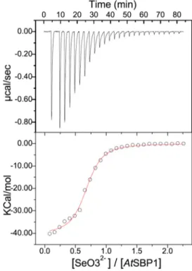
Thermodynamic Parameters of Selenium Interactions with AtSBP1—Isothermal titration calorimetry was used to study the binding of SeO32⫺ to AtSBP1. This technique allows the
determination, in one titration, of the stoichiometry, affinity, and enthalpic or entropic contributions to the Gibbs free energy. ITC curves and thermodynamic parameters obtained during the interaction of SeO3
2⫺and AtSBP1 are shown in Fig. 3
and Table 1, respectively. For each SeO32⫺injection, a release of
heat was observed (Fig. 3, upper panel) indicating a clear bind-ing event between SeO32⫺and AtSBP1. Furthermore, the
bind-ing isotherms could be fitted unambiguously to a one-site binding model (Fig. 3, lower panel), which allowed us to deter-minate an apparent binding constant in the low micromolar range (1.6M). We also estimated the number of sites as⬃0.75,
therefore confirming the 1:1 stoichiometry of the interaction determined by ICP-MS. Unexpectedly, the binding enthalpy value measured for SeO32⫺binding was⫺51,720 cal/mol, which
is extremely high only taking into account the limited number of polar interactions that a single ion could establish with
AtSBP1 (see for comparison examples of other ionic species and proteins (44 – 46)). Therefore, the strong negative enthalpy difference values obtained for SeO32⫺most likely correspond to
a combination of a binding event with a specific covalent reac-tion between the protein and SeO32⫺. This is also supported by
the fact that the calorimetric signals needed abnormally large times to recover to the baseline value (broadening of the peaks), suggesting that a covalent reaction was coupled with the bind-ing of the ion.
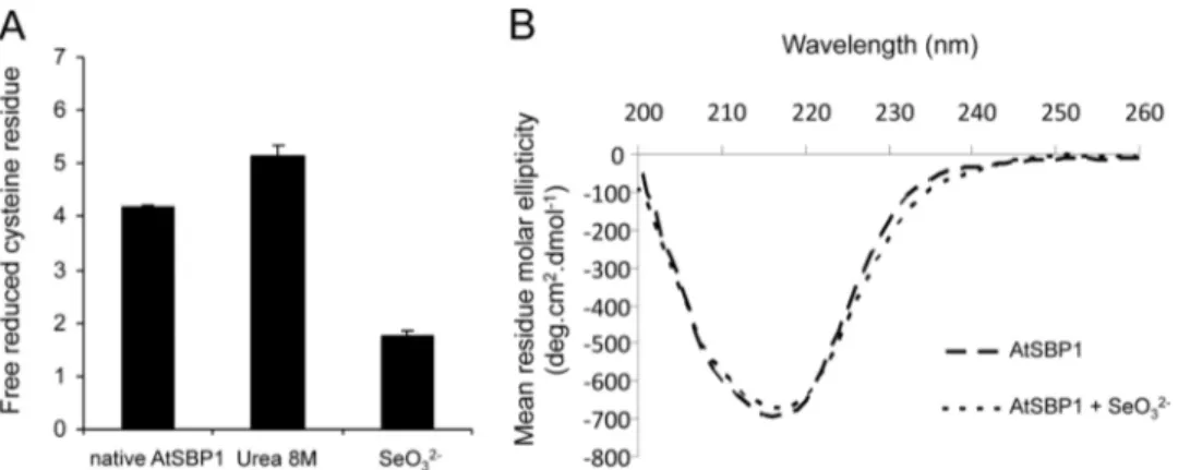
Impact of Selenium on Cysteine Titration and Secondary Structure of SBP1—Selenium can interact with cysteine (Cys) residues (47, 48). Titration of reduced and accessible Cys resi-dues was performed on AtSBP1 alone and AtSBP1 incubated with SeO32⫺ using DTNB (Fig. 4A). The AtSBP1 protein
sequence contains 7 Cys residues. In the native AtSBP1 protein, FIGURE 1. Identification of ions that stabilize A. thaliana SBP1. AtSBP1
melting temperature was determined by TSA experiments. AtSBP1 (5M) was incubated with Sypro Orange and Cd2⫹, SeO32⫺, Zn2⫹, Ni2⫹, Co2⫹, SeO42⫺, Mn2⫹, Mg2⫹, and MoO42⫺at 5 (C1) or 25M(C2) and Cu2⫹at 1 (C1) and 5M (C2). Each experiment of TSA was performed in duplicates providing two sets of values per ion concentration. Data show the mean⫾ S.D. of two indepen-dent TSA experiments. *, represents statistical difference compared with con-trol condition with no ion evaluated by Student’s t test.
FIGURE 2. In vitro binding capacity of A. thaliana SBP1 toward SeO3 2ⴚ, Cd2ⴙ, Zn2ⴙ, and Ni2ⴙ. Two nmol (80M) of recombinant AtSBP1 were incu-bated with 25 nmol (1 mM) of SeO32⫺, Cd2⫹, Zn2⫹, and Ni2⫹in a final volume of 25l. Ligand-bound AtSBP1 was separated from the free ligand by steric exclusion chromatography on a Sephadex G-25 column. The concentration of the eluted SBP1 was followed by measuring the A280(white square), and ligand content was measured by ICP-MS (black square). A, representative elu-tion profiles are shown for SeO32⫺and Cd2⫹. B, values of the ligand/AtSBP1 molar ratio evaluated on 3 independent experiments.
FIGURE 3. Isothermal titration calorimetry experiments showing the binding of SeO3
2ⴚto A. thaliana SBP1. Top panel shows titration of SeO 3 2⫺at 0.5 mMinto AtSBP1 at 50Mplaced at the sample cell. Bottom panel shows the ligand concentration dependence of the heat released upon binding after normalization. The data were fitted to one-site binding model.
4 free accessible Cys residues were titrated and 5 in the urea-treated protein indicating that one reduced Cys residue was therefore not accessible and two others would be involved in a disulfide bridge. In the presence of SeO32⫺, only two free
acces-sible Cys residues were detected, indicating that selenium bind-ing was strongly stable and would involve two accessible and reduced Cys residues.
To determine whether selenium binding results in changes to the protein secondary structure, circular dichroism (CD) spectra were recorded in the far-UV with AtSBP1 and seleni-um-bound AtSBP1 (Fig. 4B). The minimum at 217 nm indicates that the secondary structure in the two cases is predominantly -sheet (49). The binding of selenium has no large impact on the far-UV CD spectra, indicating that the overall secondary structure is not changed by the presence of SeO32⫺. However,
small changes can be observed around 230 nm that could be due either to Met or Cys side chains (50, 51), involved in local struc-tures upon ion binding. However, it is not possible to distin-guish between these two types of amino acids because they provide very similar negative signals centered on 230 nm (51). These results are well correlated with DTNB analysis suggest-ing that two Cys residues are involved in selenium bindsuggest-ing.
Mass Analysis of AtSBP1 Alone and Purified Selenium-AtSBP1 Complex by Nano-electrospray MS Analysis—To help identify the Cys residues involved in selenium binding, the sele-nium-AtSBP1 complex was investigated by mass spectrometry. Direct analysis of the intact (i.e. undigested) protein was per-formed after incubation with and without SeO32⫺, and
charge-state profiles were compared. The average mass calculated after deconvolution for AtSBP1 or selenium-AtSBP1 is presented in Fig. 5. The calculated mass was 54,184 Da for AtSBP1 (Fig. 5A), which correlates well with a protein folded with a disulfide bridge (⫺2 Da; the expected mass of the protein is 54,186 Da without a disulfide bridge). These results are supported by the DTNB analysis. In the case of selenium-AtSBP1 mass analysis, two major species were observed after deconvolution (Fig. 5B). The first peak (the minor one), with a mass of 54,185 Da, cor-responds to a portion of residual protein with no bound sele-nium, whereas the second peak (the major one) corresponds to the selenium-bound protein with a mass of 54,262 Da. The dif-ference in mass between AtSBP1 and selenium-bound AtSBP1 was 77 Da. This correlates well with the average mass of sele-nium (78.9 Da) minus the mass of 2 protons indicating that
AtSBP1 reduced SeO32⫺and lost 2 protons from 2 Cys residues.
These results further demonstrated the selenium/AtSBP1 molar ratio was around 1 as shown by ICP-MS and ITC. Fur-thermore, these data support the ITC experiments that strongly suggested a covalent binding between selenium and AtSBP1.
Sequence Alignment of SBP Proteins and Three-dimensional Model Analysis of AtSBP1 to Identify Potential Cys Residues Involved in selenium Binding—An alignment of SBPs from diverse organisms together with the 3 SBPs from A. thaliana is shown in Fig. 6. Among the three Cys residues that are 100% conserved (Cys97, Cys100, and Cys158; numbering as per AtSBP1), Cys97and Cys100belong to a CXXC motif putatively
involved in metal and cadmium binding (37, 52–54). Cys484is
highly conserved and only absent in S. tokodaii and Cys168is
conserved in all photosynthetic organisms (Fig. 5). Two addi-tional Cys residues (Cys21and Cys22) are conserved in all the photosynthetic organisms, and they aligned with a CXXC motif in mammals. We also investigated the three-dimensional struc-ture of AtSBP1 to check whether two Cys residues not adjacent in the sequence could come into contact due to protein folding. The three-dimensional model of AtSBP1 was modeled using the three-dimensional structure of SBP from S. tokodaii (PDB entry 2ECE) (Fig. 7). The model contains residues from Tyr29to
Asp487, however, the first 28 residues could not be modeled as they are predicted to be disordered and are not present in the crystal structure. The CD analysis showed that AtSBP1 second-ary structure was predominantly-sheet, which correlates well with the predicted overall fold of AtSBP1 that is a-propeller containing 7-sheets, each one composed of four antiparallel -strands (Fig. 7). DTNB (Fig. 4A) and nano-electrospray MS analysis (Fig. 5) indicated the presence of a disulfide bridge that the model predicted between Cys97and Cys158(Fig. 7). DTNB analysis also suggested that four cysteine residues would be reduced and accessible. Based on the model, Cys168would be
buried, whereas Cys21, Cys22, Cys100, and Cys484 were good
candidates to be reduced by DTNB in our experiment. We therefore predicted that in addition to Cys21and Cys22, Cys100
and Cys484could be involved in selenium binding.
Identification of AtSBP1 Amino Acids Involved in Selenium Binding by Nano-LC-MS/MS Analysis—To identify the peptide and residues involved in selenium binding in AtSBP1, fractions containing selenium-bound AtSBP1 and AtSBP1 alone were analyzed by nano-LC-MS/MS after a chymotryptic digestion made in solution. The chymotryptic peptide “TMATETE-VVAPVTVSNGGSKGCCKY” that contains the two adjacent Cys residues (Cys21and Cys22) was clearly identified as carrying
a potential (⫹selenium, ⫺2H) modification (3 spectra with Mascot score of 33, 39, and 60 identify this modified peptide) (Fig. 8, supplemental Table S1). The same analysis was con-ducted on a mutated form of the protein where Cys21and Cys22
were replaced by serine (supplemental Table S1). The mutated form of the peptide “TMATETEVVAPVTVSNGGSKGSSKY” did not show any binding with selenium. The involvement of Cys21and Cys22in selenium binding was further confirmed by
direct measurements of selenium by ICP-MS, on the wild type and mutated version of the protein after incubation with sele-nium and protein purification. Altogether, these data indicate that selenium binding to AtSBP1 involved Cys21and Cys22.
Identification of the Local Selenium Environment in AtSBP1 by XANES Analysis—The oxidation state and local environ-ment of selenium in AtSBP1 was further investigated by x-ray absorption spectroscopy. Fig. 9 compares the selenium K-edge XANES spectra for the selenium-AtSBP1 complex and for sele-TABLE 1
Thermodynamic parameters calculated from microcalorimetric experiments for the interaction between SeO3
2ⴚand A. thaliana SBP1
at 25 °C in 10 mMHEPES, pH 7.4, 150 mMNaCl
Independant experiments were carried out with 2 and 0.5 mMsolutions of SeO32⫺
titrated to AtSBP1 at 50M. Thermodynamic parameters represent the average from at least two independent experiments (estimated errors are about 5%) obtained by fitting the data to a one-site binding model. Parameters were calculated with the software Origin ITC 200. K represents the apparent binding constant.
K (Mⴚ1) ⌬G (cal/mol) ⌬H (cal/mol) ⴚT⌬S (cal/mol) ITC moyen 609,500 ⫺7,753 ⫺51,720 43,955
nium reference compounds, including SeO3
2⫺, some organic
forms of selenium, SeS2, and elemental Se(0). The shift in
energy of the main peak for Se-AtSBP1 as compared with SeO32⫺clearly indicates a reduction of SeO
3
2⫺by the protein.
The spectrum for the protein was very well reproduced by a one component fit, using 100% selenodiglutathione (R-S-Se(II)-S-R, R factor ⫽ 0.042, Fig. 9). Adding a second component slightly improved the fit quality (R factor⫽ 0.032 to 0.040, with R-Se(II)-R as a second species), but the proportion of this sec-ond species was within the precision limit of the method (4 to 8%). These results demonstrate that SeO32⫺in the presence of AtSBP1 was reduced from the ⫹4 oxidation state to the ⫹2 oxidation state and incorporated in the protein in an R-S-Se(II)-S-R-like conformation. This result is consistent with the involvement of Cys21and Cys22in selenium binding as detailed
above.
DISCUSSION
The function of SBPs among different species is not yet clearly established, although their involvement in cancer pre-vention in humans (20 –23, 25–27) and in stress response and
detoxification mechanisms in humans and plants (15–17, 35, 38, 55) is well recognized. The proteins are highly conserved between species and present many highly conserved motifs implicated in metal and selenium binding. Our data highlight the chelating properties of AtSBP1 toward selenium. In addi-tion, we demonstrate the ability of AtSBP1 to chelate various divalent metal ions. This capacity to bind different ionic species may be linked to a protein function in detoxification mecha-nisms and in essential metal homeostasis. As a first step to investigate such potential mechanisms, the identification of the ion binding site is necessary. This work led to the identification, for the first time, of the selenium-binding site in SBP1 protein homologues, the description of the oxidation state of bound selenium, and the nature of the amino acids involved. In addi-tion, the work highlights additional potential metal binding sites within the protein.
AtSBP1 Reduces SeO3
2⫺to Produce a R-S-Se(II)-S-R Bond via Cys21and Cys22—This work shows that the A. thaliana homo-logue to human SBP1 is able to bind selenium in vitro when provided as selenite (SeO32⫺) as described for human SBP1 (53).
This ability differs from the selenium binding activity of another types of selenium-binding proteins in mammals, not homologous to SBP1, that can bind selenium only when incu-bated with reduced GSH and SeO32⫺, with binding dependent
on the formation of selenodiglutathione prior to interaction with the protein (43, 44, 54). These other types of selenium-binding proteins are involved in the selenium delivery system in mammals, producing the active donor of selenium, “phosphate,” necessary for the insertion of selenium into seleno-cysteine and seleno-tRNA required for the formation of selenoproteins.
This work uses a combination of biochemical and biophysi-cal techniques that successfully led to the identification of the selenium-binding site in AtSBP1. First, ICP-MS performed on the purified selenium-AtSBP1 complex suggested that the ion to protein molar ratio was 1. This was validated by ITC and nano-electrospray MS analysis that showed that the mass of the complex selenium-AtSBP1 had gained (⫹77 Da) compared with AtSBP1 alone. This was explained by the gain of 1 selenium (⫹79) and the loss of 2 protons (⫺2) from two Cys residues. Nano-LC-MS/MS after a chymotryptic digestion of the com-FIGURE 4. Incubation of A. thaliana SBP1 with SeO32ⴚindicated modification around cysteine residues. A, quantification of free cysteine residues in AtSBP1 was performed using Ellmann’s reagent (DTNB) in the native condition, in the denaturation condition in the presence of urea, and after a 15-min incubation with SeO32⫺. Data show the mean⫾ S.D. of 6 independent measurements. B, far UV (200–260 nm) circular dichroism recorded from AtSBP1 and
AtSBP1 incubated with SeO32⫺. Both experiments were performed with a 12.5 ligand/AtSBP1 molar ratio.
FIGURE 5. Identification of a 77-Da adduct by comparative direct MS anal-ysis of A. thaliana SBP1 before and after a selenium binding assay. A and B show the deconvoluted spectra of AtSBP1 (A) and AtSBP1 after incu-bation with SeO32⫺(B). For B, selenium-bound AtSBP1 was separated from the free SeO32⫺by steric exclusion chromatography on a Sephadex G-25 column before direct MS analysis. In these experiments, selenium/AtSBP1 binding ratios were around 0.8 that correlates with the detection of a slight portion of selenium-free AtSBP1. Mass were calculated from charge states 50 to 40.
plex allowed the identification of a unique peptide carrying a (⫹selenium; ⫺2H) modification. Interestingly, this peptide contained the two adjacent cysteine residues (Cys21and Cys22
in AtSBP1) predicted as good candidates for selenium binding. Cys21 and Cys22 involvement in SBP1 selenium binding was
validated by site-directed mutagenesis. The slight shift in Tm FIGURE 6. Sequences alignment of SBP proteins from various organisms. Alignment was performed using the ClustalW method. Amino acid residues highlighted in red background are identical and blue boxes delineate highly conserved regions. Symbols and accession numbers are as follows: At_SBP1–3, A. thaliana (TAIR: At4g14030, At4g14040 and At3g23800); Zm_SBP1, Zea Mays (NP 001131338); Os_SBP1, Oryza sativa (Os01g0916400); Pt_SBP1, Populus trichocarpa (DS 017279); Ms_SBP1, Medicago sativa (CAC 67501); Mt_SBP1, Medicago truncatula (CM 001219); Lj_SBP1, L. japonicus (CAC 67492); Gm_SBP1, Glycine max (CAC 67472); Mm_SBP1, Mus musculus (NP 033176); Hs_SBP1, Homo sapiens (NP 003935); Cr_SBP1, Chlamydomonas reinhardtii (XP 001703358); St_SBP1, S. tokodaii (BAB 65016). Empty circles indicate Cys residues highly conserved. Straight lines indicate the 7 amino acids highly conserved that delineate a central hydrophilic core in AtSBP1 three-dimensional model.
FIGURE 7. Model of A. thaliana SBP1 based on the structure of S. tokodaii (PDB entry 2ECE). The-strandsarerepresentedbyarrowsandthe␣-helicesbyribbons. The-sheets of the -propeller are colored uniquely. The view of AtSBP1 after a rotation of 90° highlights the 7 Cys residues. The N and C termini of AtSBP1 appeared in blue and red, respectively. Cys residues predicted to be involved in a disulfide bridge (Cys97and Cys158), or buried (Cys168) or accessible (Cys100and Cys484) are labeled. Cys21and Cys22, predicted to be accessible, belong to the N terminus of the protein (from amino acid 1 to 28) represented in dashed lines and predicted as disordered.
FIGURE 8. Identification of a selenium adduct on the TMATETEVVAPVTVSNGGSKGCCKY GST-AtSBP1 chymotrypsic peptide after a selenium binding assay. A and B represent T1-Y25 peptide m/z spectra isotopic profiles after chymotrypsic digestion of GST-AtSBP1 (A) and selenium-bound GST-AtSBP1 (B) for z⫽ 3, (I) being the experimental profiles and (II) being the theoretical profiles of T1-Y25 peptides. For B, selenium-bound protein was separated from the free SeO32⫺by steric exclusion chromatography, on a Sephadex G-25 column before enzyme digestion and nano-LC-MS/MS analysis. We checked that the seleni-um/GST-AtSBP1 molar ratio was 1 and that GST alone was not able to bind selenium by ICP-MS and ITC. In the presence of selenium, the obtained masses are in accordance with the theoretical profile of the T1-Y25 chymotryptic peptide with an adduct of⫹77 (⫹selenium ⫺2H) at the two vicinal Cys. With no incubation with SeO32⫺, the peptide is oxidized in the level of the two Cys (the formation of this disulfide bridge is explained by the oxidizing conditions during the digestion also triggering observed Met oxidation and deamination of lysine residues).
upon selenium binding and CD analyses correlated with sele-nium binding to two adjacent Cys residues.
To characterize the oxidation state and coordination envi-ronment of the bound selenium, we used selenium K-edge XANES on purified Se-AtSBP1. These experiments demon-strated that the selenium coordination environment in AtSBP1 was identical to the selenium coordination environment in sel-enodiglutathione. Therefore, AtSBP1 reduces selenium from a (IV) oxidation state to a (II) oxidation state, and the selenium-binding site in AtSBP1 involves the two adjacent cysteine resi-dues, Cys21and Cys22that form a R-S-Se(II)-S-R-type bonding
environment. The binding of selenium (IV) to SBP1 followed by the reduction to selenium (II) explains the highly exothermic binding enthalpy measured in the ITC experiments.
This work provides the first identification of the selenium-binding site in the SBP1 family of proteins and its speciation within the Arabidopsis homologue protein. This binding site has not been characterized in the human SBP1 or in any other organism. These data demonstrate that selenium binding involves a covalent reaction via two cysteine residues in the protein. In human and mouse SBP1 sequences, the two adja-cent cysteines responsible for selenium binding in A. thaliana are absent and instead a Cys5-X
2-Cys
8motif is present. Based
on a three-dimensional model of human SBP1 using the S.
toko-daiithree-dimensional structure (PDB entry 2ECE) that was
missing the disordered N terminus of SBP1 containing the Cys5-X
2-Cys
8motif, it was predicted that Cys57was the only
candidate that could be involved on selenium binding in human SBP1 (24). However, Cys57is not conserved among SBP
homo-logues, it is not present in the mouse SBP1 homologue where selenium binding was first characterized nor has the involve-ment of Cys57in selenium binding been validated
experimen-tally. We therefore propose that in mammals, the two Cys res-idues within the Cys5-X
2-Cys
8 motif would be more likely
candidates for binding selenium that Cys57alone. This
hypoth-esis will be very interesting to test in future work.
Physiological Function of Selenium Binding in AtSBP1— Sele-nium is not essential to land plants although beneficial effects are reported (see Introduction). However, AtSBP1 has kept the ability to bind selenium in vitro, and AtSBP1 over-expression enhances tolerance to selenium (15). The selenium binding motif “Cys-Cys” in AtSBP1 is conserved in all photosynthetic organisms. Most plants and soil contain traces of selenium, and for cereals, as an example, selenium content ranges from 0.1 to 3 mol/kg of fresh weight (3). We previously showed that
AtSBP1 was expressed early during plant development and constitutively expressed in healthy adult plants (35). A similar pattern of expression was reported in Lotus japonicus (16).
AtSBP1 concentration in roots has been estimated to be around 0.09mol/kg of fresh weight and can be 5 to 10 times higher in response to inducible stresses. Based on these observations and the molar binding ratio of 1:1 between selenium and AtSBP1, it is likely that AtSBP1, by chelating selenium, acts as a selenium detoxifying mechanism, when plants are grown in soil contain-ing trace amounts of selenium. Selenium metabolism in plants is quite well described and understood due to the similarity between selenium and sulfur (7, 11, 13). Up to now, reduction of selenite to selenide in the cytosol is believed to happen nonen-zymatically via GSH (7). AtSBP1, by reducing selenite, would avoid selenite reduction by GSH, therefore preventing sele-nium from interfering with sulfur metabolism by incorporation in proteins via selenium-Met and selenium-Cys formation. Sel-enite is highly toxic and any mechanism preventing its accumu-lation may be very helpful in reducing its toxicity. The ability of
AtSBP1 to reduce selenite is another example of the functional similarity between AtSBP1 and GSH that was previously observed (see Introduction). An important question, which remains, is what is happening to selenium once bound to
AtSBP1? Is it transferred to a donor or to another cellular com-partment, which would allow SBP1 to reduce another selenium and thus increase its ability to detoxify greater concentrations of selenium? These points still need to be investigated to better understand the function of AtSBP1 in selenium metabolism and detoxification.
Physiological Function of SBP1 in Response to Metallic Stress—
This work and previous studies (35) showed that AtSBP1 was able to bind SeO3
2⫺ and Cd2⫹ that are not essential in land
plants. The apparent binding constant for selenium was 1.6M
based on ITC measurements. Using TSA and ICP-MS, we showed that AtSBP1 has chelating properties toward different essential metals, such as zinc, copper, and cobalt. Although the affinities of SBP1 for these ligands still need to be investigated, an additional function of SBP1 may therefore be linked to
SeO3 R-Se-R SeS2 (mineral) R-S-Se-S-R (Se di GSH) Gray Se(0) Absorbance (a.u.) 12680 12660 12640 12650 12670 12690 E (eV) Se-AtSBP1 100% R-S-Se-S-R 95% R-S-Se-S-R + 5% R-Se-R Se-AtSBP1
2-FIGURE 9. Normalized selenium K-edge XANES spectra for selenium ref-erence compounds, for A. thaliana SBP1 incubated in the presence of SeO3
2ⴚand linear combination fits using one (red) and two (blue) compo-nents (dashed lines). Selenium-AtSBP1 complex was purified by gel filtration before analysis and the ratio selenium/SBP1 of 1 was checked by ICP-MS. From top to bottom: SeO32⫺, selenomethylcysteine- (R-Se-R), selenium disul-fide (SeS2), selenodiglutathione (R-S-Se(II)-S-R), gray elemental selenium (gray Se(0)) and selenium-bound AtSBP1 (Se-AtSBP1) alone and with linear combination fits.
essential metal homeostasis. When we looked at the alignment of various SBP1 homologues we also noted other highly con-served putative metal binding motifs (58, 59). Among the 17 histidines present in AtSBP1, His78, His90, His91, His154, His157,
and His475are 100% conserved among all the SBP sequences. In
the three-dimensional structure of SBP1, the 7-sheets delin-eate a central hydrophilic core containing four conserved His residues (His90, His91, His154, His475), and the highly conserved
residues Tyr205, Glu270, and Gln409(data not shown). In the SBP
structure from S. tokodaii, the corresponding residues (His74,
His75, His141, His445, Tyr191, Glu255, and Gln384) are present and bound to water molecules. These seven residues facing the cav-ity are 100% conserved among all the SBPs. Although the func-tion of-propeller folded proteins is very large, the central pore is often important for ligand binding and/or catalysis of the protein (60). In Loligo vulgaris, for example, the cavity of the diisopropylfluorophosphatase (DFPase) coordinates 2 Ca2⫹,
which are important for the protein function (61, 62). The
AtSBP1 central cavity is therefore very attractive as an addi-tional potential metal-binding site.
Conclusion—SBP1 over-expression in A. thaliana enhances tolerance to various stresses including selenium. In the present work, we identify the selenium-binding site in AtSBP1. These data provide a better understanding of the putative function of
AtSBP1 in selenium metabolism and detoxification. The iden-tification of the amino acids involved in selenium binding will help clarify the link between the observed phenotype of enhanced selenium tolerance in AtSBP1 over-expressing plants. The involvement of Cys21/Cys22in selenium
accumula-tion in AtSBP1 is of great interest due to its potential use in phyto remediation processes and biofortification. Mammalian SBP1s bear a similar cysteine-rich motif at their N-terminal regions and this likely denotes the bona fide selenium-binding site.
Acknowledgments—We acknowledge the ESRF for beamtime and technical support. We thank Alexandra Kraut and Christophe Mas-selon (CEA Grenoble) for their help in mass spectrometry analyses and Sylvie Motelier (CEA Grenoble) for help in ICP-MS analyses.
REFERENCES
1. Behne, D., and Kyriakopoulos, A. (2001) Mammalian selenium-contain-ing proteins. Annu. Rev. Nutr. 21, 453– 473
2. Fu, L. H., Wang, X. F., Eyal, Y., She, Y. M., Donald, L. J., Standing, K. G., and Ben-Hayyim, G. (2002) A selenoprotein in the plant kingdom: mass spec-trometry confirms that an opal codon (UGA) encodes selenocysteine in
Chlamydomonas reinhardtiigluththione peroxidase. J. Biol. Chem. 277,
25983–25991
3. Mehdi, Y., Hornick, J. L., Istasse, L., and Dufrasne, I. (2013) Selenium in the environment, metabolism and involvement in body functions. Molecules 18,3292–3311
4. Papp, L. V., Lu, J., Holmgren, A., and Khanna, K. K. (2007) From selenium to selenoproteins: synthesis, identity, and their role in human health.
An-tioxid. Redox. Signal 9,775– 806
5. Zhu, Y. G., Pilon-Smits, E. A., Zhao, F. J., Williams, P. N., and Meharg, A. A. (2009) Selenium in higher plants: understanding mechanisms for biofortification and phytoremediation. Trends Plant Sci. 14, 436 – 442 6. Ellis, D. R., and Salt, D. E. (2003) Plants, selenium and human health. Curr.
Opin. Plant. Biol. 6,273–279
7. Sors, T. G., Ellis, D. R., and Salt, D. E. (2005) Selenium uptake,
transloca-tion, assimilation and metabolic fate in plants. Photosynth Res. 86, 373–389
8. Pilon-Smits, E. A., and LeDuc, D. L. (2009) Phytoremediation of selenium using transgenic plants. Curr. Opin. Biotechnol. 20, 207–212
9. Feng, R. W., Wei, C. Y., and Tu, S. X. (2013) The roles of selenium in protecting plants against abiotic stresses. Environ. Exp. Bot. 87, 58 – 68 10. Pilon-Smits, E. A., Quinn, C. F., Tapken, W., Malagoli, M., and Schiavon,
M. (2009) Physiological functions of beneficial elements. Curr. Opin. Plant
Biol. 12,267–274
11. Terry, N., Zayed, A. M., De Souza, M. P., and Tarun, A. S. (2000) Selenium in higher plants. Annu. Rev. Plant Physiol. Plant Mol. Biol. 51, 401– 432 12. Van Hoewyk, D. (2013) A tale of two toxicities: malformed selenoproteins
and oxidative stress both contribute to selenium stress in plants. Ann. Bot. 112,965–972
13. White, P. J., Bowen, H. C., Parmaguru, P., Fritz, M., Spracklen, W. P., Spiby, R. E., Meacham, M. C., Mead, A., Harriman, M., Trueman, L. J., Smith, B. M., Thomas, B., and Broadley, M. R. (2004) Interactions between selenium and sulphur nutrition in Arabidopsis thaliana. J. Exp. Bot. 55, 1927–1937
14. Bansal, M. P., Mukhopadhyay, T., Scott, J., Cook, R. G., Mukhopadhyay, R., and Medina, D. (1990) DNA sequencing of a mouse liver protein that binds selenium: implications for selenium’s mechanism of action in cancer prevention. Carcinogenesis 11, 2071–2073
15. Agalou, A., Roussis, A., and Spaink, H. P. (2005) The Arabidopsis seleni-um-binding protein confers tolerance to toxic levels of selenium. Funct.
Plant Biol. 32,881– 890
16. Flemetakis, E., Agalou, A., Kavroulakis, N., Dimou, M., Martsikovskaya, A., Slater, A., Spaink, H. P., Roussis, A., and Katinakis, P. (2002) Lotus japonicus gene Ljsbp is highly conserved among plants and animals and encodes a homologue to the mammalian selenium-binding proteins. Mol.
Plant Microbe Interact. 15,313–322
17. Sawada, K., Hasegawa, M., Tokuda, L., Kameyama, J., Kodama, O., Kohchi, T., Yoshida, K., and Shinmyo, A. (2004) Enhanced resistance to blast fun-gus and bacterial blight in transgenic rice constitutively expressing OsSBP, a rice homologue of mammalian selenium-binding proteins. Biosci.
Bio-technol. Biochem. 68,873– 880
18. Song, L., Zou, H., Chang, Y., Xu, W., and Wu, L. (2006) The cDNA cloning and mRNA expression of a potential selenium-binding protein gene in the scallop Chlamys farreri. Dev. Comp. Immunol. 30, 265–273
19. Ansong, E., Yang, W., and Diamond, A. M. (2014) Molecular cross-talk between members of distinct families of selenium containing proteins.
Mol. Nutr. Food Res. 58,117–123
20. Chen, G., Wang, H., Miller, C. T., Thomas, D. G., Gharib, T. G., Misek, D. E., Giordano, T. J., Orringer, M. B., Hanash, S. M., and Beer, D. G. (2004) Reduced selenium-binding protein I expression is associated with poor outcome in lung adenocarcinomas. J. Pathol. 202, 321–329
21. Huang, C., Ding, G., Gu, C., Zhou, J., Kuang, M., Ji, Y., He, Y., Kondo, T., and Fan, J. (2012) Decreased selenium-binding protein 1 enhances gluta-thione peroxidase 1 activity and downregulates HIF-1␣ to promote hep-atocellular carcinoma invasiveness. Clin. Cancer Res. 18, 3042–3053 22. Kim, H., Kang, H. J., You, K. T., Kim, S. H., Lee, K. Y., Kim, T. I., Kim, C.,
Song, S. Y., Kim, H. J., and Lee, C. (2006) Suppression of human selenium-binding protein 1 is a late event in colorectal carcinogenesis and is associ-ated with poor survival. Proteomics 6, 3466 –3476
23. Pohl, N. M., Tong, C., Fang, W., Bi, X., Li, T., and Yang, W. (2009) Tran-scriptional regulation and biological functions of selenium-binding pro-tein 1 in colorectal cancer in vitro and in nude mouse xenografts. Plos One 4,e7774
24. Raucci, R., Colonna, G., Guerriero, E., Capone, F., Accardo, M., Castello, G., and Costantini, S. (2011) Structural and functional studies of the hu-man selenium binding protein-1 and its involvement in hepatocellular carcinoma. Biochim. Biophys. Acta 1814, 513–522
25. Silvers, A. L., Lin, L., Bass, A. J., Chen, G., Wang, Z., Thomas, D. G., Lin, J., Giordano, T. J., Orringer, M. B., Beer, D. G., and Chang, A. C. (2010) Decreased selenium-binding protein 1 in esophageal adenocarcinoma re-sults from posttranscriptional and epigenetic regulation and affects che-mosensitivity. Clin. Cancer Res. 16, 2009 –2021
Suppression of selenium-binding protein 1 in gastric cancer is associated with poor survival. Hum. Pathol. 42, 1620 –1628
27. Zhang, S., Li, F., Younes, M., Liu, H., Chen, C., and Yao, Q. (2013) Reduced selenium-binding protein 1 in breast cancer correlates with poor survival and resistance to the anti-proliferative effects of selenium. Plos One 8, e63702
28. Fang, W., Goldberg, M. L., Pohl, N. M., Bi, X., Tong, C., Xiong, B., Koh, T. J., Diamond, A. M., and Yang, W. (2010) Functional and physical inter-action between the selenium-binding protein 1 (SBP1) and the glutathione peroxidase 1 selenoprotein. Carcinogenesis 31, 1360 –1366
29. Glatt, S. J., Everall, I. P., Kremen, W. S., Corbeil, J., Sásik, R., Khanlou, N., Han, M., Liew, C. C., and Tsuang, M. T. (2005) Comparative gene expres-sion analysis of blood and brain provides concurrent validation of SELENBP1 up-regulation in schizophrenia. Proc. Natl. Acad. Sci. U.S.A. 102,15533–15538
30. Kanazawa, T., Chana, G., Glatt, S. J., Mizuno, H., Masliah, E., Yoneda, H., Tsuang, M. T., and Everall, I. P. (2008) The utility of SELENBP1 gene expression as a biomarker for major psychotic disorders: replication in schizophrenia and extension to bipolar disorder with psychosis. Am. J.
Med. Genet. B Neuropsychiatr. Genet. 147B,686 – 689
31. Porat, A., Sagiv, Y., and Elazar, Z. (2000) A 56-kDa selenium-binding pro-tein participates in intra-Golgi propro-tein transport. J. Biol. Chem. 275, 14457–14465
32. Cohen, S. D., Pumford, N. R., Khairallah, E. A., Boekelheide, K., Pohl, L. R., Amouzadeh, H. R., and Hinson, J. A. (1997) Selective protein covalent binding and target organ toxicity. Toxicol. Appl. Pharmacol. 143, 1–12 33. Lanfear, J., Fleming, J., Walker, M., and Harrison, P. (1993) Different
pat-terns of regulation of the genes encoding the closely related 56-kDa sele-nium-binding and acetaminophen-binding proteins in normal-tissues and during carcinogenesis. Carcinogenesis 14, 335–340
34. Mattow, J., Demuth, I., Haeselbarth, G., Jungblut, P. R., and Klose, J. (2006) Selenium-binding protein 2, the major hepatic target for acetaminophen, shows sex differences in protein abundance. Electrophoresis 27, 1683–1691
35. Dutilleul, C., Jourdain, A., Bourguignon, J., and Hugouvieux, V. (2008) The
Arabidopsisputative selenium-binding protein family: expression study
and characterization of SBP1 as a potential new player in cadmium detox-ification processes. Plant Physiol. 147, 239 –251
36. Sarry, J. E., Kuhn, L., Ducruix, C., Lafaye, A., Junot, C., Hugouvieux, V., Jourdain, A., Bastien, O., Fievet, J. B., Vailhen, D., Amekraz, B., Moulin, C., Ezan, E., Garin, J., and Bourguignon, J. (2006) The early responses of
Ara-bidopsis thalianacells to cadmium exposure explored by protein and
me-tabolite profiling analyses. Proteomics 6, 2180 –2198
37. Pal, R., and Rai, J. P. (2010) Phytochelatins: peptides involved in heavy metal detoxification. Appl. Biochem. Biotechnol. 160, 945–963
38. Hugouvieux, V., Dutilleul, C., Jourdain, A., Reynaud, F., Lopez, V., and Bourguignon, J. (2009) Arabidopsis putative selenium-binding protein1 expression is tightly linked to cellular sulfur demand and can reduce sen-sitivity to stresses requiring glutathione for tolerance. Plant Physiol. 151, 768 –781
39. Noctor, G., Mhamdi, A., Chaouch, S., Han, Y., Neukermans, J., Marquez-Garcia, B., Queval, G., and Foyer, C. H. (2012) Glutathione in plants: an integrated overview. Plant Cell Environ. 35, 454 – 484
40. Niesen, F. H., Berglund, H., and Vedadi, M. (2007) The use of differential scanning fluorimetry to detect ligand interactions that promote protein stability. Nat. Protoc. 2, 2212–2221
41. Dupierris, V., Masselon, C., Court, M., Kieffer-Jaquinod, S., and Bruley, C. (2009) A toolbox for validation of mass spectrometry peptides identifica-tion and generaidentifica-tion of database: IRMa. Bioinformatics 25, 1980 –1981 42. Proux, O., Biquard, X., Lahera, E., Menthonnex, J. J., Prat, A., Ulrich, O.,
Soldo, Y., Trevisson, P., Kapoujyan, G., Perroux, G., Taunier, P., Grand, D., Jeantet, P., Deleglise, M., Roux, J. P., and Hazemann, J. L. (2005) FAME: a new beamline for X-ray absorption investigations of very-diluted systems
of environmental, material and biological interests. Phys. Scripta 115, 970 –973
43. Sarret, G., Avoscan, L., Carrière, M., Collins, R., Geoffroy, N., Carrot, F., Covès, J., and Gouget, B. (2005) Chemical forms of selenium in the metal-resistant bacterium Ralstonia metallidurans CH34 exposed to selenite and selenate. Appl. Environ. Microbiol. 71, 2331–2337
44. Brazier, M. W., Davies, P., Player, E., Marken, F., Viles, J. H., and Brown, D. R. (2008) Manganese binding to the prion protein. J. Biol. Chem. 283, 12831–12839
45. Lai, B., Li, Y., Cao, A., and Lai, L. (2003) Metal ion binding and enzymatic mechanism of Methanococcus jannaschii RNase HII. Biochemistry 42, 785–791
46. Crépin, T., Dias, A., Palencia, A., Swale, C., Cusack, S., and Ruigrok, R. W. (2010) Mutational and metal binding analysis of the endonuclease domain of the influenza virus polymerase PA subunit. J. Virol. 84, 9096 –9104 47. Ogasawara, Y., Lacourciere, G., and Stadtman, T. C. (2001) Formation of a
selenium-substituted rhodanese by reaction with selenite and glutathione: possible role of a protein perselenide in a selenium delivery system. Proc.
Natl. Acad. Sci. U.S.A. 98,9494 –9498
48. Ogasawara, Y., Lacourciere, G. M., Ishii, K., and Stadtman, T. C. (2005) Characterization of potential selenium-binding proteins in the sel-enophosphate synthetase system. Proc. Natl. Acad. Sci. U.S.A. 102, 1012–1016
49. Kelly, S. M., Jess, T. J., and Price, N. C. (2005) How to study proteins by circular dichroism. Biochim. Biophys. Acta 1751, 119 –139
50. Chaffotte, A. F., Guillou, Y., and Goldberg, M. E. (1992) Kinetic resolution of peptide bond and side chain far-UV circular dichroism during the fold-ing of hen egg white lysozyme. Biochemistry 31, 9694 –9702
51. Krittanai, C., and Johnson, W. C. (1997) Correcting the circular dichroism spectra of peptides for contributions of absorbing side chains. Anal.
Biochem. 253,57– 64
52. Suzuki, N., Yamaguchi, Y., Koizumi, N., and Sano, H. (2002) Functional characterization of a heavy metal binding protein CdI19 from Arabidop-sis. Plant J. 32, 165–173
53. Dian, C., Vitale, S., Leonard, G. A., Bahlawane, C., Fauquant, C., Leduc, D., Muller, C., de Reuse, H., Michaud-Soret, I., and Terradot, L. (2011) The structure of the Helicobacter pylori ferric uptake regulator Fur reveals three functional metal binding sites. Mol. Microbiol. 79, 1260 –1275 54. Sutherland, D. E., and Stillman, M. J. (2011) The “magic numbers” of
metallothionein. Metallomics 3, 444 – 463
55. Agalou, A., Spaink, H. P., and Roussis, A. (2006) Novel interaction of selenium-binding protein with glyceraldehyde-3-phosphate dehydrogen-ase and fructose-bisphosphate aldoldehydrogen-ase of Arabidopsis thaliana. Funct.
Plant Biol. 33,847– 856
56. Jeong, J. Y., Wang, Y., and Sytkowski, A. J. (2009) Human selenium binding protein-1 (hSP56) interacts with VDU1 in a selenium-dependent manner.
Biochem. Biophys. Res. Commun. 379,583–588
57. Suzuki, M., Lee, D. Y., Inyamah, N., Stadtman, T. C., and Tjandra, N. (2008) Solution NMR structure of selenium-binding protein from
Metha-nococcus vannielii. J. Biol. Chem. 283, 25936 –25943
58. Tainer, J. A., Roberts, V. A., and Getzoff, E. D. (1991) Metal-binding sites in proteins. Curr. Opin. Biotechnol. 2, 582–591
59. Tainer, J. A., Roberts, V. A., and Getzoff, E. D. (1992) Protein metal-binding sites. Curr. Opin. Biotechnol. 3, 378 –387
60. Pons, T., Gómez, R., Chinea, G., and Valencia, A. (2003)-Propellers: associated functions and their role in human diseases. Curr. Med. Chem. 10,505–524
61. Hartleib, J., Geschwindner, S., Scharff, E. I., and Rüterjans, H. (2001) Role of calcium ions in the structure and function of the di-isopropylfluoro-phosphatase from Loligo vulgaris. Biochem. J. 353, 579 –589
62. Scharff, E. I., Koepke, J., Fritzsch, G., Lücke, C., and Rüterjans, H. (2001) Crystal structure of diisopropylfluorophosphatase from Loligo vulgaris.