HAL Id: inserm-00381836
https://www.hal.inserm.fr/inserm-00381836
Submitted on 19 Jul 2011HAL is a multi-disciplinary open access archive for the deposit and dissemination of sci-entific research documents, whether they are pub-lished or not. The documents may come from teaching and research institutions in France or abroad, or from public or private research centers.
L’archive ouverte pluridisciplinaire HAL, est destinée au dépôt et à la diffusion de documents scientifiques de niveau recherche, publiés ou non, émanant des établissements d’enseignement et de recherche français ou étrangers, des laboratoires publics ou privés.
Depolarization-induced translocation of the
RNA-binding protein Sam68 to the dendrites of
hippocampal neurons.
Naïla Ben Fredj, Julien Grange, Rémy Sadoul, Stéphane Richard, Yves
Goldberg, Véronique Boyer
To cite this version:
Naïla Ben Fredj, Julien Grange, Rémy Sadoul, Stéphane Richard, Yves Goldberg, et al.. Depolarization-induced translocation of the RNA-binding protein Sam68 to the dendrites of hip-pocampal neurons.. Journal of Cell Science, Company of Biologists, 2004, 117 (Pt 7), pp.1079-90. �10.1242/jcs.00927�. �inserm-00381836�
Introduction
In neurons, a specific subset of mRNAs associated with elements of the translational machinery is transported into and along the cell processes. This phenomenon has been linked to the need for a local supply of newly synthesized protein during synaptogenesis and long-term synaptic change (Martin et al., 2000; Schuman et al., 1999). Hotspots of dendritic mRNA translation have been observed in living dendrites in response to the application of a glutamatergic agonist (Job and Eberwine, 2001). The repertoire of dendritically localized mRNAs has been largely documented these past few years, and now includes mRNAs coding for neurotrophins, and their transmembrane receptors, cytoskeletal proteins, kinases or regulators of Ca2+ signaling (Crino and Eberwine, 1996;
Garner et al., 1988; Miyashiro et al., 1994). There is strong evidence that mRNAs encoding calcium calmodulin kinase (αCaMKII) (Scheetz et al., 2000), brain-derived neurotrophic factor (BDNF) and neurotrophic tyrosine kinase (TrkB) (Tongiorgi et al., 1997), Arg3.1/Arc (activity-regulated cytoskeletal protein) (Steward et al., 1998), or FMRP (fragile
X mental retardation protein) (Weiler et al., 1997) are translated in the dendrites.
A growing number of RNA-binding proteins have also been identified in the somatodendritic compartment; some of these proteins, including testis/brain RNA-binding protein (TB-RBP)/translin (Severt et al., 1999), staufen (Kiebler et al., 1999) and FMRP (Feng et al., 1997), have been functionally implicated in the transport and translational regulation of dendritic mRNAs. In neuronal processes, both staufen and FMRP form granules that also contain polysomal mRNA. These RNA particles move along microtubules (Kohrmann et al., 1999), and overexpression of a truncated form of staufen lacking the microtubule-binding domain decreases the amount of mRNA detected in the dendrites (Tang et al., 2001). FMRP directly acts as a translational repressor (Laggerbauer et al., 2001) and genetic deficiency in FMRP results in abnormally enhanced or decreased translation of a specific range of mRNAs, presumably underlying the Fragile-X phenotype (Brown et al., 2001). While FMRP shuttles between the nucleus and the soma because of its nuclear localization and
The traffic and expression of mRNAs in neurons are modulated by changes in neuronal activity. The regulation of neuronal RNA-binding proteins is therefore currently receiving attention. Sam68 is a ubiquitous nuclear RNA-binding protein implicated in post-transcriptional processes such as signal-dependent splice site selection.
We show that Sam68 undergoes activity-responsive
translocation to the soma and dendrites of hippocampal neurons in primary culture. In unstimulated neurons transiently expressing a GFP-Sam68 fusion protein, 90% of the cells accumulated the protein exclusively in the nucleus, and 4% showed extension of GFP-Sam68 to the dendrites. This nuclear expression pattern required the integrity of the Sam68 N-terminus. When present, the dendritic GFP-Sam68 formed granules, 26% of which were colocalized with ethidium bromide-stained RNA clusters. Most of the GFP-Sam68 granules were completely stationary, but a few moved in either a retrograde or
anterograde direction. Following depolarization by 25 mM KCl, 50% of neurons displayed dendritic GFP-Sam68. GFP-Sam68 invaded the dendrites after 2 hours with high KCl, and returned to the nucleus within 3 hours after termination of the KCl treatment. A control GFP fusion derived from the SC-35 splicing factor remained fully nuclear during depolarization. No significant change was observed in the phosphorylation of Sam68 after depolarization. Translocation of Sam68 to the distal dendrites was microtubule dependent. Blockade of calcium channels with nimodipine abolished the translocation. Furthermore, inhibition of CRM-1-mediated nuclear export by leptomycin B partially prevented the depolarization-induced nuclear efflux of GFP-Sam68. These results support the possible involvement of Sam68 in the activity-dependent regulation of dendritic mRNAs.
Key words: Neuron, Depolarization, RNA-binding protein
Summary
Depolarization-induced translocation of the
RNA-binding protein Sam68 to the dendrites of
hippocampal neurons
Naïla Ben Fredj1, Julien Grange1, Rémy Sadoul1, Stéphane Richard2, Yves Goldberg1,3,*,‡ and
Véronique Boyer1,*,‡
1Neurodégénérescence et Plasticité, INSERM EMI 01-08, Institut National de la Santé et de la Recherche Médicale, Pavillon de Neurologie,
Centre Hospitalier Universitaire, 38043 Grenoble, France
2Departments of Oncology and Medicine, Lady Davis Institute for Medical Research and McGill University, Montreal, Quebec H3T 1E2, Canada 3Département de Réponse et Dynamique Cellulaires, Commissariat à l’Energie Atomique (CEA), 38054 Grenoble, France
*These authors contributed equally to this work
‡Authors for correspondence (e-mail: yves.goldberg@ujf-grenoble.fr; veronique.boyer@ujf-grenoble.fr)
Accepted 7 October 2003
Journal of Cell Science 117, 1079-1090 Published by The Company of Biologists 2004 doi:10.1242/jcs.00927
nuclear export signals (Feng et al., 1997), the bulk of staufen and FMRP is predominantly located outside of the nucleus.
We have shown elsewhere that Sam68 (Src-associated in mitosis), a ubiquitous RNA-binding protein that primarily functions in pre-mRNA processing (Hartmann et al., 1999), can also be located close to synapses (Grange et al., 2004). Splice site selection by Sam68 depends on phosphorylation by mitogen-activated protein (MAP) kinase (Matter et al., 2002), and RNA binding capacity is tyrosine phosphorylation dependent (Wang et al., 1995; Shen et al., 1999). Sam68 is also implicated in the transfer (Reddy et al., 1999) or utilization (Coyle et al., 2003) of nonspliced retroviral mRNAs in the cytoplasm. Sam68 is predominantly nuclear, but displays motifs that allow it to bind to SH2 and SH3 regions of various signaling proteins, not only in the nucleus (Derry et al., 2000) but also at the membrane (Fumagalli et al., 1994; Fusaki et al., 1997; Richard et al., 1995; Taylor and Shalloway, 1994). We have shown that in adult hippocampal and cortical tissue, a fraction of Sam68 was present in the somatodendritic compartment of neurons, and that it was associated with polysomes and specific mRNAs (Grange et al., 2004). Here, using a green fluorescent protein (GFP)-Sam68 fusion, we characterize the subcellular distribution of Sam68 in hippocampal neurons in vitro, both at rest and following depolarization.
Materials and Methods
Plasmids
The expression vectors for GFP-Sam68 or for GFP fused to N-terminally deleted (∆1-67) Sam68 have been described previously (Chen et al., 1999). Plasmids containing the cAMP responsive element (CRE)-deleted CMV promoter (upstream of GFP), the GFP-NES-Rev fusion (Kudo et al., 1999) and the GFP-SC-35 fusion were kind gifts from E. Cooper (McGill University, Montreal, Canada), M. Yoshida (RIKEN, Wako, Japan) and J. Tazi (Institute of Molecular Genetics, Montpellier, France), respectively. To obtain constructs containing the CRE-deleted CMV promoter upstream of the GFP-Sam68 (GFP-GFP-Sam68∆CREs), or the CRE-deleted CMV promoter upstream of GFP-SC-35 (GFP-SC-35∆CREs), the insert corresponding to the CRE-deleted CMV promoter was retrieved by digestion with ApaLI and NheI and subcloned into the expression vector encoding the GFP-fused proteins.
Cell culture
Cells were mechanically dissociated from hippocampi of embryonic day 20 (E20) rat embryos after treatment with freshly made 1% trypsin (type I, Worthington Biochemical Corporation) in PBS supplemented with 0.1% DNAse I (Sigma) for 8 minutes at RT. Mechanical dissociation was carried out with siliconed slim Pasteur pipettes in Neurobasal (Invitrogen) supplemented with 0.02% trypsin inhibitor (Worthington Biochemical Corporation) and 0.1% DNAse I and seeded onto 2-well Labtek chambers for fluorescence assays (at 1.2×10⁵ cells/cm2) or onto a 35-mm-diameter Petri dish for biochemical analysis (at 2×10⁵ cells/cm2) previously coated with poly(D-lysine) (50 µg/ml). Cells were incubated in Neurobasal supplemented with 1×B27 (Invitrogen), 1 mM NaPyruvate, glutamine (2 mM) and antibiotics in a humidified incubator at 37°C and 5% CO2. At day 5 of culture, 25% of freshly made complete medium was added.
Modified calcium phosphate transfection of neurons was carried out as described by Xia et al. (Xia et al., 1996). We took several steps to minimize neurotoxicity and maximize transfection efficiency.
Potential toxicity was assessed by examining each culture for morphological degeneration or nuclear fragmentation (using the DNA Hoechst 33258; Sigma). Briefly, hippocampal neurons were transfected 7 days after seeding. The conditioned culture media were removed and saved. Cells were then incubated with Neurobasal supplemented with 20 mM glucose and 1×DMKY (10 mM MgCl2/1 mM sodium kynurenate in 5 mM HEPES, pH 7.4) to reduce toxicity through ionotropic glutamate receptors and NMDA (N-methyl-D-aspartate) receptors during transfection. DNA precipitates were then formed by mixing 200 µl of 2× HEPES buffered saline (HeBs; pH 7.16) with 200 µl of DNA solution (16 µg DNA in 125 mM CaCl2) and incubating for 25 minutes at RT in the dark. The precipitates (100 µl/well) were added drop-wise and left for 1 hour at 37°C in the incubator. To improve transfection efficiency, an ‘osmotic shock’ was applied to the cells with a solution containing 1×HeBs/2% DMSO/1× DMKY for 1-2 minutes at 37°C. Precipitates were then removed by washing twice or more with Neurobasal, checking the amount of remaining precipitates under the microscope. The saved conditioned medium was added back to each well and the cells were returned to the 5% CO2incubator at 37°C. Using these conditions, we routinely obtained 10% of transfected neurons.
Cell stimulation
Cells were depolarized at 8-9 DIV, for 5 hours at 37°C using KK-medium (25 mM KCl, 1.8 mM CaCl2.2H2O, 0.8 mM MgSO4.7H2O, 110 mM NaCl, 26 mM NaHCO3, 1 mM NaH2PO4.2H2O, 0.7% D-glucose, 15 mM HEPES, pH 7.4) or control medium (5 mM KCl, 1.8 mM CaCl2.2H2O, 0.8 mM MgSO4.7H2O, 110 mM NaCl, 26 mM NaHCO3, 1 mM NaH2PO4.2H2O, 0.7% D-glucose, 15 mM HEPES, pH 7.4). For pharmacological blockade experiments, cells were pre-incubated for 30 minutes with the drug in normal culture medium. KK-medium or control medium supplemented with the drug was then used for 5 hours at 37°C. Drug concentrations were 5 µM nimodipine (Sigma), 5 µM vincristine (Sigma) and 60 ng/ml leptomycin B (kindly provided by M. Yoshida, RIKEN, Wako, Japan) (Fukuda et al., 1997).
Cell staining, RNA labeling and time-lapse imaging
At the end of the treatment (8-9 DIV), cells were fixed with 4% paraformaldehyde for 20 minutes (unless otherwise stated) at 4°C, rinsed and blocked (TBS containing 0.1%Triton X100, 3% normal goat serum) for 30 minutes at RT. Primary antibodies (glial fibrillary acidic protein (GFAP), Roche, 1 µg/ml; MAP2, Roche, 1 µg/ml; immunopurified anti-Sam68 AD1 anti-peptide antibody generated as described by Chen et al. (Chen et al., 1999), 10 µg/ml; Tau, Roche, 1 µg/ml) were diluted in blocking solution (normal goat serum at 1%), reacted for 1 hour at RT and revealed using secondary antibodies labeled with Alexa-594 (Molecular Probes). For microtubule staining, free tubulins were released by three successive incubations with OPT buffer (80 mM Pipes, 1 mM EGTA, 1 mM MgCl2, 0.5% Triton X100) for 1 minute at RT and washes with PBS. Cells were then fixed with cold methanol for 6 minutes at –20°C. Labeling was done with anti-α-tubulin antibody (kindly provided by D. Job, INSERM, Grenoble, France, 1:5000). Nuclei were stained with Hoechst 33258 (2 µg/ml; Sigma).
Labeling of RNA was carried out as described by Tang et al. (Tang et al., 2001). Briefly, 8-9 DIV neurons were fixed with 4% paraformaldehyde containing 4% sucrose for 20 minutes on ice and then with cold methanol (–20°C) for 10 minutes on ice. Fixed cells were incubated with 0.2% Triton X100 in PBS for 10 minutes on ice, and then stained with 1 µM ethidium bromide in PBS for 15 minutes at RT followed by two rinses with PBS.
Images were acquired using a Zeiss fluorescence microscope equipped with a CCD camera (Roper Scientific) or by confocal microscopy (Olympus) using a 40×(0.75 NA) or a 100×(1.3 NA) and a 63× (1.25 NA) magnification, respectively. For ethidium bromide
staining and acquisition, cells and dendrites were chosen for analysis on the basis of their GFP-Sam68 expression. In dendritic RNA quantification experiments, images were acquired with parameters that maximized the dynamic range of pixel intensity for the dendritic signal. Using these parameters, the cell body fluorescence intensity was necessarily saturated. In all experiments, there was no crosstalk between the RNA and GFP signals.
High-resolution fluorescence microscopy was performed on a Zeiss microscope equipped with a 32× (0.40 NA) and a 1.6× lens. The Labtek chamber containing GFP-Sam68-transfected neurons was transferred to a closed chamber maintained at 37°C and 5% CO2. The cells were incubated either with the KK-medium or with the control medium. Then fluorescence images were captured with a highly sensitive cooled CCD camera (Micromax 1300, Roper Scientific) and processed with Metamorph software (Universal Imaging). We used a computer-linked automatic shutter to capture images and 5000 millisecond exposures every 20 minutes over a 5-7 hour period.
Immunoprecipitation, western immunoblotting and RT-PCR
For immunoprecipitation of hippocampal neurons (9 DIV), lysis buffer contained 10 mM Tris pH 9, 150 mM NaCl, 5 mM EDTA, 1 mM DTT, 1% DOC, in the presence of phosphatase inhibitors (20 mM NaF, 0.1 mM ortho-vanadate, 100 nM microcystine-LR) and protease inhibitors (Complete, Roche). Lysates were ultracentrifuged at 100,000 g for 30 minutes at 4°C and the pH was then adjusted to 7 with Tris-HCl. Pre-immune or affinity-purified anti-Sam68 AD1 antibodies were covalently coupled to protein G-Sepharose by dimethylpimelimidate-mediated cross-linking (Sisson and Castor, 1990). Each resin was incubated for 2 hours with neuron lysates, at 4°C, washed with Tris 10 mM pH 7/1% Triton X-100 and then subjected to SDS-PAGE.
For immunoprecipitation of adult rat cortex, lysis buffer contained 10 mM Tris-HCl pH 7.5, 50 mM KCl, 1 mM sodium orthovanadate, 2 mM dithiothreitol, 100 nM microcystine-LR, protease inhibitors and 0.5% Triton-X100, 30 mM EDTA, 400 U/ml RNAsin (Promega). After clarification by centrifugation at 82,000 g for 20 minutes at 4°C, 2.5 mg of total supernatant protein was precleared by incubation with pre-immune IgG, adsorbed twice on protein G-Sepharose, divided into two portions and immunoprecipitated either with AD1 anti-Sam68 antibodies or with pre-immune protein A-purified IgG. Immune complexes were collected on protein-G Sepharose beads and washed four times with lysis buffer. To verify the quality of the immunoprecipitation, an aliquot (12.5%) of the beads was eluted into Laemmli buffer and analyzed by immunoblotting with the AD1 antibody.
For immunoblotting, protein concentration of each sample was measured in triplicate using a Bradford reagent-based assay (Bio-Rad). Fifty micrograms of each sample were boiled in Laemmli buffer, resolved by 8% SDS-PAGE and transferred onto polyvinyl-difluoridene membrane (Millipore). Primary antibodies, i.e. HRP-coupled anti-phosphotyrosine (RC20, Transduction Laboratories), anti-phosphothreonine (Transduction Laboratories), anti-Sam68 AD1 or anti-actin (Santa Cruz) antibodies were used at 1:2500, 1:1000 or 1:5000, respectively. Immune complexes were revealed by chemiluminescence with Super Signal reagent (Pierce).
For RT-PCR analysis, beads were processed for RNA extraction using the ‘Rneasy’ purification kit (Qiagen). RNA was reverse-transcribed with the universal primer GGAATTC(T)17, and the resulting cDNA was amplified with the following β-actin specific primer pair: Forward 5′-GACCTGTATCCAACACA-3′, Reverse 5′ -TCCTCATCTGCTGAAGGT-3′.
Statistical analysis
To quantitate the localization of GFP-Sam68 in cultures treated with various agents, healthy transfected neurons were identified by both
GFP fluorescence and Hoechst staining (to check DNA condensation), and the localization of GFP-Sam68 was recorded as nuclear, nuclear + somatic, or nuclear + somatic + dendritic (N, N+S, N+S+D). Occurrences of these three localization patterns were counted in samples of 200 to 300 transfected neurons in three separate experiments performed under identical conditions, and the mean percentage (± s.d.) of neurons with each pattern was calculated from the three samples. When applicable, the percentage of inhibition of dendritic Sam68 localization was calculated as the following ratio: number of transfected cells showing a N+S+D localization of Sam68 in drug-treated depolarized neurons vs. number of transfected cells showing a N+S+D localization in control depolarized neurons. The percentage of colocalization of RNA and GFP signals was calculated as the following ratio: number of yellow granules (RNA colocalized with GFP granules) in the dendrites vs. total number of GFP granules in the dendrites. The statistical significance of differences between paired conditions was evaluated with the Mann-Whitney U test, performed under StatView software.
Results
Endogenous Sam68 and transiently expressed GFP-Sam68 are present in the dendrites
We examined the expression pattern of Sam68 in cultures of hippocampal neurons maintained for 9 days in vitro, a time at which neuronal processes have differentiated into axons and dendrites, and synaptic contacts are known to be established. The subcellular distribution of endogenous Sam68 was determined by immunofluorescence, using the Sam68-specific antibody AD-1 (Chen et al., 1999). As shown in Fig. 1A (left panel), in cultured neurons, Sam68 was present mainly in nuclei, where it was concentrated in dots, presumably corresponding to the previously described Sam68/SLM nuclear bodies (Chen et al., 1999; Hartmann et al., 1999). Immunolabeling was very weak outside of the nucleus. When neurons were depolarized by raising the KCl concentration to 25 mM for 5 hours, Sam68 immunoreactivity remained strong in the nucleus but also accumulated at the perinuclear membrane and became more intense in the somatic cytoplasm, as well as in the neurites (right panel, arrows). In several neurites expressing the dendritic marker MAP2 (Fig. 1B, lower panel), Sam68 was concentrated in clusters (Fig. 1B, arrowheads, upper panel). Sam68 was never found in processes that expressed the axonal marker Tau-1. When the Sam68 content of depolarized and control neurons were compared by immunoblotting, no increase in total Sam68 expression was detected on actin-normalized western blots (data not shown). Thus, depolarization of cultured neurons induced partial delocalization of Sam68 from the nucleus to the cell body and dendrites.
We next transfected hippocampal neurons with a plasmid encoding enhanced GFP (EGFP) fused to Sam68 (GFP-Sam68). Using a modified calcium phosphate precipitation protocol as described in Materials and Methods, the transfection efficiency routinely reached 10% of the cell population, which allowed us to obtain statistically meaningful samples. In normal culture medium, GFP-Sam68 was restricted to the nucleus in a large majority of neurons (90%, N), whereas 6% displayed expression in both nucleus and soma (N+S), and 4% showed GFP-Sam68 in nucleus, soma and dendrites (N+S+D) (Fig. 2A). Nuclear staining with Hoechst dye allowed us to distinguish between nuclear (N) and somatic (S) fluorescence, and ensured that only healthy neurons (i.e.
with no signs of chromatin condensation) were counted. Immunolabeling with anti-MAP2 antibody identified neurites harboring GFP-Sam68 as dendrites (not shown). These data correspond to the previously observed distribution of endogenous Sam68 in neurons cultured in normal conditions (Fig. 1). Representative images of GFP-Sam68-overexpressing neurons are shown in Fig. 2A for each type of distribution. Note a perinuclear accumulation of GFP-Sam68 when neurons display a nuclear and somatic fluorescence (Fig. 2A, N+S) and highly fluorescent granules in the dendrites (Fig. 2A, N+S+D). Dendritic GFP-Sam68 could be detected as far as 150 µm from the nucleus. Immunolabeling of transfected cells by the anti-Sam68 anti-serum showed that GFP-anti-Sam68 colocalized with immunoreactive material (not shown).
Sam68 lacking the first 67 amino acids of the N-terminal region was found in the somatodendritic compartment (in addition to nuclear and perinuclear expression) of 50% of the transfected neurons (Fig. 2B). This result indicates that N-terminal sequences are required for proper nuclear import of GFP-Sam68 in neurons. As this result is at variance with other cell types, the possibility arises that the Sam68 N-terminus may harbor a neuron-specific nuclear localization signal.
To determine whether GFP-Sam68 granules were motile, we performed time-lapse video microscopy of transfected neurons kept in a heating CO2incubator mounted on the microscope
stage. Focusing on a single neuron selected among those displaying GFP-Sam68 granules in their dendrites (4% of the transfected cells), GFP fluorescence was repeatedly imaged at 20-minute intervals during an 8-hour recording period. A representative frame series is shown in Fig. 2C. Most granules remained stationary for the entire duration of the recording (arrowhead), suggesting that they may be anchored to
cytoskeletal structures. However, we frequently observed one or more fluorescent granules moving in a retrograde movement towards the cell body, ending by fusing with the GFP-Sam68 contained in the soma (arrows 1, 2). Occasional granules were observed moving in an anterograde fashion (arrow 3).
Sam68 is found in granules containing RNA
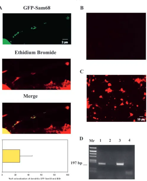
Because Sam68 is an RNA-binding protein, we sought to determine whether the distribution of GFP-Sam68 in the dendrites overlapped with that of RNA. For this, we used ethidium bromide (EtBr) to fluorescently label total cellular RNA in cultured hippocampal neurons. As shown in Fig. 3A, some of the GFP-Sam68 granules were included in EtBr-labeled spots. Quantitation indicated that 26% of the GFP-Sam68 granules colocalized with EtBr labeling. The specificity of the RNA labeling was assessed by treating fixed neurons by RNAse A or DNAse I (Fig. 3B and Fig. 3C, respectively). RNAse treatment erased all fluorescent labeling, whereas DNAse treatment did not affect the labeling, showing that in our experimental conditions, EtBr specifically labeled RNA. Thus, our data indicate that some of the dendritic GFP-Sam68 is concentrated in RNA-rich regions, supporting the possibility that Sam68 may be associated with dendritic RNAs. To further investigate this possibility, we prepared cytoplasmic extracts from rat brain, immunoprecipitated Sam68 under nondenaturing conditions and assayed the immunoprecipitates for the presence of β-actin mRNA, a transcript known to be present in neuronal dendrites (Tiruchinapalli et al., 2003) and to interact with Sam68 in vitro and in HeLa cells (Itoh et al., 2002). After reverse transcription of the coprecipitated mRNA, primers specific for the β-actin mRNA allowed amplification
Fig. 1. Nucleo-somatodendritic translocation of Sam68 in depolarized rat hippocampal neurons. (A) Immunostaining of Sam68 using the AD1 rabbit anti-serum in neurons at 9 DIV cultured at 5 mM KCl showing a nuclear labeling (left) or at 25 mM KCl showing a perinuclear staining or a somatodendritic staining (right, see arrows). (B) Confocal image of a same neuron cultured at 25 mM KCl labeled with the anti-Sam68 AD1. Sam68 was concentrated in clusters (arrowheads, red fluorescence, upper panel) and MAP2 antibodies (green fluorescence, lower panel).
of a band displaying the length expected for that mRNA, and comigrating with an amplification product obtained from the input lysate (Fig. 3D, lane 1). mRNA coprecipitation was specific, as control immunoprecipitates prepared with pre-immune antibody did not contain amplifiable β-actin mRNA (Fig. 3D, lane 2). Data to be presented elsewhere indicates that in the cortex and hippocampus, Sam68 is associated with polysomes and can be coprecipitated with a specific set of mRNAs, including other dendritic transcripts such as the αCaMKII mRNA (Grange et al., 2004). These results support the conclusion that Sam68 is associated with RNA in the somatodendritic compartment.
Membrane depolarization induces translocation of GFP-Sam68
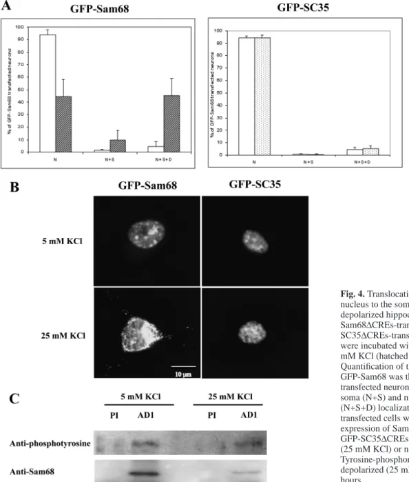
We next determined the effect of depolarization on GFP-Sam68 localization. Depolarization has been shown to activate
the CMV promoter used in our expression vector. This effect that we also observed may be due to the presence of five CRE sequences (Wheeler and Cooper, 2001). To circumvent this undesirable effect, a mutated CMV promoter devoid of the five CREs was substituted for the regular CMV in the GFP fusion constructs (which were henceforth denoted ∆CREs). GFP-Sam68 expressed from the ∆CREs vector in neurons depolarized for 5 hours delocalized from the nucleus to the cell body and neurites (Fig. 4B). GFP-Sam68 was found exclusively in nuclei in 40% of the transfected neurons; in 10%, it was nuclear and somatic, and in 50%, nuclear, somatic and dendritic (Fig. 4A, left panel). However, as a control, when neurons in parallel cultures were transfected with GFP-SC-35∆CREs (a GFP construct derived from the splicing factor SC-35), the nuclear localization of GFP-SC-35 did not change under depolarization (Fig. 4A, right panel and Fig. 4B). This result indicates that in our conditions, depolarization induced a specific change in GFP-Sam68 dynamics, rather than a general perturbation of nuclear export. Three types of images could be observed when GFP-Sam68∆CREs-transfected neurons were depolarized for 5 hours: first, most nuclei appearing less fluorescent than the surrounding cytoplasm (60%; Fig. 4B); second, nuclei displaying the same fluorescence intensity as soma, with a perinuclear concentration of fluorescent material (35%); and third, nuclei showing greater fluorescence than soma (5%). We were not able to induce a delocalization of Sam68 if the depolarization lasted for 2 hours or less (not shown). More interestingly, after 5 hours of depolarization, if the depolarizing medium was changed back to pre-depolarization medium, all the GFP-Sam68 returned to the nucleus within 3 hours (not shown). In the depolarized neurons, the proportion of dendritic GFP-Sam68 granules that were colocalized with RNA stained with ethidium bromide did not significantly differ from that observed in the few dendritic
Fig. 2. Distribution of Sam68 in GFP-Sam68-transfected hippocampal neurons.
(A) Quantification and representative images of the subcellular localization of GFP-Sam68 in nuclei (N), nuclei + soma (N+S) and in nuclei + soma + dendrites (N+S+D). Error bars represent s.d. (n=3, where 200-300 transfected cells were counted). (B) Nucleo-somatodendritic expression of the N-terminal deleted GFP-Sam68 mutant transfected neuron. (C) GFP-Sam68-transfected neuron where individual granules moving into dendrites of living hippocampal neurons could be visualized. Transfected neurons were incubated in control medium as described in Materials and Methods and individual motilities of green fluorescent granules were observed by time-lapse video microscopy for 8 hours each 20 minutes. Images taken at t=0 and t=480 minutes are shown. Arrows label moving granules, whereas the arrowhead labels a stationary granule.
granules observed in unstimulated neurons (data not shown). Taken together, these results indicate that permanent KCl depolarization induces a specific redistribution of Sam68 in favor of the distal dendrites.
Relocalization of certain splicing regulatory factors to the cell cytoplasm has previously been shown to occur in neurons and non-neuronal cells subjected to stressful stimuli; in the case of the splicing factors hnRNP-A1 (Van der Houven van Oordt et al., 2000) and hTra2-β1 (Daoud et al., 2002), this redistribution was accompanied by de novo phosphorylation of the proteins, which appeared to play an important role in their export (see Discussion). To investigate whether Sam68 similarly became hyperphosphorylated in depolarized neurons, we immunoprecipitated endogenous Sam68 from lysates of control or depolarized neurons, and we probed the phosphorylation status of the immunoprecipitated protein by western immunoblotting with phosphotyrosine or anti-phosphothreonine antibodies. This technique has previously revealed that in cell lines, Sam68 became phosphorylated on tyrosine residues in the presence of Src kinase activity (Fumagalli et al., 1994; Taylor and Shalloway, 1994) and on threonine residues following stimulation of the MAP kinase pathway (Matter et al., 2002). As shown in Fig. 4C, Sam68 was
clearly phosphorylated on tyrosine residues, even though a large majority of cells in the population were postmitotic neurons. No significant difference in tyrosine or threonine phosphorylation pattern could be observed between neurons treated with 5 mM and 25 mM KCl. We also failed to detect a change in gel mobility (Fig. 4C), even though stimulus-induced hyperphosphorylation of Sam68 has been shown to result in a shift in apparent gel electrophoretic mobility (Matter et al., 2002). In conclusion, we have so far found no evidence that Sam68 undergoes additional phosphorylation during neuronal depolarization.
The depolarization-induced translocation of Sam68 is microtubule dependent
We next determined whether microtubule integrity was necessary for the movement of GFP-Sam68 towards the distal dendrites of hippocampal neurons. Addition of the microtubule-disrupting drug nocodazole did not affect KCl-induced translocation of Sam68 (data not shown). However, staining of the neurons with an anti-tubulin antibody indicated that nocodazole had little effect on microtubule stability, an observation that may be explained by the extreme stability of
Fig. 3. Association of GFP-Sam68 granules with RNA. (A) Confocal images of dendritic localization of GFP-Sam68 in hippocampal neurons showing fluorescent granules (green fluorescence, GFP-Sam68) that colocalize in three regions (yellow fluorescence, merged image) with RNA labeling by ethidium bromide (red fluorescence). Percentage of colocalization of GFP-Sam68 and ethidium bromide. Error bar represents s.d. (n=100). (B) RNAse I treatment of the EtBr labeling of neurons. (C) DNAse I treatment of the EtBr labeling of neurons. (D) Binding of immunoprecipitated Sam68 from adult rat cortex with β-actin mRNA. RT-PCR with specific β-actin primers was carried out with AD1 anti-Sam68 antibody
immunoprecipitation (lane 1), with pre-immune IgG immunoprecipitation (lane 2), with total extract (lane 3) or with no input (lane 4).
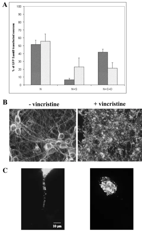
neuronal microtubules. We thus explored the effect of another microtubule-disrupting drug, vincristine, known to be very efficient in preventing tubulin polymerization in neurons (Allison et al., 2000). We applied vincristine to the GFP-Sam68∆CREs-transfected neurons 30 minutes before depolarization, and then added the drug to the depolarizing medium for 5 hours as described in Materials and Methods. As shown in Fig. 5A, the depolymerization of microtubules (verified by tubulin labeling, Fig. 5B, upper panel) affected Sam68 translocation. GFP-Sam68 fluorescence extended to the soma in 28% and to the dendrites in 21%, respectively, of neurons depolarized in the presence of vincristin, compared with 6% and 43%, respectively, of the control depolarized neurons. This result strongly suggests that in depolarized neurons treated with vincristin, Sam68 could accumulate in the somatic perikaryon, but could not efficiently proceed out to the
dendrites. Fig. 5B (lower panel) shows a vincristin-treated neuron in which the dendritic GFP-Sam68 granules were present but in a much shorter portion of the dendrites than was typical in the untreated controls. The translocation occurred through the nuclear membrane, but did not go further than 10 µm into the dendrites. These data support the notion that the motion of GFP-Sam68 towards the distal dendrites is not simply diffusive, but depends on microtubule integrity.
Sam68 translocation to the somatodendritic
compartment is dependent on calcium entry through VSCCs and partially occurs through the
CRM1/exportin1 pathway
Membrane depolarization of neurons triggers Ca2+ entry
through voltage-sensitive calcium channels (VSCCs). To
Fig. 4. Translocation of GFP-Sam68 from the nucleus to the somato-dendritic compartment in depolarized hippocampal neurons. (A) GFP-Sam68∆CREs-transfected (left panel) or GFP-SC35∆CREs-transfected (right panel) neurons were incubated with 5 mM KCl (white) or with 25 mM KCl (hatched or stippled) for 5 hours. Quantification of the subcellular localization of GFP-Sam68 was then reported as a percentage of transfected neurons showing nuclei (N), nuclei + soma (N+S) and nuclei + soma + dendrites (N+S+D) localization (n=3, where 200-300 transfected cells were counted). (B) Patterns of expression of Sam68 in GFP-Sam68∆CREs- or GFP-SC35∆CREs-transfected neurons depolarized (25 mM KCl) or not (5 mM KCl) for 5 hours. (C) Tyrosine-phosphorylation pattern of neurons depolarized (25 mM KCl) or not (5 mM KCl) for 5 hours.
determine whether Sam68 translocation was dependent on this process, GFP-Sam68∆CREs-transfected neurons were depolarized for 5 hours in the presence of the L-type channel blocker nimodipine. Under these conditions, GFP-Sam68 was completely retained in nuclei, with only 2% of transfected neurons showing the protein in the dendritic compartment (equivalent to 97% inhibition of dendritic Sam68 localization, with P<0.05; Fig. 6A). These results indicate that the delocalization of Sam68 in the somatodendritic compartment requires Ca2+ entry through L-type voltage-sensitive Ca2+
channels.
We then sought to test whether the KCl-induced nucleocytoplasmic translocation of Sam68 was passive or
active. One important mediator of active nuclear export is the general export receptor CRM1/exportin1 (Fornerod et al., 1997; Mattaj et al., 1998; Neville et al., 1997). The action of the CRM1 system can be specifically blocked by the drug leptomycin B (LMB) (Fukuda et al., 1997). To monitor the effect of LMB in our conditions, neurons were transfected with GFP-Rev-NES, a derivative of the HIV-1 Rev, which is a known substrate for CRM1. Untreated neurons localized GFP-Rev-NES in their cytoplasm; as expected, LMB caused the accumulation of GFP-Rev-NES in the nucleus (Fig. 6B, upper panel). In neurons expressing GFP-Sam68, when LMB was added during the depolarization procedure, GFP-Sam68 was retained in the nucleus in 74% of the transfected neurons (Fig. 6A). The addition of LMB thus resulted in 59% inhibition of the dendritic delocalization of GFP-Sam68 (P<0.05), as shown in Fig. 6B (lower panel). This result supports the notion that nuclear export underlies the cytoplasmic accumulation of Sam68 during depolarization; it also indicates that this export is partly mediated by the CRM1/ exportin1 active transport pathway.
Discussion
In order to track the distribution of Sam68 throughout the cell body and neurites of individual neurons, either live or fixed, we made use of a previously characterized GFP-Sam68 fusion (Chen et al., 1999). In HeLa cells and other proliferating cell lines, both the endogenous Sam68 and GFP-Sam68 were shown to reside in the same, highly specific structures (the Sam68 nuclear bodies), indicating that fusion with GFP had not altered the localization properties of the native protein, or the ability of specific cell types to generate these particular structures. Given the low transfection efficiency of neuronal cultures, it was hard to assess the overexpression level of GFP-Sam68 compared with endogenous Sam68 in transfected neurons. Staining of GFP-Sam68-expressing neurons with Hoechst dye and with antibodies raised against a variety of neuronal proteins, including the cytoskeletal MAP2 and tubulin (the present
Fig. 5. Dendritic microtubule-dependent transport of Sam68 during depolarization. (A) Effect in percent of a microtubule-disrupting drug vincristine (5 µM) in depolarized neurons. GFP-Sam68∆CREs-transfected neurons were incubated with 25 mM KCl for 5 hours in the absence (hatched) or presence (stippled) of vincristine. Error bars represent s.d. (n=3, where 200-300 transfected cells were counted). (B) Images of the α-tubulin immunostaining in depolarized neurons in the absence or presence of vincristine. (C) Images of the GFP-Sam68 expression pattern in depolarized neurons in the absence or presence of vincristine.
report); dendritic spine-associated PSD-95, NMDA receptor, AMPA receptor; and the endogeous Sam68 (our unpublished data); all indicated that the GFP-Sam68 induced no overt change in either cell morphology or survival. Our results also clearly indicate that voltage- and calcium-dependent signal transduction processes are indeed functioning in the presence of GFP-Sam68. On the basis of these premises, we assume that in neurons, GFP-Sam68 expression is unlikely to cause general perturbations of cellular functioning, and is a reliable tracer for the dynamic behavior of endogenous Sam68.
In proliferating cell lines, attempts to detect nucleocytoplasmic transport or shuttling of Sam68 by specific assays (such as transfer of nuclear Sam68 labeling in heterokaryons) have so far yielded no evidence that shuttling was taking place (Soros et al., 2001). In unstimulated hippocampal neurons, both endogenous Sam68 immunofluorescence and GFP-Sam68 were predominantly located in the nucleus. However, in HeLa cells GFP-Sam68
was nuclear in 100% of cells (Chen et al., 1999), but a small but reproducible minority of neurons (4%) expressed GFP-Sam68 in their somatodendritic compartment. Given the effect of depolarization (see below), this presence out of the nucleus might conceivably be due to episodes of spontaneous activity.
Surprisingly, somatodendritic Sam68 was increased to 50% in the case of the ∆1-67 N-terminal deletion mutant, which in HeLa cells remained fully nuclear (Chen et al., 1999). Nuclear accumulation of Sam68 in neurons may thus require specific signals at the N-terminal extremity for full efficiency. We do not know whether the N terminus provides a signal for some neuron-specific element of the nuclear import system, or whether it functions in the neuronal nucleus as a retention signal, preventing export. Further experiments will be required to clarify the localization properties of isolated Sam68 sequence elements, such as the N-terminus, within the neuronal cell. If a neuron-specific nuclear localization signal exists in the Sam68 N-terminus, it might function in conjunction with the KH domain (required for proper localization in the nucleus) (Chen et al., 1999) and with the C-terminal region (known to harbor a noncanonical nuclear localization sequence) (Ishidate et al., 1997).
When present in the dendrites, GFP-Sam68 was mostly condensed in puncta, in a fashion similar to somatodendritic RNA-binding proteins such as staufen or FMRP. Because in the dendrites, endogenous Sam68 immunoreactivity was also punctate, it is unlikely that GFP-Sam68 clusters arose from nonspecific aggregation. Interestingly, they were frequently located near dendritic branchpoints, similar to RNA granules (Rook et al., 2000). Dendritic mRNAs cluster in frequently motile granules that also include ribosomes, translation factors and specific RNA-binding proteins; they are thought to serve as vehicles for transporting the mRNAs and for maintaining them in a repressed state until needed (Krichevsky and Kosik, 2001). One fourth of the GFP-Sam68 puncta was colocalized with fluorescently labeled dendritic RNA clusters. This is in keeping with our observation that Sam68 is associated with mRNA in brain cytoplasmic extracts (Grange et al., 2004). However, the precise relationship between Sam68 and RNA transport granules remains to be defined. While some GFP-Sam68 granules were moving at time scales similar to those reported for GFP-staufen, most of the observed granules were immobile for hours, remarkably. Moreover, when entry of GFP-Sam68 in the dendrites was tracked by time-lapse recording in depolarized neurons, granules often appeared to condense in situ after the protein had traveled in a more diffuse phase, sometimes visible as a faint fluorescent background in the dendrites (data not shown). GFP-Sam68 may possibly be exchanged between a less condensed pool and recruiting granules immobilized at specific sites on the cytoskeleton. Further work will be required to precise the composition and dynamics of the GFP-Sam68-containing granules.
Depolarization by high KCl resulted in the progressive extension of GFP-Sam68 fluorescence to the somatodendritic compartment, off to the distal dendrites. This extension was detected both statistically, by a large increase (12-fold) in the proportion of neurons with somatodendritic protein, and
Fig. 6. Inhibition of Sam68 translocation during depolarization.
(A) Neurons were depolarized with 25 mM KCl for 5 hours in the absence (–) or presence of nimodipine (5 µM) or leptomycin B (LMB, 60 ng/ml) to block calcium entry through voltage-sensitive calcium channels or CRM-1 nuclear export, respectively. s.d. with n=3, where 200-300 transfected cells were counted (percentages of neurons with nuclear and somatic Sam68 expression are not shown). (B) Images of neurons transfected with GFP-Rev-NES or GFP-Sam68∆CREs treated or not with LMB (60 ng/ml) for 5 hours.
dynamically, by the surge of somatodendritic fluorescence in individual neurons. Several controls showed that the observed change in GFP-Sam68 subcellular distribution results from specific modifications of Sam68 function, rather than from some general perturbation of cellular physiology. First, endogenous Sam68 immunofluorescence was also increased in the dendrites of depolarized neurons that had not been transfected. Second, complete nuclear retention of the GFP-SC-35 fusion indicated that treatment with elevated KCl did not induce an indiscriminate transfer of all nuclear RNA-binding proteins to the cytoplasm. Third, the somatodendritic accumulation of GFP-Sam68 was reversible on return to low KCl. Moreover, at the KCl concentration used, neurons with condensed chromatin or pycnotic nuclei (indicative of apoptosis) were not numerous, and were systematically excluded from the analysis by verification with Hoechst staining. By using the CMV-∆CRE mutant promoter, we also took care to exclude a possible bias due to stimulation of GFP-Sam68 synthesis. In principle, the increase in cytoplasmic and dendritic GFP-Sam68 might be explained either by stimulation of nuclear export or by inhibition of nuclear import, or a combination of both processes. The effect of leptomycin B, a very specific inhibitor of the CRM-1/exportin nuclear export pathway, strongly suggests that KCl-mediated depolarization causes a redistribution of GFP-Sam68, at least partly by promoting nuclear export of the protein. When neurons were depolarized in the presence of leptomycin B, there was a significant increase in the frequency of neurons showing nuclear retention of GFP-Sam68. Thus, some of the cytoplasmic Sam68 molecules appear to exit the nucleus via the 1 pathway. The CRM-1/exportin 1 pathway mediates nuclear export of proteins containing a leucine-rich nuclear export signal (NES) (Fornerod et al., 1997; Mattaj et al., 1998; Neville et al., 1997). Sam68 itself does not present a leucine-rich NES sequence but may conceivably associate with CRM-1 cargoes. There is evidence for interaction between Sam68 and one such cargo, the HIV-1 shuttling protein Rev (Reddy et al., 1999); and Sam68 has recently been reported to participate in CRM-1-coupled Rev export (Li et al., 2002). However, LMB-sensitive export did not account for the totality of Sam68 accumulation in the cytoplasm. A fraction of Sam68 may possibly be transferred to the cytoplasm as part of mRNP complexes, by the mRNA export system, which is mostly distinct from the CRM-1 pathway. Data from several groups indicate that Sam68 interacts with TAP, a shuttling protein centrally implicated in mRNA export (Coyle et al., 2003; Reddy et al., 2000). Sam68 strongly stimulates the cytoplasmic utilization of retroviral mRNAs transported by TAP, raising the possibility that a fraction of Sam68 remains associated with the mRNAs during export, and modulates their translation (Coyle et al., 2003). It will be interesting to see if Sam68 can similarly affect the cytoplasmic utilization of its normal mRNA targets.
The finding that translocation of GFP-Sam68 to the dendrites required an intact microtubular network is consistent with the observed electron-microscopic localization of Sam68 immunoreactivity on microtubules in dendritic shafts (Grange et al., 2004) and with features of dendritic RNA-binding proteins (Kohrmann et al., 1999). Certain point mutations in Sam68 result in stable association of the protein with
microtubules in HeLa cells; such mutations may ‘freeze’ or amplify a normally transient binding event (Chen et al., 1999). Further work will be necessary to identify possible links between Sam68 and microtubules.
Blockade of the L-type VSCC completely prevented the depolarization-induced translocation of GFP-Sam68 to the dendrites. Calcium influx through L-type channels is known to be crucial for inducing nuclear responses such as transcription factor activation. Thus, the exit of Sam68 from the nucleus depends on a specific signaling pathway connecting neuronal activity to transcriptional responses. Because Sam68 nuclear exit was only manifest after a 2-3 hour delay, and because it was completely abolished by transcription and translation inhibitors (unpublished observations), it may be that the calcium signal acts by inducing the synthesis of macromolecules (mRNA and/or auxiliary proteins) necessary for Sam68 exit.
It is equally possible that calcium-dependent modification of Sam68 itself helps to regulate its subcellular localization. As a precedent, stress-induced activation of the p38 kinase cascade in COS cells resulted in both phosphorylation and nuclear export of the RNA-binding protein hnRNP A1 (Van der Houven van Oordt et al., 2000). In brain neurons subjected to transient ischemia in vivo, nuclear export of the splicing factors tra2-β1 and SC-35, as well as Sam68 itself, has recently been reported to take place (Daoud et al., 2002). After 6 or 24 hours of ischemic insult, the tra2-β1 translocation was accompanied by a hyperphosphorylation of the splicing factor. These published results support the notion that calcium elevation can result in efflux of Sam68 and other splicing factors such as tra2-β1 into the cytoplasm. In our conditions, SC-35 did not exit the nucleus, a difference that probably reflects the massive character of calcium mobilization in the models used by Daoud et al. (Daoud et al., 2002). Using reagents known to detect signal-induced phosphorylation of Sam68 in other cell types, we did not find evidence for a change in Sam68 phosphorylation following KCl treatment of cultured hippocampal neurons. The distribution of Sam68 in neurons may be compared with that of another splicing regulatory factor, ZBP2/KRSP. This protein is predominantly nuclear, but a minority of ZBP2 form clusters in neuronal processes, which are colocalized with mRNA (Gu et al., 2002). The function of ZBP2/KRSP is required for localizing the β-actin and MAP2 mRNAs to neuronal processes (Gu et al., 2002; Rehbein et al., 2002). We show here that in hippocampal neurons, Sam68 colocalizes with RNA clusters, and the protein immunoprecipitated from adult rat cortex binds to β-actin mRNA. Our results raise the possibility that in response to neuronal activity, Sam68 similarly accompanies, or becomes recruited to, certain dendritic mRNAs. As Sam68 can interact with signaling pathways in the cytoplasm as well as in the nucleus, it may conceivably intervene in the activity-dependent regulation of mRNA metabolism.
We thank E. Cooper, M. Yoshida and J. Tazi for providing reagents and discussions; and S. Davis, S. Laroche, C. Feuerstein and J.-L. Martiel for discussions. The authors’ work is supported by the INSERM, CEA, the Université Joseph Fourier, the Fondation pour la Recherche Médicale (FRM) and the Association pour la Recherche contre le Cancer (ARC).
References
Allison, D. W., Chervin, A. S., Gelfand, V. I. and Craig, A. M. (2000). Postsynaptic scaffolds of excitatory and inhibitory synapses in hippocampal neurons: maintenance of core components independent of actin filaments and microtubules. J. Neurosci. 20, 4545-4554.
Brown, V., Jin, P., Ceman, S., Darnell, J. C., O’Donnell, W. T., Tenenbaum, S. A., Jin, X., Feng, Y., Wilkinson, K. D., Keene, J. D. et al. (2001). Microarray identification of FMRP-associated brain mRNAs and altered mRNA translational profiles in fragile X syndrome. Cell 107, 477-487. Chen, T., Boisvert, F. M., Bazett-Jones, D. P. and Richard, S. (1999). A role
for the GSG domain in localizing Sam68 to novel nuclear structures in cancer cell lines. Mol. Biol. Cell 10, 3015-3033.
Coyle, J. H., Guzik, B. W., Bor, Y. C., Jin, L., Eisner-Smerage, L., Taylor, S. J., Rekosh, D. and Hammarskjold, M. L. (2003). Sam68 enhances the cytoplasmic utilization of intron-containing RNA and is functionally regulated by the nuclear kinase Sik/BRK. Mol. Cell. Biol. 23, 92-103. Crino, P. B. and Eberwine, J. (1996). Molecular characterization of the
dendritic growth cone: regulated mRNA transport and local protein synthesis. Neuron 17, 1173-1187.
Daoud, R., Mies, G., Smialowska, A., Olah, L., Hossmann, K. A. and Stamm, S. (2002). Ischemia induces a translocation of the splicing factor tra2-beta 1 and changes alternative splicing patterns in the brain. J. Neurosci. 22, 5889-5899.
Derry, J. J., Richard, S., Valderrama Carvajal, H., Ye, X., Vasioukhin, V., Cochrane, A. W., Chen, T. and Tyner, A. L. (2000). Sik (BRK) phosphorylates Sam68 in the nucleus and negatively regulates its RNA binding ability. Mol. Cell. Biol. 20, 6114-6126.
Feng, Y., Gutekunst, C. A., Eberhart, D. E., Yi, H., Warren, S. T. and
Hersch, S. M. (1997). Fragile X mental retardation protein:
nucleocytoplasmic shuttling and association with somatodendritic ribosomes. J. Neurosci. 17, 1539-1547.
Fornerod, M., Ohno, M., Yoshida, M. and Mattaj, I. W. (1997). CRM1 is an export receptor for leucine-rich nuclear export signals. Cell 90, 1051-1060.
Fukuda, M., Asano, S., Nakamura, T., Adachi, M., Yoshida, M., Yanagida, M. and Nishida, E. (1997). CRM1 is responsible for intracellular transport mediated by the nuclear export signal. Nature 390, 308-311.
Fumagalli, S., Totty, N. F., Hsuan, J. J. and Courtneidge, S. A. (1994). A target for Src in mitosis. Nature 368, 871-874.
Fusaki, N., Iwamatsu, A., Iwashima, M. and Fujisawa, J.-I. (1997). Interaction between Sam68 and Src family tyrosine kinases, Fyn and Lck, in T cell receptor signaling. J. Biol. Chem. 272, 6214-6219.
Garner, C. C., Tucker, R. P. and Matus, A. (1988). Selective localization of messenger RNA for cytoskeletal protein MAP2 in dendrites. Nature 336, 674-677.
Grange, J., Boyer, V., Fabian-Fine, R., Benfredj, N., Sadoul, R. and Goldberg, Y. (2004). Somatodendritic localization and mRNA association of the splicing regulatory protein Sam68 in the hippocampus and cortex. J. Neuroscience Res. (in press).
Gu, W., Pan, F., Zhang, H., Bassell, G. J. and Singer, R. H. (2002). A predominantly nuclear protein affecting cytoplasmic localization of beta-actin mRNA in fibroblasts and neurons. J. Cell Biol. 156, 41-51. Hartmann, A. M., Nayler, O., Schwaiger, F. W., Obermeier, A. and Stamm,
S. (1999). The interaction and colocalization of Sam68 with the splicing-associated factor YT521-B in nuclear dots is regulated by the Src family kinase p59(fyn). Mol. Biol. Cell 10, 3909-3926.
Ishidate, T., Yoshihara, S., Kawasaki, Y., Roy, B. C., Toyoshima, K. and Akiyama, T. (1997). Identification of a novel nuclear localization signal in Sam68. FEBS Lett. 409, 237-241.
Itoh, M., Haga, I., Li, Q. H. and Fujisawa, J. (2002). Identification of cellular mRNA targets for RNA-binding protein Sam68. Nucleic Acids Res. 30, 5452-5464.
Job, C. and Eberwine, J. (2001). Identification of sites for exponential translation in living dendrites. Proc. Natl. Acad. Sci. USA 98, 13037-13042. Kiebler, M. A., Hemraj, I., Verkade, P., Kohrmann, M., Fortes, P., Marion, R. M., Ortin, J. and Dotti, C. G. (1999). The mammalian staufen protein localizes to the somatodendritic domain of cultured hippocampal neurons: implications for its involvement in mRNA transport. J. Neurosci. 19, 288-297.
Kohrmann, M., Luo, M., Kaether, C., DesGroseillers, L., Dotti, C. G. and Kiebler, M. A. (1999). Microtubule-dependent recruitment of Staufen-green fluorescent protein into large RNA-containing granules and subsequent dendritic transport in living hippocampal neurons. Mol. Biol. Cell 10, 2945-2953.
Krichevsky, A. M. and Kosik, K. S. (2001). Neuronal RNA granules: a link between RNA localization and stimulation-dependent translation. Neuron 32, 683-696.
Kudo, N., Taoka, H., Toda, T., Yoshida, M. and Horinouchi, S. (1999). A novel nuclear export signal sensitive to oxidative stress in the fission yeast transcription factor Pap1. J. Biol. Chem. 274, 15151-15158.
Laggerbauer, B., Ostareck, D., Keidel, E. M., Ostareck-Lederer, A. and Fischer, U. (2001). Evidence that fragile X mental retardation protein is a negative regulator of translation. Hum. Mol. Genet. 10, 329-338. Li, J., Liu, Y., Kim, B. O. and He, J. J. (2002). Direct participation of Sam68,
the 68-kilodalton Src-associated protein in mitosis, in the CRM1-mediated Rev nuclear export pathway. J. Virol. 76, 8374-8382.
Martin, K. C., Barad, M. and Kandel, E. R. (2000). Local protein synthesis and its role in synapse-specific plasticity. Curr. Opin. Neurobiol. 10, 587-592.
Mattaj, I. W., Englmeier, L., Stutz, F., Lee, L., Davis, L. I. and Rosbash, M. (1998). Nucleocytoplasmic transport: the soluble phase. Annu. Rev. Biochem. 67, 265-306.
Matter, N., Herrlich, P. and Konig, H. (2002). Signal-dependent regulation of splicing via phosphorylation of Sam68. Nature 420, 691-695.
Miyashiro, K., Dichter, M. and Eberwine, J. (1994). On the nature and differential distribution of mRNAs in hippocampal neurites: implications for neuronal functioning. Proc. Natl. Acad. Sci. USA 91, 10800-10804. Neville, M., Stutz, F., Lee, L., Davis, L. I. and Rosbash, M. (1997). The
importin-beta family member Crm1p bridges the interaction between Rev and the nuclear pore complex during nuclear export. Curr. Biol. 7, 767-775. Reddy, T. R., Xu, W., Mau, J. K., Goodwin, C. D., Suhasini, M., Tang, H., Frimpong, K., Rose, D. W. and Wong-Staal, F. (1999). Inhibition of HIV replication by dominant negative mutants of Sam68, a functional homolog of HIV-1 Rev. Nat. Med. 5, 635-642.
Reddy, T. R., Tang, H., Xu, W. and Wong-Staal, F. (2000). Sam68, RNA helicase A and Tap cooperate in the post-transcriptional regulation of human immunodeficiency virus and type D retroviral mRNA. Oncogene 19, 3570-3575.
Rehbein, M., Wege, K., Buck, F., Schweizer, M., Richter, D. and Kindler, S. (2002). Molecular characterization of MARTA1, a protein interacting with the dendritic targeting element of MAP2 mRNAs. J. Neurochem. 82, 1039-1046.
Richard, S., Yu, D., Blumer, K. J., Hausladen, D., Olszowy, M. W., Connelly, P. A. and Shaw, A. S. (1995). Association of p62, a multifunctional SH2-and SH3-domain-binding protein, with src family tyrosine kinases, Grb2, SH2-and phospholipase C gamma-1. Mol. Cell. Biol. 15, 186-197.
Rook, M. S., Lu, M. and Kosik, K. S. (2000). CaMKIIalpha 3′untranslated
region-directed mRNA translocation in living neurons: visualization by GFP linkage. J. Neurosci. 20, 6385-6393.
Scheetz, A. J., Nairn, A. C. and Constantine-Paton, M. (2000). NMDA receptor-mediated control of protein synthesis at developing synapses. Nat. Neurosci. 3, 211-216.
Schuman, E. M., Crino, P. B. and Eberwine, J. (1999). mRNA trafficking and local protein synthesis at the synapse. Neuron 23, 645-648.
Severt, W. L., Biber, T. U., Wu, X., Hecht, N. B., DeLorenzo, R. J. and Jakoi, E. R. (1999). The suppression of testis-brain RNA binding protein and kinesin heavy chain disrupts mRNA sorting in dendrites. J. Cell Sci. 112, 3691-3702.
Shen, Z., Batzer, A., Koehler, J. A., Polakis, P., Schlessinger, J., Lydon, N. B. and Moran, M. F. (1999). Evidence for SH3 domain directed binding and phosphorylation of Sam68 by Src. Oncogene 18, 4647-4653. Sisson, T. H. and Castor, C. W. (1990). An improved method for
immobilizing IgG antibodies on protein A-agarose. J. Immunol. Methods 127, 215-220.
Soros, V. B., Carvajal, H. V., Richard, S. and Cochrane, A. W. (2001). Inhibition of human immunodeficiency virus type 1 Rev function by a dominant-negative mutant of Sam68 through sequestration of unspliced RNA at perinuclear bundles. J. Virol. 75, 8203-8215.
Steward, O., Wallace, C. S., Lyford, G. L. and Worley, P. F. (1998). Synaptic activation causes the mRNA for the IEG Arc to localize selectively near activated postsynaptic sites on dendrites. Neuron 21, 741-751.
Tang, S. J., Meulemans, D., Vazquez, L., Colaco, N. and Schuman, E. (2001). A role for a rat homolog of staufen in the transport of RNA to neuronal dendrites. Neuron 32, 463-475.
Taylor, S. J. and Shalloway, D. (1994). An RNA-binding protein associated with Src through its SH2 and SH3 domains in mitosis. Nature 368, 867-871. Tiruchinapalli, D. M., Oleynikov, Y., Kelic, S., Shenoy, S. M., Hartley, A., Stanton, P. K., Singer, R. H. and Bassell, G. J. (2003). Activity-dependent
trafficking and dynamic localization of zipcode binding protein 1 and beta-actin mRNA in dendrites and spines of hippocampal neurons. J. Neurosci. 23, 3251-3261.
Tongiorgi, E., Righi, M. and Cattaneo, A. (1997). Activity-dependent dendritic targeting of BDNF and TrkB mRNAs in hippocampal neurons. J. Neurosci. 17, 9492-9505.
Van der Houven van Oordt, W., Diaz-Meco, M. T., Lozano, J., Krainer, A. R., Moscat, J. and Caceres, J. F. (2000). The MKK(3/6)-p38-signaling cascade alters the subcellular distribution of hnRNP A1 and modulates alternative splicing regulation. J. Cell Biol. 149, 307-316.
Wang, L. L., Richard, S. and Shaw, A. S. (1995). p62 association with RNA is regulated by tyrosine phosphorylation. J. Biol. Chem. 270, 2010-2013.
Weiler, I. J., Irwin, S. A., Klintsova, A. Y., Spencer, C. M., Brazelton, A. D., Miyashiro, K., Comery, T. A., Patel, B., Eberwine, J. and Greenough, W. T. (1997). Fragile X mental retardation protein is translated near synapses in response to neurotransmitter activation. Proc. Natl. Acad. Sci. USA 94, 5395-5400.
Wheeler, D. G. and Cooper, E. (2001). Depolarization strongly induces human cytomegalovirus major immediate-early promoter/enhancer activity in neurons. J. Biol. Chem. 276, 31978-31985.
Xia, Z., Dudek, H., Miranti, C. K., Greenberg, M. E. and Domenici, L. (1996). Calcium influx via the NMDA receptor induces immediate early gene transcription by a MAP kinase/ERK-dependent mechanism. J. Neurosci. 16, 5425-5436.