149
Minireview
Epididymosomes and Prostasomes:
Their Roles in Posttesticular Maturation
of the Sperm Cells
FABRICE SAEZ, GILLES FRENETTE, AND ROBERT SULLIVAN
From the Centre de recherche en biologie de la
reproduction et de´partement d’Obste´trique-Gyne´cologie, Universite´ Laval, Sainte-Foy, Que´bec, Canada
G1V-4G2.
The occurrence of membrane vesicles along the male re-productive tract and in the ejaculated semen appears as a common feature among different species, including hu-mans. Indeed, prostasomes (prostate-derived vesicles) were first described in human semen in 1978 (Ronquist et al, 1978), and vesicular structures similar to prosta-somes were also found in the seminal plasma of rabbit (Davis, 1978), ram (Breitbart and Rubinstein, 1982), and stallion (Arienti et al, 1998; Minelli et al, 1998). Fur-thermore, ‘‘prostasome-like’’ particles are present in the epididymal fluid of rat (Fornes and De Rosas, 1991), hamster (Yanagimachi et al, 1985), and bull (Frenette and Sullivan, 2001) and are also secreted by the bull seminal vesicles (Agrawal and Vanha-Perttula, 1987). The bio-chemical composition of prostasomes purified from hu-man semen has been well documented. These are multi-lamellar lipoprotein membrane particles with a diameter of 50 to 500 nm. Their cholesterol-phospholipid ratio reaches 2, sphingomyelin being the major phospholipid. Many proteins are associated with prostasomes, some of them having a catalytic activity (Review: Kravets et al, 2000; Ronquist and Nilsson, 2002).
This review will focus on the physiological implication of these membranous structures with regard to the post-testicular sperm maturation.
Epididymosomes and Epididymal Maturation
Epididymal transit confers the mammalian sperm cells their fertilizing ability, a process that is under androgenic control (Cooper et al, 1986). Among the complex modi-fications that spermatozoa undergo in the epididymis is
Correspondence to: Dr Robert Sullivan, Unite´ d’Ontoge´nie-Reproduc-tion, Centre de Recherche, Centre Hospitalier de l’Universite´ Laval, 2705 Blvd Laurier, Ste-Foy, Que´bec, Canada G1V-4G2 (e-mail: robert. sullivan@crchul.ulaval.ca).
Received for publication July 15, 2002; accepted for publication Oc-tober 15, 2002.
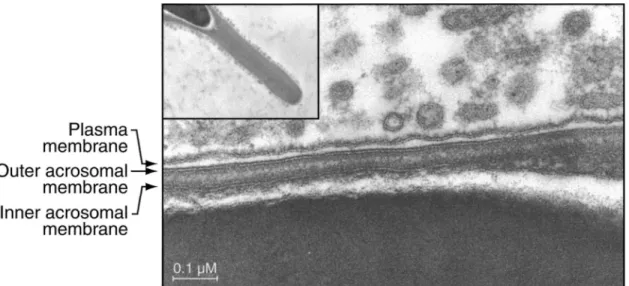
the process whereby proteins are produced by the epidid-ymal epithelium and then secreted and transferred to the sperm cells, thereby generating functional male gametes (Jones, 1998). However, there is evidence that proteins without any signal peptide are also acquired by sperm cells during their epididymal transit, implying an unusual secretion pathway. In fact, a family of orthologous epi-didymis-specific proteins was characterized in different mammalian species and named P26h, P25b, and P34H in hamster, bull, and man, respectively (Sullivan, 1999). Each of them is acquired by the sperm cells during epi-didymal transit, and the hamster protein, P26h, has no signal peptide as deduced from its mRNA sequence. This hamster protein is located on the sperm surface, is at-tached via a glycosyl-phosphatidylinositol (GPI) anchor, and is involved in the zona pellucida recognition (Sulli-van, 1999). The epididymal membranous particles present in the lumen, ‘‘prostasome-like particles’’ or epididymo-somes, are responsible for anchoring the P26h to the sperm surface (Le´gare´ et al, 1999). Furthermore, epidi-dymosomes of the Chinese hamster have been shown to interact in vivo with the sperm plasma membrane (Yan-agimachi et al, 1985; Figure 1). Epididymosomes are thus directly involved in the epididymal maturation process of the sperm cells.
Like P26h, the bovine ortholog protein P25b is GPI anchored, and its transfer from the epididymal epithelial cells to spermatozoa also implies the intervention of ep-ididymosomes (Frenette et al, 2002). The transfer of P25b was demonstrated between epididymosomes isolated from the cauda epididymidis and spermatozoa from the caput epididymidis, on which the initial quantity of P25b was null. This transfer is fundamental for the fertility of the animals, as bull subfertility is associated with low levels of P25b (Parent et al, 1999). In order to test the hypoth-esis of possible transfer of other proteins from the epi-didymosomes to sperm cells in the bull epididymis, the proteins exposed at the surface of epididymosomes from the cauda epididymidis can be biotinylated and then in-cubated with caput spermatozoa. After 4 washing steps to get rid of the residual epididymosomes, the sperm pro-teins are extracted and Western blotted with peroxidase-conjugated neutravidin. Under these conditions, only a fraction of the proteins associated with epididymosomes are transferred to spermatozoa (Frenette et al, 2002). This
Figure 1. Electron photomicrographs showing Chinese hamster epididymosomes surrounding the plasma membrane of a spermatozoon. The inset shows the general appearance of the acrosome surrounded by these vesicles. Original photos were kindly provided by Dr Yanagimachi (University of Hawaii).
transfer of selected proteins can either mean that only these proteins possess the ability to be transferred or that epididymosomes are not transferred totally, as an intact entity, on spermatozoa. Another possibility is that the population of epididymosomes is heterogeneous, with a different protein composition, as has already been report-ed for rat epididymal vesicles (Fornes et al, 1995). Protein transfer is dependent on the pH and the zinc concentra-tion: indeed, it is optimum at a pH of 6.0 to 6.5, the variation being 2.5-fold between pH 6.0 and 7.5 (Frenette et al, 2002). This is in accordance with the fact that the fusion between human prostasomes and ejaculated sper-matozoa is favored at a slightly acidic pH (Arienti et al, 1997a), thus showing a tendency toward a conserved mechanism. Zinc is also important for the transfer, favor-ing this process when rangfavor-ing between 0.1 and 1.5 mM, whereas magnesium and calcium have no effect. Consid-ering that the epididymal intraluminal pH is 6.5 and that high zinc concentrations are found in the epididymis, it is apparent that these are the physiologically relevant con-ditions for the in vitro transfer of protein.
The biotynilated proteins of epididymosomes are trans-ferred to the acrosomal cap and the midpiece of sper-matozoa, with this transfer being temperature-dependent (the maximum transfer occurs between 328C and 378C). These facts may reflect the importance of the lipid com-position and the membrane fluidity of spermatozoa and/ or epididymosomes. Thus, the transfer would only be pos-sible on certain microdomains of the sperm plasma mem-brane, which undergoes important composition
modifi-cations during epididymal transit (Parks and
Hammerstedt, 1985). The evolution of the structure and composition of the sperm plasma membrane during the transit would permit the acquisition of important proteins,
via the epididymosomes, at the right time and location in the excurrent duct.
The mechanism responsible for the protein transfer via the vesicles has not yet been elucidated. However, differ-ent hypotheses have been proposed for the cell-to-cell transfer of GPI-anchored molecules and have been re-viewed by Ilangumaran et al (1996). Briefly, the acqui-sition could be mediated in one of 3 ways: 1) by the intervention of plasma lipid-carrier proteins, 2) by inter-actions between a donor and an acceptor membrane via a ‘‘flip-over’’ phenomenon, or 3) by vesicles that could be processed by endocytosis and the carried proteins sent back to the plasma membrane of the acceptor cell. The 2 latter propositions would be in complete accordance with the epididymosomes–spermatozoa interaction.
The identity (or identities) and precise function(s) of the transferred protein(s) remain to be investigated but could lead to a better understanding of the complex cess of epididymal sperm maturation. A number of pro-teins other than P25b and its orthologs, generally with no signal peptide, are also secreted by the mammalian epi-didymis or other organs from the male genital tract and are associated with vesicular structures closely resembling the epididymosomes (Table). The direct relation between these proteins and fertility has not always been demon-strated but confirms the importance of the mechanism, the purpose of which could be to protect important proteins from proteolytic digestion, as suggested by several au-thors (Sutovsky et al, 2001; Rejraji et al, 2002).
Because of the availability of a large amount of bio-logical material, the bull model allows researchers to characterize more precisely the implication of epididy-mosomes in the mammalian epididymal sperm maturation process. The study of such mechanisms in humans is very
Characteristics of selected proteins associated with vesicles secreted by the male genital tract of different mammalian species
Protein Origin and Species Function Observations Reference
Epididymosomes: P34H
P26h P25b
Epididymis of humans, hamsters, and bulls, respectively
Zona pellucida recog-nition and binding
P26h and P25b present on epididymosomes; trans-ferred to spz*
Sullivan, 1999
GPX5 Mouse epididymis Glutathion peroxidase Associated with
epididymo-somes and transferred to spz
Rejraji et al, 2002 Macrophage migration
inhibitory factor
Rat epididymis T-cell cytokine but
un-known function here
Associated with epididymo-somes, no signal peptide
Eickhoff et al, 2001
Ubiquitin Bovine epididymal
vesi-cles
Elimination of defective spz
Secreted and transferred to spz Fraile et al, 1996; Sutov-sky et al, 2001 Prostasomes: CD55 CD59 CD46
Prostasomes from hu-man ejaculated semen
Complement inhibition CD59 and CD46 transferred to spz in vitro
Rooney et al, 1993, 1996
HE5 (CD52) Human and monkey
prostasomes
Unknown function here Acquired by spz during epi-didymal transit Rooney et al, 1996; Yeung et al, 1997 Other vesicles: Transglutaminase, carbonic anhydrase II
Rat coagulating gland Coagulatory plug, pH adjustment
No signal peptide, apocrine secretion, androgen de-pendence, interaction with spz after ejaculation
Aumuller et al, 1999
Mouse vas deferens protein
Mouse vas deferens Aldose reductase but unknown function here
Associated with vesicles in the vas deferens and transferred to spz, no sig-nal peptide
Manin et al, 1995
* spz indicates spermatozoa.
difficult from a technical point of view because of the quasi-impossibility of disposing of human epididymis in sufficient amounts to purify both epididymosomes and ep-ididymal spermatozoa. However, the P26h/P25b human ortholog protein P34H is also related to fertility and has been proposed as an indicator of the sperm fertilizing ability (Sullivan, 1999). We can thus hypothesize that the same mechanisms, or relatively close ones, also occur in the human epididymis. Membranous vesicles are impor-tant mediators in the epididymis-related maturational events of mammalian spermatozoa. The involvement of epididymosomes in the acquisition of new proteins by the male gamete during epididymal transit can explain why many of these surface proteins behave as integral mem-brane proteins (Cooper, 1998).
Prostasomes and Postejaculatory Sperm Modifications
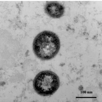
As mentioned earlier, the first described vesicles were re-ported by Ronquist et al in 1978 and were isolated from human seminal plasma by an ultracentrifugation method. Figure 2 shows an electron photomicrograph of prosta-somes purified from human seminal plasma. Further stud-ies showed that these so-called ‘‘prostasomes’’ are se-creted by the human prostate and mixed in the semen at
the moment of ejaculation. The precise physiological function of these vesicles still remains unclear, but many different in vitro functions have been related to them, such as blood coagulation activity (Fernandez et al, 1997), antibacterial activity (Carlsson et al, 2000), serine-protease activity via the enzyme dipeptidyl-peptidase IV (CD26, Arienti et al, 1997b), and antioxidant activity (Saez et al, 1998, 2000). Many different proteins have been shown to be present on prostasomes, some of which possess enzymatic properties (Review: Kravets et al, 2000; Ronquist and Nilsson, 2002).
The effects of prostasomes on the posttesticular matu-ration of spermatozoa include an immunosuppressive ac-tivity (Rooney et al, 1991, 1993, 1996), an enhancement of sperm motility (Stegmayr and Ronquist, 1982; Fabiani et al, 1994a,b; Carlsson et al, 1997), and an influence on the sperm capacitation process (Cross, 1996a,b; Cross and Mahasreshti, 1997).
The immunosuppressive activity of prostasomes arises from the presence of several complement inhibitory mol-ecules (eg, CD55 [decay accelerating factor] and CD46 [membrane cofactor protein], both of which inhibit the C3-convertase) and from the presence of CD59 (protec-tin), which inhibits the formation of the membrane attack
Figure 2. Electron photomicrographs of prostasomes purified from hu-man semen. Three prostasomes appear as electron-dense material and show their size heterogeneity. The original photo was kindly provided by Dr Kemeny and Dr Guy (Centre Hospitalier Universitaire de Clermont-Ferrand, 63000 France).
complex. CD55 and CD59 are GPI-anchored proteins, whereas CD46 is a transmembrane protein. Prostasomes have the ability to transfer CD59 to spermatozoa in vitro, and this mechanism also exists between prostasomes and red blood cells or fibroblasts (Rooney et al, 1993, 1996). It has also been demonstrated that the transfer of CD59 from prostasomes to CD59-deficient red blood cells re-sulted in protection against complement-mediated hemo-lysis, thus showing that the functional protein is trans-ferred (Babiker et al, 2002). The transmembrane protein CD46 is also transferred from prostasomes to red blood cells, with the same efficiency, thus showing a complex mechanism, as both GPI-anchored and transmembrane proteins can be transferred (Rooney et al, 1993). The pos-sible role of these molecules would be to protect sper-matozoa, once in the female genital tract, from being phagocytosed by the white blood cells. It should be noted that the human epididymal protein HE5 (CD52), which is also a GPI-anchored protein, is acquired by sperm cells during their epididymal transit in humans as well as in cynomolgus monkeys (Yeung et al, 1997). This protein was previously shown to be present on prostasomes (Roo-ney et al, 1996), thus suggesting an intervention of epi-didymosomes in the transfer of this protein from epithe-lial cells to spermatozoa.
Prostasomes also have an influence on sperm motility under several conditions in vitro. First, they enhance the progressive motility of spermatozoa, measured after 1
washing step of whole semen (Stegmayr and Ronquist, 1982). This effect could be due to the modifications of the sperm microenvironment by prostasomes, since these vesicles contain a calcium-dependent ATPase. Second, prostasomes favor the recovery of sperm motility after their immobilization by NaCl washes (Fabiani et al, 1994b). Finally, prostasomes also stimulate the rate of motile sperm recovery following the swim-up technique (Fabiani et al, 1994a). This effect is higher than the effect of albumin and seems to be dependent on the particular lipid composition of the prostasomes, since they keep these properties after a heat treatment of 5 minutes at 1008C. This enhancement of motile sperm recovery after swim-up is also applicable if the semen has been sub-mitted to freezing and thawing according to classical cry-oconservation protocols (Carlsson et al, 1997). The mo-lecular mechanism underlying these effects still remains unclear but is an important part of the prostasomes’ ac-tion. Indeed, sperm motility and movement quality are important factors in the movement of sperm in the female genital tract (eg, crossing the cervical mucus as well as penetrating the zona pellucida). If prostasomes keep the same functions in vivo, they could promote these various steps by their effects on sperm motility.
Another effect of prostasomes on sperm function is that they have an influence on the capacitation step, a prereq-uisite for fertilization to occur. Indeed, one of the known inhibitory factors of capacitation, as determined by sperm response to progesterone, is cholesterol (Cross, 1996a). Prostasomes are very rich in cholesterol and represent approximately 40% of the total cholesterol present in seminal plasma. They were shown to inhibit the proges-terone-stimulated acrosome reaction of human spermato-zoa in vitro (Cross, 1996b). According to Cross and Ma-hasreshti (1997), the most likely hypothesis is that cho-lesterol could transfer from prostasomes to the sperm cells. Whatever the mechanism, the particular lipid com-position and structure of the prostasomes are related to this function and, as mentioned earlier, could also be in-volved in the protein transfer within certain precise mem-brane domains of the sperm cells.
Taken together, the different functions of prostasomes seem to have the common aim to protect spermatozoa after ejaculation in order to preserve them in the most proper state, with their full functional capacities, prior to their encounter with the oocyte.
Epididymosomes and Prostasomes: An Unusual
Secretion Pathway?
The transfer of proteins to spermatozoa thus appears as a common feature of epididymosomes and prostasomes. Sev-eral of these proteins do not possess a signal peptide or are GPI anchored and transferred with their functional anchor, which implies an unusual secretion pathway. One possibility
is that epididymosomes and prostasomes are released in the intraluminal compartment by apocrine secretion. In contrast to mesocrine secretion, this type of secretion does not in-volve the Golgi apparatus or the fusion of secretory vesicles with the plasma membrane prior to protein secretion. Apo-crine secretion implies the formation of apical blebs con-taining selected organelles, including vesicles of various siz-es. These blebs detach from the cell surface, and one hy-pothesis is that their content could be released when the blebs undergo fragmentation (Hermo and Robaire, 2002). They could also appear as whole entities in the luminal fluid and show properties similar to those of the isolated vesicles or represent 2 different types of secretion. This blebbing phenomenon was first studied in the rat coagulating gland, as well as in the prostate and seminal vesicles (Aumuller, 1979; Aumuller and Adler, 1979), and was also very well documented, in terms of photographic studies, in the bull reproductive tract (Agrawal and Vanha-Perttula, 1988). The studies by Aumuller were undertaken to test whether the apical blebs secreted by these organs (apocrine secretion) were real or just artifacts due to tissue fixation problems. However, the improvement of fixation techniques could not get rid of these blebs, thus suggesting a real phenomenon. Two enzymes, transglutaminase and carbonic anhydrase II, are secreted in an apocrine way by the rat coagulating gland and do not possess a signal peptide (Seitz et al, 1991; Wil-helm et al, 1998). This secretion pathway thus seems to be specific for certain proteins, mainly GPI-anchored proteins and proteins without a signal peptide.
Apical blebs have also been described as a feature of principal and narrow cells of the epididymis of many mammalian species, including humans. As mentioned earlier, the occurrence of prostasomes or epididymosomes in the lumen of the different ducts could derive from the fragmentation of the released ‘‘blebs,’’ thus allowing their interaction with spermatozoa (Hermo and Robaire, 2002). It is very likely that proteins of the cytoskeleton (mainly actin) are involved in the mechanism of apocrine secre-tion at the step of bleb release, although much work still needs to be done to define this precisely (Aumuller et al, 1999). This secretion pathway is very common in the re-productive tract but is also present in the mammary glands and sweat glands (Aumuller et al, 1997).
The maturation of sperm cells in the epididymis or after ejaculation is thus related to the presence of vesicles in their environment, which is probably the result of apo-crine secretions by the male genital tract organs. This pro-cess seems to be involved in the constant development of the sperm plasma membrane, playing a major role in the acquisition of particular proteins by spermatozoa.
Conclusion
The occurrence of extracellular vesicular structures in the biological fluids surrounding spermatozoa is a
character-istic of the male reproductive tract. These vesicles are present as early as the epididymal transit, when they are tightly related to the sperm maturation process and the acquisition of fertilizing ability. Then, after ejaculation, prostasomes coming from the prostate or from other or-gans, depending on species, are mixed together in semen. This new environment protects spermatozoa and keeps them functional in association with other soluble factors. A common feature of these different vesicles is their abil-ity to transfer new biologically active proteins to sper-matozoa and, also, probably new lipids such as choles-terol. During their journey from the testis to the female genital tract, the development of the complex lipid and protein pattern of the sperm plasma membrane relies at least in part on these vesicles. Their maturation properties in the epididymis are changed to a ‘‘reservoir’’ function concerning the prostasomes.
Prostasomes and epididymosomes can therefore be considered a new insight in the posttesticular maturation process of the mammalian spermatozoa.
Acknowledgments
The authors would like to thank Dr Yanagimachi (University of Hawaii) and Dr Kemeny and Dr Guy (Centre Hospitalier Universitaire de Cler-mont-Ferrand) for the photographic illustrations included in this paper. Work from the authors’ laboratory has been supported by grants from CIHR and NSERC of Canada.
References
Agrawal Y, Vanha-Perttula T. Effect of secretory particles in bovine sem-inal vesicle secretion on sperm motility and acrosome reaction. J Re-prod Fertil. 1987;79:409–419.
Agrawal Y, Vanha-Perttula T. Electron microscopic study of the secretion process in bovine reproductive organs. J Androl. 1988;9:307–316. Arienti G, Carlini E, De Cosmo AM, Di Profio P, Palmerini CA.
Prostasome-like particles in stallion semen. Biol Reprod. 1998;59:309–313. Arienti G, Carlini E, Palmerini CA. Fusion of human sperm to
prosta-somes at acidic pH. J Membr Biol. 1997a;155:89–94.
Arienti G, Polci A, Carlini E, Palmerini CA. Transfer of CD26/dipeptidyl peptidase IV (E.C.3.5.4.4) from prostasomes to sperm. FEBS Lett. 1997b;410:343–346.
Aumuller G. Prostate gland and seminal vesicles. In: Teil VL, ed. Hand-buch der mikroskopischen Anatomie des Menschen. Vol 7. Band, Harn-und Geschlechtsapparat. Part 6. New York, NY: Springer-Ver-lag; 1979.
Aumuller G, Adler G. Experimental studies of apocrine secretion in the dorsal prostate epithelium of the rat. Cell Tissue Res. 1979;198:145–158. Aumuller G, Renneberg H, Schiemann PJ, Wilhelm B, Seitz J, Konrad
L, Wennemuth G. The role of apocrine released proteins in the post-testicular regulation of human sperm function. Adv Exp Med Biol. 1997;424:193–219.
Aumuller G, Wilhelm B, Seitz J. Apocrine secretion—fact or artifact? Anat Anz. 1999;181:437–446.
Babiker AA, Ronquist G, Nilsson UR, Nilsson BO. Transfer of prosta-somal CD59 to CD59-deficient red blood cells results in protection against complement-mediated hemolysis. Am J Reprod Immunol. 2002;47:183–192.
Breitbart H, Rubinstein S. Characterization of Mg21- and Ca21-ATPase activity in membrane vesicles from ejaculated ram seminal plasma. Arch Androl. 1982;9:147–157.
Carlsson L, Pahlson C, Bergquist M, Ronquist G, Stridsberg M. Anti-bacterial activity of human prostasomes. Prostate. 2000;44:279–286. Carlsson L, Ronquist G, Stridsberg M, Johansson L. Motility stimulant effects of prostasome inclusion in swim-up medium on cryopreserved human spermatozoa. Arch Androl. 1997;38:215–221.
Cooper TG. Interactions between epididymal secretions and spermatozoa. J Reprod Fertil. 1998;53(suppl):119–136.
Cooper TG, Waites GM, Nieschlag E. The epididymis and male fertility. A symposium report. Int J Androl. 1986;9:81–90.
Cross NL. Effect of cholesterol and other sterols on human sperm acro-somal responsiveness. Mol Reprod Dev. 1996a;45:212–217. Cross NL. Human seminal plasma prevents sperm from becoming
acro-somally responsive to the agonist, progesterone: cholesterol is the major inhibitor. Biol Reprod. 1996b;54:138–145.
Cross NL, Mahasreshti P. Prostasome fraction of human seminal plasma prevents sperm from becoming acrosomally responsive to the agonist progesterone. Arch Androl. 1997;39:39–44.
Davis BK. Uterine fluid from progesterone treated rabbits contains sub-cellular membranes. Experientia. 1978;34:350–351.
Eickhoff R, Wilhelm B, Renneberg H, et al. Purification and character-ization of macrophage migration inhibitory factor as a secretory pro-tein from rat epididymis: evidences for alternative release and transfer to spermatozoa. Mol Med. 2001;7:27–35.
Fabiani R, Johansson L, Lundkvist O, Ronquist G. Enhanced recruitment of motile spermatozoa by prostasome inclusion in swim-up medium. Hum Reprod. 1994a;9:1485–1489.
Fabiani R, Johansson L, Lundkvist O, Ulmsten U, Ronquist G. Promotive effect by prostasomes on normal human spermatozoa exhibiting no forward motility due to buffer washings. Eur J Obstet Gynecol Re-prod Biol. 1994b;57:181–188.
Fernandez JA, Heeb MJ, Radtke KP, Griffin JH. Potent blood coagulant activity of human semen due to prostasome-bound tissue factor. Biol Reprod. 1997;56:757–763.
Fornes MW, De Rosas JC. Interactions between rat epididymal epithelium and spermatozoa. Anat Rec. 1991;231:193–200.
Fornes WM, Sosa MA, Bertini F, Burgos MH. Vesicles in rat epididymal fluid. Existence of two populations differing in ultrastructure and en-zymatic composition. Andrologia. 1995;27:233–237.
Fraile B, Martin R, De Miguel MP, Arenas MI, Bethencourt FR, Peinado F, Paniagua R, Santamaria L. Light and electron microscopic immu-nohistochemical localization of protein gene product 9.5 and ubiquitin immunoreactivities in the human epididymis and vas deferens. Biol Reprod. 1996;55:291–297.
Frenette G, Lessard C, Sullivan R. Selected proteins of ‘‘prostasome-like particles’’ from epididymal cauda fluid are transferred to epididymal caput spermatozoa in bull. Biol Reprod. 2002;67:308–313.
Frenette G, Sullivan R. Prostasome-like particles are involved in the transfer of P25b from the bovine epididymal fluid to the sperm sur-face. Mol Reprod Dev. 2001;59:115–121.
Hermo R, Robaire B. Epididymal cell types and their functions. In: Robaire B, Hinton B, eds. The epididymis: From Molecules to Clinical Practice. New York, NY: Kluwer Academic/Plenum Publishers; 2002:81–102. Ilangumaran S, Robinson PJ, Hoessli DC. Transfer of exogenous
glyco-syl-phosphatidylinositol (GPI)-linked molecules to plasma mem-branes. Trends Cell Biol. 1996;6:163–167.
Jones R. Plasma membrane structure and remodelling during sperm mat-uration in the epididymis. J Reprod Fertil. 1998;53(suppl):73–84. Kravets FG, Lee J, Singh B, Trocchia A, Pentyala SN, Khan SA.
Pros-tasomes: current concepts. Prostate. 2000;43:169–174.
Le´gare´ C, Berube B, Boue F, Lefievre L, Morales CR, El-Alfy M,
Sul-livan R. Hamster sperm antigen P26h is a phosphatidylinositol-an-chored protein. Mol Reprod Dev. 1999;52:225–233.
Manin M, Lecher P, Martinez A, Tournadre S, Jean C. Exportation of mouse vas deferens protein, a protein without a signal peptide, from mouse vas deferens epithelium: a model of apocrine secretion. Biol Reprod. 1995;52:50–62.
Minelli A, Moroni M, Martinez E, Mezzasoma I, Ronquist G. Occurrence of prostasome-like membrane vesicles in equine seminal plasma. J Reprod Fertil. 1998;114:237–243.
Parent S, Lelie`vre L, Brindle Y, Sullivan R. Bull subfertility is associated with low levels of a sperm membrane antigen. Mol Reprod Dev. 1999; 52:57–65.
Parks JE, Hammerstedt RH. Development changes occurring in the lipids of ram epididymal spermatozoa plasma membrane. Biol Reprod. 1985;32:653–668.
Rejraji H, Vernet P, Drevet JR. GPX5 is present in the mouse caput and cauda epididymidis lumen at three different locations. Mol Reprod Dev. 2002;63:96–103.
Ronquist G, Brody I, Gottfries A, Stegmayr B. An Mg21- and Ca21-stimulated adenosine triphosphatase in human prostatic fluid—part II. Andrologia. 1978;10:427–433.
Ronquist G, Nilsson BO, eds. Wenner-Gren International Series. Vol 81. Prostasomes. London: Portland Press; 2002.
Rooney IA, Atkinson JP, Krul ES, Schonfeld G, Polakoski K, Saffitz JE, Morgan BP. Physiologic relevance of the membrane attack complex in-hibitory protein CD59 in human seminal plasma: CD59 is present on extracellular organelles (prostasomes), binds cell membranes, and inhibits complement-mediated lysis. J Exp Med. 1993;177:1409–1420. Rooney IA, Davies A, Griffiths D, Williams JD, Davies M, Meri S,
Lach-mann PJ, Morgan BP. The complement-inhibiting protein, protectin (CD59 antigen), is present and functionally active on glomerular ep-ithelial cells. Clin Exp Immunol. 1991;83:251–256.
Rooney IA, Heuser JE, Atkinson JP. GPI-anchored complement regula-tory proteins in seminal plasma. An analysis of their physical con-dition and the mechanisms of their binding to exogenous cells. J Clin Invest. 1996;97:1675–1686.
Saez F, Motta C, Boucher D, Grizard G. Antioxidant capacity of prosta-somes in human semen. Mol Hum Reprod. 1998;4:667–672. Saez F, Motta C, Boucher D, Grizard G. Prostasomes inhibit the NADPH
oxidase activity of human neutrophils. Mol Hum Reprod. 2000;6:883– 891.
Seitz J, Keppler C, Huntemann S, Rausch U, Aumuller G. Purification and molecular characterization of a secretory transglutaminase from coag-ulating gland of the rat. Biochim Biophys Acta. 1991;1078:139–146. Stegmayr B, Ronquist G. Promotive effect on human sperm progressive
motility by prostasomes. Urol Res. 1982;10:253–257.
Sullivan R. Interaction between sperm and epididymal secretory proteins. In: Gagnon C, ed. The Male Gamete From Basic to Clinical Appli-cations. Vienna, Ill: Cache River Press; 1999:93–104.
Sutovsky P, Moreno R, Ramalho-Santos J, Dominko T, Thompson WE, Schatten G. A putative, ubiquitin-dependent mechanism for the rec-ognition and elimination of defective spermatozoa in the mammalian epididymis. J Cell Sci. 2001;114:1665–1675.
Wilhelm B, Keppler C, Hoffbauer G, Lottspeich F, Linder D, Meinhardt A, Aumuller G, Seitz J. Cytoplasmic carbonic anhydrase II of rat coagulating gland is secreted via the apocrine export mode. J Histo-chem CytoHisto-chem. 1998;46:505–511.
Yanagimachi R, Kamiguchi Y, Mikamo K, Suzuki F, Yanagimachi H. Maturation of spermatozoa in the epididymis of the Chinese hamster. Am J Anat. 1985;172:317–330.
Yeung CH, Schroter S, Wagenfeld A, et al. Interaction of the human epididymal protein CD52 (HE5) with epididymal spermatozoa from men and cynomolgus monkeys. Mol Reprod Dev. 1997;48:267–275.