Publisher’s version / Version de l'éditeur:
Vous avez des questions? Nous pouvons vous aider. Pour communiquer directement avec un auteur, consultez la première page de la revue dans laquelle son article a été publié afin de trouver ses coordonnées. Si vous n’arrivez pas à les repérer, communiquez avec nous à PublicationsArchive-ArchivesPublications@nrc-cnrc.gc.ca.
Questions? Contact the NRC Publications Archive team at
PublicationsArchive-ArchivesPublications@nrc-cnrc.gc.ca. If you wish to email the authors directly, please see the first page of the publication for their contact information.
https://publications-cnrc.canada.ca/fra/droits
L’accès à ce site Web et l’utilisation de son contenu sont assujettis aux conditions présentées dans le site LISEZ CES CONDITIONS ATTENTIVEMENT AVANT D’UTILISER CE SITE WEB.
The Journal of Biological Chemistry, 2017-02-22
READ THESE TERMS AND CONDITIONS CAREFULLY BEFORE USING THIS WEBSITE. https://nrc-publications.canada.ca/eng/copyright
NRC Publications Archive Record / Notice des Archives des publications du CNRC :
https://nrc-publications.canada.ca/eng/view/object/?id=7fa45627-206c-465e-8616-06acc30fe7e8 https://publications-cnrc.canada.ca/fra/voir/objet/?id=7fa45627-206c-465e-8616-06acc30fe7e8
NRC Publications Archive
Archives des publications du CNRC
This publication could be one of several versions: author’s original, accepted manuscript or the publisher’s version. / La version de cette publication peut être l’une des suivantes : la version prépublication de l’auteur, la version acceptée du manuscrit ou la version de l’éditeur.
For the publisher’s version, please access the DOI link below./ Pour consulter la version de l’éditeur, utilisez le lien DOI ci-dessous.
https://doi.org/10.1074/jbc.M116.768754
Access and use of this website and the material on it are subject to the Terms and Conditions set forth at
An engineered TGF-β monomer that functions as a dominant negative
to block TGF-β signaling
Kim, Sun Kyung; Barron, Lindsey; Hinck, Cynthia S.; Petrunak, Elyse M.;
Cano, Kristin E.; Thangirala, Avinash; Iskra, Brian; Brothers, Molly; Vonberg,
Machell; Leal, Belinda; Richter, Blair; Kodali, Ravindra; Taylor, Alex B.; Du,
Shoucheng; Barnes, Christopher O.; Sulea, Traian; Calero, Guillermo; Hart,
P. John; Hart, Matthew J.; Demeler, Borries; Hinck, Andrew P.
Engineered TGF-b Monomer that Blocks TGF-b Signaling
1
An Engineered TGF-b Monomer that Functions as a Dominant Negative to Block TGF-b signaling Sun Kyung Kim, Lindsey Barron, Cynthia S. Hinck, Elyse M. Petrunak, Kristin E. Cano, Avinash Thangirala, Brian Iskra, Molly Brothers, Machell Vonberg, Belinda Leal, Blair Richter, Ravindra Kodali,
Alexander B. Taylor, Shoucheng Du, Christopher O. Barnes, Traian Sulea, Guillermo Calero, P. John Hart, Matthew J. Hart, Borries Demeler, and Andrew P. Hinck
From the Department of Structural Biology, University of Pittsburgh School of Medicine, Pittsburgh, PA 15260 U.S.A, the Department of Biochemistry, the Department of Cell and Structural Biology, and Center for Innovative Drug Discovery, University of Texas Health Science Center at San Antonio, San
Antonio, TX 78229-3900 U.S.A., and the National Research Council, Human Health Therapeutics Portfolio, Montréal, Quebec, H4P2R2 Canada
Running title: Engineered TGF-b Monomer that Blocks TGF-b Signaling To whom correspondence should be addressed: Prof. Andrew P. Hinck, Department of Structural Biology, University of Pittsburgh School of Medicine, Biomedical Science Tower 3, Room 1035, 3501 Fifth Avenue, Pittsburgh, PA 15260, U.S.A, Telephone: (412) 648-8533, FAX: (412) 648-9008, E-mail: ahinck@pitt.edu
Keywords: transforming growth factor beta (TGF-b), dominant negative, protein engineering, inhibitor, cell signaling, cancer, fibrosis
ABSTRACT
The transforming growth factor beta isoforms, TGF-β1, -β2, and –β3 are small secreted homodimeric signaling proteins with essential roles in regulating the adaptive immune system and maintaining the extracellular matrix. However, dysregulation of the TGF-b pathway is
responsible for promoting the progression of several human diseases, including cancer and fibrosis. In spite of the known importance of TGF-bs in promoting disease progression, no inhibitors have been approved for use in humans. Herein, we describe an engineered TGF-b monomer, lacking the heel helix, a structural motif essential for binding the TGF-b type I receptor, TbRI, but dispensible for binding the other receptor required for TGF-b signaling, the TGF-b type II receptor, TbRII, as an alternative therapeutic modality for blocking TGF-b signaling in humans. As shown through binding studies and crystallography, the engineered monomer retained the same overall structure of native TGF-b monomers and bound TbRII in an identical manner. Cell-based luciferase assays showed that the engineered
monomer functioned as a dominant negative to inhibit TGF-β signaling with a Ki of 20 – 70 nM.
Investigation of the mechanism showed that the high affinity of the engineered monomer for TβRII, coupled with its reduced ability to non-covalently dimerize and its inability to bind and recruit TbRI, enabled it to bind endogenous TbRII, but
prevented it from binding and recruiting TbRI to form a signaling complex. Such engineered monomers provide a new avenue to probe and manipulate TGF-β signaling, and may inform similar modifications of other TGF-β family members.
The transforming growth factor beta isoforms, TGF-b1, -b2, and -b3, are small secreted signaling proteins. Their overall structures are similar and consist of two cystine-knotted
monomers tethered together by a single inter-chain disulfide bond (5). They coordinate wound healing, modulate immune cell function, maintain the extracellular matrix, and regulate epithelial and
http://www.jbc.org/cgi/doi/10.1074/jbc.M116.768754 The latest version is at
JBC Papers in Press. Published on February 22, 2017 as Manuscript M116.768754
at National Research Council Canada on September 5, 2017
http://www.jbc.org/
Downloaded from
at National Research Council Canada on September 5, 2017
http://www.jbc.org/
Downloaded from
at National Research Council Canada on September 5, 2017
http://www.jbc.org/
Downloaded from
at National Research Council Canada on September 5, 2017
http://www.jbc.org/
Downloaded from
at National Research Council Canada on September 5, 2017
http://www.jbc.org/
Downloaded from
at National Research Council Canada on September 5, 2017
http://www.jbc.org/
Downloaded from
at National Research Council Canada on September 5, 2017
http://www.jbc.org/
Downloaded from
at National Research Council Canada on September 5, 2017
http://www.jbc.org/
Downloaded from
at National Research Council Canada on September 5, 2017
http://www.jbc.org/
Downloaded from
at National Research Council Canada on September 5, 2017
http://www.jbc.org/
Downloaded from
at National Research Council Canada on September 5, 2017
http://www.jbc.org/
Downloaded from
at National Research Council Canada on September 5, 2017
http://www.jbc.org/
Engineered TGF-b Monomer that Blocks TGF-b Signaling endothelial cell growth and differentiation (6). The
TGF-bs are synthesized as pre-pro proteins and after maturation, secretion, and release from their pro-domains (7), the mature homodimeric growth factors (GFs) bind and bring together two single-pass transmembrane receptors, known as TbRI and TbRII, to form the signaling-competent TbRI2-TbRII2 heterotetramer (8,9). TGF-b GFs
assemble TbRI2-TbRII2 heterotetramer in a
sequential manner, first by binding TbRII followed by recruitment of TbRI (10,11). The stepwise assembly of TbRII and TbRI into a heterotetramer is driven by binding of TbRI to a composite TGF-b:TbRII interface (12,13) (Fig. 1A).
The disruption or dysregulation of the TGF-b pathway is responsible for several human diseases. These include connective tissue disorders, such as Marfan’s disease and Loeys-Dietz
syndrome, which are caused by increased or decreased signaling due to mutations in the matrix protein fibrillin-1 or TbRII, respectively (2,14). The dysregulation of the pathway is also responsible for fibrotic disorders (1) and soft tissue cancers (4). The fibrotic disorders are a result of hyperactive TGF-b signaling following tissue injury or disease progression that leads to the accumulation of extracellular matrix proteins. TGF-b’s role in cancer is complex, with loss of its potent growth inhibitory activity being responsible for cancer initiation (15), and excessive TGF-b signaling, in the context of growth refractory advanced cancers, potently stimulating cancer progression and metastasis (4).
TGF-b’s disease promoting activities, together with animal studies that have
demonstrated beneficial effects of inhibiting TGF-b in models of cancer and fiTGF-brosis (16-23), have made them important targets for the development of inhibitors. However, in spite of clinical trials ongoing for nearly two decades using receptor kinase inhibitors, neutralizing antibodies, and other approaches, no TGF-b inhibitors have been approved for clinical use in humans (24,25). One of the main challenges involves finding the correct dosing and pharmacodynamics for the particular disease to enable an effective therapeutic response, but sparing or minimally impacting TGF-b
signaling, or other signaling pathways, in normal cells and tissues. TGF-b kinase inhibitors have posed some challenges in this respect as they have
significant inhibitory activity against other type I receptors of the TGF-b superfamily, as well as other related kinases (26-28), and may further lead to rapid development of resistance (29). Pan-isoform TGF-b neutralizing antibodies, such as Sanofi’s humanized mouse mononclonal antibody, GC1008, are specific, though tissue residence times are long and some concerning side effects, such as keratoacanthoma and squamous cell carcinoma, have been reported in clinical trials (30).
Thus, alternative approaches are needed to target the TGF-b pathway. The objective of this study was to investigate whether it might be possible to design an engineered TGF-b GF that functioned as a dominant negative to potently and specifically inhibit TGF-b signaling. This
approach offers several potential advantages over existing therapies. Relative to kinase inhibitors, engineered GFs would be expected to have much higher specificity, especially if they function by binding and blocking TbRII, which is known to only bind and transduce signals for TGF-b1, -b2, and -b3, but not other TGF-b family GFs (5,31). Another potential advantage over kinase inhibitors is increased bioavailability, since unlike the kinase inhibitors, engineered GFs would not have to cross the plasma membrane to reach their target.
Relative to monoclonal antibodies, the engineered GFs, because of their smaller size, would be expected to have shorter tissue lifetimes, which would limit sustained inhibition in normal cells and tissues and may alleviate undesirable side effects. The smaller size of engineered GFs may also lead to improved penetration of diseased tissues, particularly solid tumors, relative to 150 kDa monoclonal antibody molecules (32,33). Engineered ligands have been successfully used to target other signaling pathways, such as the VEGF pathway (34), and thus represent a largely
undeveloped, but potentially very effective therapeutic modality for treating disease.
Through previous studies, monomeric forms of TGF-b1 and TGF-b3, formed by substituting the cysteine residue that forms the inter-chain disulfide to serine (C77S), were shown to have diminished signaling activity compared to their disulfide-linked counterparts, but nonetheless were still quite potent, with EC50s for stimulation
of TGF-b reporter gene activity in the range of 100 pM (11,35). Amatayakul-Chantler and
at National Research Council Canada on September 5, 2017
http://www.jbc.org/
Engineered TGF-b Monomer that Blocks TGF-b Signaling workers (35), and later Zúñiga and co-workers
(11), suggested this residual activity might arise from assembly of a dimeric complex of a GF homodimer and two bound TbRIs and two bound TbRIIs, but without the disulfide linkage between the GF monomers. This model was attractive for two reasons – first, structures of the TGF-bs show there are in fact extensive hydrophobic contacts between the TGF-b monomers that could promote non-covalent self-association of the monomers (Fig. 1B) (36,37) – once formed, these non-covalent dimers would be stabilized as the
receptors bind, since crystal structures show that at least one them, TbRI, binds by straddling the TGF-b homodimer interface (Fig. 1A, C) (12,13).
The objective of this study was to design an engineered TGF-b monomer that still retained its full capacity to bind the high affinity TGF-b receptor, TbRII, but was fully impaired in its ability to bind and recruit TbRI. This type of engineered monomer would be expected to function as a dominant negative, and thus inhibit TGF-b signaling, since it would bind and thus occupy cell surface TbRII, but in turn be unable to recruit TbRI to form a signaling complex. The results presented here document the generation of such an engineered monomer and demonstrate that such monomers function as potent inhibitors of TGF-b signaling in cultured cells. The results further show that unlike dimeric TGF-bs, as well as their C77S monomeric counterparts, engineered monomers are highly soluble. These properties, together with the high intrinsic specificity of TGF-bs for TbRII, should engender this novel inhibitor with favorable properties for treating human diseases, such as Marfan’s disease, fibrotic
disorders, and soft tissue cancers that are driven by excessive TGF-b signaling.
RESULTS
Design of engineered mini monomeric TGF-b (mmTGF-b) - The structures of the TGF-b receptor complexes (12,13), as well as
accompanying binding and crosslinking studies with TGF-b3 C77S (11,12,38), suggested that the signaling capacity of monomeric TGF-bs (TGF-b1 C77S or b1 and TGF-b3 C77S or mTGF-b3) arise from their ability to non-covalently dimerize and in turn bind their receptors. (Fig. 1A,
C). This led to our hypothesis that it should be
possible to diminish or completely eliminate receptor complex assembly with monomeric TGF-bs by removing or altering residues responsible for dimer formation and binding of TbRI. The
structural motif that likely contributes the greatest to self-association of the monomers is the ‘heel’ a-helix, a-helix 3 (Fig. 1A). This helix is highly amphiphatic and has numerous hydrophobic interactions with residues that line the ‘palm’ of the opposing monomer (Fig. 1B). This helix also forms a large portion of the binding surface for TbRI (Fig. 1C). Thus, it was hypothesized that elimination of a-helix 3 should interfere with both self-association of the monomers and binding of TbRI, but should not impair TbRII binding as this occurs through the ligand fingertips far away from a-helix 3 (Fig. 1A).
To evaluate this hypothesis, bacterial expression constructs were generated for TGF-b1, TGF-b2, and TGF-b3 in which residues 52 – 71 were eliminated and Cys77 was substituted with serine. This corresponds to deletion of all of a-helix 3, as well as five flanking residues on the N-terminal end and three flanking residues on the C-terminal end (Fig. 1D). The length of the deletion was chosen so as to leave a sufficient number of residues between the last residue of b-strand 4 (G48) and the first residue of b-strand 5 (C77/S77) to form an unconstrained loop that bridges b-strands 4 and 5. Though a secondary consideration, either two (TGF-b2) or three (TGF-b1 and -b3) of the loop forming residues were also substituted so as to increase the net overall charge at pH 7.0 for the full-length TGF-b1, -b2, and -b3 monomers from -0.9, +1.1, and +4.4 to -3.1, +3.9, and +6.1 for the constructs in which a-helix 3 was deleted (Fig. 1D). The rationale for this was that the solubility of the monomers, which like the homodimers are poor from pH 4.5 to 9.5 (see Fig. 4A-B below), might be improved by both
removing hydrophobic a-helix 3 and by artificially increasing the net charge at pH 7.0.
Isolation and physical characterization of mmTGF-β2 - The TGF-b1, -b2, and –b3 ‘mini
monomers’ described above, designated mmTGF-b1, mmTGF-b2, and mmTGF-b3, were expressed in E. coli and accumulated in the form of insoluble inclusion bodies. The inclusion bodies were isolated and after reconstitution and purification in denaturant, the mini monomers were renatured by dilution into CHAPS-containing buffer at pH 9.0
at National Research Council Canada on September 5, 2017
http://www.jbc.org/
Engineered TGF-b Monomer that Blocks TGF-b Signaling as previously described (39). The folding of the
mini monomers differed greatly: a large portion of the mmTGF-b2 remained soluble during the folding and yielded large amounts of monomeric protein after purification by cation exchange chromatography, while only a small amount of mmTGF-b1 and mmTGF-b3 remained soluble during the folding and either no monomeric protein (TGF-b1) or a very small amount of monomeric protein (TGF-b3) was obtained after purification by cation exchange chromatography. This pattern mirrors that previously observed for the folding of TGF-b homodimers from full-length wild type monomers (39) and likely reflects differences in the intrinsic propensity of the monomers to properly form the four
intramolecular disulfides characteristic of each monomer.
Though mmTGF-b2 was the least desired variant, due to expected low intrinsic affinity for binding TbRII, this was nonetheless considered something that could be relatively easily addressed. This follows based on our prior studies which demonstrated that substitution of the three residues in TGF-b2’s interface with TbRII that differ from those in TGF-b1 and TGF-b3 was sufficient to engender TGF-b2 with the ability to bind TbRII with high affinity (40,41).
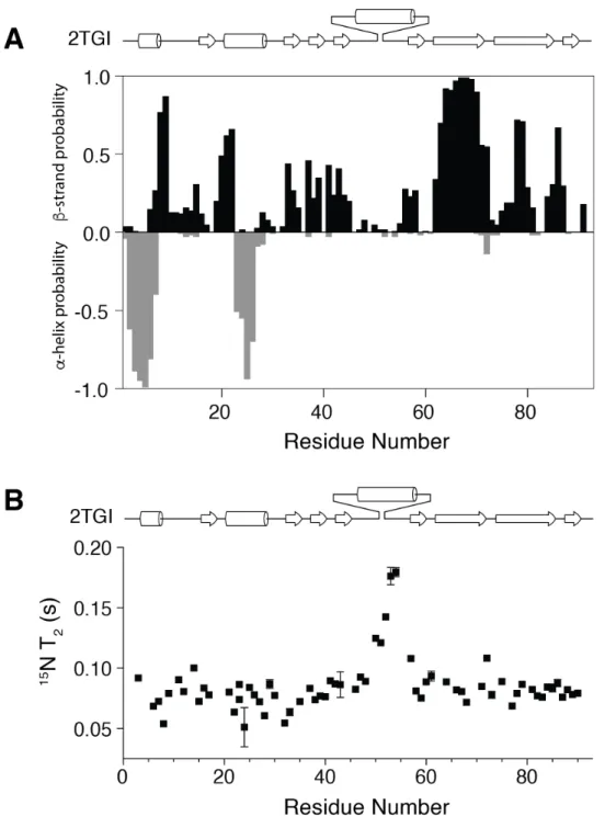
To determine if mmTGF-b2 was suitable for further development in the manner described above, it was characterized in terms of its folding, solubility, and receptor binding properties. To assess folding, a 15N-labeled sample of mmTGF-b2 was prepared and examined by recording a two-dimensional 1H-15N shift correlation spectrum (Fig. 2A). This revealed a highly dispersed
spectrum characteristic of natively folded protein. The spectrum could be fully assigned and analysis of the assigned chemical shifts to identify
secondary structure propensities showed that the protein had the expected secondary structure, particularly in the palm region formed by the cystine knot and the finger region where TbRII binds (Supplemental Fig. S2A). This analysis further showed that the newly created loop
between residues 47 - 56 had near zero probability of forming either an a-helix or b-strand,
suggesting that it is likely flexible as would be expected for a loop of this length connecting two antiparallel b-strands. This was directly confirmed by an analysis of backbone 15N T2 values. These
provide information about motions on fast (ns-ps) and intermediate (µs-ms) timescales and were significantly elevated in the region corresponding to the newly created loop relative to the other parts of the protein (Supplemental Fig. S2B), which except for the N-terminus and the short loop connecting a-helix 1 and b-strand 1, are expected to be structurally well-ordered.
To directly examine the three-dimensional structure, mmTGF-b2 was crystallized and its structure was determined to a resolution of 1.8 Å using molecular replacement (Table 1). The overall fold of mmTGF-b2 was shown to be highly similar to that previously determined for TGF-b2, with the exception of the newly created loop, which was shown to take the place of a-helix 3 as anticipated (Fig. 2B). Superposition of the mmTGF-b2 with the monomer from the structure of TGF-b2 shows that there is a systematic displacement of up to about 1.5 Å of the finger region of mmb2 relative to TGF-b2. Such differences appear to be a result of bending of the monomer near the center of the finger region, not a change in the structure of the finger region, as superimposition of the fingers alone show that they correspond closely, with a backbone RMSD of under 0.2 Å and similar orientations of the sidechains of several residues that pack and stabilize the fingers (Fig. 2D). Such bending is also supported by an overlay of the two molecules of mmTGF-b2 present in the
crystallographic asymmetric unit, which also exhibit a smaller but still noticeable displacement of the finger regions relative to one another (Fig. 2C). Consistent with the NMR analysis, not only was the electron density noticeably weaker in the region corresponding to the newly created loop, but also it was shown to adopt different
orientations for the two molecules from the asymmetric unit (Fig. 2C).
The similar folding of mmTGF-b2 relative to TGF-b2, especially in the TbRII-binding finger region, suggested that it would also bind TbRII in a similar manner. To evaluate this, surface plasmon resonance (SPR) experiments were performed in which the same concentration series of TbRII was injected over TGF-b2 and mmTGF-b2 immobilized on separate flow cells (Figure 3A,
B). Though it was not possible to quantitate
affinity due to weak binding, the sensorgrams nonetheless showed similar shapes and
at National Research Council Canada on September 5, 2017
http://www.jbc.org/
Engineered TGF-b Monomer that Blocks TGF-b Signaling concentration dependence. These sensorgrams
show that mmTGF-b2 binds TbRII weakly, consistent with earlier reports (40), and that it does so in a manner qualitatively similar to TGF-b2.
The solubility of mmTGF-b2 appeared to be significantly better than that of TGF-b2 and the full-length TGF-b2 monomer, mTGF-b2, as samples of the former could be readily prepared at concentrations of 2 – 3 mg mL-1 without
noticeable precipitation at pH 7.0, whereas samples of the latter two proteins were completely precipitated under these same conditions. To quantitate solubility, TGF-b2, mTGF-b2, and mmTGF-b2 were prepared as concentrated stocks in 100 mM acetic acid (pH 2.9) where they were readily soluble and then diluted into PBS at pH 7.4. The light scattering at 340 nM was measured to assess precipitation, and then the samples were centrifuged and the absorbance at 280 nM was measured to assess the protein concentration. This demonstrated that TGF-b2 and mTGF-b2 were both effectively insoluble at neutral pH over the entire concentration range evaluated (7 – 100 µM) (Fig. 4A-B). This is consistent with the known poor solubility of the TGF-b homodimers (42), but shows that this property also extends to full-length monomeric TGF-bs. The mini monomeric TGF-b2, mmTGF-b2, in contrast, exhibited modest light scattering and a corresponding modest reduction in the amount of soluble protein relative to that expected when the protein concentration was 40 µM or higher, indicating that indeed mmTGF-b2 was reasonably soluble at neutral pH, although not perfectly so. This was reflected in NMR spectra which showed that although 100 – 200 µM 15N mmTGF-b2 samples could be readily prepared, the spectrum was nonetheless poor, with the only detectable signals arising from residues in the flexible parts of the protein, namely the N- terminus, the exposed loop between a-helix 1 and strand 1, and the newly created loop between b-strands 4 and 5. The fact that signals could only be detected from the flexible parts of the protein suggested that mmTGF-b2 forms large soluble aggregates under these conditions. Through trial and error, it was found that these soluble
aggregates could be eliminated by addition of the zwitterionic detergent CHAPS, with the majority of the NMR signals appearing at concentration of 5 mM CHAPS and all of the NMR signals appearing at 10 mM CHAPS. Thus, all NMR
spectra, including that shown in Fig. 2A, were recorded in the presence of 10 mM CHAPS.
Isolation and physical characterization of mmTGF-β2-7M - The results presented above
show that while mmTGF-b2 is natively folded, it nonetheless possesses low intrinsic affinity for binding TbRII. In order to confer mmTGF-b2 with the ability to bind TbRII with high affinity
comparable to that of TGF-b1 and TGF-b3, the three residues in mouse TGF-b2 shown previously to differ in the interface with TbRII, K25, I92, and N94 (41,43), were substituted with the
corresponding residues from TGF-b1 and -b3, R25, V92, and R94 (Fig. 1E, F). In previous studies, substitution of these three residues was shown to be sufficient to confer TGF-b2 with a TbRII binding affinity comparable to TGF-b1 and TGF-b3 (40,41). In spite of this, four additional residues peripheral to the TbRII binding site that differed in TGF-b2 relative to TGF-b1 were also substituted with the corresponding residues from TGF-b1 (R26K, L89V, T95K, I98V) (Fig. 1E, F). Though previous results suggested this was not strictly necessary, it was nonetheless done to ensure that the precise orientation of residues in mmTGF-b2’s binding site for TbRII matched as closely as possible with that in the high affinity TGF-b isoforms, TGF-b1 and TGF-b3. The resulting construct bearing these seven amino acid substitutions, designated mmTGF-b2-7M (Fig. 1E, Supplemental Fig. S1, Supplemental Table S1), was expressed in E. coli in the form of insolulble inclusion bodies. As with mmTGF-b2, most of the protein remained in solution after reconstitution and dilution into native folding buffer, and large amounts of homogenous monomer could be isolated (4 – 5 mg per liter of E. coli culture medium).
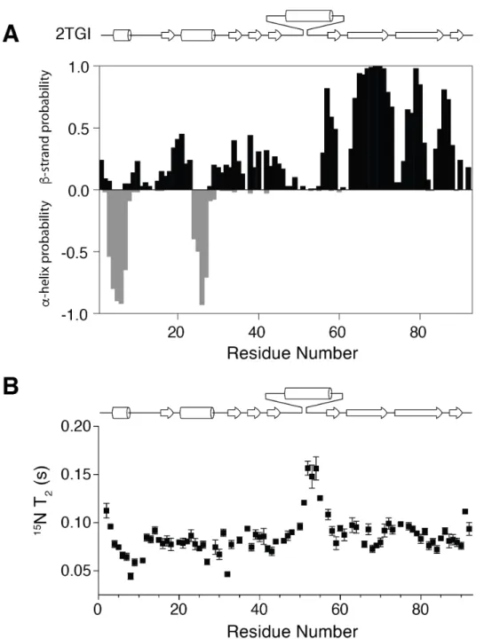
The folding and homogeneity of the isolated mmTGF-b2-7M was evaluated by NMR, and as with mmTGF-b2, the protein was found to have the expected number of signals in a 2D 1
H-15N shift correlation spectrum (Fig. 5A) as well as
secondary structure, as determined by an analysis of the NMR secondary shifts (Supplemental Fig. S3A). The solubility of mmTGF-b2-7M was evaluated as before, and as shown, its behavior was comparable or perhaps slightly better than that of mmTGF-b2 (Fig. 4C, D). This slight
improvement in the macroscopic solubility did not
at National Research Council Canada on September 5, 2017
http://www.jbc.org/
Engineered TGF-b Monomer that Blocks TGF-b Signaling however change the microscopic solubility as
NMR analysis showed that it was still necessary to include 10 mM CHAPS in the sample buffer in order to detect signals from all of the backbone amide resonances in the protein.
The three-dimensional structure of mmTGF-b2-7M was determined by
crystallography to a resolution of 2.75 Å (Table 1), and as before the overall fold was preserved relative to TGF-b2, with the only difference being a slight hinge bending of the monomer as
described for mmTGF-b2 (Fig. 5B, C). The increase in the 15N T2 relaxation times in the
region corresponding to the newly formed loop in mmTGF-b2-7M was comparable to that in mmTGF-b2 (Supplemental Fig. S3B). This suggested that the missing density in the region corresponding to the newly formed loop in mmTGF-b2-7M, which among the three
molecules in the asymmetric unit was observed for part of chain A and most of chain C, was not due to increased dynamics, but other factors, most likely the lower resolution of the mmTGF-b2-7M structure compared to the mmTGF-b2 structure (Table 1).
To determine whether mmTGF-b2-7M bound TbRII with high affinity, variants of mmTGF-b2-7M and TGF-b3 were produced bearing an N-terminal avitag, and after biotinylation and immobilization onto a streptavidin-coated SPR sensor, their binding affinity for TbRII was measured by performing kinetic SPR experiments (Fig. 3C, D). The sensorgrams obtained differed greatly from that previously obtained for mmTGF-b2 and TGF-b2, in that they exhibited a clear pattern of saturation. The sensorgrams were furthermore shown to have similar shapes as well as fitted parameters, including KD values (Table 2), which were within
experimental error of one another and consistent, although on the high end, of KD values reported
earlier for TbRII binding to TGF-b1 and TGF-b3 (38,40,41).
To determine if the interactions that enabled high affinity TbRII binding were preserved in mmb2-7M compared to TGF-b1 and TGF-b3, the mmTGF-b2-7M:TbRII complex was crystallized and its structure was determined to a resolution of 1.88 Å (Table1). The overall structure of the mmTGF-b2-7M:TbRII complex is shown to be very similar to that of one
of the TbRII-bound monomers from the structure of the TGF-b3:TbRII:TbRI complex, with TbRII bound to the mmTGF-b2-7M fingertips in a manner that is essentially indistinguishable from that of TGF-b3 (Fig. 5D). The interactions known to contribute most significantly to high affinity binding are furthermore shown to be fully preserved in the mmTGF-b2-7M:TbRII complex relative to TGF-b1:TbRII and TGF-b3:TbRII complexes that have been previously determined (the TGF-b3:TbRII complex determined to 1.8 Å (43) is shown as this is the highest resolution structure determined to date) (Fig. 5E). This includes the packing of I53 from TbRII in the hydrophobic pocket between the TGF-b fingers, and the hydrogen-bonded ion pairs formed between TGF-b R25 and R94 on the tips of the loops connecting fingers 1/2 and 3/4, respectively, and the carboxylate groups of E119 and D32 on TbRII (Fig. 5E).
Inhibitory activity of mmTGF-b2-7M and
the underlying mechanism - The results presented
above show that mmTGF-b2-7M possesses one of the essential attributes required to function as a dominant negative inhibitor of TGF-b signaling, that is the ability to bind TbRII with high affinity, comparable to that of TGF-b1 and TGF-b3. To directly assess whether mmTGF-b2-7M might signal, and if not, whether it might function as an inhibitor, TGF-b signaling was assessed by treating HEK293 cells stably transfected with a TGF-b luciferase reporter under the control of a CAGA12 promoter (44) with increasing
concentrations of TGF-bs. The results showed that dimeric TGF-b1 (TGF-b1) and full-length
monomeric TGF-b3 (mTGF-b3) resulted in a sigmoidal increase in the luciferase response, with concentrations of roughly 10 pM TGF-b1 and 100 pM mTGF-b3 leading to no further increase in the measured luciferase response. This is consistent with earlier reports which showed that (full-length) monomeric TGF-b1 and -b3 were 5 – 15 fold less potent than their dimeric counterparts (11,35). The normalized luciferase responses could be readily fitted to a standard model for ligand-dependent activation and yielded EC50 values of 12.4 ± 1.5
pM for TGF-b1 and 182 ± 16 pM for mTGF-b3. The values for TGF-b1 and mTGF-b3 are in close accord with the values previously reported by Amatayakul-Chantler for TGF-b1 (35) and by
at National Research Council Canada on September 5, 2017
http://www.jbc.org/
Engineered TGF-b Monomer that Blocks TGF-b Signaling Zúñiga and coworkers for mTGF-b3 (11). The
potent sub-nanomolar signaling activity observed for TGF-b1 and mTGF-b3 stands in contrast to that of mmTGF-b2-7M, which had no detectable signaling activity at the concentration that led to a saturating response for mTGF-b3 (ca. 200 pM) or at concentrations that were up to four orders of magnitude higher (Fig 6A). Thus, mmTGF-b2-7M was either completely devoid of signaling activity, or it possessed signaling activity, but with a potency more than a 10000-fold less than that of mTGF-b3.
To further investigate the properties of mmTGF-b2-7M, a competition experiment was performed in which the same HEK293 luciferase reporter cell line was stimulated with a constant sub-EC50 concentration of dimeric TGF-b1 (8.0
pM) and increasing concentrations of mTGF-b3 or mb2-7M. The results showed that mTGF-b3 further stimulated signaling with a midpoint concentration similar to that of mTGF-b3 alone (Fig. 6B). The fitted EC50 values confirm this, with
an EC50 of 182 ± 16 pM for the data shown in Fig.
6A and EC50 of 194 ± 36 pM for the data shown in
Fig. 6B. The behavior of mmTGF-b2-7M was very different, with no detectable change in the signaling activity when added up to concentrations of 10 nM, but with a sharp decrease to no
detectable signaling activity when the
concentration was increased to 100 nM (Fig. 6B). This shows that mmTGF-b2-7M indeed possesses no signaling activity and that it can function to completely block and inhibit TGF-b signaling. The normalized luciferase responses could be readily fitted to a standard model for ligand-dependent inhibition and yielded an IC50 value of
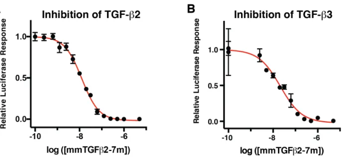
68 ± 7 nM. Similar experiments showed that mmTGF-b2-7M also functioned as a potent competitive inhibitor against the other TGF-b isoforms, TGF-TGF-b2 and TGF-TGF-b3, with measured IC50 values (TGF-b2 IC50 19 ± 3 nM and TGF-b3
IC50 21 ± 8 nM) within a factor of 2 -3 of that
measured for TGF-b1 (Supplement Fig. S4A, B). These IC50 values are on the lower end of the
range of affinities that have been reported for binding of the high affinity TGF-b isoforms to TbRII, including mmTGF-b2-7M reported here (Table 2). This suggests that mmTGF-b2-7M functions to inhibit TGF-b signaling in the manner anticipated, that is by binding to and blocking
endogenous TbRII. The fact that the measured potency is greater than the greatest affinity previously reported for TGF-b1 and TGF-b3 binding to TbRII (140 nM) (13), suggest that other factors, such as non-specific association of
mmTGF-b2-7M with the plasma membrane, may serve to potentiate its inhibitory activity.
The finding that mmTGF-b2-7M possesses no apparent signaling activity, and in fact functions as low nM inhibitor of TGF-b signaling, suggests that the elimination of a-helix 3 in fact diminished non-covalent association of the monomers and greatly attenuated or abrogated TbRI binding. To assess this directly, SPR experiments were performed to determine if mmTGF-β2-7M could recruit TbRI in the
presence of TbRII. To accomplish this, increasing concentrations of TβRI, as well as the same concentration series of TβRI in the presence of near-saturating amount of TβRII (2 µM) were injected over the same TGF-β3 and mmTGF-β2-7M SPR chip surfaces used for the TbRII binding measurements described above. This showed that TβRI alone binding is negligible to both TGF-β3 and mmTGF-β2-7M (Figures 3E, F), but unlike TGF-β3, TβRII-bound mmTGF-β2-7M is unable to recruit TβRI (Figures 3G, H). This is consistent with the earlier result reported by Huang and co-workers that TbRII-bound mTGF-b3 was
significantly or completely impaired in terms of its ability to bind and recruit TbRI (38). This also provides further evidence that TbRII-bound TGF-b monomers are incapaTGF-ble of TGF-binding and
recruiting TbRI, but because the mmTGF-b2-7M was immobilized on the surface of the sensor, it alone does not provide any insight as to whether mmTGF-b2-7M might be capable of non-covalently dimerizing and binding and recruiting TbRI.
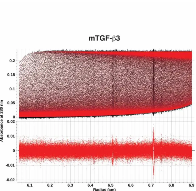
To address these questions directly, two solution based techniques were used, analytical ultracentrifugation (AUC) and time-resolved fluorescence resonance energy transfer (TR-FRET). The AUC experiments were performed by measuring the total UV absorbance at 280 nm as a function of the radial position and time as mTGF-b3, mmTGF-b2, and mmTGF-b2-7M were sedimented under acidic conditions (pH 3.8) where the monomers are fully soluble. The AUC data revealed parabolically-shaped van Holde-Weischet sedimentation coefficient distribution
at National Research Council Canada on September 5, 2017
http://www.jbc.org/
Engineered TGF-b Monomer that Blocks TGF-b Signaling plots for all three monomers (not shown),
consistent with each undergoing reversible self-association to form a dimer or other higher order oligomer. To determine more precisely which species might be present in solution, the data was fitted to the simplest model possible, a discrete monomer-dimer equilibrium, using finite element analysis as described in Experimental Procedures. The fitting procedure resulted in near-perfect fits for all three monomers to the simple monomer-dimer model, as shown by a) the close overlays between the fitted curves (red) with the raw data, after the time- and radially invariant noise was removed (black) and b) the absence of any
systemic deviations in the residuals (Supplemental Figs. S5 – S7). The fitted parameters further showed that KD for self-association was one order
of magnitude greater for mTGF-b3 compared to mmTGF-b2 and mmTGF-b2-7M. Thus, the removal of the heel helix, a3, does diminish self-association of the monomers to form dimers, but it does not completely abrogate dimer formation.
TR-FRET was used to assess the ability of dimeric and monomeric TGF-bs to bind and bring TbRI and TbRII together. This was accomplished by generating differentially tagged forms of TbRII and TbRI and in turn binding to these tags with proteins labeled with fluorescent donors and acceptors: TbRII was tagged with a C-terminal histag and was bound by a terbium cryptate-labeled Anti-His monoclonal antibody fluorescent donor and TbRI was tagged with an N-terminal avitag, which after enzymatic biotinylation, was bound to a dye-labeled (XL-665) streptavidin fluorescent acceptor (Fig. 7A). The addition of TGF-b to the tagged receptors brings them together and leads to a large increase in the DF value, which is defined as the ratio of the acceptor and donor emission fluorescent intensities. The TR-FRET assay is demonstrated by the data presented in Supplemental Figure S8 and was used here to compare the ability of the TGF-b3 full-length monomer, mTGF-b3, and the TGF-b2 mini monomer that binds TbRII with high affinity, mmTGF-b2-7M, to bind and bring TbRI and TbRII together. The TR-FRET signal for mTGF-β3 was shown to be comparable to that of TGF-b3 and this did not depend on whether the TGF-b concentration was 100 nM or 250 nM (Fig. 7B). The TR-FRET signal of mmTGF-b2-7M was, in contrast, within the error limits of the buffer
control and this also did not depend on the TGF-b concentration (Fig. 7B). These results demonstrate that under these conditions, mTGF-β3 retains full capacity to assemble a non-covalent dimeric complex with TbRI and TbRII, while under these same conditions, mmTGF-b2-7M has no capacity to do so. These results, together with the AUC results, suggest that the removal of the heel helix had the effects anticipated: its removal appears to have reduced, although not eliminated dimer formation, and even though dimers are still formed, they are evidently unable to bind and recruit TbRI.
DISCUSSION
The TGF-bs are responsible for promoting the progression of numerous human diseases (1-4), yet in spite of nearly two decades of preclinical studies and clinical trials, no inhibitors have been approved for use in humans. The results presented here demonstrate that an engineered TGF-b
monomer, lacking Cys77 and the heel a-helix (a3), functions to potently block and inhibit signaling of the TGF-b1, -b2, and –b3 with IC50s in the range
of 20 – 70 nM (Fig. 6B, Supplemental Fig. S4). This novel inhibitor has several attributes that may overcome limitations that have been encountered with other classes of inhibitors – for example, the natural high specificity of TGF-b, and thus the inhibitor, for TbRII may engender it with much greater specificity, and thus fewer undesirable side effects, compared to the much more promiscuous TGF-b kinase inhibitors. The small size of the inhibitor (ca. 10 kDa) may further engender it with a much greater ability to penetrate tumors and other dense tissues where the TGF-bs drive disease progression, a distinct advantage compared to IgG antibodies, which are much larger (ca. 150 kDa) and tend to occupy only the vascular and interstitial space of well-perfused organs (32,33). The other advantages of this novel inhibitor include its high intrinsic stability, owing to the four intramolecular disulfide bonds that tie the four fingers together, and the fact that it is highly soluble in water at neutral pH, unlike native TGF-b dimers or full-length TGF-TGF-b monomers.
The structures of TGF-b receptor complexes, together with the previous published chemical crosslinking data, suggested that the potent signaling activity of TGF-b1 C77S and TGF-b3 C77S was due to the ability of the monomers to non-covalently dimerize and in turn
at National Research Council Canada on September 5, 2017
http://www.jbc.org/
Engineered TGF-b Monomer that Blocks TGF-b Signaling assemble a (TbRI:TbRII)2 heterotetramer. The
results presented here, namely the AUC experiments which were used to assess non-covalent dimer formation, and the TR-FRET experiments which were used to assess assembly of complexes with TbRI and TbRII, provided further evidence for this. The AUC data showed that full length monomeric TGF-b3, mTGF-b3, self-associates to form dimers with a dimerization constant of 4.1 µM (Table 3). The TR-FRET data showed that at a concentration of 0.1 or 0.25 µM and in the presence of comparable concentrations of the TbRI and TbRII ectodomains, mTGF-b3 assembles TbRI:TbRII complexes to the same extent as dimeric TGF-b3 (Fig. 7B). That this occurs, even under conditions where the mTGF-b3 concentrations (0.1 – 0.25 µM, Fig. 7B) were more than an order of magnitude below the KD for
self-association (4.1 µM, Table 3), indicates that receptor binding also contributes significantly to assembly of TbRI:TbRII complexes. The assembly of TbRI:TbRII complexes with mTGF-b3, and presumably mTGF-b1 as well, therefore appears to be a cooperative process, much like protein folding, in which multiple weaker interactions, including monomer-monomer, non-covalent dimer-receptor, and receptor-receptor interactions, cooperate to enable formation of a thermodynamically stable TGF-b:TbRI:TbRII complex. This manner of cooperative assembly is likely responsible for the ability of mTGF-b1 and mTGF-b3 to induce signaling at concentrations that are more than four orders of magnitude below the KD for self-association of the monomers (EC50s
of about 0.1 nM vs. KDs for self-association of 4.1
µM).
The elimination of the heel helix from the TGF-b monomer was shown to be very effective in terms of blocking the cooperative assembly of TbRI:TbRII complexes as shown by the TR-FRET data (Fig. 7B) and the cell based signaling data (Fig. 6A-B). The AUC data showed that
elimination of the heel helix led to the weakening of the monomer-monomer interaction by one order of magnitude (Table 3). The SPR data shown in Figures 6G-H further showed that the TbRII-bound form of mmTGF-b2-7M was incapable of binding and recruiting TbRI, which is completely expected based on published structures of TGF-b receptor complexes which show that TbRI binds
to a composite interface formed by both chains of TGF-b, as well as TbRII (12,13). Thus, the data show that the reduced propensity of the engineered monomer to self-associate, together with what would be expected to be very weak binding of TbRI to any dimers that do form, is responsible for the inability of mmTGF-b2-7M to assemble a TbRI:TbRII complex. This accounts for the lack of signaling activity, and this together with the retention of high affinity TbRII binding, accounts for the inhibitory activity.
The other type II receptors of the family, ActRII, ActRIIB, BMPRII, and AMHRII, have either been shown or are predicted to bind the GF knuckle, not the GF fingertips as does TbRII (5). They nonetheless share the same property as TbRII in that they bind only by contacting residues from a single GF monomer, not both monomers as has been shown or is predicted for all type I receptors of the family (5). This, together with the structures reported here that show that it is possible to remove a3 without affecting the overall structure of the monomer (Figs. 2B-D,
5B-E), suggests that it might be possible to generate
monomers of other GFs of the family lacking the heel helix that function as inhibitors. These types of inhibitors have numerous potential applications, ranging from research tools for probing roles of specific ligands in vivo to clinically useful inhibitors for treating disease which are driven by hyperactive signaling by other ligands of the family, such as cancer cachexia by activin (45).
EXPERIMENTAL PROCEDURES Protein expression and purification -
TGF-b1 was expressed as a secreted protein bound to its prodomain in stably transfected CHO cells. The cell line used to produce TGF-b1, and the accompanying procedure to isolate the mature disulfide-linked TGF-b1 homodimer from the conditioned medium has been previously
described (46), and was kindly provided from Dr. Peter Sun (NIAID, Rockville, MD). Mouse homodimeric TGF-b2 (TGF-b2), human homodimeric TGF-b3 (TGF-b3), and variants, including homodimeric N-terminal avi-tagged (47) TGF-b3 (avi-TGF-b3), monomeric TGF-b2 (mTGF-b2), monomeric TGF-b3 (mTGF-b3), mini monomeric TGF-b1 (mmTGF-b1), mini monomeric TGF-b2 (mmTGF-b2), mini monomeric TGF-b3 (mmTGF-b3), mini
at National Research Council Canada on September 5, 2017
http://www.jbc.org/
Engineered TGF-b Monomer that Blocks TGF-b Signaling monomeric TGF-b2 with seven substitutions to
enable high affinity TbRII binding (mmTGF-b2-7M), and mini monomeric N-terminal avi-tagged (47) TGF-b2 with seven substitutions to enable high affinity TbRII binding (avi-mmTGF-b2-7M) were expressed in E. coli, refolded from inclusion bodies into native folded disulfide-linked
homodimers (TGF-b2, TGF-b3, avi-TGF-b3) or monomers (mTGF-b1, mTGF-b2, mTGF-b3, b1, b2, b3, mmTGF-b2-7M, avi-mmTGF-b2-7M), and purified to homogeneity using high resolution cation exchange chromatography (Source Q, GE Healthcare, Piscataway, NJ) as previously described (39). The nomenclature and features of the dimeric and monomeric TGF-bs used in this study are summarized in the Supplemental Table S1 and the complete sequences are shown in Supplemental Figure S1.
The human TbRI ectodomain (TbRI), spanning residues 1-101 of the mature receptor, or a variant spanning residues 1-88 of the mature receptor with a 15 amino acid avitag (47) appended to the C-terminus (TbRI-DC-Avi) was expressed in E. coli, refolded from inclusion bodies, and purified to homogeneity as previously described (11). The human TbRII ectodomain (TbRII), spanning residues 15-136 of the mature receptor, or the same but with a C-terminal hexahistidine tag (TbRII-His) was expressed in E. coli, refolded from inclusion bodies, and purified to
homogeneity as previously described (48).
Solubility Assays - TGF-b dimers and
monomers were prepared in 100 mM acetic acid to concentrations of 300 µM or higher and diluted to the desired concentration in either 100 mM acetic acid or phosphate buffered saline (PBS, 10 mM Na2HPO4, 1.8 mM KH2PO4, 137 mM NaCl, 2.7
mM KCl, pH 7.4). The pH of the samples diluted into PBS were adjusted with small aliquots of NaOH to ensure a final pH of 7.4. The light scattering at 340 nm of the samples were measured in a 1 cm quartz cuvette using a HP 8452 diode array spectrophotometer (HP, Palo Alto, CA). The samples were transferred to a microfuge tube, centrifuged at 20000 x g for 5 minutes and the absorbance at 280 nm of the supernatant was measured using a Nanodrop spectrophotomer (ThermoFisher, Waltham, MA).
NMR Spectroscopy. mmTGF-b2 and
mmTGF-b2-7M samples isotopically labeled with 15N or 15N
and 13C for NMR were prepared by growing bacterial cells in M9 media containing 0.1 % (w/v)
15
NH4Cl or 0.1 % (w/v) 15NH4Cl and 0.03% (w/v) 13
C labeled glucose. All NMR samples were prepared in 10 mM sodium phosphate, 10 mM 3-
[(3-cholamidopropyl)dimethylammonio]-1-propanesulfonate (CHAPS), and 5% 2H2O at a
protein concentration of 0.2 mM, pH 4.7. All NMR data was acquired at a sample temperature of 37 °C at either 700 or 800 MHz using Bruker AV-I or AV-II spectrometers equipped with a 5 mm 1H-{13C,15N} TCI cryogenically cooled probe (Bruker, Billerica, MA). Backbone resonance assignments of mmTGF-b2 and mmTGF-b2-7M were obtained by collecting and analyzing sensitivity-enhanced HNCACB (49),
CBCA(CO)NH (50), C(CO)NH (51), HNCO (52), data sets with 25% non-uniform sampling (NUS) of the points in the 13C,15N acquisition grid. Backbone amide 15N T2 relaxation parameters
were measured in an interleaved manner at 300 °K at a 15N frequency of 70.95 MHz using 1
H-detected pulse schemes previously described (53). The T2 data sets were each collected using 8 - 10
delay times, varying between 16 -192 ms. The T2
relaxation times were obtained by fitting relative peak intensities as a function of the T2 delay time
to a two parameter decaying exponential. Data was processed using NMRPipe (54), with the SMILE algorithm used for prediction of the missing points in the 13C and 15N dimensions of the NUS data sets (55). Data analysis was performed using
NMRFAM-SPARKY (56).
SPR binding measurements - SPR
measurements with TGF-b2 and mmTGF-b2 shown in Fig. 3A, B were performed using a Biacore 3000 SPR (G.E. Healthcare, Piscataway, NJ) instrument with direct immobilization of TGF-b2 or mmTGF-TGF-b2 on the surface of a CM5 sensor chip (G.E. Healthcare, Piscataway, NJ) using an amine (carbodiimide-based) coupling kit (G.E. Healthcare, Piscataway, NJ). SPR experiments shown in Figure 3C, E, and G and Figure 3D, F, and H with TGF-b3 and mmTGF-b2-7M, respectively, were performed using a Biacore X100 SPR instrument (G.E. Healthcare,
Piscataway, NJ) with biotinylated ligands captured at a moderate density (50 – 200 RU) onto a streptavidin-coated CM5 sensor chip (GE
Healthcare, Piscataway, NJ). Biotinylated TGF-b3 or mmTGF-b2-7M were generated by expressing
at National Research Council Canada on September 5, 2017
http://www.jbc.org/
Engineered TGF-b Monomer that Blocks TGF-b Signaling TGF-b3 or mmTGF-b2-7M with a N-terminal 15
amino acid avitag (47). TGF-b3-avi or mmTGF-b2-7M-avi was bound to TbRII in 10 mM bicine at pH 8.0 and biotinylated by incubating with a catalytic amount of bacterially expressed BirA recombinase, biotin, and ATP at 37 ° for 2 hr as described (39). Biotinylated avi-tagged TGF-b3 or avi-tagged TGF-b2-7M were bound to a C4 reverse phase column equilibrated with 94.9% water/5% acetonitrile/0.1% triflouroacetic acid and eluted with a linear acetonitrile gradient. SPR measurements shown in Figure 3A-F were performed in HBS-EP buffer (10 mM Hepes, pH 7.4, 150 mM NaCl, 3 mM EDTA, 0.005% surfactant P20 (GE Healthcare, Piscataway, NJ) with the receptor indicated injected over a series of two-fold dilutions over the concentration range shown. Injections were carried out in duplicates and included 10 buffer blank injections at the start of the experiment. Binding was allowed to
associate for 2 - 3 minutes at a flow rate of 100 µL min-1, followed by dissociation for 1 minute or longer. Each cycle of injection was followed by a 30 sec injection of 4 M guanidine•HCl, 2 M NaCl. Data was processed by subtracting both the response from a blank flow cell as well as buffer blanks using the program Scrubber2 (Biologic software, Campbell, Australia). Kinetic fitting of the data was performed with Scrubber2 assuming a simple 1:1 binding model. SPR measurements shown in Figure 3G, H were performed similarly, except 2 µM TbRII was included in both the running buffer and the injected samples.
Crystallization, structure determination and refinement - Crystals of mmTGF-b2 were
formed in sitting drops at 25 °C by combining 0.2 µL of a 7.9 mg mL-1 protein stock solution in 10 mM MES pH 5.5 with 0.2 µL of the precipitant from the well, 20 % PEG 3350, 0.2 M sodium thiocyanate. Harvested crystals were mounted in undersized nylon loops with excess mother liquor wicked off, followed by flash-cooling in liquid nitrogen prior to data collection. Data were acquired at the Advanced Photon Source NE-CAT beamline 24-ID-C and integrated and scaled using XDS (57). The structure was determined by the molecular replacement method implemented in PHASER (58) using a truncated version of PDB entry 2TGI (59) as the search model. Coordinates were refined using PHENIX (60), including simulated annealing with torsion angle dynamics,
and alternated with manual rebuilding using COOT (61). Data collection and refinement statistics are shown in Table 1.
Crystals of the mmTGF-β2-7M:TβRII complex were formed in hanging drops at 25 °C by
combining 1.0 µL of a 7.4 mg mL-1 stock solution of the complex in 10 mM Tris, pH 7.4 with 1.0 µL of 0.1 M Hepes, pH 7.5, 60 % v/v (+/-)-2-methyl-2,4-pentanediol. Harvested crystals were mounted in nylon loops, followed by flash-cooling in liquid nitrogen prior to data collection. Data were acquired at the Advanced Photon Source 22-ID-D and integrated and scaled using HKL2000 (62). The structure was determined by the molecular replacement method implemented in PHASER (58) using TbRII (PDB 1M9Z (63)) and mmTGF-β2 as search models. Coordinates were refined using PHENIX (60), alternated with manual rebuilding using COOT (61). Data collection and refinement statistics are shown in Table 1.
Crystals of mmTGF-b2-7M were formed in hanging drops at 25 °C by combining 1.0 µL of a 10 mg mL-1 protein stock solution in 20 mM acetic acid with 0.8 µL of the precipitant from the well, 100 mM sodium acetate dibasic trihydrate, pH 4.6, 25% 2-propanol, and 400 mM calcium chloride dehydrate, and 0.2 µL 5% n-ocyl-b-D-glucoside. Harvested crystals were mounted in nylon loops and cryoprotected in well buffer containing 20% glycerol and flash-cooled in a nitrogen stream. Data was collected at 100 K using a Rigaku FR-E Superbright generator equipped with a Saturn 944 CCD detector and processed using MOSFLM (64) in CCP4 (65). The structure of mmTGF-b2-7M was solved via molecular replacement using the structure of mmTGF-b2-7M from its co-crystal structure with TbRII. Iterative model building and refinement were performed using COOT (61) and PHENIX4, respectively. Data collection and refinement statistics are shown in Table 1.
Luciferase assays - HEK293 cells stably
transfected with the CAGA12 TGF-β reporter were
used for the luciferase reporter assays (44) and were maintained in Dulbecco’s modified eagles medium (DMEM) containing 10% fetal bovine serum (FBS) and 1% penicillin/streptomycin. Cells were treated for 16 hours with a TGF-β (TGF-b1, mTGF-b3, or mmTGF-b2-7M) concentrations series or a mmTGF-b2-7M concentration series in the presence of a constant sub-saturating concentration of TGF-b (TGF-β1 –
at National Research Council Canada on September 5, 2017
http://www.jbc.org/
Engineered TGF-b Monomer that Blocks TGF-b Signaling 8 pM, TGF-b2 – 20 pM, or TGF-b3 – 10 pM).
Proteins were diluted in Dulbecco’s Modified Eagle’s medium (DMEM) containing 0.1% w/v BSA. After 16 hours cells were lysed with Tropix lysis buffer (ThermoFisher, Waltham, MA) and luciferase activity was read with a Promega GloMax luminometer (Promega, Madison, WI). Luciferase activity was normalized to total protein levels determined by bicinchoninic acid (BCA) protein assay. Graphpad Prism 6 was used to fit the data to standard models for ligand activity (EC50) and ligand inhibitory activity (IC50)
(Graphpad, LaJolla, CA).
Time-resolved FRET assays - The
following purified proteins were used to address the ligand requirements for the formation of complexes containing TbRI and TbRII: TGF-b3, mTGF-b3, mmTGF-b2-7M, biotinylated TbRI-DC-Avi and TbRII-His. Initially 20 µM binary complexes of TGF-b3:TbRII-His, mTGF-b3:TbRII-His, and mmTGF-b2-7M:TbRII-His were formed in a 50 mM Tris, pH 7.5 buffer and stored at 4° C. A time-resolved fluorescence resonance energy transfer (TR-FRET) assay based on the proximity-dependent transfer of
fluorescence from the donor terbium cryptate labeled anti-His mAb (Tb-anti-His, CisBio, Bedford, MA) to the acceptor XL665 labeled streptavidin (SA-665, CisBio, Bedford, MA) was used to monitor the assembly of ternary
ligand:TbRII-His:biotinylated TbRI-DC-Avi complexes. 50 µL assays containing 100 nM or 250 nM TGF-b3:TbRII-His (1:2), mTGF-b3:TbRII-His (1:1), and mmTGF-b2-7M:TbRII-His (1:1) complexes were incubated with 50 nM biotinylated TbRI-DC-Avi. Each 50ul ternary complex formation assay also contained 2 nM Tb-anti-His and 30nM SA-665 and was incubated at room temperature for 2 h. Each condition was tested in replicates of six. Buffer control (n=6) contained only 2 nM Tb-anti-His and 30 nM SA-XL665. The buffer conditions for each assay were 50 mM Tris, 50 mM NaCl, pH 7.5. The assays were performed in Corning black 384 well low flange microplates (ThermoFisher, Waltham, MA). After a 2 h incubation, the assay plate was
measured for terbium/XL-665 TR-FRET on a BMG Labtech Pherastar FS multimode plate reader (BMG Labtech Inc., Cary, NC). An optic module containing 337, 490 and 665 nm filters was used to monitor TR-FRET producing raw data
for 337/490 (terbium emission) and 337/665 (XL-665) emission. The ratio of 665 emission/490 emission was determined for each condition and was subsequently used to calculate DF, which is a measure that reflects the signal of the sample versus the background. DF was calculated using the following equation: (Ratiosignal
-Rationegative/Rationegative) x 100. The Ratiosignal refers
to the assays containing the trimeric complexes or buffer control. The Rationegative refers to two buffer
control assays (2 nM Tb-anti-His and 30 nM SA-665). For the buffer control, 2 out of the 6 replicates were assigned as negative controls for the purpose of calculating DF. DF was calculated for the remaining 4 buffer control replicates.
Analytical ultracentrifugation - mTGF-b3,
mmTGF-b2, and mmTGF-b2-7M were analyzed by sedimentation velocity to establish equilibrium constants for self-association of monomeric TGF-bs to form homodimers. mTGF-b3, mmTGF-b2, and mmTGF-b2-7M were each measured at 280 nm in an epon two channel centerpiece fitted with quartz windows, and centrifuged at 20°C and 42,000 rpm for 27 hours in a 15 mM sodium phosphate buffer adjusted to pH 3.8, containing 100 mM NaCl. 300 scans were collected in intensity mode on a Beckman Optima XL-I analytical ultracentrifuge at the CAUMA facility at the UTHSCSA. Data analysis was performed with UltraScan release 2142 (66,67), calculations were performed at the San Diego Supercomputing Center on Comet and Gordon. The sedimentation velocity data were initially fitted with the two-dimensional spectrum analysis as described in (66) to remove time- and radially invariant noise from the raw data, and to fit the meniscus position. Subsequently, the data were fitted to a discrete monomer-dimer model using the adaptive space-time finite element method (67) and genetic algorithms for the parameter optimization (68). The monomer-dimer model accounts for mass action and the reversible association behavior, fitting the thermodynamic and hydrodynamic parameters, as well as the partial specific volume while assuming the predicted molar mass for either wildtype or mutant. A Monte Carlo analysis (69) with 100 iterations was performed for each dataset to obtain fitting statistics. Buffer density and viscosity were estimated with UltraScan based on buffer composition and all hydrodynamic values were corrected for standard conditions
at National Research Council Canada on September 5, 2017
http://www.jbc.org/
Engineered TGF-b Monomer that Blocks TGF-b Signaling (20°C and water). The fitting results provided an
excellent fit with random residuals and very low
RMSD values (see Supplementary Material, Figs. 4, 5, and 6). All results are summarized in Table 3.
ACKNOWLEDGEMENTS: The authors would like to thank Dr. Peter Sun NIH NIAID, for providing
the stably transected CHO cell line overexpressing TGF-b1, Dr. William Furey for guidance on the refinement of the mmTGF-b2-7M and mmTGF-b2-7M:TbRII structures, Doowon Lee for assistance with the X-ray instrumentation, Mike Delk for assistance with the NMR instrumentation, and Liping Wang for technical assistance with the analytical ultracentrifugation measurements. The assigned chemical shifts for mmTGF-b2 and mmTGF-b2-7M were deposited to BioMagResBank (BMRB) under accession codes 26943 and 26944, respectively. The structures of mmTGF-b2, 7M, and the mmTGF-b2-7M:TbRII complex were submitted to the RCSB Protein Data Bank (PDB) under accession codes 5TX2, 5TX6, and 5TX4 respectively.
CONFLICT OF INTEREST: The content is solely the responsibility of the authors and does not
necessarily represent the official views of the National Institutes of Health. A.P.H. and T.S. are co-inventors of a provisional patent (application US 62/423,920) that covers the dominant negative TGF-b, mmTGF-b2-7M.
AUTHOR CONTRIBUTIONS: SKK crystallized mmTGF-b2 and the mmTGF-b2-7M:TbRII complex,
performed the solubility measurements, performed a portion of the SPR experiments, and wrote the initial draft of the paper. LB and CSH performed the luciferase assays. CSH together with MB, MV, BL, BR, and KEC established the expression and purification of mmTGF-b2-7M. AT, BI, and KEC performed the NMR assignment of mmTGF-b2. EMP crystallized and determined the structure of mmTGF-b2-7M. ABT and PJH determined the structure of mmTGF-b2. RK performed some of the SPR experiments. SD, COB, and GC determined the structure of mmTGF-b2-7M:TbRII. MJH performed the TR-FRET
experiments and BD performed the AUC experiments. APH conceived the design of the dominant negative TGF-b inhibitors, in consultation with TS. APH also performed the NMR assignment of mmTGF-b2-7M and wrote the final draft of the paper. All authors reviewed the results and approved the final version of the manuscript.
REFERENCES
1. Biernacka, A., Dobaczewski, M., and Frangogiannis, N. G. (2011) TGF-beta signaling in fibrosis. Growth Factors 29, 196-202
2. Dietz, H. C., Cutting, G. R., Pyeritz, R. E., Maslen, C. L., Sakai, L. Y., Corson, G. M.,
Puffenberger, E. G., Hamosh, A., Nanthakumar, E. J., Curristin, S. M., and et al. (1991) Marfan syndrome caused by a recurrent de novo missense mutation in the fibrillin gene. Nature 352, 337-339
3. Loeys, B. L., Mortier, G., and Dietz, H. C. (2013) Bone lessons from Marfan syndrome and related disorders: fibrillin, TGF-B and BMP at the balance of too long and too short. Pediatr Endocrinol Rev 10 Suppl 2, 417-423
4. Massague, J. (2008) TGFbeta in Cancer. Cell 134, 215-230
5. Hinck, A. P., Mueller, T. D., and Springer, T. A. (2016) Structural Biology and Evolution of the TGF-beta Family. Cold Spring Harb Perspect Biol
6. Massague, J. (1998) TGF-beta signal transduction. Annu Rev Biochem 67, 753-791
7. Robertson, I. B., and Rifkin, D. B. (2013) Unchaining the beast; insights from structural and evolutionary studies on TGFbeta secretion, sequestration, and activation. Cytokine Growth Factor Rev 24, 355-372
8. Wrana, J. L., Attisano, L., Carcamo, J., Zentella, A., Doody, J., Laiho, M., Wang, X. F., and Massague, J. (1992) TGF beta signals through a heteromeric protein kinase receptor complex. Cell 71, 1003-1014
at National Research Council Canada on September 5, 2017
http://www.jbc.org/
Engineered TGF-b Monomer that Blocks TGF-b Signaling 9. Wrana, J. L., Attisano, L., Wieser, R., Ventura, F., and Massague, J. (1994) Mechanism of
activation of the TGF-beta receptor. Nature 370, 341-347
10. Laiho, M., Weis, F. M., Boyd, F. T., Ignotz, R. A., and Massague, J. (1991) Responsiveness to transforming growth factor-beta (TGF-beta) restored by genetic complementation between cells defective in TGF-beta receptors I and II. J Biol Chem 266, 9108-9112
11. Zuniga, J. E., Groppe, J. C., Cui, Y., Hinck, C. S., Contreras-Shannon, V., Pakhomova, O. N., Yang, J., Tang, Y., Mendoza, V., Lopez-Casillas, F., Sun, L., and Hinck, A. P. (2005) Assembly of TbetaRI:TbetaRII:TGFbeta ternary complex in vitro with receptor extracellular domains is cooperative and isoform-dependent. J Mol Biol 354, 1052-1068
12. Groppe, J., Hinck, C. S., Samavarchi-Tehrani, P., Zubieta, C., Schuermann, J. P., Taylor, A. B., Schwarz, P. M., Wrana, J. L., and Hinck, A. P. (2008) Cooperative assembly of TGF-beta superfamily signaling complexes is mediated by two disparate mechanisms and distinct modes of receptor binding. Mol Cell 29, 157-168
13. Radaev, S., Zou, Z., Huang, T., Lafer, E. M., Hinck, A. P., and Sun, P. D. (2010) Ternary complex of transforming growth factor-beta1 reveals isoform-specific ligand recognition and receptor recruitment in the superfamily. J Biol Chem 285, 14806-14814
14. Loeys, B. L., Schwarze, U., Holm, T., Callewaert, B. L., Thomas, G. H., Pannu, H., De Backer, J. F., Oswald, G. L., Symoens, S., Manouvrier, S., Roberts, A. E., Faravelli, F., Greco, M. A., Pyeritz, R. E., Milewicz, D. M., Coucke, P. J., Cameron, D. E., Braverman, A. C., Byers, P. H., De Paepe, A. M., and Dietz, H. C. (2006) Aneurysm syndromes caused by mutations in the TGF-beta receptor. N Engl J Med 355, 788-798
15. Markowitz, S. D., and Roberts, A. B. (1996) Tumor suppressor activity of the TGF-beta pathway in human cancers. Cytokine & growth factor reviews 7, 93-102
16. Bandyopadhyay, A., Agyin, J. K., Wang, L., Tang, Y., Lei, X., Story, B. M., Cornell, J. E., Pollock, B. H., Mundy, G. R., and Sun, L. Z. (2006) Inhibition of pulmonary and skeletal metastasis by a transforming growth factor-beta type I receptor kinase inhibitor. Cancer Res 66, 6714-6721
17. Ganapathy, V., Ge, R., Grazioli, A., Xie, W., Banach-Petrosky, W., Kang, Y., Lonning, S., McPherson, J., Yingling, J. M., Biswas, S., Mundy, G. R., and Reiss, M. (2010) Targeting the Transforming Growth Factor-beta pathway inhibits human basal-like breast cancer metastasis. Mol Cancer 9, 122
18. Muraoka, R. S., Dumont, N., Ritter, C. A., Dugger, T. C., Brantley, D. M., Chen, J., Easterly, E., Roebuck, L. R., Ryan, S., Gotwals, P. J., Koteliansky, V., and Arteaga, C. L. (2002) Blockade of TGF-beta inhibits mammary tumor cell viability, migration, and metastases. J Clin Invest 109, 1551-1559
19. Nam, J. S., Terabe, M., Mamura, M., Kang, M. J., Chae, H., Stuelten, C., Kohn, E., Tang, B., Sabzevari, H., Anver, M. R., Lawrence, S., Danielpour, D., Lonning, S., Berzofsky, J. A., and Wakefield, L. M. (2008) An anti-transforming growth factor beta antibody suppresses metastasis via cooperative effects on multiple cell compartments. Cancer Res 68, 3835-3843
20. Yang, Y. A., Dukhanina, O., Tang, B., Mamura, M., Letterio, J. J., MacGregor, J., Patel, S. C., Khozin, S., Liu, Z. Y., Green, J., Anver, M. R., Merlino, G., and Wakefield, L. M. (2002) Lifetime exposure to a soluble TGF-beta antagonist protects mice against metastasis without adverse side effects. J Clin Invest 109, 1607-1615
21. Miao, Z. F., Zhao, T. T., Wang, Z. N., Miao, F., Xu, Y. Y., Mao, X. Y., Gao, J., Wu, J. H., Liu, X. Y., You, Y., Xu, H., and Xu, H. M. (2014) Transforming growth factor-beta1 signaling blockade attenuates gastric cancer cell-induced peritoneal mesothelial cell fibrosis and alleviates peritoneal dissemination both in vitro and in vivo. Tumour Biol 35, 3575-3583
22. Di Sabatino, A., Jackson, C. L., Pickard, K. M., Buckley, M., Rovedatti, L., Leakey, N. A., Picariello, L., Cazzola, P., Monteleone, G., Tonelli, F., Corazza, G. R., MacDonald, T. T., and Pender, S. L. (2009) Transforming growth factor beta signalling and matrix metalloproteinases in the mucosa overlying Crohn's disease strictures. Gut 58, 777-789
at National Research Council Canada on September 5, 2017
http://www.jbc.org/
Engineered TGF-b Monomer that Blocks TGF-b Signaling 23. Yamada, M., Kuwano, K., Maeyama, T., Yoshimi, M., Hamada, N., Fukumoto, J., Egashira, K.,
Hiasa, K., Takayama, K., and Nakanishi, Y. (2007) Gene transfer of soluble transforming growth factor type II receptor by in vivo electroporation attenuates lung injury and fibrosis. J Clin Pathol 60, 916-920
24. Connolly, E. C., Freimuth, J., and Akhurst, R. J. (2012) Complexities of TGF-beta targeted cancer therapy. Int J Biol Sci 8, 964-978
25. Hawinkels, L. J., and Ten Dijke, P. (2011) Exploring anti-TGF-beta therapies in cancer and fibrosis. Growth Factors 29, 140-152
26. Ge, R., Rajeev, V., Subramanian, G., Reiss, K. A., Liu, D., Higgins, L., Joly, A., Dugar, S., Chakravarty, J., Henson, M., McEnroe, G., Schreiner, G., and Reiss, M. (2004) Selective inhibitors of type I receptor kinase block cellular transforming growth factor-beta signaling. Biochem Pharmacol 68, 41-50
27. Singh, J., Ling, L. E., Sawyer, J. S., Lee, W. C., Zhang, F., and Yingling, J. M. (2004)
Transforming the TGFbeta pathway: convergence of distinct lead generation strategies on a novel kinase pharmacophore for TbetaRI (ALK5). Curr Opin Drug Discov Devel 7, 437-445
28. Yingling, J. M., Blanchard, K. L., and Sawyer, J. S. (2004) Development of TGF-beta signalling inhibitors for cancer therapy. Nat Rev Drug Discov 3, 1011-1022
29. Connolly, E. C., Saunier, E. F., Quigley, D., Luu, M. T., De Sapio, A., Hann, B., Yingling, J. M., and Akhurst, R. J. (2011) Outgrowth of drug-resistant carcinomas expressing markers of tumor aggression after long-term TbetaRI/II kinase inhibition with LY2109761. Cancer research 71, 2339-2349
30. Lonning, S., Mannick, J., and McPherson, J. M. (2011) Antibody targeting of TGF-beta in cancer patients. Curr Pharm Biotechnol 12, 2176-2189
31. Hinck, A. P. (2012) Structural studies of the TGF-betas and their receptors - insights into evolution of the TGF-beta superfamily. FEBS Lett 586, 1860-1870
32. Meibohm, B. (2012) Pharmacokinetics and Half-Life of Protein Therapeutics. in Therapeutic Proteins: Strategies to Modulate Their Plasma Half-Lives (Kontermann, R. ed.), Wiley-Blackwell, Weinheim, Germany. pp 23-38
33. Meibohm, B., and Braeckman, R. A. (2007) Pharmacokinetics and Pharmacodynamics of
Peptides and Proteins. in Pharmaceutical Biotechnology: Concepts and Applications (Crommelin, D. J. A., Sindelar, R. D., and Meibohm, B. eds.). pp 95-123
34. Papo, N., Silverman, A. P., Lahti, J. L., and Cochran, J. R. (2011) Antagonistic VEGF variants engineered to simultaneously bind to and inhibit VEGFR2 and alphavbeta3 integrin. Proc Natl Acad Sci U S A 108, 14067-14072
35. Amatayakul-Chantler, S., Qian, S. W., Gakenheimer, K., Bottinger, E. P., Roberts, A. B., and Sporn, M. B. (1994) [Ser77]transforming growth factor-beta 1. Selective biological activity and receptor binding in mink lung epithelial cells. J Biol Chem 269, 27687-27691
36. Hinck, A. P., Archer, S. J., Qian, S. W., Roberts, A. B., Sporn, M. B., Weatherbee, J. A., Tsang, M. L., Lucas, R., Zhang, B. L., Wenker, J., and Torchia, D. A. (1996) Transforming growth factor beta 1: three-dimensional structure in solution and comparison with the X-ray structure of
transforming growth factor beta 2. Biochemistry 35, 8517-8534
37. Mittl, P. R., Priestle, J. P., Cox, D. A., McMaster, G., Cerletti, N., and Grutter, M. G. (1996) The crystal structure of TGF-beta 3 and comparison to TGF-beta 2: implications for receptor binding. Protein Sci 5, 1261-1271
38. Huang, T., David, L., Mendoza, V., Yang, Y., Villarreal, M., De, K., Sun, L., Fang, X., Lopez-Casillas, F., Wrana, J. L., and Hinck, A. P. (2011) TGF-beta signalling is mediated by two autonomously functioning TbetaRI:TbetaRII pairs. EMBO J 30, 1263-1276
39. Huang, T., and Hinck, A. P. (2016) Production, Isolation, and Structural Analysis of Ligands and Receptors of the TGF-beta Superfamily. Methods Mol Biol 1344, 63-92
40. Baardsnes, J., Hinck, C. S., Hinck, A. P., and O'Connor-McCourt, M. D. (2009) TbetaR-II discriminates the high- and low-affinity TGF-beta isoforms via two hydrogen-bonded ion pairs. Biochemistry 48, 2146-2155
at National Research Council Canada on September 5, 2017
http://www.jbc.org/