Model of the Human Large Intestine and Interactions with Probiotic
Yeasts and Resident Microbiota
Jonathan Thévenot,a,bLucie Etienne-Mesmin,a,bSylvain Denis,aSandrine Chalancon,aMonique Alric,aValérie Livrelli,b,c Stéphanie Blanquet-Diota
Clermont Université, Université d’Auvergne, Centre de Recherche en Nutrition Humaine Auvergne, EA 4678 CIDAM, Conception Ingénierie et Développement de l’Aliment et du Médicament, Clermont-Ferrand,aClermont Université, Université d’Auvergne, Centre de Recherche en Nutrition Humaine Auvergne, M2iSH, Microbes, Intestin, Inflammation et Susceptibilité de l’Hôte UMR INSERM/Université d’Auvergne U1071 USC-INRA 2018, Clermont-Ferrand,bCHU Clermont-Ferrand, Service de Bactériologie, Clermont-Ferrand,cFrance
This is the first report on the fate of enterohemorrhagic Escherichia coli O157:H7 in simulated human colonic conditions. The pathogen was progressively eliminated from the bioreactor and did not modify the major populations of resident microbiota. The coadministration of the Saccharomyces cerevisiae CNCM I-3856 probiotic strain led to a significant increase in acetate pro-duction but did not reduce pathogen viability.
E
nterohemorrhagic Escherichia coli (EHEC) is a major food-borne zoonotic agent associated with outbreaks worldwide. Human contamination occurs mainly after consumption of raw or undercooked ground beef, vegetables, water, and dairy prod-ucts contaminated by bovine feces (1). EHEC causes illnesses ranging from uncomplicated diarrhea to life-threatening compli-cations, such as hemolytic-uremic syndrome. Most of the out-breaks and sporadic cases are caused by EHEC strains belonging to serotype O157:H7, but other serotypes can be involved. For in-stance, EHEC of the O104:H4 serotype was responsible for a large outbreak in June 2011 in Europe (2,3). The virulence of EHEC strains is mainly associated with their ability to damage intestinal epithelial cells and produce Shiga toxins Stx1 and/or Stx2.Although the terminal ileum and colon are assumed to be the main sites of EHEC colonization and pathology in humans (4,5), data on bacterial survival and regulation of virulence genes in the human intestine are scant. Human studies are obviously unethical when pathogenic microorganisms are involved, and no animal model can reproduce the physiopathology of EHEC infection as a whole (6). Hence, for technical, economic, and ethical reasons, in
vitro models offer a relevant alternative to in vivo studies (7,8). EHEC survival was recently evaluated in a dynamic in vitro system reproducing the luminal conditions of the human stomach and small intestine (9), but no data are available on their viability in human large intestinal conditions. Recent studies have suggested that human intestinal microbiota may play a key role in the out-come of EHEC infection (10,11). de Sablet et al. (10) showed, in
vitro and ex vivo, using the cecal contents of human digestive
mi-crobiota-associated rats, that soluble factors released by the hu-man microbiota repressed Stx synthesis in EHEC O157:H7. Con-versely, DNase colicins produced by colicinogenic flora may enhance Stx production (11). Moreover, Kendall et al. (12) showed that ethanolamine, a major component of mammalian and bacterial membranes found in the large intestine, regulates O157:H7 virulence gene expression.
Since antibiotic therapy to treat EHEC infection is controver-sial (13, 14), probiotics are being investigated as an alternative strategy. Probiotics are defined as live microorganisms that resist digestion and reach the colon alive and that when administered in
adequate amounts, confer a health benefit on the host (15). They can exert protective effects against enteric pathogens via direct antagonism, immunomodulation, and/or competitive exclusion (16). Probiotic yeasts have already shown beneficial effects in the control of EHEC infection. Saccharomyces cerevisiae var. boulardii (S. boulardii) reduces O157:H7 growth in ruminal fluids (17) and decreases proinflammatory pathways in EHEC-infected T84 hu-man colonic cells (18,19). Using an in vitro model of the human gastrointestinal tract, Etienne-Mesmin et al. (9) also showed that another probiotic yeast, S. cerevisiae CNCM I-3856, significantly decreases EHEC O157:H7 growth resumption in the distal parts of the small intestine, possibly through the combined action of eth-anol and other inhibitory substances.
This study used the in vitro model of the human large intestine ARCOL (artificial colon) to (i) evaluate the survival of an EHEC O157:H7 strain in simulated human colonic conditions and (ii) investigate the effect of probiotic treatment with S. cerevisiae strains. In addition, the influence of both pathogen and probiotics on the main populations of resident microbiota was also assessed.
Fermentation in the ARCOL. ARCOL is a one-stage
fermen-tation system (Applikon, Schiedam, The Netherlands) that inte-grates the main parameters of the in vivo human colonic environ-ment (20), including pH, temperature, supply of ileal effluents, retention time, anaerobiosis maintained by the sole activity of resident microbiota, and passive absorption of water and fermen-tation metabolites by dialysis fibers (Table 1). The bioreactor was inoculated with fresh feces from a healthy volunteer who had no history of antibiotic treatment 3 months before the study, and it was used under semicontinuous conditions.
Received 26 October 2012 Accepted 27 November 2012 Published ahead of print 30 November 2012
Address correspondence to Stéphanie Blanquet-Diot, stephanie.blanquet @udamail.fr.
Copyright © 2013, American Society for Microbiology. All Rights Reserved. doi:10.1128/AEM.03303-12
Two fermentations were carried out (Fig. 1). Both started after a 3-day stabilization phase (phase A) and were divided into three different 3-day phases (B, C, and D). In fermentation 1 (F1), E. coli O157:H7 (an isogenic mutant of the reference strain EDL 933 lacking the stx1gene but harboring stx2) (21) was first
adminis-tered alone (phase B), then coadminisadminis-tered with S. boulardii (Bio-codex, Gentilly, France) (phase C), and finally coadministered with S. cerevisiae CNCM I-3856 (Lesaffre Human Care, Milwau-kee, WI) (phase D). In fermentation 2 (F2), phases C and D were inverted to ensure that there was no carryover of treatment effects between phases. An aerobic culture (LB, 24 h, 37°C) of E. coli O157:H7 was introduced in the vessel (105CFU/ml of colonic medium) (9), whereas yeasts were supplied in active dried powder form (107CFU/ml of colonic medium) (22). Samples were regu-larly collected from the colonic medium and plated onto selective media (Table 2) to determine O157:H7 and probiotic yeast sur-vival kinetics. Sursur-vival rates were assessed by comparing the pro-files obtained for bacteria and yeasts with that of a theoretical marker which simulates the behavior of an inert (i.e., nonde-graded and nonabsorbed) compound. Its removal from the bio-reactor was described by Ct⫽ C0⫻ e(⫺t/), where Ctis the
concen-tration of the marker at time t, C0its initial concentration, t the
time of fermentation, and the residence time (23). In parallel, the main bacterial populations of human intestinal microbiota were measured by plate counts (Table 2) and real-time quantitative PCR (qPCR) analysis. Total genomic DNA was extracted from 250 l colonic medium by using the two first steps of Yu and Morri-son’s protocol (24) and the QIAamp DNA stool minikit (Qiagen, Courtaboeuf, France) according to the manufacturer’s instruc-tions. Specific primers used in this study are listed inTable 3. PCR amplification and detection were performed with a Stratagene Mx3005P QPCR system (Agilent Technologies, Massy, France).
Ethanol concentrations in the colonic medium were determined by an enzymatic UV test (Biosentec, Toulouse, France). Dialysis outflow was daily sampled to determine short-chain fatty acid (SCFA) production by gas chromatography, as described by Gérard-Champod et al. (30). For all survival kinetics, significant differences between treatments were tested by repeated-measure two-way analysis of variance (ANOVA) followed by a Bonferroni correction for multiple comparisons. For data on intestinal bac-terial populations and SCFA production, a Kruskal-Wallis test was performed followed, if the results were significant, by a Mann-Whitney test for pairwise comparisons (GraphPad Software, Inc., La Jolla, CA). Values of P⬍ 0.05 were taken as indicating statisti-cally significant differences.
Survival of E. coli O157:H7 in simulated human colonic con-ditions. This study is the first report of EHEC O157:H7 survival
kinetics in a one-stage fermentation system reproducing human large intestinal conditions (Fig. 2). The pathogen was progres-sively eliminated from the bioreactor, more rapidly than the the-oretical transit marker. After 36 h fermentation, 4.2⫾ 0.03 log10
CFU/ml of viable bacteria (n⫽ 2) was recovered in the bioreactor, i.e., less than 5% of the initial intake versus 35% for the marker. These results indicate that the pathogen did not persist in this environment at a level similar to that of the marker, probably due to the barrier effect of intestinal microbiota. Resident microbiota can prevent the establishment of intestinal pathogen by producing antimicrobial substances, such as bacteriocins, or through com-petition for nutrients or ecological niches (31). In particular, E. TABLE 1 Parameters of in vitro fermentations in the ARCOL system
Parameters of in vitro fermentation Exptl conditions
Temp 37°C
Continuous stirring 400 rpm
pH 6.3
Pressure 1.1 bar
Vol of fermentative medium 450 ml
Retention time 36 h
Redox potential ⫺400 mV
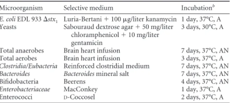
FIG 1 Schedule of in vitro fermentations in the ARCOL system. TABLE 2 Selective media useda
Microorganism Selective medium Incubationb
E. coli EDL 933⌬stx1 Luria-Bertani⫹ 100 g/liter kanamycin 1 day, 37°C, A
Yeasts Sabouraud dextrose agar⫹ 50 mg/liter chloramphenicol⫹ 10 mg/liter gentamicin
3 days, 30°C, A
Total anaerobes Brain heart infusion 7 days, 37°C, AN Total aerobes Brain heart infusion 3 days, 37°C, A
Clostridia/Eubacteria Reinforced clostridial medium 7 days, 37°C, AN
Bacteroides Bacteroides mineral salt 7 days, 37°C, AN
Bifidobacteria Beerens 4 days, 37°C, AN
Enterobacteriaceae MacConkey 1 day, 37°C, A
Enterococci D-Coccosel 2 days, 37°C, A
a
Overview of the selective media used to enumerate E. coli O157:H7, probiotic yeasts, and the main cultivable bacterial populations of the human intestinal microbiota.
b
coli strains producing colicins or microcins have been reported to
be effective in vitro in inhibiting E. coli O157:H7 (32,33). EHEC is considered to be a colonic pathogen. However, in the present study, E. coli O157:H7 does not persist in the colonic medium. Previous findings (4,9,34,35) suggest that the distal small intes-tine would play a major role in EHEC infection, whence bacteria can spread to the colon as colonization becomes established.
Yeast viability in the ARCOL system. As survival in the
hu-man gastrointestinal tract is a key feature of probiotic strains, yeast viability was followed during large intestine in vitro fermentations. Both strains were rapidly eliminated from the bioreactor (Fig. 3). However, S. cerevisiae CNCM I-3856 was significantly (P⬍ 0.05) more affected by colonic conditions than S. boulardii, no viable yeast being recovered after 36 h and 60 h, respectively. S. boulardii is a variant of S. cerevisiae with specific genomic and phenotypic TABLE 3 Primer and probe sequences used in real-time qPCR assays
Name Sequence 5=–3= Target
Annealing
temp (°C) Reference SYBR green
BAC338F ACTCCTACGGGAGGCAG Total bacteria 58 25
BAC516F GTATTACCGCGGCTGCTG
789cfbF CRAACAGGATTAGATACCCT Bacteroidetes 61 26
cfb967R GGTAAGGTTCCTCGCGTAT
Act920F3 TACGGCCGCAAGGCTA Actinobacteria 61 26
Act1200R TCRTCCCCACCTTCCTCCG
928F-Firm TGAAACTYAAAGGAATTGACG Firmicutes 61 26
1040FirmR ACCATGCACCACCTGTC
Eco1457F CATTGACGTTACCCGCAGAAGAAGC Enterobacteriaceae 63 27
Eco1652R CTCTACGAGACTCAAGCTTGC
F_Lacto05 AGCAGTAGGGAATCTTCCA Lactobacillus/Pediococcus/Leuconostoc 60 28
R_Lacto04 CGCCACTGGTGTCTYTCCATATA
TaqMan
F_Bact 1369 CGGTGAATACGTTCCCGG
P_TM1389F FAM-CTTGTACACACCGCCCGTC-TAMRA Total bacteria 60 28
R_Prok1492R TACGGCTACCTTGTTACGACTT
E. coli-F CATGCCGCGTGTATGAAGAA
E. coli-P FAM-TATTAACTTTACTCCCTTCCTCCCCGCTGAA-TAMRA Escherichia coli 60 29
E. coli-R CGGGTAACGTCAATGAGCAAA
F_Bifid 09c CGGGTGAGTAATGCGTGACC
P_Bifid FAM-CTCCTGGAAACGGGTG-TAMRA Bifidobacteria 60 28
R_Bifid 06 TGATAGGACGCGACCCCA
F_Bacter 11 CCTWCGATGGATAGGGGTT
P_Bac303 YY-AAGGTCCCCCACATTG-TAMRA Bacteroides/Prevotella 60 28
R_Bacter 08 CACGCTACTTGGCTGGTTCAG
FIG 2 Survival of E. coli O157:H7 during in vitro fermentations in the ARCOL
system and influence of probiotic treatment. The curves obtained for bacteria alone (䊐) or coadministered with S. boulardii (⫻) or S. cerevisiae CNCM I-3856 (p) were compared with that of the theoretical transit marker (). Results are expressed as means (log10CFU/ml)⫾ standard deviations (n ⫽ 2
for bacteria, n⫽ 6 for theoretical transit marker).
FIG 3 Survival kinetics of S. boulardii (䊐) and S. cerevisiae CNCM I-3856 (p)
during in vitro fermentations in the ARCOL system. The results obtained for yeasts were compared with that of the theoretical transit marker (). Results are expressed as means (log10CFU/ml)⫾ standard deviations (n ⫽ 2 for
characteristics, including optimal growth at body temperature. Consistent with our results in colonic conditions, S. boulardii seems more resistant than S. cerevisiae in a simulated human gas-tric environment (36,37). The extensive elimination of yeasts ob-served in vitro has already been described in humans (38,39), supporting the usefulness of the ARCOL model. To maximize the number of viable yeasts in the human intestine and so maybe potentiate their probiotic effect, repeated administration of yeasts could be considered (20).
Influence of probiotic treatment on EHEC survival. When
yeasts were coadministered with the pathogen, the survival rate of
E. coli O157:H7 in the ARCOL (Fig. 2) was not modified (P⬎ 0.05). Accordingly, the antagonistic effect observed in vitro with S.
cerevisiae CNCM I-3856 in the distal small intestine (9) was not found here in human colonic conditions. This difference may be explained by (i) the low survival of yeasts in colonic medium, (ii) the lack of resident microbiota in the gastric and small intestinal model used by Etienne-Mesmin et al. (9), and (iii) higher concen-trations in ethanol, which may act as an inhibitory agent (40), in the small intestinal compartments of this model (up to 0.6 g/liter) compared with those measured in colonic medium in the present study (0.05 g/liter).
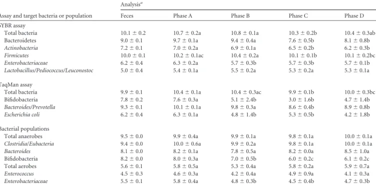
Influence of E. coli O157:H7 and probiotic treatment on the main populations of human intestinal microbiota. The major
phyla of gut microbiota and their main members (41,42) were counted throughout fermentations in the ARCOL (Table 4). Dur-ing the stabilization phase (phase A), their concentrations were similar to those described in humans (43,44) or in other in vitro models of the human gut (45,46).
No study had hitherto evaluated the influence of EHEC
O157:H7 on human intestinal microbiota. In phase B of both assays (F1 and F2), no change was observed by cultural and mo-lecular approaches after addition of EHEC O157:H7, except for bifidobacteria, Enterobacteriaceae, and E. coli (P⬍ 0.05). Each administration of E. coli O157:H7 was followed by a decrease in concentrations of Enterobacteriaceae (Fig. 4). Our results suggest that EHEC colonization of the large intestine is not associated with any major modifications in the profile of dominant bacterial pop-ulations. The decrease in Enterobacteriaceae may be explained by Shiga toxin-encoding phage transfer from E. coli O157:H7 to commensal E. coli resulting in bacterial lysis. According to Gam-age et al. (47), about 10% of the normal E. coli in the human intestine is sensitive to infection by Shiga-toxin-encoding phage. TABLE 4 Influence of E. coli O157:H7 and probiotic treatment on human intestinal microbiota
Assay and target bacteria or population
Analysisa
Feces Phase A Phase B Phase C Phase D
SYBR assay
Total bacteria 10.1⫾ 0.2 10.7⫾ 0.2a 10.8⫾ 0.1a 10.3⫾ 0.2b 10.4⫾ 0.3ab
Bacteroidetes 9.0⫾ 0.1 9.7⫾ 0.1a 9.4⫾ 0.4a 7.6⫾ 0.5b 8.1⫾ 0.8b
Actinobacteria 7.2⫾ 0.1 7.0⫾ 0.2a 6.9⫾ 0.1a 6.5⫾ 0.2b 6.2⫾ 0.3b
Firmicutes 10.0⫾ 0.1 10.2⫾ 0.1ac 10.4⫾ 0.2a 10.1⫾ 0.1b 10.1⫾ 0.2bc
Enterobacteriaceae 6.2⫾ 0.4 6.3⫾ 0.2a 5.7⫾ 0.3b 5.7⫾ 0.3b 5.7⫾ 0.1b
Lactobacillus/Pediococcus/Leuconostoc 5.0⫾ 0.4 5.4⫾ 0.1a 5.5⫾ 0.2a 5.3⫾ 0.2a 5.3⫾ 0.1a
TaqMan assay
Total bacteria 9.9⫾ 0.1 10.4⫾ 0.1a 10.4⫾ 0.3ac 9.9⫾ 0.1b 10.0⫾ 0.3bc
Bifidobacteria 7.8⫾ 0.2 7.6⫾ 0.3a 5.1⫾ 2.4b 3.0⫾ 1.6b 4.7⫾ 1.4b
Bacteroides/Prevotella 9.3⫾ 0.1 10.1⫾ 0.1a 9.8⫾ 0.3a 8.6⫾ 0.4b 8.9⫾ 0.8b
Escherichia coli 6.2⫾ 0.4 6.3⫾ 0.1a 4.8⫾ 1.4b 5.3⫾ 0.5b 4.2⫾ 1.8b
Bacterial populations
Total anaerobes 9.5⫾ 0.0 9.9⫾ 0.4a 9.9⫾ 0.1a 9.8⫾ 0.1a 10.0⫾ 0.1a
Clostridia/Eubacteria 9.4⫾ 0.0 10.0⫾ 0.6a 9.9⫾ 0.2a 9.8⫾ 0.1a 10.0⫾ 0.1a
Bacteroides 8.1⫾ 0.0 8.2⫾ 0.1a 7.8⫾ 0.5a 8.2⫾ 0.0a 8.5⫾ 1.0a
Bifidobacteria 8.2⫾ 0.0 8.0⫾ 0.3a 7.0⫾ 0.5b 6.0⫾ 0.2c 6.1⫾ 0.2c
Total aerobes 5.6⫾ 0.1 5.8⫾ 0.5a 5.3⫾ 0.4a 5.8⫾ 0.2a 5.9⫾ 0.7a
Enterococcus 4.5⫾ 0.3 4.6⫾ 0.3a 4.2⫾ 0.4a 4.9⫾ 0.9a 4.1⫾ 0.3a
Enterobacteriaceae 5.5⫾ 0.1 5.8⫾ 0.4a 4.8⫾ 0.3b 4.5⫾ 0.4b 4.7⫾ 0.3b
aMajor phyla of gut microbiota and their main members were measured by plate counts (results shown in 1og
10CFU/ml) and/or real-time qPCR analysis (results shown in log10 mean copy number/ml). Data are means⫾ standard deviations for the 3 days of each phase during F1 and F2 assays (n ⫽ 6 for phases A, B, C, and D). Samples with different letters in a row are significantly different according to Kruskal-Wallis test followed, if the results were significant, by Mann-Whitney test; P⬍ 0.05. Phase A, stabilization; phase B, E. coli O157:H7; phase C, E. coli O157:H7 plus S. boulardii; phase D, E. coli O157:H7 plus S. cerevisiae CNCM I-3856.
FIG 4 Survival kinetics of E. coli O157:H7 and Enterobacteriaceae during in vitro
fermentations in the ARCOL system. E. coli O157:H7 and Enterobacteriaceae were regularly counted (log10CFU/ml) during phases A and B of F1 (closed symbol)
and F2 (open symbol) assays.ⴱ, addition of E. coli O157:H7 (105CFU/ml of
When probiotic yeasts were coadministered with the pathogen (phases C and D), no profound change was observed in gut mi-crobiota compared to phase B. The only modifications identified by qPCR were slight decreases in the levels of Bacteroidetes,
Acti-nobacteria, and Firmicutes (P⬍ 0.05). Our results are consistent
with those obtained in healthy human volunteers, showing that S.
boulardii and S. cerevisiae do not alter radically the dominant
bac-terial groups of fecal microbiota (39,48).
Influence of E. coli O157:H7 and probiotic treatment on the metabolic activity of human intestinal microbiota. To further
investigate the effects of both pathogen and probiotics on intesti-nal microbiota, its metabolic activity was followed by assessing the production of major and minor SCFAs in the dialysis outflow of the ARCOL (Table 5). Whichever the treatment, similar trends were observed for SCFA production and acetate was the predom-inant SCFA, followed by propionate and butyrate. The molar ra-tios of acetate-propionate-butyrate obtained in vitro were consis-tent with that measured in humans (49,50). The coadministration of S. boulardii (phase C) led to no significant change in SCFA production. In contrast, when EHEC O157:H7 and S. cerevisiae CNCM I-3856 were coadministered (phase D), the production of acetate was significantly increased (P⬍ 0.01) and that of butyrate significantly decreased (P⬍ 0.05) compared with phases A and B. Hitherto, S. boulardii was known to have no effect on SCFA pro-files in healthy humans but was found to increase butyrate and acetate production in patients with diarrhea who were on long-term total enteral nutrition (51). Although the increase in acetate production with S. cerevisiae CNCM I-3856 was not linked to any decrease in E. coli O157:H7 viability, appropriate manipulation of the SCFA levels through probiotic treatment may be a potentially useful approach in the fight against EHEC infection. Indeed, in mice, the anti-infectious activity of bifidobacteria against E. coli O157:H7 has been thought to be related to an increase in acetate production, leading to either (i) a lower gut pH (52) or (ii) inhi-bition of Stx production and translocation (53). Interestingly, S.
cerevisiae CNCM I-3856, when coadministered with E. coli O157:
H7, led to a decrease in butyrate concentrations, while this SCFA has been shown to enhance the expression of virulence-associated genes in EHEC (35). The ARCOL system would potentially enable us to determine what experimental conditions could lead to high
acetate and low butyrate production when probiotic yeasts are added, with the aim of inhibiting EHEC O157:H7.
In conclusion, this study is the first report on the fate of EHEC O157:H7 in simulated human colonic conditions. Our experi-ments provide new information on EHEC survival in the human gastrointestinal tract and its interactions with resident microbi-ota, which is essential for a full understanding of EHEC pathogen-esis. There is growing interest in developing new strategies, such as the use of probiotics, in the fight against this pathogen. In our experimental conditions, S. cerevisiae strains did not exert any clear-cut antagonistic effect against E. coli O157:H7, but the pres-ent results open new fields in research on probiotics. The ARCOL system could be advantageously used to investigate the effect of resident microbiota on EHEC virulence or to screen probiotic strains for their ability to modulate pathogen infectivity in the human gastrointestinal tract. In a more holistic view of EHEC behavior in the human digestive environment, the ARCOL model should be used in combination with gastric and small intestinal systems (20,54), if possible including a resident microbiota (55).
ACKNOWLEDGMENTS
This work was supported by grants from the French Ministère de l’Education Nationale, de l’Enseignement Supérieur et de la Recherche to J.T., EA 4678 CIDAM, and UMR INSERM/Université d’Auvergne U1071 USC-INRA 2018.
We thank Jean-Michel Cardot for help in statistical analysis and the Lesaffre Company for providing the S. cerevisiae CNCM I-3856 strain.
REFERENCES
1. Pennington H. 2010. Escherichia coli O157. Lancet 376:1428 –1435. 2. Frank C, Werber D, Cramer JP, Askar M, Faber M, an der Heiden M,
Bernard H, Fruth A, Prager R, Spode A, Wadl M, Zoufaly A, Jordan S, Kemper MJ, Follin P, Muller L, King LA, Rosner B, Buchholz U, Stark K, Krause G. 2011. Epidemic profile of Shiga-toxin-producing Escherichia
coli O104:H4 outbreak in Germany. N. Engl. J. Med. 365:1771–1780. 3. King LA, Nogareda F, Weill FX, Mariani-Kurkdjian P, Loukiadis E,
Gault G, Jourdan-DaSilva N, Bingen E, Mace M, Thevenot D, Ong N, Castor C, Noel H, Van Cauteren D, Charron M, Vaillant V, Aldabe B, Goulet V, Delmas G, Couturier E, Le Strat Y, Combe C, Delmas Y, Terrier F, Vendrely B, Rolland P, de Valk H. 2012. Outbreak of Shiga
toxin-producing Escherichia coli O104:H4 associated with organic fenu-greek sprouts, France, June 2011. Clin. Infect. Dis. 54:1588 –1594. 4. Chong Y, Fitzhenry R, Heuschkel R, Torrente F, Frankel G, Phillips
TABLE 5 Influence of E. coli O157:H7 and probiotic treatment on SCFA productiona
SCFA
Amt during phaseb:
A B C D
mmol/h % mmol/h % mmol/h % mmol/h %
Totalc 9.4⫾ 0.7a 8.3⫾ 1.0a 8.2⫾ 1.4a 9.4⫾ 0.4a
Acetate 5.8⫾ 0.3a 62.2␣ 5.3⫾ 0.8a 63.9␣ 5.4⫾ 1.1a 65.2␣ 6.6⫾ 0.3b 70.2
Propionate 1.9⫾ 0.3a 19.7␣ 1.5⫾ 0.3a 17.9␣ 1.4⫾ 0.2a 17.5␣ 1.5⫾ 0.2a 16.1␣
Butyrate 1.7⫾ 0.3a 18.1␣ 1.5⫾ 0.2a 18.2␣ 1.4⫾ 0.3ab 17.3␣ 1.3⫾ 0.1b 13.7
iso-Butyrate 0.2⫾ 0.02a 0.2⫾ 0.01a 0.2⫾ 0.03a 0.2⫾ 0.01a
iso-Valerate 0.3⫾ 0.04a 0.3⫾ 0.01a 0.3⫾ 0.1a 0.3⫾ 0.01a
Valerate 0.4⫾ 0.05a 0.4⫾ 0.03a 0.3⫾ 0.05a 0.4⫾ 0.01a
Hexanoic acid 0.3⫾ 0.05a 0.3⫾ 0.1a 0.4⫾ 0.1a 0.4⫾ 0.1a
Heptanoic acid 0.1⫾ 0.03a 0.1⫾ 0.02a 0.2⫾ 0.04a 0.2⫾ 0.03a
aSCFA in the dialysis outflow were measured by gas chromatography. Data are means⫾ standard deviations for the 3 days of each phase during F1 and F2 assays (n ⫽ 6 for phases
A, B, C, and D). Samples with different letters in a row (a, b, and c for values in mmol/h;␣ and  for percentages) are significantly different according to Kruskal-Wallis test followed, if the results were significant, by Mann-Whitney test; P⬍ 0.05.
b
Phase A, stabilization; phase B, E. coli O157:H7; phase C, E. coli O157:H7 plus S. boulardii; phase D, E. coli O157:H7 plus S. cerevisiae CNCM I-3856.
AD. 2007. Human intestinal tissue tropism in Escherichia coli O157: H7–
initial colonization of terminal ileum and Peyer’s patches and minimal colonic adhesion ex vivo. Microbiology 153:794 – 802.
5. Shigeno T, Akamatsu T, Fujimori K, Nakatsuji Y, Nagata A. 2002. The clinical significance of colonoscopy in hemorrhagic colitis due to entero-hemorrhagic Escherichia coli O157:H7 infection. Endoscopy 34:311–314. 6. Naylor SW, Gally DL, Low JC. 2005. Enterohaemorrhagic E. coli in
veterinary medicine. Int. J. Med. Microbiol. 295:419 – 441.
7. Guerra A, Etienne-Mesmin L, Livrelli V, Denis S, Blanquet-Diot S, Alric
M. 2012. Relevance and challenges in modeling human gastric and small
intestinal digestion. Trends Biotechnol. 30:591– 600.
8. Payne AN, Zihler A, Chassard C, Lacroix C. 2012. Advances and per-spectives in in vitro human gut fermentation modeling. Trends Biotech-nol. 30:17–25.
9. Etienne-Mesmin L, Livrelli V, Privat M, Denis S, Cardot JM, Alric M,
Blanquet-Diot S. 2011. Effect of a new probiotic Saccharomyces cerevisiae
strain on survival of Escherichia coli O157:H7 in a dynamic gastrointestinal model. Appl. Environ. Microbiol. 77:1127–1131.
10. de Sablet T, Chassard C, Bernalier-Donadille A, Vareille M, Gobert AP,
Martin C. 2009. Human microbiota-secreted factors inhibit Shiga toxin
synthesis by enterohemorrhagic Escherichia coli O157:H7. Infect. Immun.
77:783–790.
11. Toshima H, Yoshimura A, Arikawa K, Hidaka A, Ogasawara J, Hase A,
Masaki H, Nishikawa Y. 2007. Enhancement of Shiga toxin production in
enterohemorrhagic Escherichia coli serotype O157:H7 by DNase colicins. Appl. Environ. Microbiol. 73:7582–7588.
12. Kendall MM, Gruber CC, Parker CT, Sperandio V. 2012. Ethanolamine controls expression of genes encoding components involved in interking-dom signaling and virulence in enterohemorrhagic Escherichia coli O157: H7. mBio 3:e00050 –12. doi:10.1128/mBio.00050-12.
13. Menne J, Nitschke M, Stingele R, Abu-Tair M, Beneke J, Bramstedt J,
Bremer JP, Brunkhorst R, Busch V, Dengler R, Deuschl G, Fellermann K, Fickenscher H, Gerigk C, Goettsche A, Greeve J, Hafer C, Hagen-muller F, Haller H, Herget-Rosenthal S, Hertenstein B, Hofmann C, Lang M, Kielstein JT, Klostermeier UC, Knobloch J, Kuehbacher M, Kunzendorf U, Lehnert H, Manns MP, Menne TF, Meyer TN, Michael C, Munte T, Neumann-Grutzeck C, Nuernberger J, Pavenstaedt H, Ramazan L, Renders L, Repenthin J, Ries W, Rohr A, Rump LC, Samuelsson O, Sayk F, Schmidt BM, Schnatter S, Schocklmann H, Schreiber S, von Seydewitz CU, Steinhoff J, Stracke S, Suerbaum S, van de Loo A, Vischedyk M, Weissenborn K, Wellhoner P, Wiesner M, Zeissig S, Buning J, Schiffer M, Kuehbacher T. 2012. Validation of
treatment strategies for enterohaemorrhagic Escherichia coli O104:H4 in-duced haemolytic uraemic syndrome: case-control study. BMJ 345:e4565. doi:10.1136/bmj.e4565.
14. Wong CS, Jelacic S, Habeeb RL, Watkins SL, Tarr PI. 2000. The risk of the hemolytic-uremic syndrome after antibiotic treatment of Escherichia coli O157:H7 infections. N. Engl. J. Med. 342:1930 –1936.
15. FAO/WHO. 2002. Report of a joint FAO/WHO working group on draft-ing guidelines for the evaluation of probiotics in food. Food and Agricul-ture Organization of the United Nations and World Health Organization, London, Ontario, Canada.
16. Preidis GA, Hill C, Guerrant RL, Ramakrishna BS, Tannock GW,
Versalovic J. 2011. Probiotics, enteric and diarrheal diseases, and global
health. Gastroenterology 140:8 –14.
17. Bach SJ, McAllister TA, Veira DM, Gannon VPJ, Holley RA. 2003. Effects of a Saccharomyces cerevisiae feed supplement on Escherichia coli O157:H7 in ruminal fluid in vitro. Anim. Feed Sci. Technol. 104:179 –189. 18. Dahan S, Dalmasso G, Imbert V, Peyron JF, Rampal P, Czerucka D. 2003. Saccharomyces boulardii interferes with enterohemorrhagic Esche-richia coli-induced signaling pathways in T84 cells. Infect. Immun. 71: 766 –773.
19. Dalmasso G, Loubat A, Dahan S, Calle G, Rampal P, Czerucka D. 2006. Saccharomyces boulardii prevents TNF-alpha-induced apoptosis in EHEC-infected T84 cells. Res. Microbiol. 157:456 – 465.
20. Blanquet-Diot S, Denis S, Chalancon S, Chaira F, Cardot JM, Alric M. 2012. Use of artificial digestive systems to investigate the biopharmaceu-tical factors influencing the survival of probiotic yeast during gastrointes-tinal transit in humans. Pharm. Res. 29:1444 –1453.
21. Gobert AP, Vareille M, Glasser AL, Hindre T, de Sablet T, Martin C. 2007. Shiga toxin produced by enterohemorrhagic Escherichia coli inhibits PI3K/NF-kappaB signaling pathway in globotriaosylceramide-3-negative human intestinal epithelial cells. J. Immunol. 178:8168 – 8174.
22. McFarland LV. 2010. Systematic review and meta-analysis of Saccharo-myces boulardii in adult patients. World J. Gastroenterol. 16:2202–2222. 23. Danckwerts PV. 1953. Continuous flow systems: distribution of residence
times. Chem. Eng. Sci. 2:1–13.
24. Yu Z, Morrison M. 2004. Improved extraction of PCR-quality commu-nity DNA from digesta and fecal samples. Biotechniques 36:808 – 812. 25. Yu Y, Lee C, Kim J, Hwang S. 2005. Group-specific primer and probe sets
to detect methanogenic communities using quantitative real-time poly-merase chain reaction. Biotechnol. Bioeng. 89:670 – 679.
26. Bacchetti De Gregoris T, Aldred N, Clare AS, Burgess JG. 2011. Im-provement of phylum- and class-specific primers for real-time PCR quan-tification of bacterial taxa. J. Microbiol. Methods 86:351–356.
27. Bartosch S, Fite A, Macfarlane GT, McMurdo ME. 2004. Characteriza-tion of bacterial communities in feces from healthy elderly volunteers and hospitalized elderly patients by using real-time PCR and effects of antibi-otic treatment on the fecal microbiota. Appl. Environ. Microbiol. 70: 3575–3581.
28. Furet JP, Firmesse O, Gourmelon M, Bridonneau C, Tap J, Mondot S,
Dore J, Corthier G. 2009. Comparative assessment of human and farm
animal faecal microbiota using real-time quantitative PCR. FEMS Micro-biol. Ecol. 68:351–362.
29. Huijsdens XW, Linskens RK, Mak M, Meuwissen SG,
Vandenbroucke-Grauls CM, Savelkoul PH. 2002. Quantification of bacteria adherent to
gastrointestinal mucosa by real-time PCR. J. Clin. Microbiol. 40:4423– 4427.
30. Gerard-Champod M, Blanquet-Diot S, Cardot JM, Bravo D, Alric M. 2010. Development and validation of a continuous in vitro system repro-ducing some biotic and abiotic factors of the veal calf intestine. Appl. Environ. Microbiol. 76:5592–5600.
31. Lievin-Le Moal V, Servin AL. 2006. The front line of enteric host defense against unwelcome intrusion of harmful microorganisms: mucins, anti-microbial peptides, and microbiota. Clin. Microbiol. Rev. 19:315–337. 32. Eberhart LJ, Deringer JR, Brayton KA, Sawant AA, Besser TE, Call DR.
2012. Characterization of a novel microcin that kills enterohemorrhagic Escherichia coli O157:H7 and O26. Appl. Environ. Microbiol. 78:6592– 6599.
33. Schamberger GP, Diez-Gonzalez F. 2005. Assessment of resistance to colicinogenic Escherichia coli by E. coli O157:H7 strains. J. Appl. Micro-biol. 98:245–252.
34. Etienne-Mesmin L, Chassaing B, Sauvanet P, Denizot J, Blanquet-Diot
S, Darfeuille-Michaud A, Pradel N, Livrelli V. 2011. Interactions with M
cells and macrophages as key steps in the pathogenesis of enterohemor-ragic Escherichia coli infections. PLoS One 6:e23594. doi:10.1371/journal .pone.0023594.
35. Nakanishi N, Tashiro K, Kuhara S, Hayashi T, Sugimoto N, Tobe T. 2009. Regulation of virulence by butyrate sensing in enterohaemorrhagic Escherichia coli. Microbiology 155:521–530.
36. Edwards-Ingram L, Gitsham P, Burton N, Warhurst G, Clarke I, Hoyle
D, Oliver SG, Stateva L. 2007. Genotypic and physiological
characteriza-tion of Saccharomyces boulardii, the probiotic strain of Saccharomyces cerevisiae. Appl. Environ. Microbiol. 73:2458 –2467.
37. Fietto JL, Araujo RS, Valadao FN, Fietto LG, Brandao RL, Neves MJ,
Gomes FC, Nicoli JR, Castro IM. 2004. Molecular and physiological
comparisons between Saccharomyces cerevisiae and Saccharomyces boular-dii. Can. J. Microbiol. 50:615– 621.
38. Klein SM, Elmer GW, McFarland LV, Surawicz CM, Levy RH. 1993. Recovery and elimination of the biotherapeutic agent, Saccharomyces boulardii, in healthy human volunteers. Pharm. Res. 10:1615–1619. 39. Pecquet S, Guillaumin D, Tancrede C, Andremont A. 1991. Kinetics of
Saccharomyces cerevisiae elimination from the intestines of human volun-teers and effect of this yeast on resistance to microbial colonization in gnotobiotic mice. Appl. Environ. Microbiol. 57:3049 –3051.
40. Chiou RY, Phillips RD, Zhao P, Doyle MP, Beuchat LR. 2004. Ethanol-mediated variations in cellular fatty acid composition and protein profiles of two genotypically different strains of Escherichia coli O157:H7. Appl. Environ. Microbiol. 70:2204 –2210.
41. Arumugam M, Raes J, Pelletier E, Le Paslier D, Yamada T, Mende DR,
Fernandes GR, Tap J, Bruls T, Batto JM, Bertalan M, Borruel N, Casellas F, Fernandez L, Gautier L, Hansen T, Hattori M, Hayashi T, Kleerebezem M, Kurokawa K, Leclerc M, Levenez F, Manichanh C, Nielsen HB, Nielsen T, Pons N, Poulain J, Qin J, Sicheritz-Ponten T, Tims S, Torrents D, Ugarte E, Zoetendal EG, Wang J, Guarner F, Pedersen O, de Vos WM, Brunak S, Dore J, Antolin M, Artiguenave F,
Blottiere HM, Almeida M, Brechot C, Cara C, Chervaux C, Cultrone A, Delorme C, Denariaz G, Dervyn R, Foerstner KU, Friss C, van de Guchte M, Guedon E, Haimet F, Huber W, van Hylckama-Vlieg J, Jamet A, Juste C, Kaci G, Knol J, Lakhdari O, Layec S, Le Roux K, Maguin E, Merieux A, Melo Minardi R, M’Rini C, Muller J, Oozeer R, Parkhill J, Renault P, Rescigno M, Sanchez N, Sunagawa S, Torrejon A, Turner K, Vandemeulebrouck G, Varela E, Winogradsky Y, Zeller G, Weissenbach J, Ehrlich SD, Bork P. 2011. Enterotypes of the human gut
microbiome. Nature 473:174 –180.
42. Eckburg PB, Bik EM, Bernstein CN, Purdom E, Dethlefsen L, Sargent
M, Gill SR, Nelson KE, Relman DA. 2005. Diversity of the human
intestinal microbial flora. Science 308:1635–1638.
43. Finegold SM, Attebery HR, Sutter VL. 1974. Effect of diet on human fecal flora: comparison of Japanese and American diets. Am. J. Clin. Nutr. 27: 1456 –1469.
44. Marteau P, Pochart P, Dore J, Bera-Maillet C, Bernalier A, Corthier G. 2001. Comparative study of bacterial groups within the human cecal and fecal microbiota. Appl. Environ. Microbiol. 67:4939 – 4942.
45. Marzorati M, Verhelst A, Luta G, Sinnott R, Verstraete W, Van de
Wiele T, Possemiers S. 2010. In vitro modulation of the human
gastro-intestinal microbial community by plant-derived polysaccharide-rich di-etary supplements. Int. J. Food Microbiol. 139:168 –176.
46. Sannasiddappa TH, Costabile A, Gibson GR, Clarke SR. 2011. The influence of Staphylococcus aureus on gut microbial ecology in an in vitro continuous culture human colonic model system. PLoS One 6:e23227. doi:10.1371/journal.pone.0023227.
47. Gamage SD, Strasser JE, Chalk CL, Weiss AA. 2003. Nonpathogenic Escherichia coli can contribute to the production of Shiga toxin. Infect. Immun. 71:3107–3115.
48. Swidsinski A, Loening-Baucke V, Verstraelen H, Osowska S, Doerffel Y.
2008. Biostructure of fecal microbiota in healthy subjects and patients with chronic idiopathic diarrhea. Gastroenterology 135:568 –579. 49. Cummings JH, Pomare EW, Branch WJ, Naylor CP, Macfarlane GT.
1987. Short chain fatty acids in human large intestine, portal, hepatic and venous blood. Gut 28:1221–1227.
50. Topping DL, Clifton PM. 2001. Short-chain fatty acids and human co-lonic function: roles of resistant starch and nonstarch polysaccharides. Physiol. Rev. 81:1031–1064.
51. Schneider SM, Girard-Pipau F, Filippi J, Hebuterne X, Moyse D,
Hinojosa GC, Pompei A, Rampal P. 2005. Effects of Saccharomyces
boulardii on fecal short-chain fatty acids and microflora in patients on long-term total enteral nutrition. World J. Gastroenterol. 11:6165– 6169.
52. Asahara T, Shimizu K, Nomoto K, Hamabata T, Ozawa A, Takeda Y. 2004. Probiotic bifidobacteria protect mice from lethal infection with Shiga toxin-producing Escherichia coli O157:H7. Infect. Immun. 72: 2240 –2247.
53. Fukuda S, Toh H, Hase K, Oshima K, Nakanishi Y, Yoshimura K, Tobe
T, Clarke JM, Topping DL, Suzuki T, Taylor TD, Itoh K, Kikuchi J, Morita H, Hattori M, Ohno H. 2011. Bifidobacteria can protect from
enteropathogenic infection through production of acetate. Nature 469: 543–547.
54. Hatanaka M, Nakamura Y, Maathuis AJ, Venema K, Murota I,
Yamamoto N. 2012. Influence of Bacillus subtilis C-3102 on microbiota in
a dynamic in vitro model of the gastrointestinal tract simulating human conditions. Benef. Microbes 3:229 –236.
55. Possemiers S, Marzorati M, Verstraete W, Van de Wiele T. 2010. Bacteria and chocolate: a successful combination for probiotic delivery. Int. J. Food Microbiol. 141:97–103.