The Rockefeller University Press $30.00
IL-10 is an important immune-regulatory cyto-kine (Moore et al., 2001), and IL-10 produced by T cells is important for the prevention of auto-immune disease and immunopathology upon chronic infections (Groux et al., 1997; Asseman et al., 1999; Roers et al., 2004; Anderson et al., 2007; Jankovic et al., 2007; Couper et al., 2008). Two main subsets of IL-10–producing T cells with immune-regulatory functions have been described: “natural” and “adaptive” T reg cells (Bluestone and Abbas, 2003). Both subsets
have been shown to prevent autoimmune dis-eases and limit immune responses (Groux et al., 1997; Roncarolo and Levings, 2000; Maloy and Powrie, 2001; Vieira et al., 2004), and lack IL-2–producing capacities (Groux et al., 1997; Sakaguchi, 2004; Vieira et al., 2004; Scheffold et al., 2005). They can be generated from naive T cells following different protocols of tolero-genic priming (Groux et al., 1997; Seddon and Mason, 1999; Jonuleit et al., 2000; Walker et al., 2003; Apostolou and von Boehmer, 2004), CORRESPONDENCE
Jens Geginat: [email protected]
Abbreviations used: CLA, cuta-neous lymphocyte-associated antigen; mDC, myeloid DC; pDC, plasmacytoid DC; TT, tetanus toxoid.
P. Gruarin and B. Häringer contributed equally to this paper. L. Rivino’s present address is Dept. of Microbiology and Immunology Programme, National University of Singapore, Singapore 117456
CCR6 is expressed on an IL-10–producing,
autoreactive memory T cell population
with context-dependent regulatory function
Laura Rivino,
1,2Paola Gruarin,
3Barbara Häringer,
2Svenja Steinfelder,
2Laura Lozza,
1,2Bodo Steckel,
2Anja Weick,
2Elisa Sugliano,
3David Jarrossay,
1Anja A. Kühl,
4Christoph Loddenkemper,
4,5Sergio Abrignani,
3Federica Sallusto,
1Antonio Lanzavecchia,
1and Jens Geginat
1,2,3 1Institute for Research in Biomedicine, 6500 Bellinzona, Switzerland2Charité Research Center for ImmunoSciences and German Rheumatism Research Center, Campus Charité Mitte, 10117 Berlin, Germany
3Istituto Nazionale di Genetica Molecolare, 20122 Milan, Italy
4Charité Research Center for ImmunoSciences/Institute of Pathology, Campus Benjamin Franklin, 12200 Berlin, Germany 5Institute of Pathology, Technische Universität München, 81625 Munich, Germany
Interleukin (IL)-10 produced by regulatory T cell subsets is important for the prevention of autoimmunity and immunopathology, but little is known about the phenotype and function of IL-10–producing memory T cells. Human CD4+CCR6+ memory T cells contained
comparable numbers of IL-17– and IL-10–producing cells, and CCR6 was induced under both Th17-promoting conditions and upon tolerogenic T cell priming with transforming growth factor (TGF)–. In normal human spleens, the majority of CCR6+ memory T cells
were in the close vicinity of CCR6+ myeloid dendritic cells (mDCs), and strikingly, some of
them were secreting IL-10 in situ. Furthermore, CCR6+ memory T cells produced
suppres-sive IL-10 but not IL-2 upon stimulation with autologous immature mDCs ex vivo, and secreted IL-10 efficiently in response to suboptimal T cell receptor (TCR) stimulation with anti-CD3 antibodies. However, optimal TCR stimulation of CCR6+ T cells induced expression
of IL-2, interferon-, CCL20, and CD40L, and autoreactive CCR6+ T cell lines responded
to various recall antigens. Notably, we isolated autoreactive CCR6+ T cell clones with
context-dependent behavior that produced IL-10 with autologous mDCs alone, but that secreted IL-2 and proliferated upon stimulation with tetanus toxoid. We propose the novel concept that a population of memory T cells, which is fully equipped to participate in secondary immune responses upon recognition of a relevant recall antigen, contributes to the maintenance of tolerance under steady-state conditions.
© 2010 Rivino et al. This article is distributed under the terms of an Attribu-tion–Noncommercial–Share Alike–No Mirror Sites license for the first six months after the publication date (see http://www.rupress.org/terms). After six months it is available under a Creative Commons License (Attribution–Noncommercial– Share Alike 3.0 Unported license, as described at http://creativecommons.org/ licenses/by-nc-sa/3.0/).
The Journal of Experimental Medicine
on March 30, 2010
jem.rupress.org
constitutively in lymphoid and nonlymphoid tissues and up-regulated upon inflammation, but the CCR6–CCL20 axis appears to be particularly important for migration of immune cells to the gut and the skin (Cook et al., 2000; Schutyser et al., 2003). Studies on CCR6-deficient mice indicated a nonredundant role for CCR6 in gut lymphoid tissue homeostasis (Cook et al., 2000; Varona et al., 2001). Furthermore, CCR6-deficient mice have altered CD4+
T cell responses, including reduced contact hypersensitiv-ity and enhanced delayed type hypersensitivhypersensitiv-ity responses (Lukacs et al., 2001; Varona et al., 2001, 2005). CCL20 and CCR6 are further involved in several autoimmune diseases including psoriasis (Homey et al., 2000), inflammatory bowel disease (Varona et al., 2003; Kaser et al., 2004), ex-perimental autoimmune encephalomyelitis (Kleinewietfeld et al., 2005), and rheumatoid arthritis (Matsui et al., 2001; Ruth et al., 2003). Furthermore, we and others have shown that CCR6 is expressed on human Th17 cells Rodriguez et al., 2007; Annunziato et al., 2007).
In this paper, we show that CCR6+ human memory
T cells have a low stimulation threshold for IL-10 production and, consequently, secrete IL-10 after suboptimal stimulation by autologous DCs ex vivo and in the spleen in situ. These autoreactive memory T cells are distinct from the effector-like cells with regulatory activity that we identified previously (Häringer et al., 2009). They produce IL-2 and IFN-, and proliferate after strong stimulation with recall antigens, suggesting that they can have a context-dependent function depending on the strength of TCR stimulation.
RESULTS
TGF- promotes CCR6 expression on primed human CD4+ T cells
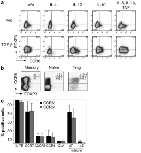
The chemokine receptor CCR6 is expressed on Th17 and T reg cells (Kleinewietfeld et al., 2005; Acosta-Rodriguez et al., 2007; Annunziato et al., 2007). We observed that TGF- promoted CCR6 expression after naive (CD45RA+
CCR7+CCR6CD25) human CD4+ T cell priming with
anti-CD3 and anti-CD28 antibodies (Fig. 1 a) or allogenic DCs (not depicted). Naive T cells were labeled with CFSE for these experiments to exclude undivided cells from the analy-sis. TGF-–dependent CCR6 expression was inhibited by the Th1- or Th2-polarizing cytokines IL-4 or IL-12 but not by IL-10. IL-4 was highly efficient in preventing CCR6 ex-pression, because it completely prevented TGF-–dependent CCR6 induction (in six different donors in independent ex-periments from 22 ± 12 to 2 ± 2%; P < 0.05), whereas IL-12 also reduced the mean CCR6 expression significantly but less efficiently (to 10 ± 4%). Consistent with previous results (Acosta-Rodriguez et al., 2007), the proinflammatory cyto-kines TNF, IL-1, and IL-6 also promoted CCR6 expres-sion and synergized with TGF-, indicating that CCR6 can be induced under either tolerogenic or Th17-promoting conditions. In the absence of polarizing or proinflammatory cytokines, TGF- promoted FOXP3 expression as expected (Fantini et al., 2004), but a fraction of CCR6+ cells remained
and TGF- is required for IL-10 production from both subsets in vivo (Maynard et al., 2007). Natural T reg cells have been characterized in detail both in mice and humans. Thus, nat-ural T reg cells can already mature in the thymus and express CD25, the high affinity receptor for IL-2, (Shevach, 2001; Sakaguchi, 2004), and the transcription factor FOXP3 (Sakaguchi, 2004). The maintenance and function of natural CD25+ T reg cells relies on IL-2 that is produced by activated
Th cells (Scheffold et al., 2005), consistent with the autoim-mune phenotype of IL-2–deficient mice. However, IL-2 also has some immunostimulatory effects, which might be induced at higher levels of IL-2 production (Scheffold et al., 2005), because it can promote memory T cell generation (Dooms et al., 2007) and autoimmunity (Waithman et al., 2008). Less is known about adaptive T reg cells because their phenotype and characteristics vary between different experimental set-tings. However, we have recently identified CD4+FOXP3
effector-like cells in human blood that coproduce IL-10 and IFN- and suppress T cell activation via IL-10 (Häringer et al., 2009). Notably, these Tr1-like cells are distinct from memory cells, because they are activated in vivo, have lost IL-7R expression, produce little IL-2, and do not respond to vaccination antigens.
The strength of TCR stimulation depends on the avidity of the TCR/peptide–MHC interaction and on engagement of co-stimulatory receptors, and is an important parameter of T cell activation to discriminate between tolerance and immunity (Lanzavecchia and Sallusto, 2002). In the thymus, developing T cells with a low affinity for self-MHC mole-cules are selected for survival (van den Boorn et al., 2006), whereas those with a higher affinity for self-peptides become CD25+ T reg cells (Jordan et al., 2001). In the periphery,
low-level TCR stimulation of mature naive T cells by self-MHC is essential for their survival and antigen responsive-ness in the mouse (Brocker, 1997; Stefanová et al., 2002). Importantly, this TCR “tickling” by self-MHC is also important for mouse CD4+ memory T cells, because they
become nonfunctional upon transfer into MHC class II– deficient hosts (Kassiotis et al., 2002). However, suboptimal TCR stimulation in the absence of co-stimulation can also induce a state of unresponsiveness called anergy in T cells (Schwartz, 1997), or lead to a state of unfitness and abortive proliferation in both mouse and human T cells (Gett et al., 2003). The strength of TCR stimulation also regulates dif-ferent functional responses, because clones derived from human CD8+ memory T cells have different stimulation
thresholds for cytotoxicity, IFN- production, and prolifer-ation (Valitutti et al., 1996). In the case of CD4+ T cells, it
was shown that IL-2 and IL-10 are preferentially induced by different co-stimulatory receptors (Hutloff et al., 1999; Blair et al., 2000), but whether the secretion of these two key cytokines is differentially regulated by the strength of TCR stimulation has not been investigated.
CCR6 is a chemokine receptor expressed on B cells, a fraction of T cells, and immature DCs (Schutyser et al., 2003). The ligand of CCR6, CCL20, is widely expressed
on March 30, 2010
jem.rupress.org
Low stimulation threshold for IL-10 production in CCR6+ memory T cells
Human memory T cells have different activation thresholds for the expression of activation-induced surface receptors. Thus, total memory cells could be efficiently activated by suboptimal stimulation with anti-CD3 antibodies to ex-press the CD69 activation marker but required CD28 stimulation for the up-regulation of CD40L (Fig. 2 a). Also, the induction of cytokines had different stimulation thresh-olds. Thus, memory T cells secreted IL-10 efficiently at late time points with anti-CD3 antibodies alone, whereas IL-2 secretion and optimal IFN- production occurred earlier and required CD28 co-stimulation (Fig. 2 b).
We previously showed that CCR6 is expressed on human blood Th17 cells but not on Th2 cells, whereas IL-2– and IFN-–producing cells are present among both CCR6+
and CCR6 T cells (not depicted; Acosta-Rodriguez et al.,
2007). We investigated the IL-10–producing capacities of CCR6+ and CCR6 memory T cells in response to anti-CD3
FOXP3, showing that cells that acquire CCR6 upon
tolero-genic priming do not necessarily become FOXP3+ T reg cells.
Consistently, as shown in Fig. 1 b, CCR6 in human blood was found not only on the majority of FOXP3+ T reg cells
but also on a large fraction of CD25FOXP3 memory cells,
whereas phenotypic naive cells were largely CCR6.
Vir-tually all (99%) CD25CCR6+ cells expressed the memory
marker IL-7R, but they were heterogeneous for CCR7, integrin 7, and cutaneous lymphocyte-associated antigen (CLA), indicating that they contained central memory T, ef-fector memory T, and gut- and skin-homing cells (Fig. 1 c). A similar heterogeneity was observed among CCR6 cells
for all analyzed surface markers with the exception of the integrin E subunit (also regulated by TGF-; Hadley et al., 1997), which was selectively expressed on a small fraction of CCR6+ cells (Fig. 1 c). Collectively, these results suggest
that CCR6 is expressed on antigen-experienced T cells that could have been primed under either tolerogenic or Th17-promoting conditions.
Figure 1. CCR6 is induced by TGF- and expressed on populations of CD25+ T reg cells and CD4+ memory T cells. (a) CFSE-labeled peripheral blood naive CD4+ T cells were stimulated with anti-CD3 and anti-CD28 antibodies for 4 d in the presence or absence of the indicated cytokines. Cells were stained at day 5, and CFSE cells were analyzed for CCR6 and FOXP3 expression by flow cytometry. (b) Peripheral blood CD4+ T cell populations were analyzed for CCR6 and FOXP3 expression by flow cytometry ex vivo. Numbers indicate percentages. (c) Peripheral blood CD4+CD45RACD25 memory T cells were stained with antibodies specific for CCR6, IL-7R, CCR7, CLA, CXCR5, and integrin 7 and E, and were analyzed by flow cytometry. The mean percentage of CCR6+ or CCR6 cells expressing the relevant surface marker of three different donors is shown.
on March 30, 2010
jem.rupress.org
CD3 antibodies alone (4.3 ± 2.6 vs. 0.7 ± 0.4%; P < 0.05; Fig. 2 c). Similar results were obtained by analyzing superna-tants of stimulated cells by ELISA (unpublished data). The fraction of CCR6+ T cells producing IL-10 with anti-CD3
was relatively small, but it was similar to the fraction of Th17 cells (4.6 ± 2.8% of IL-17A+ cells upon CD28 co-stimulation;
Fig. 2 c). Conversely, a major fraction of CCR6+ T cells
pos-sessed CCL20-producing capacities, including not only vir-tually all Th17 cells, as expected, but also many IL-10–secreting antibodies in the absence or presence of CD28 co-stimulation.
Because IL-7R is a memory cell marker and IL-7R cells are
effector-like cells with high IL-10–producing capacities (Häringer et al., 2009), we also purified CCR6+ and CCR6
memory T cell populations according to IL-7R expression. Interestingly, although variable fractions of both CCR6+ and
CCR6 memory T cells produced IL-10 in response to
anti-CD3 in the presence of CD28 co-stimulation, only CCR6+
T cells secreted IL-10 efficiently upon stimulation with
anti-Figure 2. Ex vivo cytokine production of CCR6+ and CCR6 memory T cells after weak or strong TCR stimulation. (a) Up-regulation of CD69 and CD40L after stimulation of total antigen-experienced Th cells (CD45RACD25) with anti-CD3 alone (left) or anti-CD3 plus anti-CD28 (right). One experiment out of three is shown. (b) Production of IL-10 versus IL-2 (left) or versus IFN- (right) at 6 (top) and 30 (bottom) h after stimulation with anti-CD3 alone or anti-anti-CD3 plus anti-CD28. One representative experiment out of eight is shown. (c) Expression of CCL20 and IL-10 (left) or IL-17 (right) of CCR6+ and CCR6 memory T cells (IL-7R+CD25CD45RA) after stimulation with anti-CD3 antibodies in the absence or presence of CD28 co-stimulation. Two representative donors out of six analyzed in three independent experiments are shown. Numbers indicate percentages.
on March 30, 2010
jem.rupress.org
proliferate or secrete cytokines in response to autologous DCs. We used circulating DCs isolated ex vivo, because in healthy subjects they could present self-antigens as well as physiologically relevant innocuous foreign antigens that they are exposed to in the steady state. Allogenic DCs were used in parallel as a positive control. CFSE-labeled CCR6+ and
CCR6 T cells proliferated vigorously with allogenic DCs, as
expected, but weakly (CCR6+) or not at all (CCR6) with
autologous DCs (Fig. 3 a). However, CCR6+ but not CCR6
memory T cells proliferated vigorously with autologous DCs when neutralizing anti–IL-10 antibodies were added, whereas an isotype-matched control antibody had no cells (Fig. 2 c) that lack IL-17–producing capacities (not
depicted; Häringer et al., 2009). Thus, unlike CCR6
mem-ory T cells, CCR6+ cells could attract other CCR6+ cells via
CCL20 and secrete IL-10 efficiently upon weak TCR stimulation. Conversely, both CCR6+ and CCR6
mem-ory cells produce CD40L, IL-2, IL-10, and IFN- after optimal activation.
CCR6+ T cells produce suppressive IL-10 upon contact
with autologous DCs
We then analyzed whether memory T cell populations differed in autoreactivity, which we defined as the capacity to
Figure 3. CCR6+ memory T cells proliferate with autologous DCs upon IL-10 neutralization. (a) Purified CCR6+ and CCR6 T cells were incubated with ex vivo–isolated autologous or allogenic mDCs in the presence or absence of neutralizing anti–IL-10 antibodies. On day 7, CFSE profiles of T cells were analyzed by flow cytometry. One experiment out of six is shown. Numbers indicate percentages. (b) CCR6+ T cells and autologous mDCs were puri-fied after 24 h following co-culture or culture in medium alone. IL-10 mRNA levels relative to 18S rRNA were quantipuri-fied by quantitative real-time RT-PCR. Results are expressed in arbitrary units (AU) of IL-10 mRNA/18S rRNA. Error bars represent means of three different experiments analyzing different do-nors. (c) CCR6+ T cells were co-cultured with autologous or allogenic mDCs, and culture supernatants were assessed by ELISA for the presence of IL-2 and IL-10. The mean of three independent experiments is shown.
on March 30, 2010
jem.rupress.org
proliferation via IL-10 was limited to conditions of weak TCR stimulation and limited IL-2 availability. Consistently, addition of exogenous IL-2 to autologous cultures induced vigorous CCR6+ T cell proliferation in the absence of IL-10
neutralization, and CCR6 T cells also proliferated under this
condition, but to a lower extent (unpublished data). In sum-mary, these findings indicate that autologous DCs provide a suboptimal TCR stimulation of CCR6+ T cells, which is
sufficient for IL-10 production that, in a negative feedback loop, inhibits autoreactive T cell proliferation.
Immature mDCs activate autoreactive CCR6+ T cells efficiently
The extent of the CCR6+ autoreactive T cell proliferation
with different subsets of autologous APCs was associated with the well-established capacities of these APC subsets to acti-vate CD4+ T cells. Thus, autoreactive proliferation was highest
with mDCs, lower with plasmacytoid DCs (pDCs), and very low or absent with monocytes (unpublished data). Surpris-ingly, however, mDCs were less stimulatory for autoreactive T cells after LPS-induced maturation, although mature mDCs more potently activated allogenic T cells, as expected (Fig. 4 a). Interestingly, the reduced capacity of mDCs to activate au-tologous T cells upon LPS maturation was associated with the down-regulation of the transcription factor AIRE (Fig. 4 b). Previous work has shown that AIRE allows presentation of tissue-restricted antigens by specialized APC subsets predom-inantly in the thymus (Kyewski and Klein, 2006) but also in effect (not depicted). CCR6+ memory T cell proliferation
with autologous DCs shared several features with conven-tional antigenic proliferation, as it was inhibited by neutraliz-ing antibodies to MHC class II and by natural T reg cells (unpublished data). CD25+ T reg cells could also proliferate with
autologous DCs but required the addition of exogenous IL-2, whereas IL-10 neutralization had no effect (unpublished data).
To understand whether DCs or T cells produced the suppressive IL-10 in autologous cultures, we resorted the two cell types after co-culture and measured the IL-10 mRNA. Notably, CCR6+ T cells constitutively expressed
IL-10 mRNA, and IL-10 was superinduced in CCR6+ T cells
upon co-culture with DCs (Fig. 3 b). Conversely, IL-10 mRNA was neither detected in myeloid DCs (mDCs) ex vivo nor induced after co-culture with CCR6+ T cells,
showing that the suppressive IL-10 was derived exclusively from CCR6+ T cells. Consistent with the mRNA
expres-sion, supernatants from autologous CCR6+ T cell–DC
co-cultures contained considerable amounts of IL-10 but little IL-2 (Fig. 3 c), consistent with the notion that autologous DCs provide only weak stimulation for autologous T cells in the absence of exogenous antigens. Conversely, both IL-10 and IL-2 were detected in allogenic co-cultures. Notably, although comparable amounts of IL-10 were produced in autologous and allogenic cultures, IL-10 neutralization had no clear effect on alloreactive T cell proliferation (Fig. 3 a). Thus, the capacity of CCR6+ T cells to suppress T cell
Figure 4. Immature mDCs most efficiently induce autoreactive T cell proliferation. (a) Purified, CFSE-labeled CCR6+ memory T cells were cultured with autologous or allogenic immature or LPS-matured mDCs in the presence of neutralizing anti–IL-10 antibodies, and proliferation was assessed on day 7. One experiment out of three is shown. (b) AIRE mRNA expression in purified monocytes, or immature or LPS-matured mDCs was measured by quantitative RT-PCR and normalized on 18S rRNA. AIRE expression in immature mDCs was set to 100%. The mean of three independent experiments is shown. (c) Purified blood monocytes, pDCs, and mDCs were stained for CCR6 surface expression in three healthy donors. Numbers indicate percentages.
on March 30, 2010
jem.rupress.org
located close to CD11c+ DCs (Fig. 5, inset, IL-10 CD11c).
A considerable fraction of the IL-10+ T cells coexpressed
CCR6 (Fig. 5, bottom right, yellow), but they were largely negative for FOXP3 (inset, FOXP3 IL-10; and Table I). These results are consistent with the view that immature mDCs activate autoreactive CCR6+ memory T cells in the
steady state to produce IL-10.
Context-dependent cytokine profile of CCR6+
tetanus-specific T cell clones
We next analyzed specificities of CCR6+ and CCR6
memory T cell populations to self- and recall antigens (Fig. 6 a). Responses to the melanocyte self-antigen MelanA were detected exclusively among CCR6+ T cells in six out of eight
donors, whereas one donor did not respond and another one with a very high response among CCR6+ cells had a lower
the periphery (Ramsey et al., 2006). Notably, human blood mDCs, but not pDCs or monocytes, expressed CCR6 ex vivo (Fig. 4 c), and they down-regulated CCR6 upon matu-ration with LPS (not depicted). In summary, immature mDCs that express CCR6 and AIRE activate autoreactive CCR6+
memory T cells most efficiently.
To understand whether CCR6+ T cells could be
acti-vated by immature mDCs in vivo, we analyzed normal human spleen sections for localization of CCR6+ T cells
and mDCs. As shown in Fig. 5, CCR6+ T cells (yellow
ar-rows) were present in T cell zones of the human spleen, and the majority of them were in close proximity to CD11c+
DCs, i.e., mDCs (inset, CCR6 CD11c; and Table I). Strik-ingly, we could identify IL-10–producing T cells in the hu-man spleen in situ. Scattered IL-10+ cells were detected
within the periarteriolar T cell areas, and these cells were also
Figure 5. CCR6+ T cells colocalize with CD11c+ DCs and produce IL-10 in the human spleen. (top) Spleen sections showing anti-CCR6–labeled CCR6+ cells (red) in the anti-CD3–stained (green) T cell areas and B cell follicles with a proportion of CCR6/CD3 double-positive cells (arrows). (bottom left) Double immunohistochemical staining displays the distribution of CD11c+ DCs (red) predominantly in the periarteriolar CD3+ (brown) T cell areas (T) but also in the red pulp (RP) and the marginal zone (MZ) of the B cell follicles (B). CCR6+ cells (red) in the T cell areas are often found close to CD11c+ DCs (brown; inset, CCR6 CD11c). (middle right) Within the periarteriolar T cell area, scattered IL-10+ cells (brown) can be identified, and costaining with anti-CD11c (red) indicates that some are in close contact to DCs (inset, IL-10 anti-CD11c), whereas the large majority of IL-10+ cells were negative for FOXP3 (red, nuclear; inset, FOXP3 IL-10). (bottom right) Immunofluorescence shows that proportions of CD3+ or CCR6+ cells (green) coexpress IL-10 (red), resulting in a yellow overlay (merge). Stainings are representative of five samples each, and the cell numbers are summarized in Table I. Bars: (top; and bottom left) 100 µm; (middle right) 50 µm; (bottom right) 10 µm.
on March 30, 2010
jem.rupress.org
stimulation at higher anti-CD3 concentration (>4 ng/ml). We conclude that autoreactive CCR6+ memory T cells have
a context-dependent behavior, because cells derived from the same memory precursor proliferate and secrete IL-2, IFN-, and IL-10 upon strong stimulation with recall an-tigens, whereas they produce mainly IL-10 upon weak stim-ulation with autologous DCs.
DISCUSSION
The presence of IL-10–producing memory cells in humans has been known for a long time, but their function and phe-notype has remained unclear. In this study, we showed that human CCR6+ memory T cells secrete IL-10 efficiently in
response to suboptimal TCR stimulation, whereas they produce immunostimulatory cytokines at higher levels of stimulation. These different activation thresholds allow auto-reactive CCR6+ memory T cells to secrete suppressive IL-10
in response to autologous DCs, whereas they produce IFN-, IL-2, and CD40L upon strong activation, as can be provided by a recall antigen. Thus, memory T cells that cross react with antigens presented by autologous mDCs could have a context-dependent function and inhibit autoreactivity in the steady state, but contribute to recall responses upon infections or vaccinations.
IL-10 production by T cells inhibits autoimmunity and immunopathology, and the induction of IL-10–producing capacities in mouse Th cells requires TGF- (Maynard et al., 2007). We found that tolerogenic priming with TGF- induced CCR6 on naive T cells in vitro and that the polariz-ing cytokines IL-12 and in particular IL-4 inhibited CCR6 expression. CCR6 was also induced by proinflammatory cytokines and is expressed on human blood Th17 cells (Acosta-Rodriguez et al., 2007), raising the question of whether the IL-10–producing CCR6+ memory cells might
belong to the Th17 lineage. However, we recently showed that IL-17 and IL-10 coproducing cells in human blood are largely absent from the memory compartment (Häringer et al., response among CCR6 cells, confirming that autoreactive
T cells express CCR6 in healthy subjects (14 vs. 3% prolifer-ating cells; P < 0.05; Fig. 6 b). Conversely, in four vitiligo autoimmune patients that have lost tolerance to derived antigens, MelanA-specific cells were mostly or exclusively CCR6 (2 vs. 14%; Fig. 6 b). Consistent with
previous results, tetanus-specific cells were detectable in both CCR6+ and CCR6 populations (Acosta-Rodriguez et al.,
2007). We next investigated whether autoreactive and recall-specific T cells exclusively represented two distinct popula-tions within CCR6+ T cells, or if CCR6+ T cells contained
recall antigen–specific cells that cross reacted with antigens presented by autologous DCs. CCR6+ T cells that had
divided extensively with autologous DCs in the presence of anti–IL-10 were isolated and analyzed for responsiveness to recall antigens. A variable fraction of these autoreactive cells in all donors analyzed responded to various recall antigens, including tetanus toxoid (TT) and other nonpersistent vacci-nation antigens (Fig. 6 c and Table II). Clones derived from single autoreactive cells were then analyzed for proliferation and cytokine production after stimulation with autologous APCs in the absence or presence of TT. Several autoreactive CCR6+ clones from two different donors were obtained that
proliferated vigorously with TT and produced high amounts of IL-2, IL-10, and IFN- (Fig. 7, a and b), but not IL-17 (not depicted). Notably, when cells from the same tetanus-specific clones were stimulated with autologous mDCs alone, they produced IL-10 but not IL-2 and proliferated poorly and only upon IL-10 neutralization (Fig. 7 b). Conversely, two CCR6 tetanus-specific clones from the same donors
did not respond to mDCs alone (Fig. 7, a and b). Finally, autoreactive, tetanus-specific CCR6+ T cell clones had
dif-ferent activation thresholds for IL-2 and IL-10 production, as expected. Thus, they produced IL-10 only with anti-CD3 alone (unpublished data) or after CD28 co-stimulation at low anti-CD3 concentrations (1–2 ng/ml; Fig. 7 c). Conversely, they produced IL-2 together with IL-10 after CD28
co-Table I. Number of T cells interacting with mDCs, expressing CCR6, FOXP3, or IL-10 in human spleen sections
T cell area Total CD3+ CD3+ close to
DCs Total CCR6
+ CCR6+ close
to DCs Total FOXP3
+ CD3+ IL-10+ FOXP3+ IL-10+ CCR6+ IL-10+
1A 137 42 21 14 5 13 1 4 1B 124 38 17 12 4 11 0 2 2A 175 61 35 23 12 15 1 5 2B 225 95 63 37 8 22 2 7 3A 105 31 14 11 3 8 0 3 3B 92 28 9 7 4 9 0 1 4A 80 26 7 6 3 7 0 2 4B 76 20 7 5 5 5 0 3 5A 119 42 13 9 6 11 0 2 5B 85 31 10 4 3 6 0 1 Mean 122 ± 47 41 ± 22 20 ± 17 13 ± 10 5 ± 3 11 ± 5 0.4 ± 0.7 3 ± 2 % 34 (CD3+) 16 (CD3+) 66 (CCR6+) 4 (CD3+) 9 (CD3+) 4 (IL-10+) 28 (IL-10+)
Cells staining positive for CD3, CCR6, IL-10, or FOXP3 in the vicinity or not of CD11c mDCs were counted in two T cell areas (A and B) in five different spleen samples (1–5), and the mean numbers, percentages, and standard deviations were calculated.
on March 30, 2010
jem.rupress.org
in the vicinity of CCR6+ mDCs. Obviously, we cannot
exclude that chemokines other than CCL20 are responsible for this colocalization; in particular, CCR4 ligands are known to be constitutively expressed by immature mDCs, and a fraction of IL-10–producing CCR6+ T cells coexpresses
CCR4 (unpublished data). Importantly, we could show that some of the CCR6+ T cells in the spleen were actively
secre-ting IL-10 and interacted with mDCs, but were mostly FOXP3. Mouse T cells with IL-10–producing capacities
have been previously detected at low frequency in the spleen of mice and are enriched in the gut (Maynard et al., 2007). In humans, IL-10–producing T cells have been previously visualized in the gut (Uhlig et al., 2006), but to our knowl-edge this is the first study that characterizes IL-10–producing T cells in the human spleen in situ. Collectively, the in situ findings are consistent with the view that autoreactive CCR6+
memory T cells are activated by mDCs to secrete IL-10 under steady-state conditions. Moreover, autologous immature mDCs specifically activated CCR6+ memory T cells to
produce suppressive IL-10 ex vivo. These circulating mDCs were isolated ex vivo, because in contrast to in vitro–generated DCs, they present physiologically relevant self- and innoc-uous foreign antigens. Immature mDCs expressed AIRE and down-regulated this transcription factor upon matura-tion with LPS, consistent with the view that immature DCs are tolerogenic and might be able to present tissue-restricted self-antigens (Steinman and Nussenzweig, 2002; Kyewski and Klein, 2006). Consistently, in some donors the T cells that had proliferated upon contact with immature mDCs responded to the self-antigen MelanA, which is also ex-pressed in AIRE+ medullary thymic epithelial cells. It should
be noted that the peptides that activate CCR6+ T cells in the
steady state are not necessarily derived from self-antigens but might also be derived from innocuous foreign antigens to which normal individuals are continuously exposed. Finally, inhibitory cytokines secreted upon DC maturation could also reduce the proliferation of autologous T cells.
CCR6+ memory T cells were IL-7R+ and possessed
both IL-2– and IL-10–producing capacities, and are thus dif-ferent from previously described professional T reg cell sub-sets (Groux et al., 1997; Sakaguchi, 2004; Vieira et al., 2004; 2009). Moreover, the autoreactive, tetanus-specific clones
analyzed did not produce IL-17, consistent with the view that IL-10–producing CCR6+ memory cells are distinct from
Th17 cells. Nevertheless, a considerable fraction of IL-10– producing CCR6+ memory T cells possessed CCL20-
producing capacities and could thus attract other CCR6+
cells, including immature mDCs (Vanbervliet et al., 2002). Although CCL20 production was low in response to weak TCR stimulation, these low amounts might be nevertheless physiologically relevant. Consistently, in situ analysis revealed that CCR6+ T cells in normal human spleens were enriched
Figure 6. CCR6+ T cells respond to self- and recall antigens in
healthy donors. (a) Sorted CFSE-labeled CCR6+ and CCR6 T cells from healthy individuals were cultured with autologous monocytes with or without MelanA or TT. CFSE profiles of CD3+CD14 cells were analyzed on day 7 by flow cytometry. One representative donor out of eight analyzed in independent experiments is shown. (b) Percentage of proliferating (CFSElo) CCR6+ and CCR6 T cells in eight healthy donors and six vitiligo patients. Horizontal bars indicate the mean percentages. (c) Autoreactive T cell lines were generated by stimulating CCR6+ T cells with autologous mDCs plus anti–IL-10 and sorting of cells that had lost CFSE on day 7. Cells were then incubated with autologous monocytes with or without MelanA, TT, or other recall antigens for 24 h in the presence of brefeldin A, and intracellular staining of cytokines was detected with specific antibodies (Table II). Forward scatter (FSC) and staining for IL-2 of autore-active cells of one donor are shown. Numbers indicate percentages.
Table II. Antigen specificities of autoreactive T cell lines
TT PPD Flu MelanA Donor 1 + n.d. + n.d. Donor 2 ++ ++ ++ Donor 3 ++ n.d. + + Donor 4 + + + Donor 5 n.d. n.d. + n.d.
CCR6+ T cells that had completely lost CFSE labeling after stimulation with autologous DCs and anti–IL-10 antibodies for 7 d were sorted, expanded with recombinant IL-2, and tested for cytokine production (TNF, IFN-, or IL-2) after restimulation with autologous monocytes in the absence or presence of TT, purified protein derivate (PPD), influenza hemagglutinin (Flu), or MelanA. , cytokine responses <0.1% over control; +, cytokine responses between 0.1 and 1% over control; and ++, cytokine responses >1% over control. n.d., not done.
on March 30, 2010
jem.rupress.org
lular heterogeneity, but single memory cells have the potential to secrete different cytokines depending on the conditions of antigenic stimulation and thus exert different functions. It should be noted that the autoreactive memory clones we iso-lated are distinct from conventional autoreactive T cell clones that are fully activated by autologous APCs alone (Kitani et al., 2000), but they might be related to the ones that spontaneously grow out from PBMCs and react with multiple self-antigens (Cai and Hafler, 2007).
Our data raise the question of why the immune system harbors context-dependent regulatory memory T cells when it already possesses different types of professional T reg cells. In the thymus and the periphery, T cells are selected to recognize self-antigens with low affinity, and highly autoreactive naive T cells that escape negative selection are probably immediately deleted or converted to profes-sional T reg cell cells in the periphery. Conversely, many slightly autoreactive T cells that are not sufficiently activated by immature DCs in the naive state might be suboptimally activated after differentiation to memory cells, because memory cells are characterized by a lower activation threshold (Pihlgren et al., 1996). In this scenario, newly generated memory T cells that are activated by immature Scheffold et al., 2005; Häringer et al., 2009). It was previously
reported that IL-2 and IL-10 are differentially regulated by the co-stimulatory receptors inducible T cell co-stimulator, CD28, and CD40L (Hutloff et al., 1999; Blair et al., 2000). We found that in human memory T cells, IL-10 was efficiently induced by low-level stimulation with anti-CD3 in the ab-sence of CD28 co-stimulation. We believe that IL-10 produc-tion in response to suboptimal stimulaproduc-tion is physiologically relevant, because mDCs, which are poorly stimulatory for autologous T cells in the absence of foreign antigens as a consequence of thymic selection, induced IL-10 quite effi-ciently. Conversely, secretion of IFN- and especially of IL-2 as well as expression of CD40L required strong activation that could be provided with anti-CD3/CD28 co-stimulation or allogenic DCs. Importantly, we found that different acti-vation thresholds for IL-10 and IL-2 were also present in clones derived from single autoreactive CCR6+ memory T cells. In
particular, different cytokine profiles and proliferative re-sponses were induced in these clones with antigens associated with steady-state conditions (i.e., ex vivo–isolated mDCs) and recall responses (i.e., TT). Thus, the different cytokine profiles detected after weak or strong stimulation of poly-clonal memory T cell populations do not simply reflect
cel-Figure 7. Autoreactive CCR6+ T cell clones produce IL-2 with recall antigens. Clones were generated by expanding single cells from CCR6+ auto-reactive T cell lines or from CCR6 TT-specific control lines and screened for tetanus specificity by [3H]thymidine incorporation after stimulation with TT. (a) Culture supernatants of TT-specific clones were assessed for the presence of IL-2, IFN-, and IL-10 by ELISA. Shown are concentrations of IL-10, IFN-, and IL-2 produced by three different autoreactive CCR6+ clones (gray bars, A–C) and two different CCR6 control clones (black bars, D and E) derived from the same two donors. (b) Proliferation measured by [3H]thymidine incorporation of CCR6+ (A and B) and CCR6 (E) TT-specific clones derived from the same donor in response to autologous monocytes (left) in the absence (open bars) or presence (shaded bars) of TT, and to autologous mDCs (right) with (shaded bars) or without (open bars) IL-10 neutralization. (c) Production of IL-2 (black lines) and IL-10 (red lines) of the same two CCR6+ TT-specific T cell clones (A and B; continuous and dotted lines, respectively) in response to different concentrations of anti-CD3 antibodies in the presence of CD28 co-stimulation.
on March 30, 2010
jem.rupress.org
mRNA extraction and quantitative RT-PCR. IL-10 and AIRE mRNA
induction was analyzed by real-time quantitative RT-PCR. After culture, DCs and T cells were reseparated by cell sorting and total RNA was extracted using the TRIzol method (Invitrogen) according to the manufac-turer’s instructions. cDNA synthesis was performed by RT-PCR by using random hexamers and a Moloney murine leukemia virus transcriptase kit (Agilent Technologies). IL-10 transcripts were quantified by real-time quan-titative PCR on a sequence detector (ABI PRISM 7700; Applied Biosys-tems) with predesigned gene expression assays and reagents (TaqMan; Applied Biosystems). For each sample, the mRNA abundance was nor-malized to the amount of 18S rRNA and is expressed in arbitrary units.
Immunohistochemistry. 2–3-µm-thick sections of formalin-fixed,
paraffin-embedded tissue from well-preserved splenectomy specimens after trauma were cut, deparaffinized, and subjected to a heat-induced epitope retrieval step. Slides were rinsed in cool running water and washed in Tris-buffered saline, pH 7.4, before incubation with primary rabbit anti-CD3 (clone N1580; 1:10; Dako), mouse anti-CD3 (clone LN10; 1:100; Novocastra), mouse anti-CCR6 (clone 53103; 1:20; R&D Systems), mouse anti-CD11c (clone 5D11; 1:20; Novocastra), rabbit anti–IL-10 (clone 500-P20; 1:100; PeproTech), or rat FOXP3 (clone PCH101; 1:100; eBioscience) anti-bodies for 30 min. For detection, donkey anti–rabbit (Dianova), rabbit anti–rat (Dako), or donkey anti–mouse (Dianova) secondary antibodies were used, followed by the streptavidinPO kit (Dako), the streptavidinAP kit (Dako), the EnvisionPO kit (Dako), or the alkaline phosphatase anti–alkaline phosphatase method. Alkaline phosphatase was revealed by Fast Red as chromogen, and peroxidase was developed with a highly sensitive diami-nobenzidine chromogenic substrate for 10 min. Negative controls were per-formed by omitting the primary antibodies. For anti–IL-10 labeling, EBV-positive classical Hodgkin lymphoma served as a positive control (Herbst et al., 1996). Additional negative controls were performed by block-ing the IL-10 antibody with recombinant human IL-10 (Sigma-Aldrich). For double immunofluorescence labeling, sections were incubated with rabbit anti-CD3 or mouse anti-CCR6 antibody followed by Alexa Fluor 488– conjugated anti–rabbit or anti–mouse antibody (1:100; Invitrogen), washed three times in PBS, and incubated with mouse anti-CCR6 or rabbit anti–IL-10 followed by Alexa Fluor 555–conjugated anti–mouse or anti–rabbit antibody (1:100; Invitrogen). Nuclei were counterstained with DAPI (1:1,500; Roche), and slides were mounted in Fluoromount-G (SouthernBiotech). Images were acquired using a fluorescence microscope (AxioImager Z1) equipped with a charge-coupled device camera (AxioCam MRm) and processed with AxioVision software (all purchased from Carl Zeiss, Inc.).
Statistics. Statistical significance was calculated with a two-tailed Student’s t test. P < 0.05 was regarded as statistically significant.
We thank E. Berg and S. Spieckermann for excellent technical assistance with the immunohistochemical and immunofluorescence stainings, and S. Wirths and A. Hauser for critical reading and discussion.
This work was supported by the Swiss National Foundation (grant 31000A0-104168) and the Deutsche Forschungsgemeinschaft (Junior Group grants SFB650 TPN01 and SFB650 Z3).
The authors have no conflicting financial interests. Submitted: 8 May 2009
Accepted: 4 February 2010 REFERENCES
Acosta-Rodriguez, E.V., L. Rivino, J. Geginat, D. Jarrossay, M. Gattorno, A. Lanzavecchia, F. Sallusto, and G. Napolitani. 2007. Surface phe-notype and antigenic specificity of human interleukin 17-producing T helper memory cells. Nat. Immunol. 8:639–646. doi:10.1038/ni1467 Anderson, C.F., M. Oukka, V.J. Kuchroo, and D. Sacks. 2007.
CD4+CD25Foxp3 Th1 cells are the source of IL-10–mediated im-mune suppression in chronic cutaneous leishmaniasis. J. Exp. Med. 204:285–297. doi:10.1084/jem.20061886
DCs could be educated by TGF- to acquire CCR6 and IL-10–producing capacities. The CCR6+ memory T cells could
then be activated in the steady state and secrete low levels of IL-10 to inhibit autoreactive T cells in a paracrine manner, or increase their own activation threshold by an negative feedback loop. This strategy would allow the immune system to maintain a broad TCR repertoire for pathogen recognition and at the same time limit the intrinsic risk of autoimmunity caused by T cell memory.
MATERIALS AND METHODS
Cell culture. CD4+ T cells were isolated from PBMCs from healthy do-nors as described previously (Rivino et al., 2004). Human primary cell protocols were approved by the Federal Office of Public Health. PBMCs from vitiligo patients were obtained from C. Mainetti (San Giovanni Hospital, Bellinzona, Switzerland). T cell populations were purified by cell sorting based on expression of CD45RA, CD25, and CCR6 to a pu-rity of >95% and labeled with CFSE. Antibodies used to determine T cell phenotype were anti-CXCR5 and anti-CCR7 (both purchased from R&D Systems), anti-CLA, anti–IL-7Ra, and anti–integrin 7 and E (Beckman Coulter). Cells were cultured in complete RPMI 1640 me-dium containing 5% pooled human serum or, in some experiments, autologous plasma. Monocytes were purified with anti-CD14 beads (Miltenyi Biotec), whereas circulating pDCs and mDCs were isolated by cell sorting after enrichment for BDCA-2–PE+ and CD1c-FITC+ cells, respectively, with ant-FITC and anti-PE beads (Miltenyi Biotec), exclud-ing CD19+ B cells and CD14+ monocytes with allophycocyanin-labeled antibodies. 2 × 10⁴ CFSE-labeled CD4+ T cells were cultured with DCs in 96 round-bottom wells at a 5:1 ratio. Cloning of cells proliferating with autologous DCs was performed by plating 0.5 cells/well in 96 U-bottom wells in the presence of 106 cells/ml of irradiated allogeneic PBMCs, 1 µg/ml PHA (Sigma-Aldrich), and 1,000 U/ml IL-2. Antigen specificity of purified CD4+ T cell populations was assessed by co-culturing CFSE-labeled T cells with irradiated autologous monocytes at a 1:1 ratio in the presence or absence of the recombinant antigens MelanA (1 µg/ml; Prospec), TT (1 µg/ml; Novartis), influenza hemagglutinin Texas (Prospec), purified protein derivate (Statens Institute), or a lysate of CMV-infected cells (provided by G. Gerna, Fondazione Istituto di Ricovero e Cura a Carattere Scientifico Policlinico San Matteo, Pavia, Italy). On day 7, CFSE dilution was assessed by flow cytometry. In some experiments, proliferating cells were briefly restimulated in the presence of bredfeldin A (Sigma-Aldrich) with autologous monocytes in the ab-sence or preab-sence of various antigens to control antigen specificity by in-tracellular cytokine staining. Recombinant cytokines were used at the concentrations of 25 ng/ml (TGF- was purchased from R&D Systems; IL-2, IL-10, IL-4, and IL-12 were purchased from BD), whereas neutral-izing antibodies to IL-10 or MHC class II (BD) were used at the concen-tration of 10 µg/ml.
Cytokine production. Cytokine production of purified T cell populations
was assessed after stimulation of 25 × 103 cells in 100 µl for 24 h in wells coated with optimal amounts (2 µg/ml) of anti-CD3 alone or with an optimal combination of anti-CD3 plus anti-CD28 antibodies (0.1 and 6 µg/ml, respectively; both purchased from BD). Cell-culture supernatants were assessed for the presence of cytokines by ELISA and were analyzed with the Softmax program. Intracellular cytokines were detected after stimulating cells for 6 h in the presence of 10 µg/ml brefeldin A for the last 2 h of culture. Cells were fixed with 4% formaldehyde and permeabilized with saponin, and nonspecific binding was blocked with 10% FCS. Cells were stained with labeled antibodies for IL-10, IL-2, IFN-, TNF (all purchased from BD), IL-17A (purchased from eBioscience), or CCL20 (purchased from R&D Systems), washed, and analyzed by flow cytometry. Intracellular FOXP3 was detected with a staining kit by following the manufacturer’s instruc-tions (eBioscience).
on March 30, 2010
jem.rupress.org
Jankovic, D., M.C. Kullberg, C.G. Feng, R.S. Goldszmid, C.M. Collazo, M. Wilson, T.A. Wynn, M. Kamanaka, R.A. Flavell, and A. Sher. 2007. Conventional T-bet+Foxp3 Th1 cells are the major source of host-protective regulatory IL-10 during intracellular protozoan infection. J. Exp. Med. 204:273–283. doi:10.1084/jem.20062175 Jonuleit, H., E. Schmitt, G. Schuler, J. Knop, and A.H. Enk. 2000.
Induc-tion of interleukin 10–producing, nonproliferating CD4+ T cells with regulatory properties by repetitive stimulation with allogeneic im-mature human dendritic cells. J. Exp. Med. 192:1213–1222. doi:10 .1084/jem.192.9.1213
Jordan, M.S., A. Boesteanu, A.J. Reed, A.L. Petrone, A.E. Holenbeck, M.A. Lerman, A. Naji, and A.J. Caton. 2001. Thymic selection of CD4+CD25+ regulatory T cells induced by an agonist self-peptide.
Nat. Immunol. 2:301–306. doi:10.1038/86302
Kaser, A., O. Ludwiczek, S. Holzmann, A.R. Moschen, G. Weiss, B. Enrich, I. Graziadei, S. Dunzendorfer, C.J. Wiedermann, E. Mürzl, et al. 2004. Increased expression of CCL20 in human inflammatory bowel disease.
J. Clin. Immunol. 24:74–85. doi:10.1023/B:JOCI.0000018066.46279.6b
Kassiotis, G., S. Garcia, E. Simpson, and B. Stockinger. 2002. Impairment of immunological memory in the absence of MHC despite survival of memory T cells. Nat. Immunol. 3:244–250. doi:10.1038/ni766 Kitani, A., K. Chua, K. Nakamura, and W. Strober. 2000. Activated
self-MHC-reactive T cells have the cytokine phenotype of Th3/T regula-tory cell 1 T cells. J. Immunol. 165:691–702.
Kleinewietfeld, M., F. Puentes, G. Borsellino, L. Battistini, O. Rötzschke, and K. Falk. 2005. CCR6 expression defines regulatory effector/ memory-like cells within the CD25(+)CD4+ T-cell subset. Blood. 105:2877–2886. doi:10.1182/blood-2004-07-2505
Kyewski, B., and L. Klein. 2006. A central role for central tolerance.
Annu. Rev. Immunol. 24:571–606. doi:10.1146/annurev.immunol.23
.021704.115601
Lanzavecchia, A., and F. Sallusto. 2002. Progressive differentiation and se-lection of the fittest in the immune response. Nat. Rev. Immunol. 2:982– 987. doi:10.1038/nri959
Lukacs, N.W., D.M. Prosser, M. Wiekowski, S.A. Lira, and D.N. Cook. 2001. Requirement for the chemokine receptor CCR6 in allergic pulmonary inflammation. J. Exp. Med. 194:551–555. doi:10.1084/jem.194.4.551 Maloy, K.J., and F. Powrie. 2001. Regulatory T cells in the control of
immune pathology. Nat. Immunol. 2:816–822. doi:10.1038/ni0901- 816
Matsui, T., T. Akahoshi, R. Namai, A. Hashimoto, Y. Kurihara, M. Rana, A. Nishimura, H. Endo, H. Kitasato, S. Kawai, et al. 2001. Selective recruitment of CCR6-expressing cells by increased production of MIP-3 alpha in rheumatoid arthritis. Clin. Exp. Immunol. 125:155–161. doi:10.1046/j.1365-2249.2001.01542.x
Maynard, C.L., L.E. Harrington, K.M. Janowski, J.R. Oliver, C.L. Zindl, A.Y. Rudensky, and C.T. Weaver. 2007. Regulatory T cells expressing interleukin 10 develop from Foxp3+ and Foxp3 precur-sor cells in the absence of interleukin 10. Nat. Immunol. 8:931–941. doi:10.1038/ni1504
Moore, K.W., R. de Waal Malefyt, R.L. Coffman, and A. O’Garra. 2001. Interleukin-10 and the interleukin-10 receptor. Annu. Rev. Immunol. 19:683–765. doi:10.1146/annurev.immunol.19.1.683
Pihlgren, M., P.M. Dubois, M. Tomkowiak, T. Sjögren, and J. Marvel. 1996. Resting memory CD8+ T cells are hyperreactive to antigenic challenge in vitro. J. Exp. Med. 184:2141–2151. doi:10.1084/jem.184.6.2141 Ramsey, C., S. Hässler, P. Marits, O. Kämpe, C.D. Surh, L. Peltonen, and
O. Winqvist. 2006. Increased antigen presenting cell-mediated T cell activation in mice and patients without the autoimmune regulator. Eur.
J. Immunol. 36:305–317. doi:10.1002/eji.200535240
Rivino, L., M. Messi, D. Jarrossay, A. Lanzavecchia, F. Sallusto, and J. Geginat. 2004. Chemokine receptor expression identifies pre–T helper (Th)1, pre–Th2, and nonpolarized cells among human CD4+ central memory T cells. J. Exp. Med. 200:725–735. doi:10.1084/jem.20040774 Roers, A., L. Siewe, E. Strittmatter, M. Deckert, D. Schlüter, W. Stenzel,
A.D. Gruber, T. Krieg, K. Rajewsky, and W. Müller. 2004. T cell– specific inactivation of the interleukin 10 gene in mice results in enhanced T cell responses but normal innate responses to lipopolysaccharide or skin irritation. J. Exp. Med. 200:1289–1297. doi:10.1084/jem.20041789 Annunziato, F., L. Cosmi, V. Santarlasci, L. Maggi, F. Liotta, B. Mazzinghi,
E. Parente, L. Filì, S. Ferri, F. Frosali, et al. 2007. Phenotypic and func-tional features of human Th17 cells. J. Exp. Med. 204:1849–1861. doi:10 .1084/jem.20070663
Apostolou, I., and H. von Boehmer. 2004. In vivo instruction of suppres-sor commitment in naive T cells. J. Exp. Med. 199:1401–1408. doi:10 .1084/jem.20040249
Asseman, C., S. Mauze, M.W. Leach, R.L. Coffman, and F. Powrie. 1999. An essential role for interleukin 10 in the function of regulatory T cells that inhibit intestinal inflammation. J. Exp. Med. 190:995–1004. doi:10 .1084/jem.190.7.995
Blair, P.J., J.L. Riley, D.M. Harlan, R. Abe, D.K. Tadaki, S.C. Hoffmann, L. White, T. Francomano, S.J. Perfetto, A.D. Kirk, and C.H. June. 2000. CD40 ligand (CD154) triggers a short-term CD4+ T cell activa-tion response that results in secreactiva-tion of immunomodulatory cytokines and apoptosis. J. Exp. Med. 191:651–660. doi:10.1084/jem.191.4.651 Bluestone, J.A., and A.K. Abbas. 2003. Natural versus adaptive regulatory
T cells. Nat. Rev. Immunol. 3:253–257. doi:10.1038/nri1032
Brocker, T. 1997. Survival of mature CD4 T lymphocytes is dependent on major histocompatibility complex class II–expressing dendritic cells.
J. Exp. Med. 186:1223–1232. doi:10.1084/jem.186.8.1223
Cai, G., and D.A. Hafler. 2007. Multispecific responses by T cells expanded by endogenous self-peptide/MHC complexes. Eur. J. Immunol. 37:602– 612. doi:10.1002/eji.200636787
Cook, D.N., D.M. Prosser, R. Forster, J. Zhang, N.A. Kuklin, S.J. Abbondanzo, X.D. Niu, S.C. Chen, D.J. Manfra, M.T. Wiekowski, et al. 2000. CCR6 mediates dendritic cell localization, lymphocyte homeostasis, and immune responses in mucosal tissue. Immunity. 12: 495–503. doi:10.1016/S1074-7613(00)80201-0
Couper, K.N., D.G. Blount, M.S. Wilson, J.C. Hafalla, Y. Belkaid, M. Kamanaka, R.A. Flavell, J.B. de Souza, and E.M. Riley. 2008. IL-10 from CD4CD25Foxp3CD127 adaptive regulatory T cells modu-lates parasite clearance and pathology during malaria infection. PLoS
Pathog. 4:e1000004. doi:10.1371/journal.ppat.1000004
Dooms, H., K. Wolslegel, P. Lin, and A.K. Abbas. 2007. Interleukin-2 en-hances CD4+ T cell memory by promoting the generation of IL-7R– expressing cells. J. Exp. Med. 204:547–557. doi:10.1084/jem.20062381 Fantini, M.C., C. Becker, G. Monteleone, F. Pallone, P.R. Galle, and M.F.
Neurath. 2004. Cutting edge: TGF-beta induces a regulatory pheno-type in CD4+CD25 T cells through Foxp3 induction and down-regulation of Smad7. J. Immunol. 172:5149–5153.
Gett, A.V., F. Sallusto, A. Lanzavecchia, and J. Geginat. 2003. T cell fitness determined by signal strength. Nat. Immunol. 4:355–360. doi:10 .1038/ni908
Groux, H., A. O’Garra, M. Bigler, M. Rouleau, S. Antonenko, J.E. de Vries, and M.G. Roncarolo. 1997. A CD4+ T-cell subset inhibits antigen-specific T-cell responses and prevents colitis. Nature. 389:737–
742. doi:10.1038/39614
Hadley, G.A., S.T. Bartlett, C.S. Via, E.A. Rostapshova, and S. Moainie. 1997. The epithelial cell-specific integrin, CD103 (alpha E integ-rin), defines a novel subset of alloreactive CD8+ CTL. J. Immunol. 159:3748–3756.
Häringer, B., L. Lozza, B. Steckel, and J. Geginat. 2009. Identification and characterization of IL-10/IFN-–producing effector-like T cells with regulatory function in human blood. J. Exp. Med. 206:1009–1017. doi:10.1084/jem.20082238
Herbst, H., H.D. Foss, J. Samol, I. Araujo, H. Klotzbach, H. Krause, A. Agathanggelou, G. Niedobitek, and H. Stein. 1996. Frequent expres-sion of interleukin-10 by Epstein-Barr virus-harboring tumor cells of Hodgkin’s disease. Blood. 87:2918–2929.
Homey, B., M.C. Dieu-Nosjean, A. Wiesenborn, C. Massacrier, J.J. Pin, E. Oldham, D. Catron, M.E. Buchanan, A. Müller, R. deWaal Malefyt, et al. 2000. Up-regulation of macrophage inflammatory protein-3 alpha/CCL20 and CC chemokine receptor 6 in psoriasis. J. Immunol.
164:6621–6632.
Hutloff, A., A.M. Dittrich, K.C. Beier, B. Eljaschewitsch, R. Kraft, I. Anagnostopoulos, and R.A. Kroczek. 1999. ICOS is an inducible T-cell co-stimulator structurally and functionally related to CD28.
Nature. 397:263–266. doi:10.1038/16717
on March 30, 2010
jem.rupress.org
Roncarolo, M.G., and M.K. Levings. 2000. The role of different subsets of T regulatory cells in controlling autoimmunity. Curr. Opin. Immunol. 12:676–683. doi:10.1016/S0952-7915(00)00162-X
Ruth, J.H., S. Shahrara, C.C. Park, J.C. Morel, P. Kumar, S. Qin, and A.E. Koch. 2003. Role of macrophage inflammatory protein-3alpha and its ligand CCR6 in rheumatoid arthritis. Lab. Invest. 83:579–588. Sakaguchi, S. 2004. Naturally arising CD4+ regulatory T cells for
im-munologic self-tolerance and negative control of immune responses.
Annu. Rev. Immunol. 22:531–562. doi:10.1146/annurev.immunol
.21.120601.141122
Scheffold, A., J. Hühn, and T. Höfer. 2005. Regulation of CD4+CD25+ regulatory T cell activity: it takes (IL-)two to tango. Eur. J. Immunol. 35:1336–1341. doi:10.1002/eji.200425887
Schutyser, E., S. Struyf, and J. Van Damme. 2003. The CC chemokine CCL20 and its receptor CCR6. Cytokine Growth Factor Rev. 14:409– 426. doi:10.1016/S1359-6101(03)00049-2
Schwartz, R.H. 1997. T cell clonal anergy. Curr. Opin. Immunol. 9:351–357. doi:10.1016/S0952-7915(97)80081-7
Seddon, B., and D. Mason. 1999. Peripheral autoantigen induces regula-tory T cells that prevent autoimmunity. J. Exp. Med. 189:877–882. doi:10.1084/jem.189.5.877
Shevach, E.M. 2001. Certified professionals: CD4+CD25+ suppressor T cells. J. Exp. Med. 193:F41–F46. doi:10.1084/jem.193.11.F41 Stefanová, I., J.R. Dorfman, and R.N. Germain. 2002. Self-recognition
promotes the foreign antigen sensitivity of naive T lymphocytes. Nature. 420:429–434. doi:10.1038/nature01146
Steinman, R.M., and M.C. Nussenzweig. 2002. Avoiding horror autotoxi-cus: the importance of dendritic cells in peripheral T cell tolerance. Proc.
Natl. Acad. Sci. USA. 99:351–358. doi:10.1073/pnas.231606698
Uhlig, H.H., J. Coombes, C. Mottet, A. Izcue, C. Thompson, A. Fanger, A. Tannapfel, J.D. Fontenot, F. Ramsdell, and F. Powrie. 2006. Characterization of Foxp3+CD4+CD25+ and IL-10-secreting CD4+CD25+ T cells during cure of colitis. J. Immunol. 177 :5852–5860.
Valitutti, S., S. Müller, M. Dessing, and A. Lanzavecchia. 1996. Different responses are elicited in cytotoxic T lymphocytes by different
levels of T cell receptor occupancy. J. Exp. Med. 183:1917–1921. doi:10.1084/jem.183.4.1917
Vanbervliet, B., B. Homey, I. Durand, C. Massacrier, S. Aït-Yahia, O. de Bouteiller, A. Vicari, and C. Caux. 2002. Sequential in-volvement of CCR2 and CCR6 ligands for immature dendritic cell recruitment: possible role at inflamed epithelial surfaces. Eur.
J. Immunol. 32:231–242. doi:10.1002/1521-4141(200201)32:1<231::
AID-IMMU231>3.0.CO;2-8
van den Boorn, J.G., I.C. Le Poole, and R.M. Luiten. 2006. T-cell avidity and tuning: the flexible connection between tolerance and autoimmu-nity. Int. Rev. Immunol. 25:235–258. doi:10.1080/08830180600743081 Varona, R., R. Villares, L. Carramolino, I. Goya, A. Zaballos, J. Gutiérrez,
M. Torres, C. Martínez-A, and G. Márquez. 2001. CCR6-deficient mice have impaired leukocyte homeostasis and altered contact hyper-sensitivity and delayed-type hyperhyper-sensitivity responses. J. Clin. Invest. 107:R37–R45. doi:10.1172/JCI11297
Varona, R., V. Cadenas, J. Flores, C. Martínez-A, and G. Márquez. 2003. CCR6 has a non-redundant role in the development of inflamma-tory bowel disease. Eur. J. Immunol. 33:2937–2946. doi:10.1002/ eji.200324347
Varona, R., V. Cadenas, L. Gómez, C. Martínez-A, and G. Márquez. 2005. CCR6 regulates CD4+ T-cell-mediated acute graft-versus-host disease responses. Blood. 106:18–26. doi:10.1182/blood-2004-08-2996 Vieira, P.L., J.R. Christensen, S. Minaee, E.J. O’Neill, F.J. Barrat, A.
Boonstra, T. Barthlott, B. Stockinger, D.C. Wraith, and A. O’Garra. 2004. IL-10-secreting regulatory T cells do not express Foxp3 but have comparable regulatory function to naturally occurring CD4+CD25+ regulatory T cells. J. Immunol. 172:5986–5993.
Waithman, J., T. Gebhardt, G.M. Davey, W.R. Heath, and F.R. Carbone. 2008. Cutting edge: enhanced IL-2 signaling can convert specific T cell response from tolerance to autoimmunity. J. Immunol.
180:5789–5793.
Walker, M.R., D.J. Kasprowicz, V.H. Gersuk, A. Benard, M. Van Landeghen, J.H. Buckner, and S.F. Ziegler. 2003. Induction of FoxP3 and acquisition of T regulatory activity by stimulated human CD4+CD25 T cells. J. Clin. Invest. 112:1437–1443. on March 30, 2010
jem.rupress.org