Abstract A rapid and highly sensitive receptor immuno-assay for botulinum toxin (BT) has been developed using ganglioside-incorporated liposomes. Botulism outbreaks are relatively rare, but their results can be very severe, usually leading to death from respiratory failure. To exert their tox-icity, the biological toxins must first bind to receptors on the cell surface, and the trisialoganglioside GT1b has been identified as the cell receptor for BT. Therefore, in this study, GT1b was used to prepare the ganglioside–lipo-somes by spontaneous insertion into the phospholipid bi-layer. In a sandwich-based, hybrid receptor immunoassay, BT is detected as a colored band on a nitrocellulose mem-brane strip, where BT bound to the GT1b-liposomes are captured by anti-BT antibodies immobilized in a band across the strip. The intensity of the colored band can be vi-sually estimated, or measured by densitometry using com-puter software. The limit of detection (LOD) for BT in the lateral-flow assay system was 15 pg mL–1, which is compa-rable to the limits of detection achieved with the most sen-sitive assays previously reported. However, this rapid assay can be completed in less than 20 min. These results demon-strate that the sandwich assay using GT1b-liposomes for detection of BT is rapid and very sensitive, suggesting the possibility for detecting BT in field screening, simply and reliably, without the need for complex instrumentation. Keywords Botulinum toxin · Ganglioside · Liposomes · Ganglioside–liposomes · Immunoliposomes · Receptor immunoassay
Introduction
Biological toxins, viruses, and hormones must first bind to cell surface receptors in order to act inside the cells.
Af-ter binding to the receptors, these biologically active mol-ecules penetrate through the cell membrane, usually via endocytosis, and then exert their activity inside the cell. Carbohydrates, existing as glycolipids or glycoproteins on the cell surface, have long been implicated as major re-ceptors for biological toxins [1] and as rere-ceptors for hor-mones and other small molecules. Gangliosides, sialic acid-containing glycosphingolipids, are present in the plasma membranes of most vertebrate cells. The various func-tions of gangliosides have been studied, including their use as receptors for biological toxins [2, 3]. Since van Heyningen et al. reported that brain gangliosides bound and deactivated cholera toxin [4], the toxin deactivation effect of gangliosides has been studied with other toxins such as botulinum toxin [5] and tetanus toxin [6], which also suggested the function of gangliosides as toxin recep-tors. Gangliosides contain both hydrophilic and hydropho-bic regions and carry a negative charge. The hydrophohydropho-bic portion, ceramide, consists of a long-chain fatty acid linked to the amino alcohol sphingosine through an amide bond. The hydrophilic carbohydrate moiety is composed of hexoses, N-acetylated hexosamines, and at least one sialic acid molecule. In the membrane, the ceramide por-tion is imbedded in the lipid bilayer, while the hydrophilic oligosaccharide chain is exposed to the outer environment [7]. This structure makes gangliosides well suited as a surface receptor for toxins. The structure of trisialoglioside GT1b is shown in Fig. 1. As toxin receptors, gan-gliosides have been used in model membrane systems where the gangliosides were incorporated into liposome bilayers or lipid monolayers. Since gangliosides are nat-ural cell membrane receptors, these ganglioside-incorpo-rated liposomes can be a useful biomimetic model system to study the interaction between biological toxins and cell-surface gangliosides. Improved techniques to prepare ganglioside–liposomes have also been applied to the de-velopment of toxin detection assays that take advantage of the strong and specific interactions between toxins and gangliosides [8, 9, 10].
Liposomes, spherical vesicles composed of a phospho-lipid bilayer surrounding an aqueous cavity, were originally Soohyoun Ahn-Yoon · Thomas R. DeCory ·
Richard A. Durst
Ganglioside–liposome immunoassay for the detection of botulinum toxin
DOI 10.1007/s00216-003-2365-4
Received: 17 August 2003 / Revised: 17 October 2003 / Accepted: 22 October 2003 / Published online: 13 November 2003
PA P E R I N F O R E F R O N T
S. Ahn-Yoon · T. R. DeCory · R. A. Durst (✉) Department of Food Science and Technology,
Bioanalytical Research Laboratory, Cornell University, Geneva, New York 14456–0462, USA
e-mail: rad2@cornell.edu © Springer-Verlag 2003
developed to study cell membranes. However, because of their ability to carry various water-soluble agents in their aqueous cavity, liposomes have been used in clinical di-agnostics, drug delivery, and even in the cosmetics and food industries [11]. The use of liposomes in diagnostics has several advantages over enzyme-linked assays. Lipo-somes have the sites for ligands exposed on their surface and relatively large volumes for containing dye or other markers in their cavity, thus providing greatly enhanced signals. Liposomes utilized in sandwich assay detection systems mostly exist as immunoliposomes with antibod-ies on their surface, or as nucleic acid-tagged liposomes. Despite having specificity and strong affinity for biologi-cal toxins comparable to those of antibodies, gangliosides have not been widely used as receptors in liposome-based assays until recently. Ganglioside-incorporated liposomes have advantages over immunoliposomes because of the amphiphilicity of the gangliosides. Gangliosides contain the hydrophobic ceramide, which can be spontaneously incorporated into a lipid bilayer structure, while antibod-ies need several chemical steps for covalent conjugation to the liposome structure.
Botulinum neurotoxin (BT) produced by Clostridium botulinum is the most toxic substance known: as little as 0.05–0.1µg is a lethal dose in humans. Patients with bot-ulism show neurological symptoms of flaccid muscular paralysis, with death resulting from respiratory failure if left untreated. In addition, the high probability of bioter-rorists using biological toxins as agents of mass destruc-tion, makes these toxins of even more serious concern [12]. Therefore, the development of rapid and sensitive detection methods for BT is urgently needed. At present, the mouse bioassay is the commonly accepted “gold-stan-dard” method for the detection of BT [13]. Although it is highly sensitive, with a detection limit of 10–20 pg mL–1, the mouse bioassay is costly, time-consuming, and requires the use of animals. To date, several detection methods have been developed for BT as alternatives to the mouse bioas-say: immunoassays, enzyme activity-based assays, and
polymerase chain reaction (PCR)-based assays [14, 15, 16, 17]. In this study, BT was detected using ganglioside– liposomes containing the intensely red sulforhodamine B (SRB) dye as the visual marker, and the trisialoganglio-side GT1b receptor for BT was used for the preparation of the liposomes. Anti-BT antibodies were immobilized in narrow zones on plastic-backed nitrocellulose (NC) mem-brane sheets, which were then cut into test strips. In this sandwich assay system, BT was first bound to the GT1b on the liposomes and these were then captured by the an-tibodies in the analytical zone during capillary migration through the test strip. The presence of BT was observed as a colored band in the analytical zone on the strip. The in-tensity of the dye color in the band was measured either by visual estimation or by densitometry utilizing a com-puter scanner. As described below, in addition to its speed and specificity, the method has very high sensitivity, com-parable to ELISA and the mouse bioassay, thereby pro-viding a promising alternative detection approach.
Materials and methods
Materials
Dipalmitoyl phosphatidylcholine (DPPC), dipalmitoyl phosphati-dylethanolamine (DPPE), and dipalmitoyl phosphatidylglycerol (DPPG) were purchased from Avanti Polar Lipids, Inc. (Alabaster, AL). N-(κ-maleimidoundecanoyloxy)sulfosuccinimide ester (sulfo-KMUS), N-succinimidyl-S-acetylthiopropionate (SATA), hydrox-ylamine hydrochloride, and N-ethylmaleimide were purchased from Pierce (Rockford, IL). Trisialoganglioside (GT1b), sulforhodamine B (SRB), cholesterol, N-acetylneuraminic acid (NANA), and all other chemicals were purchased from Sigma Chemical Co. (St. Louis, MO). To avoid biological hazards, commercialized toxin subunits or toxoids (formaldehyde-inactivated toxins) were used, if available, in this study. Botulinum neurotoxin type A heavy chain, tetanus toxoid, diphtheria toxoid, and Escherichia coli heat-stable toxin (STa) were obtained from List Biological Laboratories, Inc. (Campbell, CA). Cholera toxin B subunit was purchased from Sigma Chemical Co. Affinity-purified rabbit polyclonal antibodies to botulinum toxin subtype A were purchased from Biogenesis (Poole, England). Nitrocellulose (NC) membranes with plastic back-ing (10-µm pore size) were obtained from Millipore (Bedford, MA). Polycarbonate (PC) filter membranes of 0.2-µm pore size came from Whatman International Ltd. (Maidstone, England). The STa used in this study is an intact toxin, so it requires handling precau-tions. Appropriate laboratory attire should be worn, including a lab coat, gloves, and safety glasses. In case of exposure, the area of the
Fig. 1 Structure of the trisialoganglioside GT1b, which is one of
the natural receptors for botulinum neurotoxin. Abbreviations: Glc, glucose; Gal, galactose; GalNAc, N-acetylgalactosamine; NANA, N-acetylneuraminic acid (sialic acid)
body that comes into contact with STa should be washed thor-oughly. STa can be inactivated by 0.04 mM dithiothreitol or 0.1 M β-mercaptoethanol. STa-contaminated materials can be inactivated by autoclaving at 121°C and 15 psi.
Preparation of GT1b-liposomes
GT1b-liposomes were prepared by the extrusion method, after repetitive freeze–thaw cycles [18], from a mixture of DPPC, DPPG, cholesterol, and GT1b in a molar ratio of 40.3:4.2:40.9:1.3. An 86.7-µmol aliquot of the lipid mixture was completely dis-solved in a 100-mL round-bottom flask by swirling in 7 mL of a chloroform/methanol mixture (6:1, v/v). The dissolved lipid mix-ture was dried by evaporation under vacuum on a rotary evaporator to form a thin lipid film on the flask wall. Four mL of a 150 mM aqueous SRB solution, in 20 mM HEPES buffer (pH 7.5) contain-ing 0.01% sodium azide, were added to the dry lipid mixture. Af-ter gentle swirling, 5 cycles of freezing and thawing were per-formed, by alternating placement of the flask in a dry ice/acetone bath and a 50°C water bath. The hydrated liposomes were extruded through a 0.2-µm pore size PC filter membrane using a mini-ex-truder (Avanti Polar Lipids, Inc.). The resulting liposomes were gel-filtered through a 1.5×25 cm Sephadex G-50 column to re-move unencapsulated dye.
The phospholipid concentration in the resulting liposomes was determined by quantitation of phosphorus using Bartlett’s phospho-rus assay [19]. The mean diameter of the liposomes was measured by laser-diffraction particle-size analysis with an LS particle-size analyzer (Coulter Scientific Instruments, Hialeah, FL). The ganglio-side concentration in the liposomes was quantified by the method of Hikita et al. [20] using NANA as the standard. Liposome con-centration, receptor concentration and the number of SRB mole-cules per liposome were determined as described previously [21].
Preparation of immunoliposomes
Preparation of DPPE-ATA
For conjugation of antibodies to liposomes, DPPE-ATA was prepared from DPPE and a thiolating reagent, SATA (21). DPPE (7.2µmol) was dissolved in 1 mL of 0.7% (v/v) triethylamine in chloroform. SATA (14.3µmol) was added to the DPPE solution
and sonicated for 1 min under nitrogen gas. The flask containing the mixture was capped and stirred for 20 min at room tempera-ture. Addition of 3 mL of chloroform followed by evaporation was repeated until all traces of triethylamine were completely removed. The final product was dissolved in 1 mL of chloroform.
Preparation of liposomes
Liposomes were prepared by the extrusion method described above. For the immunoliposomes, DPPE-ATA (3.6µmol) was used in place of GT1b. Hydroxylamine solution (0.5 M hydroxylamine, 25 mM EDTA in 0.1 M HEPES buffer, pH 7.5) was added to the final li-posome solution (1:10, v/v) and the mixture was incubated in the dark for 2 h at room temperature.
Modification of antibodies for conjugation
Polyclonal antibodies to botulinum subtype A were dialyzed over-night against PBS (pH 7.4), containing 1 mM ethylenediaminete-traacetic acid (EDTA) and 0.01% sodium azide. Sulfo-KMUS was added to the dialyzed antibodies at a molar ratio of 15:1 (sulfo-KMUS:antibody), and the mixture was incubated with shaking for 3 h at room temperature in the dark. The reaction was stopped by adding 0.5 M Tris (pH 7.8) at 20 times the molar ratio of the sulfo-KMUS used. The reaction mixture was incubated for an additional 15 min at room temperature and then dialyzed in a DispoDialyzer (molecular weight cut-off 15,000, Spectrum Lab. Inc., Rancho Dominguez, CA) against Tris-buffered saline (TBS, pH 7.4) con-taining 0.01% sodium azide.
Conjugation of maleimide-derivatized antibodies to liposomes
Conjugation was achieved by incubating derivatized antibodies with thiolated liposomes for 3.5 h at room temperature and then overnight at 4°C. The reaction was carried out in the dark under ni-trogen gas. After the incubation, the immunoliposomes were treated with 100 mM N-ethylmaleimide in PBS (pH 7.4) for 30 min at room temperature to quench the remaining thiol groups and then filtered through a 1.5×17 cm Sepharose CL-4B column, equili-brated with TBS containing 0.01% sodium azide, to remove un-conjugated antibodies.
Fig. 2 The test strip assay
for-mat. The BT in the reaction mixture binds to the ganglio-sides on the liposome surface. The BT–GT1b-liposome com-plex migrates through the ni-trocellulose test strip by capil-lary action until it reaches the analytical zone, where toxins in the complexes are captured by immobilized antibodies. This binding is shown as a
Preparation of test strips
Test strips were prepared as reported previously [21], with modifi-cations. Plastic-backed NC membrane sheet was cut to the desired size (20×5 cm or 20×8 cm), pre-wetted with 10% (v/v) methanol in phosphate-buffered saline (PBS, pH 7.4) containing 0.01% sodium azide, and dried under vacuum at room temperature. Antibodies to botulinum neurotoxin subtype A (concentration of 1 mg mL–1) in
PBS were applied to the analytical zone (approximately 2 cm from one end of the membrane) of the NC membranes using a Linomat IV TLC Sampler (Camag Scientific, Wrightsville Beach, NC). The antibody-immobilized membranes were dried for 1.5 h at room temperature, then incubated in the blocking solution containing 2% polyvinylpyrrolidone (PVP), 0.01% gelatin, 0.002% Tween 20 in PBS, for 1 h with constant shaking, and dried overnight under vacuum at room temperature. After drying, the membranes were cut into test strips (5×50 mm or 5×80 mm) and a filter paper pad was attached to the top of the test strip to provide additional ab-sorbency for the migration process.
Assay formats
The assay was performed by adding 60µL of GT1b-liposome stock solution, diluted with TBS, to 40µL of the sample in glass test tubes (10×75 mm). The total volume of the reaction mixture was 100µL. After the contents were mixed briefly by swirling, the test strip was inserted into the mixture and left in the tube until all of the mixture solution was drawn from the bottom of the test tube. This capillary migration process took approximately 15–20 min. The assay format is depicted in Fig. 2.
Detection and quantitation
The signal color on the test strips can be detected visually. For quantitation of the signal intensity, grayscale densitometry can be used. The test strips were scanned using an Expresstion 636 color image scanner (Epson, Torrance, CA), and the scanned images were converted into grayscale readings. The intensity of each signal was quantified with Scan Analysis densitometry software (Biosoft, Ferguson, MO)
Results and discussion
Preparation and characterization of GT1b-liposomes
Following repetitive freeze–thaw cycles, the GT1b-incor-porated liposomes, encapsulating SRB, were prepared by the extrusion method. To decide the optimal amount of GT1b, several batches of liposomes were prepared with different amounts of GT1b in the lipid mixture, and the binding of BT to those liposomes was compared. The highest binding was observed when GT1b was approximately 1–2 mol% of the total lipids in the lipid mixture (data not shown). Higher concentrations of GT1b made the liposomes un-stable because of the large carbohydrate moiety. There-fore, to achieve the highest stability, the concentration of GT1b was maintained at less than 2 mol% of total lipids in the lipid mixture. The DMB assay showed that about 40% of the GT1b in the lipid mixture was incorporated into the resulting liposomes.
The characteristics of the liposomes used in these stud-ies are shown in Table 1. The mean diameter of GT1b-li-posomes was measured by a particle-size analyzer to be 197 nm. This result seems reasonable because the
lipo-somes were extruded through 0.2-µm pore size PC mem-brane filter and, without any sonication during prepara-tion, the size of liposomes are only affected by the pore size of the membrane filter used during the extrusion [22]. With the assumption that the thickness of the bilayer is 4 nm [23], the internal volume of a single liposome can be calculated from the diameter. From the diameter of a lipo-some, the lipid concentration, and the concentration of en-capsulated SRB, all other characteristics could be calcu-lated as described previously [21]. The mean diameter, volume, and SRB content of a single GT1b-liposome molecule were comparable to those of liposomes prepared for previous studies of liposomal test strip assay [24]. The number of ganglioside molecules on the liposome surface, 2.1×10⁴, was similar to the number of 1.9×10⁴, reported by Singh et al. [9]. The stability of the liposomes was de-termined by measuring the fluorescence of SRB that leaked out of the liposomes during storage, and no significant changes in liposome stability were observed over 9 months of storage at 4°C in the dark. All buffers used in the lipo-some preparation and the assay were adjusted to have the same osmolarity as the encapsulated SRB, in order to pre-vent osmotic pressure-related swelling or crenation.
Development of the test strip assay for BT detection
The test strip assay format used in this study is depicted in Fig. 2. The assay is based on the strong binding between the sample BT and GT1b on the liposome surface, capil-lary migration on a nitrocellulose strip, and detection of the captured BT–liposome complex in an analytical zone. To minimize the nonspecific binding of GT1b-liposomes to the test strip, the NC membrane was treated with a blocking solution containing PVP. Of the blocking agents tested, PVP, bovine serum albumin (BSA), and gelatin showed consistently low backgrounds (data not shown). However, in the test strips blocked with BSA and gelatin, the reaction mixture appeared to migrate more slowly. Since the objective of this study was to develop a rapid detec-tion method, PVP was chosen as the blocking reagent, with only a small amount (0.01%) of gelatin added. Tween-20 was also added to the blocking solution for uni-form migration of liposomes, but its concentration was only 0.002% to avoid the lysis of the liposomes.
In the assay, BT in the sample binds strongly and specifically to GT1b on the liposomes to form BT–GT1b-liposome complexes that can migrate through the NC test Table 1 Characteristics of GT1b-liposomes
Parameter Value
Mean diameter (nm) 197±33
Volume (µL) 4.0×10⁻¹²
Liposome concentration (liposomes mL–1) 5.3×10¹¹
SRB concentration (mM) 150
Number of SRB per liposome (molecules liposome–1) 3.2×10⁵
strip by capillary action. These complexes are then cap-tured by immobilized anti-BT antibodies in the analytical zone of the strips and can be observed as a colored band in this zone due to the SRB encapsulated inside the trans-parent liposomes. The appearance of the actual test strips showing the colored bands due to different concentrations of BT is illustrated in Fig. 3. With various concentrations of BT (0–10µg mL–1) in buffer, the intensity of the SRB signal in the analytical zone can be visually detected. The intensity of the band is proportional to the amount of toxin in the sample, and the visual detection limit for BT is ap-proximately 100 pg mL–1.
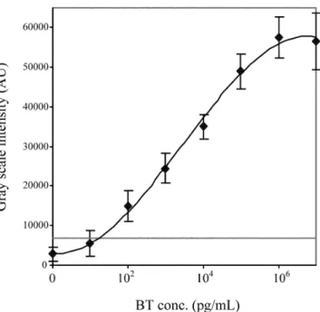
The analytical sensitivity and detection limit of the test strip assay for BT detection was determined from a dose– response curve (Fig. 4). Dose–response data were ob-tained by scanning densitometry of the test strips, which were run in various concentrations of BT. The limit of de-tection (LOD) is defined as the lowest concentration of toxin producing a signal intensity 3 times higher than the standard deviation of the intensity of the sample without toxin (i.e., the negative control). By this definition, the in-strumental (densitometry) LOD of the current assay for BT is estimated to be 15 pg mL–1, which is about 3 mLD
50 (mouse 50% lethal dose). The visual detection limit is about a factor of 10 higher. In the dose–response curve for BT, the intensity of the binding signal increases with in-creasing concentration of BT in the sample, providing a dynamic analytical range between approximately 101and 106pg mL–1, or about 5 orders of magnitude. Several re-search groups reported that their detection assays for bio-logical toxins, including BT, utilizing ganglioside–lipo-somes showed high sensitivity [8, 9, 25]. However, these assays detected the color change due to conformational change in the liposomes [8] or the signal from
dye-mark-ers or enzymes on the liposome surface [9, 25]. In this study, we used dye-encapsulating liposomes, which, be-cause of the much larger number of dye molecules con-tained in each liposome, produced a much higher signal intensity, thereby resulting in higher sensitivity. The LOD of 3 mLD50is comparable to the most sensitive BT detec-tion assays previously reported [15, 16, 26, 27] for which the detection limits reach the level of the mouse bioassay. Fig. 3 Scanned images of representative nitrocellulose test strips.
Strips were run at room temperature, as described in “Materials and methods”. Each strip was inserted into the test tube containing 100µL of the reaction mixture of GT1b-liposomes and BT at the indicated concentrations: A negative control, B 1×10⁻⁵µg mL–1, C 1×10⁻⁴µg mL–1, D 1×10⁻³µg mL–1, E 1×10⁻²µg mL–1, F 1×
10–1µg mL–1, G 1µg mL–1, H 10µg mL–1
Fig. 4 Dose–response curve for botulinum neurotoxin, generated
from test strip assays using GT1b-liposomes. The solid line repre-sents the third-order polynomial curve fit, with an R2value of
0.996. The straight horizontal line indicates the limit of detection, defined as the color intensity 3 times higher than the standard de-viation of the background (negative control) signal. Each point represents four replicates of grayscale values in the analytical zone
Table 2 Sensitivity of various detection assays for botulinum
neu-rotoxina
Method LOD of Assay
Refer-toxin type A time ence
(mLD50)b
Mouse bioassay 1–2 3–4 days [32]
RPHA 1.3–1.6 5–6 h [33] ELISA 1–5 h 2 [34] 9 [35] 1–2 [15] ELCA <1 >18 hc [27] Enzymatic assayd 0.5 5–6 h [16]
This study 3 20 min
aAbbreviations: mLD
50, mouse 50% lethal dose; RPHA, reversed
passive hemagglutination; ELISA, enzyme-linked immunosorbent assay, ELCA, enzyme-linked coagulation assay
b1 mLD
50 for chromatographically purified type A botulinum
neu-rotoxin has been calculated as approximately 6 pg mL–1[32]. At
1 mLD50, 50% of mice injected with 1 mL will die cIncluding the immunobinding phase
These previously reported assays, based on ELISA or the enzymatic activity of BT, are time-consuming and need to be performed by well-trained personnel. In contrast, the as-say developed in this study can be completed within 20 min and is very easy to perform. Therefore, this result suggests a BT detection assay using GT1b-liposomes can replace existing methods. In Table 2, the GT1b-liposome assay is compared to other previously reported BT detection as-says.
Comparison of the sensitivity of GT1b-liposomes to immunoliposomes for BT detection
The sensitivity of the BT detection assay using GT1b-li-posomes was compared to immunoliGT1b-li-posomes with anti-bodies to BT on the liposome surface (Fig. 5). The LOD for BT in the immunoliposome assay system was esti-mated from the dose–response curve to be 40 pg mL–1. At concentrations equal to or higher than 100 pg mL–1, the signal could be visually detected. This result suggests that the binding of the ganglioside to the toxin is as strong and specific as the binding of antibodies to the toxin. The sen-sitivity of the immunoliposome assay system is slightly lower, the detection limit a little higher (LOD=40 pg mL–1 versus 15 pg mL–1), and the signal intensity decreased more at higher concentrations of toxin as compared to the GT1b-liposome assay system. This poorer performance of
the immunoliposome assay can possibly be explained by the format used in this study. In the sandwich assay for-mat of the immunoliposome assay, the same antibodies were used for coating the analytical zone on test strips and for conjugation onto the liposome surface. This would re-sult in these antibodies competing for the same epitopes on the toxin, which could ultimately lead to the lower sen-sitivity of the assay. This problem could possibly be miti-gated by using antibodies to two different BT epitopes, one for use on the test strip analytical zone and the other for conjugation to liposomes. However, considering the complex and time-consuming (at least 2 days) process re-quired to prepare immunoliposomes, ganglioside–lipo-somes have a distinct advantage over immunolipoganglioside–lipo-somes in their ease of preparation.
Specificity of a capillary migration test strip assay for BT detection
To evaluate the specificity of the test strip assay for BT, the assay was performed substituting various other gan-glioside-binding toxins for BT. For this purpose, cholera toxin (CT) from Vibrio cholerae, diphtheria toxin (DT) from Corynebacterium diphtheriae, E. coli heat-stable toxin (STa), and tetanus toxin (TT) from Clostridium tetani were used. For safety purposes, commercially available toxoids or subunits of toxin were used in this study, ex-cept for STa. Each toxin was added to the assay system at high concentration (10µg mL–1), and the signal intensity was measured, as described in “Materials and methods”. As shown in Fig. 6, the BT detection assay using GT1b-li-posomes showed a high signal intensity for BT over the
Fig. 5 Comparison of the dose–response curves for
immunolipo-somes (E) with GT1b-liposomes (K) for botulinum neurotoxin de-tection. The solid line represents the curve fit for immunolipo-somes, and the dashed line represents the curve fit for GT1b-lipo-somes (as shown in Fig. 4), with R2values of 0.997 and 0.996,
re-spectively. The straight horizontal line indicates the limit of detec-tion for the immunoliposomes, defined as the color intensity 3 times higher than the standard deviation of the background (negative control) signal. Each point represents four replicates of grayscale values in the analytical zone
Fig. 6 Specificity of the BT detection assay using
GT1b-lipo-somes. A 10µg mL–1aliquot of each toxin was dissolved in TBS
and used in the assay. The data shown are an average of 3 repli-cates. Abbreviations: BT, botulinum toxin; CT, cholera toxin; DT, diphtheria toxin; STa, E. coli heat-stable toxin; TT, tetanus toxin
other toxins, which suggests specificity of the assay for BT detection. In this experiment, TT showed a little higher binding signal than the other toxins and this could be the result of the similarity of TT and BT in terms of structure and amino acid sequence [28, 29]. In addition, it has been reported that tetanus toxin binds to gangliosides, specifically to the disialoganglioside GD1b and GT1b, as membrane receptors [30, 31]. Despite the fact that TT in the sample can also bind to GT1b on the liposomes, the result from this study suggests that the antibodies to BT, immobilized on the test strip, can provide enough speci-ficity to distinguish BT from TT. The high specispeci-ficity of the detection assay, which requires binding to two sepa-rate and distinct receptors, provides an advantage over other immunological detection methods, which show the problems of false-positive signals from cross-reactivity, especially in sandwich-type assays.
Conclusions
In this study, GT1b-liposomes were used in a sandwich test strip assay for botulinum neurotoxin detection. We demonstrated that GT1b-liposomes can interact with BT at least as strongly as immunoliposomes. The assay de-veloped in this study provides detection levels compara-ble to the mouse bioassay and most of the other previ-ously reported assays, but has the added advantages of simplicity and rapidity. These results show that a capillary migration test strip assay can be an alternative assay sys-tem for BT detection, which can also be applied to the field screening of food or environmental samples. This as-say system could be applied to the detection of other bio-logical toxins that use gangliosides as their cell receptors. However, for the application of this assay to field screen-ing, the effect of food matrices will be evaluated. Prelim-inary studies on a variety of vegetable and seafood sam-ples have demonstrated only a moderate loss in sensitivity with the exception of certain fish, such as salmon, that are very high in fatty acids that appear to interfere with lipo-some integrity. Also, the effect of other organisms present in the sample on the sensitivity of this assay will be stud-ied.
Acknowledgments The authors acknowledge partial support for
this research from Innovative Biotechnologies International, Inc., Grand Island, NY, and the Cornell University Center for Biotech-nology, a New York State Center for Advanced Technology of the New York State Office of Science, Technology, and Academic Re-search, under whose auspices this publication was developed. This research also was supported in part by the Cornell University Agri-cultural Experiment Station Federal Formula Funds, Project No. NYG 623498, received from Cooperative State Research, Educa-tion, and Extension Service, US Department of Agriculture. Any opinions, finding, conclusions, or recommendations expressed in this publication are those of the authors and do not necessarily re-flect the view of the US Department of Agriculture.
References
1. Eidels L, Proia RL, Hart DA (1983) Membrane receptors for bacterial toxins. Microbial Rev 47:596–620
2. Holmgren J, Elwig H, Fredman P, Strannegard O, Svenner-holm L (1980) Gangliosides as receptors for bacterial toxins and Sendai virus. Adv Exp Med Biol 125:453–470
3. Fishman PH, Pacuszke T, Orlandi PA (1993) Gangliosides as receptors for bacterial enterotoxins. Adv Lipid Res 25:165–187 4. Van Heyningen WE, Carpenter CC, Pierce NF, Greenough WB III (1971) Deactivation of cholera toxin by ganglioside. J Infect Dis 124:415–418
5. Kitamura M, Iwamori M, Nagai Y (1980) Interaction between
Clostridium botulinum neurotoxin and gangliosides. Biochim
Biophys Acta 628:328–335
6. Ledley FD, Lee G, Kohn LD, Habig WH, Hardgree MC (1977) Tetanus toxin interactions with thyroid plasma membranes. Im-plications for structure and function of tetanus toxin receptors and potential pathophysiological significance. J Biol Chem 252:4049–4055
7. Fishman PH (1982) Role of membrane gangliosides in the binding and action of bacterial toxins. J Membr Biol 69:85–97 8. Pan JJ, Charych D (1997) Molecular recognition and colori-metric detection of cholera toxin by poly(diacetylene)liposomes incorporating Gm1ganglioside. Langmuir 13:1365–1367
9. Singh AK, Harrison SH, Schoeniger JS (2000) Gangliosides as receptors for biological toxins: development of sensitive fluo-roimmunoassays using ganglioside-bearing liposomes. Anal Chem 72:6019–6024
10. Ahn-Yoon S, DeCory T, Baeumner AJ, Durst RA (2003) Gan-glioside-liposome immunoassay for the ultrasensitive detection of cholera toxin. Anal Chem 75:2256–2261
11. Lasic DD, Papahadjopoulos D (1995) Liposomes revisited. Science 267:1275–1276
12. Arnon SS, Schechter R, Inglesby TV, Henderson DA, Bartlett JG, Ascher MS, Eitzen E, Fine AD, Hauer J, Layton M, Lilli-bridge S, Osterholm MT, O’Toole T, Parker G, Pearl TM, Rus-sell PK, Swerdlow DL, Tonat K (2001) Botulinum toxin as a biological weapon: medical and public health management. JAMA 285:1059–1070
13. Kautter DA, Solomon HM (1976) Collaborative study of a method for the detection of Cl. botulinum and its toxins in foods. J Assoc Anal Chem 60:541–545
14. Hatheway CL, Ferreira JL (1996) Detection and identification of Clostridium botulinum neurotoxins. Adv Exp Med Biol 391: 481–498
15. Ekong TAN, McLellan K, Sesardic D (1995) Immunological detection of Clostridium botulinum toxin type A in therapeutic preparations. J Immunol Methods 180:181–191
16. Wictome M, Newton KA, Jameson K, Dunnigan P, Clarke S, Gaze J, Tauk A, Foster KA, Shone CC (1999) Novel assays for the detection of botulinum toxins in foods. Dev Biol Stand 101:141–145
17. Szabo ZA, Pemberton JM, DesMarchelier PM (1993) Detec-tion of the genes encoding botulinum neurotoxin types A to E by the polymerase chain reaction. Appl Environ Microbiol 59: 3011–3020
18. Mayer LD, Hope MJ, Cullis PR (1986) Vesicles of variable sizes produced by a rapid extrusion procedure. Biochim Bio-phys Acta 858:161–168
19. Bartlett GR (1959) Phosphorus assay in column chromatogra-phy. J Biol Chem 234:466–468
20. Hikita T, Tadano-Aritomi K, Iida-Tanaka N, Toyoda H, Suzuki A, Toida T, Imanari T, Abe T, Yanagawa Y, Ishizuka I (2000) Determination of N-acetyl- and N-glycolylneuraminic acids in gangliosides by combination of neuraminidase hydrolysis and fluorometric high-performance liquid chromatography using a GM3 derivative as an internal standard. Anal Biochem 281: 193–201
21. Siebert STA, Reeves SG, Durst RA (1993) Liposome im-munomigration field assay device for Alachlor determination. Anal Chim Acta 282:297–305
22. Szoka F Olson F, Heath T, Vail W, Mayhew E (1980) Prepara-tion of unilamellar liposomes of intermediate size (0.1–0.2µm) by a combination of reverse phase evaporation and extrusions through polycarbonate membranes. Biochim Biophys Acta 601:559–571
23. Israelachvili J N, Mitchell DJ (1975) A model for the packing of lipids in bilayer membranes. Biochim Biophys Acta 389: 13–19
24. Park S, Durst RA (2000) Immunoliposome sandwich assay for the detection of Escherichia coli O157:H7. Anal Biochem 280: 151–158
25. Alfonta L, Willner I, Throckmorton DJ, Singh AK (2001) Elec-trochemical and quartz crystal microbalance detection of the cholera toxin employing horseradish peroxidase and GM1-functionalized liposomes. Anal Chem 73:5287–5295
26. Shone C, Wilson-Smith P, Appleton N, Hambleton P, Modi N, Gatley S, Melling J (1985) Monoclonal antibody-based im-munoassay for type A Clostridium botulinum toxin is compara-ble to the mouse bioassay. Appl Environ Microbiol 50:63–67 27. Doellgast G, Triscott MX, Beard GA, Bottoms JD, Cheng T,
Roh BH, Roman MG, Hall PA, Brown JE (1993) Sensitive en-zyme-linked immunosorbent assay for detection of Clostridium
botulinum neurotoxins A, B, and E using signal amplication via
enzyme-linked coagulation assay. J Clin Microbiol 31:2402– 2409
28. Sathyamoorthy V, DasGupta BR (1985) Partial amino acid se-quence of the heavy and light chains of botulinum neurotoxin type E. Biochem Biophys Res Commun 127:768–772 29. Halpern JL, Smith LA, Seamon KB, Groover KA, Habig WH
(1989) Sequence homology between tetanus and botulinum toxins detected by an antipeptide antibody. Infect Immun 57: 18–22
30. Van Heyningen WE (1974) Gangliosides as membrane recep-tors for tetanus toxin, cholera toxin and serotonin. Nature 249:415–417
31. Helting TB, Zwisler O, Wiegandt H (1977) Structure of tetanus toxin II. Toxin binding to ganglioside. J Biol Chem 252:194– 198
32. Schantz EJ, Kautter DA (1978) Standardized assay for
Clostridium botulinum toxins. J Assoc Off Anal Chem 61:96–
99
33. Johnson HM, Brenner K, Angelotti R, Hall HE (1966) Sero-logical studies of types A, B, and E botulinal toxins by passive hemagglutination and betonite flocculation. J Bacteriol 91: 967–973
34. Ransom GM, Lee WH, Elliot EL, Lattuada CP (1993) In: DasGupta BR (ed) Enzyme-linked immunosorbent assays (ELISAs) to detect botulinum toxins using high titer rabbit an-tisera in Botulinum and Tetanus Neurotoxins. Plenum Press, New York, pp 449–461
35. Potter MD, Meng J, Kimsey P (1993) An ELISA for detection of botulinal toxin types A, B, and E in inoculated food samples. J Food Protect 56:856–861