Regulatory regions of the paraoxonase 1 (
PON1)
gene are associated with neovascular age-related
macular degeneration (AMD)
Jadwiga Oczos&Christian Grimm&Daniel Barthelmes& Florian Sutter&Moreno Menghini&
Barbara Kloeckener-Gruissem&Wolfgang Berger
Received: 23 March 2012 / Accepted: 20 August 2012 / Published online: 7 September 2012 # American Aging Association 2012
Abstract Physiological stress response and oxidative damage are factors for aging processes and, as such, are thought to contribute to neovascular age-related macular degeneration (AMD). Paraoxonase 1 (PON1) is an en-zyme that plays an important role in oxidative stress and aging. We investigated association of DNA sequence variants (SNP) within the upstream regulatory region of the PON1 gene with neovascular AMD in 305 patients
and 288 controls. Four of the seven tested SNPs (rs705379, rs705381, rs854573, and rs757158) were more frequently found in AMD patients compared to controls (P00.0099, 0.0295, 0.0121, and 0.0256, respec-tively), and all but one (SNP rs757158) are in linkage disequilibrium. Furthermore, haplotype TGGCCTC con-ferred protection (odds ratio (OR)00.76, (CI)00.60– 0.97) as it was more frequently found in control individ-DOI 10.1007/s11357-012-9467-x
Barbara Kloeckener-Gruissem and Wolfgang Berger contributed equally to this work.
Electronic supplementary material The online version of this article (doi:10.1007/s11357-012-9467-x) contains supplementary material, which is available to authorized users.
J. Oczos
:
B. Kloeckener-Gruissem (*):
W. Berger (*) Institute of Medical Molecular Genetics,University of Zurich, Schwerzenbach, Switzerland e-mail: kloeckener@medgen.uzh.ch e-mail: berger@medgen.uzh.ch
J. Oczos
:
C. Grimm Lab for Retinal Cell Biology,Department of Ophthalmology, University of Zurich, Zurich, Switzerland
J. Oczos
:
C. Grimm:
W. BergerZurich Center for Integrative Human Physiology (ZIHP), University of Zurich,
Zurich, Switzerland
C. Grimm
:
W. BergerZurich Center of Neuroscience (ZNZ), Zurich, Switzerland
D. Barthelmes
:
F. Sutter:
M. MenghiniDepartment of Ophthalmology, University Hospital Zurich, Zurich, Switzerland
D. Barthelmes
Save Sight Institute, University of Sydney, Sydney, Australia
B. Kloeckener-Gruissem
Department of Biology, ETH Zurich, Zurich, Switzerland
uals, while haplotype CGATGCT increased the risk (OR01.55, CI01.09–2.21) for AMD. These results were also reflected when haplotypes for the untranscribed and the 5′untranslated regions (5′UTR) were analyzed sepa-rately. To assess haplotype correlation with levels of gene expression, the three SNPs within the 5′UTR were tested in a luciferase reporter assay. In retinal pigment epithelium-derived ARPE19 cells, we were able to mea-sure significant differences in reporter levels, while this was not observed in kidney-derived HEK293 cells. The presence of the risk allele A (SNP rs705381) caused an increase in luciferase activity of approximately twofold. Our data support the view that inflammatory reactions mediated through anti-oxidative activity may be relevant to neovascular age-related macular degeneration.
Keywords Paraoxonase 1 . Gene regulation . SNP association . Age-related macular degeneration (AMD)
Introduction
Age-related macular degeneration (AMD) is a pro-gressive degenerative disease, which in its advanced stages leads to severe visual impairment. It is the major cause of loss of vision in elderly patients in developed countries (Deangelis et al.2011). Advanced stages of AMD manifest either as atrophic changes in the macula (dry/atrophic/nonexudative AMD) or as cho-roidal neovascularization (wet/neovascular/exudative AMD). Although the neovascular AMD represents only 10–20 % of all AMD cases, it accounts for the vast majority of cases with severe and rapid vision loss (~90 %) (Jager et al.2008).
The etiologic complexity of AMD arises from an interplay of genetic and environmental factors. Recent progress in AMD genetics has established several impor-tant risk loci, among them the complement factor H (CFH) on chromosome 1 (1q31) (Edwards et al.
2005; Hageman et al.2005,2006; Hughes et al.2006; Klein et al. 2005; Li et al. 2006) and the age-related maculopathy susceptibility 2 (ARMS2/HTRA1) on chro-mosome 10 (10q26) (Dewan et al. 2006; Jakobsdottir et al. 2005; Rivera et al. 2005; Schmidt et al. 2006; Tanimoto et al. 2007; Weger et al. 2007; Yang et al.
2006). Both loci combined are thought to account for more than 50 % of AMD cases (Edwards et al.2005; Klein et al.2005; Maller et al.2006; Swaroop et al.2007; Thakkinstian et al.2006). Their association with AMD
has been replicated across multiple ethnic groups world-wide and further validated through a meta-analysis of whole genome scans (Fisher et al.2005). The discovery of CFH as a genetic risk factor for AMD was supported by functional studies revealing a role of the alternative complement pathway in drusen formation (Hageman et al.2005). Following the discovery of genetic susceptibil-ity loci, an impressive number of further genes has been reported that either increase or lower the risk towards AMD development (Katta et al.2009). Despite the strong associations established, it is still not possible to predict the onset or course of AMD (Swaroop et al. 2007). Hence, it is necessary to identify other genes involved in the pathogenesis and understand their interactions.
To date, several additional candidate genes have been studied for their association with AMD, including lipid metabolism and oxidative stress genes (APOE (Baird et al.2004b; Pang et al.2000), VLDLR (Conley et al.2005; Haines et al.2006), LRP5 (Kloeckener-Gruissem et al.
2011), LRP6 (Haines et al. 2006), PON1 (Baird et al.
2004a; Brion et al.2011; Esfandiary et al.2005; Ikeda et al.2001; Pauer et al.2010), LIPC (Neale et al.2010), and SOD2 (Gotoh et al. 2008; Kimura et al. 2000). Discordant results across multiple studies indicate the existence of additional factors that influence the devel-opment of AMD in different populations.
Oxidative stress is implicated in the development of AMD (AREDS2001; Beatty et al.2000; Khandhadia and Lotery2010); thus, identification of enzymes con-ferring antioxidant protection is of interest. During the last decade, paraoxonase 1 (PON1), an enzyme with antioxidant properties, has been extensively investigated in several disorders, including age-related pathologies such as hyperlipidemia, atherosclerosis, coronary artery disease, metabolic syndrome, type 2 diabetes, as well as Alzheimer's disease (Androutsopoulos et al. 2011; Mackness et al.1998b,c; Paragh et al. 1998; Senti et al.2003). PON1 is a protein mainly synthesized in the liver, and most studies have focused their analysis on this fact. The enzyme is secreted into the blood where it binds high-density lipoprotein (HDL) (Aviram 2004). One of the functions of PON1 is to protect low-density lipoprotein (LDL) from oxidative damage and to inacti-vate oxidized LDL by hydrolyzing its oxidized phos-pholipids (Deakin and James2004).
PON1 serum concentration and activity are highly variable across the general population (Deakin and James2004). This variability is attributed to environmen-tal factors, such as smoking, diet, alcohol consumption,
but also to the genetic polymorphisms in the PON1 gene. There are two annotated missense SNPs within the PON1 coding region: p.L55M (rs854560), where the major allele L leads to elevated PON1 protein levels, and p.Q192R (rs662), affecting substrate specificity as well as enzyme activity (Garin et al. 1997; Humbert et al.
1993). These two missense polymorphisms affect also the ability of HDL to protect LDL from oxidative mod-ifications, with MM/QQ genotype being most effective (Mackness et al.1998a). The level of PON1 concentra-tion in serum is also affected by sequence variants within the promoter of the PON1 gene. In particular, the C allele at position−107 (SNP rs705379) yields the highest level of PON1 in the serum among all alleles that have been investigated so far (Leviev and James2000).
The antioxidant properties of PON1 stimulated re-search for association of DNA sequence variants within the PON1 coding region and AMD (Baird et al.2004a; Brion et al.2011; Esfandiary et al. 2005; Ikeda et al.
2001; Pauer et al. 2010). Much focus has been on studying the effects of the two above-mentioned poly-morphisms, p.L55M and p.Q192R. In a Japanese AMD patient cohort, the 55L and the 192R variants were found more frequently (Ikeda et al.2001), while in one Caucasian cohort, the 55Q variant was associated with the disease (Pauer et al. 2010). This association could not be confirmed in further three Caucasian patient groups suggesting the influence of other factors as well (Baird et al.2004a; Brion et al.2011; Esfandiary et al.
2005). No information has been published on associa-tions of sequence variants within upstream regulatory regions of the gene and diseases. It was our goal to expand the knowledge of effects of PON1 gene sequen-ces beyond protein isoforms and to investigate the in-fluence of the upstream regulatory regions on AMD. We report here the identification of protective and risk hap-lotypes with effects on gene expression, which displays cell-type specificity.
Materials and methods
Patients
Patients with neovascular AMD were diagnosed at the Department of Ophthalmology at the University Hospital Zurich. Inclusion criteria regarding age, visual acuity, and lesion type (minimally classic, pre-dominantly classic, and occult) were based on the
MARINA (Rosenfeld et al. 2006) and ANCHOR (Brown et al. 2006) trials. The patients were given a full explanation of the nature and purpose of the study. The study was conducted according to the Declaration of Helsinki and was approved by the local ethics committee. Clinical and demographic description as well as the analysis of several common AMD risk factors of this patient cohort has been published pre-viously (Kloeckener-Gruissem et al.2011). Data from 305 neovascular AMD patients are reported in the current study. The average age of the patients was 79.41 years (±6.97); female to male ratio, 1.95. All were of European descent, living in Switzerland. DNA of 288 individuals representing the general population was used as the control group (Moskvina et al.2005). These individuals were not age-matched and did not undergo clinical assessment for the present study. They were of European descent, living in Switzerland. The female/male ratio was 2.1.
Genotyping
Venous blood was collected in EDTA tubes, and ge-nomic DNA was extracted as described (Kloeckener-Gruissem et al.2011). DNA concentration was adjusted to 10 ng/μl. SNP genotyping was performed either by DNA sequencing (primer 1 CCTCCCCGACTGGAC-TAGG and primer 2 AGGGAGTGAGGAGGAC-GAAG were used for genotyping SNPs rs705379, rs705380, and rs705381; primer 3 CCAAAAGCCTT-GAGAAGGAA and primer 4 TGCTCTAGGTGATG-CATGTG were used for genotyping SNPs rs854571 and rs854572) or by TaqMan technology (SNPs rs854571, rs854573, and rs757158), using ABI chemistry (Ap-plied Biosystems, Inc. [ABI], Rotkreuz, Switzerland). Sequencing primers were designed by Primer3 (v. 0.4.0;
http://frodo.wi.mit.edu/primer3, provided in the public domain by Massachusetts Institute of Technology, Cambridge, MA) and synthesized by Microsynth (http://www.microsynth.ch; Balgach, Switzerland). Annealing temperatures varied between 54 and 60 °C. For PCR, 50-ng genomic DNA and primers were cycled 35 times, each cycle lasting 1 min. For TaqMan assay, probes from ABI were used. Genotype calling was performed on SDS2.2 software, ABI. The allele calling of SNP assay was verified by DNA sequencing analysis for 10 % of all samples, yielding 100 % concordance. In addition, genotyping of 10 % of all samples was repeated for each SNP.
Selection of SNPs and haplotype analysis
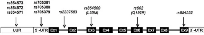
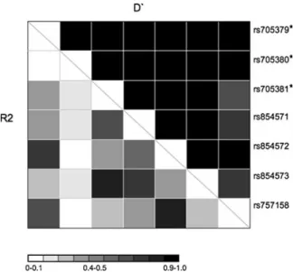
Seven SNPs, namely rs705379, rs705380, rs705381, rs854571, rs854572, rs854573, and rs757158 (Fig. 1
and Table1), within PON1 gene (NG_008779.1) were chosen for association studies based on the linkage dis-equilibrium (LD) data in the HapMap Caucasion (CEU) population panel, using a tagging criteria of R2>0.8. Since there is no LD information available for SNPs in the 5′UTR (rs705379, rs705380, and rs705381), we in-cluded them in the analysis.
Individual haplotypes and their estimated population frequencies were inferred using PHASE program, v2.1.1 (http://stephenslab.uchicago.edu/software.html), with all parameters set at the default values (Stephens and Donnelly2003; Stephens et al.2001).
In silico analysis of 5′UTR variants on RNA folding Putative RNA folding structures were predicted using the Mfold online software with standard settings (
http://mfold.rna.albany.edu/?q0mfold/RNA-Folding-Form) (Zuker 2003). RNA structures were predicted for the PON1 transcript (transcript: PON1-001 ENST00000222381) containing eight possible combina-tions of alleles at the SNPs within 5′UTR.
Cloning
A 400-bp fragment of PON1 exon 1 containing the 5′ UTR was PCR-amplified (primer 1 and primer 2) and cloned into the intermediate pJET1.2/blunt cloning vec-tor using the CloneJET PCR Cloning Kit (Fermentas International Inc, Glen Burnie, MD, USA). Fragments containing four of the eight possible haplotypes were obtained directly through PCR from patients' genomic DNA (CCA, CGA, CGG, and TGG; SNPs' order from left to right, rs705379, rs705380, and rs705381). DNA fragments containing the other four haplotypes were obtained by site-directed mutagenesis with the primer
extension approach. For all substitutions, primer 1 and primer 2 served as outside flanking primers. Each mutation was obtained using additional pairs of inside primers (sequences are written from 5′to 3′) as primer 5 C C G A C C C G G C G G G G A G G G G T G G G G C G G GCC with primer 6 GCAGCGCCGATTGGCCC GCCCCACCCCTCCC, primer 7 GGGGCTGA CCGCAAGCCGCGCCTTCTGTGC with primer 8 G A C C A G G T G C A C A G A A G G C G C G G C T TGCGG, primer 9 GGGGCTGACCGCAAGC CACGCCTTCTGTGC with primer 10 GACCAGGTG CACAGAAGGCGTGGCTTGCGGTC, and primer 11 TGGTCGGCCCAGCTAGCTGCCGACCCGGCGGG with primer 12 CCACCCCTCCCCGCCGGGTCGG CAGCTAGC. Subsequently, the PON1 fragments repre-senting each of the haplotype were cloned into pGL3-Control vector (Promega, Madison, WI, USA). In the first step, PCR reaction was performed to create a HindIII restriction site upstream of the three 5′UTR SNPs using primer 2 and primer 13 TATAAGCTTGTTGGAAG GAGCAAAATG. In the second step, 238-bp fragments spanning from the constructed HindIII site to the trans-lational start codon ATG (NcoI site) were cloned into the pGL3-Control vector using HindIII and NcoI restriction enzymes (Fermentas). DNA sequences of all constructs were verified by Sanger sequencing.
Expression studies
HEK293 cells were seeded on 24-well plates (105cells/ well) in 1 ml DMEM, 10 % FBS, and 1 % penicillin/ streptomycin and incubated under standard conditions (37 °C, 5 % CO2). After 24 h, cells were transfected using 1.5-μl branched polyethylenimine, PEI (1 μg/μl), 600-ng pFirefly construct, and 15-ng pRenilla construct per well. Twenty-four hours later, the cells were har-vested, and luciferase activities were analyzed using Dual-Glo Luciferase Assay System according to the protocol provided by Promega and measured on a Bio-Tek Synergy microplate reader (BioBio-Tek Instruments,
Fig. 1 Schematic representation of the PON1 gene. Positions of SNPs that were analyzed in this study are printed in bold. They map to the upstream untranscribed region (UUR) and the 5 ′un-translated region (5′UTR). SNPs in exon 3 (Ex3), exon 6 (Ex6),
and exon 9 (Ex9), investigated by other groups, are printed in italics (Baird et al.2004a; Brion et al.2011; Esfandiary et al.2005; Ikeda et al.2001; Pauer et al.2010)
Winooski, VT, USA). Data were normalized for trans-fection efficiency by Renilla luciferase activity. The ex-periment was performed three times with three replicates each. ARPE19 cells (ATCC, Manassas, VA, USA) were seeded on 24-well plates (8×10⁴cells/well) in 1 ml DMEM, 10 % FBS, 1 % penicillin/streptomycin, and 7.5 % Na2CO3and cultured under standard conditions (37 °C, 5 % CO2). Twenty-four hours later, transfection was performed; for each well, 0.75-μl X-tremeGENE 9 transfection reagent (Roche Diagnostics, Indianapolis, IN, USA), 250-ng pFirefly construct, and 6.25-ng pRe-nilla construct were used. Luciferase activities were measured and analyzed 24 h after transfection as described above. The experiment was repeated four times. Three of these repeats included three technical replicates while one of them included eight.
Statistical analysis
Pairwise LD analysis was performed on phased haplo-type data generated from the control group according to the following formulas: R20D2/(pA1pA2pB1pB2), where D0pA1B1pA2B2−pA1B2pA2B1; D′0D/Dmax, with Dmax0 min(pA1pB2, pA2pB1) if D>0 and Dmax0min(−pA1pB1, -pA2pB2) if D<0 (Mueller2004). For association studies, odds ratios and significance were calculated using 2×2 contingency table provided on an open access Internet portal (http://faculty.vassar.edu/lowry/odds2x2.html). Odds ratios were displayed with 95 % confidence inter-vals and Chi-square and P values according to Pearson. Statistical significance was assumed at P<0.05. For
expression studies, eight technical replicas were mea-sured and averages with confidence intervals at 95 % were computed. Fold-change differences were deter-mined after normalizing the data to those obtained for the TCG haplotype. This haplotype was chosen because it yielded the lowest luciferase activity in the ARPE19 cell line. The Shapiro–Wilk test was applied to assess normal distribution of the data. For statistical analysis, the two-tailed t test was used.
Results
Linkage disequilibrium revealed two DNA blocks within the upstream regulatory region of PON1
Linkage disequilibrium data are crucial for the design of association studies. If two SNPs share high linkage disequilibrium, with R2>0.8 one of them can be used as tagged SNP. The region of PON1 investigated in our study contains seven annotated SNPs (Fig. 1 and Table1). LD data on the HapMap website are available only for four of them (rs854571, rs854572, rs854573, and rs757158). To complete this information, we per-formed LD analysis on phased genotype data of the control individuals (Table 2). Our analysis confirms already annotated data in a larger group of individuals (haplotype number n0500 in our study, n0224 in Hap-Map) and also provides pairwise R2 and D′ values for SNPs which are not included in HapMap. Among the seven analyzed SNPs, six belong to one haplotype block Table 1 Allele frequencies of SNPs in the upstream regulatory region of PON1
Marker Major/minor allele Position Region MAF P value OR CI (95 %)
HapMap AMD (n0305) Controls (n0288)
rs705379 T/C −107 5′UTR 0.425 0.517 0.440 0.0099 1.36 1.08–1.73 rs705380 G/C −126 5′UTR 0.042 0.056 0.044 0.3566 1.29 0.75–2.21 rs705381 G/A −162 5′UTR 0.167 0.253 0.199 0.0295 1.37 1.03–1.81 rs854571 C/T −831 UUR 0.302 0.284 0.257 0.3009 1.14 0.89–1.49 rs854572 C/G −908 UUR 0.425 0.471 0.419 0.0759 1.23 0.98–1.56 rs854573 T/C −1075 UUR 0.230 0.255 0.194 0.0121 1.42 1.08–1.88 rs757158 C/T −1740 UUR 0.376 0.444 0.380 0.0256 1.30 1.03–1.64
Comparison between patients with neovascular AMD (n0305) and control individuals (n0288). Three SNPs map to the 5′untranslated region while four of the SNPs lie within the upstream untranscribed region (Fig.1). Minor allele frequencies for a Caucasian population were taken from the HapMap website (http://www.hapmap.org) and were calculated for our patient (AMD) and control groups. Statistical analyses are displayed as odds ratios (OR) with confidence intervals (CI) and P values, withα00.05
as they revealed high pairwise D′ values (rs705379, rs705380, rs705381, rs854571, rs854572, and rs854573). SNP rs757158 belongs to a separate haplo-type block (Fig. 2). Among the analyzed SNPs, only two pairs (rs705381 with rs854573, and rs854572 with rs757158) fulfill tagging criteria of R2>0.8 (Table S1).
Association studies showed risk and protection potential for AMD
Genotypes of seven SNPs within regulatory, non-coding regions of the PON1 locus were determined from 305 patients with neovascular AMD and compared to those from 288 control individuals (Table 1). The patients' AMD phenotype has been reported in detail previously (Kloeckener-Gruissem et al.2011). Three SNPs within the 5′UTR and four SNPs within the upstream untran-scribed region (UUR) (Fig. 1) were genotyped and analyzed. Note that as the transcriptional promoter re-gion has not been defined experimentally, we chose to name this region“UUR”, rather than “promoter region”. Statistically significant association was found at two SNP loci within the 5′UTR and two within the UUR: rs705379 (OR01.36, CI01.08–1.73, P00.0099), rs705381 (OR01.37, CI01.03–1.81, P00.0295), rs854573 (OR01.42, CI01.08–1.88, P00.0121), and rs757158 (OR01.30, CI 01.03–1.64, P 00.0256) (Table1).
To strengthen the significance of the association, we included analyses of haplotypes. For the seven SNP haplotypes (rs705379 at −107, rs705380 at −126 and
rs705381 at−162, rs854571 at −831, rs854572 at −908, rs854573 at −1075, and rs757158 at −1740), PHASE predicted 18 different combinations. Haplotypes with fewer than five counts in PHASE (11 haplotypes) were not taken into account for statistical analyses. The haplo-type TGGCCTC, comprising exclusively major alleles, was more frequently found in the control group, suggest-ing a protective function (OR00.76, CI00.60–0.97, P0 0.0293), whereas the haplotype CGATGCT was more frequent in AMD patients (OR01.55, CI01.09–2.21, P0 0.0141), posing a risk for the disease (Table2). Similar results were found when SNPs from the 5′UTR and the upstream untranscribed region (UUR) were analyzed separately. The PHASE program revealed the presence of four possible haplotypes for SNPs in the 5′UTR (TGG, CGG, CGA, CCA; order of SNPs from left to right, rs705379 at −107, rs705380 at −126, and rs705381 at −162). Carriers of the haplotype TGG, representing the major alleles within a population, were more frequently found in the control group compared to the patient group, and, hence, this haplotype confers a protection against AMD (OR00.72, CI00.57–0.91, P00.0063), while con-versely the haplotype CGA indicates a risk for AMD (OR01.35, CI00.99–1.85, P00.0564) (Table3). For the four SNPs in the UUR, PHASE predicted seven different haplotypes. Three haplotypes showed statistically signif-icant differences between patient and control group Table 2 Haplotype analysis for all seven SNPs
Haplotype AMD (n0556) n (%) Controls (n0500) n (%) P value OR CI (95 %) TGGCCTC 264 (47.5) 271 (54.2) 0.0293 0.76 0.60–0.97 CGGCGTT 100 (18.0) 74 (14.8) 0.1637 1.26 0.91–1.75 CGATGCT 94 (16.9) 58 (11.6) 0.0141 1.55 1.09–2.21 CCATGCT 25 (4.5) 22 (4.4) 0.9203 1.02 0.57–1.84 CGGCCTC 26 (4.7) 18 (3.6) 0.3833 1.31 0.71–2.43 CGGTGTT 17 (3.1) 25 (5.0) 0.1069 0.60 0.32–1.12 CGATGCC 12 (2.2) 14 (2.8) 0.5023 0.77 0.35–1.67
SNP order for each haplotype, from left to right is as follows: -107, -126, -162, -831, -908, -1075, and−1740. Haplotype counts (n) and percentages (%) are given. Haplotypes with less than five counts in PHASE were not taken into account for statistical analysis. Odds ratios (OR) with confidence intervals (CI) and P values (alpha00.05) are shown
Fig. 2 Pairwise linkage analysis of SNPs within the PON1 5′ UTR and upstream untranscribed region. R2 and D′ values are depicted by the grayscale color code according to the legend. Asterisks indicate SNPs for which no data are available on the HapMap website
frequencies (Table3). The haplotype TGCT (rs854571 at −831, rs854572 at −908, rs854573 at −1075, and rs757158 at −1740) was associated with the disease (OR01.44, CI01.07–1.95, P00.0165), whereas the two haplotypes CCTC and TGTT were more frequently found in the control group (OR00.79, CI00.63–0.99, P00.0492 and OR00.48, CI00.27–0.84, P00.0092, respectively) (Table3).
Sequence variants in the 5′UTR alter predicted structure of the PON1 mRNA
It is known that SNPs within the 5′UTR of genes can affect its mRNA folding and consequently change trans-lational efficiency (Zuercher et al.2010). We simulated the influence of the 5′UTR polymorphisms on secondary RNA structures using the web server Mfold. RNA struc-tures of the entire PON1 mRNA (5′UTR, coding sequen-ces, and 3′UTR) were predicted (Fig.3). The predicted structure of the reference sequence carrying the minor alleles at three analyzed positions (−107C, -126C, and -162A) was not different from that containing only one major T allele at position −107 (Fig. 3a). In contrast, predicted folding of RNA containing the major alleles at position −126 and −162, singly, or in combination, assumed a strikingly different structure (Fig.3b). Based on these bioinformatic predictions, we hypothesized that
SNPs within the 5′UTR of PON1 may have different effects on protein synthesis.
AMD risk haplotype altered reporter gene activity in cell culture
To correlate our association data and the structural pre-dictions with potential effects on gene expression, we designed experiments to test whether different PON1 5′ UTR haplotypes influence luciferase reporter gene ex-pression in cell culture. We generated the eight possible haplotypes in DNA constructs, containing the PON1 5′ UTR region fused to the Luciferase coding region, both transcribed from the SV40 promoter. Since tissue-specific effects on gene expression are likely to occur, we included two different cell lines in this test: the embryonic kidney-derived cell line HEK293 and the retinal pigment epithelium-derived cell line ARPE19. Both cell lines were transiently transfected with the various PON1 5′UTR haplotypes in front of the lucifer-ase reporter. In ARPE19 cells, we found statistically significant differences in luciferase levels (Fig. 4). To assess contribution of each individual SNP to increased protein expression, we performed a pairwise compari-son of luciferase levels between haplotypes differing at only one position (Table4). This analysis revealed that SNP rs705381 at position−162 is mostly responsible for Table 3 Haplotype analysis of the upstream untranscribed and untranslated region
Region Haplotype AMD n (%) Controls n (%) P value OR CI (95 %)
5′UTRa TGG 280 (48.4) 297 (56.7) 0.0063 0.72 0.57–0.91 CGG 150 (26.0) 121 (23.1) 0.2713 1.17 0.89–1.54 CGA 116 (20.1) 82 (15.6) 0.0564 1.35 0.99–1.85 CCA 32 (5.5) 24 (4.6) 0.4708 1.22 0.71–2.10 UURb CCTC 308 (52.4) 320 (58.2) 0.0492 0.79 0.63–0.99 TGCT 128 (21.8) 89 (16.2) 0.0165 1.44 1.07–1.95 CGTT 109 (18.5) 87 (15.8) 0.2253 1.21 0.89–1.65 TGTT 19 (3.2) 36 (6.5) 0.0092 0.48 0.27–0.84 TGCC 19 (3.2) 17 (3.1) 0.8875 1.05 0.54–2.04
Four haplotypes in the 5′untranslated region (5'UTR) and five haplotypes in the upstream untranscribed region (UUR) were analyzed in our patient and control population. The order of SNPs, from left to right is−107, -126, and −162 (5′UTR) and −831, -908, -1075, and −1740 (UUR). Haplotype counts (n) and percentages (%) are given. Odds ratios (OR) with confidence intervals (CI) and P values (alpha00.05) are shown. Haplotypes with fewer than five counts in PHASE were not taken into account for statistical analysis
a
AMD, n0578; controls, n0524
b
the observed difference in luciferase activity. The pres-ence of the minor allele A leads to increase in the reporter activity up to 2.1-fold. Small contribution to the protein expression regulation has been found for two other SNPs (rs705379 at−107, and rs705380 at −126), with minor allele C at position−107 and minor allele C at position −126, causing up to 1.6- and 1.4-fold increase in luciferase level, respectively. Interestingly, in HEK293 cells, we did not find statistically significant differences in luciferase activity between the different constructs (data not shown), supporting our initial as-sumption of cell-specific effects on gene expression.
Discussion
This is the first report to show association of SNPs in the upstream regulatory region of PON1 with neovascular AMD. Previous studies have investigated association between SNPs within the PON1 coding region and AMD (Brion et al.2011; Baird et al.2004a; Esfandiary et al.2005; Pauer et al. 2010; Ikeda et al. 2001). The focus was on two missense polymorphisms p.L55M and p.Q192R. Conflicting results were obtained when com-paring different patient cohorts; while association with wet AMD has been found for both variants in a Japanese population (Ikeda et al.2001), only the p.Q192R variant was associated in only one of the four Caucasian cohorts (Pauer et al.2010). Furthermore, in the Japanese study, the arginine at position 192 of the PON1 protein has been found more frequently in the patient group,
whereas in Caucasians, the glutamine (192Q) variant has been associated with the disease. Possible explana-tions encounter different allele frequencies in the differ-ent ethnic groups but also a diverse phenotype of AMD (Bird 2003; Oshima et al.2001). Specifically for SNP p.Q192R in Caucasians, the minor variant is R with a frequency of 0.332, whereas in Japanese, the arginine is the major variant with an allele frequency of 0.706 (HapMap). As there is no linkage disequilibrium between p.Q192R variant and the promoter polymorphisms (Lev-iev and James 2000) (HapMap), a direct comparison between our data and those reported from other Cauca-sian patient cohorts, which examined p.Q192R (Baird et al.2004a; Brion et al.2011; Esfandiary et al.2005; Pauer et al.2010) is not meaningful.
In contrast to p.Q192R, the variant 55L is in linkage disequilibrium with the allele -107C (Leviev and James
2000), which is associated with AMD in our study. It has been shown that these two alleles lead to increased levels of PON1 in serum (Leviev and James 2000). Thus, it may be difficult to understand how higher levels of a protein that is known to have antioxidant properties can be associated with AMD. For a possible explanation, we would like to emphasize that variant p.L55M has been reported to affect PON1 activity towards lipid peroxides, with the LL genotype being less effective than MM at protecting LDL against oxidation (Mackness et al.
1998a). Since alleles at position -107C and -162A are in linkage disequilibrium with variant 55L, they are likely to be also associated with the less effective protection against oxidation, thus can confer the risk for AMD and Fig. 3 Predicted PON1
mRNA foldings. a The structure represents folding of the PON1 mRNA containing the minor alleles CCA in the 5′untranslated region. It was indistinguish-able from the allele combi-nation TCA. b The structure depicts mRNA folding in the presence of the other six allelic combinations: CGA, TGA, CCG, TCG, CGG, and TGG (SNP order from left to right, -107,-126, and−162)
be responsible for increased oxLDL levels (Ikeda et al.
2001) in AMD patients.
To strengthen the significance of the association found with individual SNPs, we performed haplotype associa-tion analysis. We discovered two seven-SNP haplotypes
associated with AMD. The most abundant haplotype (54.2 % in controls), TGGCCTC, was more frequently found in controls conferring a protection against AMD, whereas haplotype CGATGCT was a risk factor. Inter-estingly, the two haplotypes CGATGCC and CCATGCT, each differing at only one position in comparison to CGATGCT, appeared not to be associated with the dis-ease. These data imply a possible interplay between the analyzed loci and underlines the importance of haplotype analysis to assess risk factors. Similar results were obtained when analyzing haplotypes within the 5′UTR and UUR separately.
It is known that sequence variants within the 5′UTR can regulate gene expression at the level of protein translation. We asked whether this would also apply to the PON1 gene. To support our notion, we performed in silico RNA folding predictions for the different allelic combinations and found that the presence of the major alleles for two SNPs in the 5′UTR would lead to a striking difference in the folding properties of the entire PON1 transcript. Furthermore, these alleles also appear to have an effect on reporter gene expression as mea-sured by luciferase enzyme activity. Specifically, in the retinal pigment epithelium-derived cell line ARPE19, the protective TGG haplotype yielded the lowest level of luciferase activity, while the CCA hap-lotype resulted in the highest reported expression levels. When comparing those haplotypes differing at only one position, we discovered that the SNP at position−162 provided the largest contribution, with the A nucleotide causing up to 2.1-fold increase in luciferase reporter activity. Previous reports have shown that this allele Fig. 4 Fold increase of
luciferase activity for the eight PON1 5′UTR haplo-types in ARPE19 cells. The luciferase activity of the re-porter construct containing the TCG haplotype was set to 1. SNP order in haplotypes from left to right: -107, -126, and−162. Error bars show confidence inter-vals, CI095 %. Displayed are results from a represen-tative experiment with eight technical replicates
Table 4 Effects of 5′UTR haplotypes on luciferase reporter activity
SNP Haplotypea Fold increase
in luciferase activityb P valuec 1 2 −107 CCA TCA 1.4 <0.001 CCG TCG 1.4 0.016 CGA TGA 1.3 0.030 CGG TGG 1.0 0.984 −126 CCA CGA 1.6 <0.001 CCG CGG 1.2 0.094 TCA TGA 1.4 <0.001 TCG TGG 0.9 0.242 −162 CCA CCG 2.1 <0.001 CGA CGG 1.6 <0.001 TCA TCG 2.0 <0.001 TGA TGG 1.2 0.030
The comparison is based on haplotypes that differ at a single position. Displayed are results for a representative experiment shown in Fig.4
a
SNP order in haplotype, from left to right is−107, -126, and −162
b
Fold increase in luciferase activity, haplotype 1 versus haplotype 2
c
caused up-regulation of transcription in cultured kidney and liver cell lines (Brophy et al. 2001). These data, together with our findings, indicate that the position at −162 has regulatory function at both steps of gene ex-pression, transcription, as well as translation. Alternative-ly, a sole effect on transcription of the luciferase reporter cannot be excluded. In our study, the two other investi-gated SNPs at positions −107 and −126 caused only minor changes in luciferase activity. In the literature, no effect of the SNP at −126 has been reported, but the minor allele C at position −107 has been shown to increase transcription of a luciferase reporter gene in HepG2 and HEK293 cells (Brophy et al.2001). Taking these data together, we conclude that the three SNPs support different activities for PON1 gene expression.
It is important to note that the luciferase reporter assays did not yield the same results in the two different cell lines tested. As cell and tissue-specific regulation of gene expression is a well-known phenomenon, it is not surprising that gene expression control potential within the 5′UTR of PON1 is manifested in ARPE19 cells, but not in HEK293 cells. The latter cells, derived from kid-ney, do not seem to be implicated in the AMD pathology, while ARPE19 cells, derived from retinal pigment epi-thelium, do (Kinnunen et al.2012). In this context, it is plausible that the relevant SNP at position−162 exerts a cell type-specific effect in favor of AMD. Since Pon1 is expressed in the retinal pigment epithelium in mice (data not shown), further studies will provide crucial informa-tion to clarify these assumpinforma-tions.
A number of factors such as blood pressure, usage of antihypertensive medication, blood cholesterol, or blood levels of high-density lipoprotein were shown to be associated with the development of neovascular AMD (Hyman et al. 2000). As the mean age of the population studied here is 79.5 years, it can be expected that some of the factors (e.g., increased arterial blood pressure) are present in the study population—assumingly randomly distributed across the participants. An influence of the proposed cardio-vascular risk factors on the outcome of the treatment of neovascular AMD using ranibizumab has not yet been shown. Hence, it remains speculative whether or not such risk factors may have had an impact in the current study population and the reported outcomes.
Many open questions remain on the involvement of PON1 in AMD pathology. As our patients were diag-nosed with the end stage neovascular AMD, our results cannot account for a potential role of PON1 as a trigger
for the onset of AMD or as an accelerator during the progression of the disease, or maybe both. Our and other previous association studies imply the involvement of PON1 at the late stage of the disease. An alternative mechanism may exist, where PON1 enzyme changes are secondary to other events, present at early stages of AMD. For example, paraoxonase activity in serum is decreased in AMD patients. As AMD is considered to be a chronic inflammatory disease and the PON1 tran-scription levels in liver are known to be down-regulated in response to infections, it seems feasible that PON1 down-regulation and decreased activity are the outcome of primarily inflammation events. Cross talk between PON1 levels and inflammatory response could be medi-ated by the oxLDL, which can induce expression of genes involved in an inflammation by stimulating various transcription factors (Maziere and Maziere 2009). Our results provide insights in the action of PON1, but further investigation is required for a deeper understanding of the contribution of PON1 in the pathology of AMD.
Acknowledgments We would like to thank Marijana Samardzija for help with Excel macros to ease data entry in PHASE. We appreciate the genomic DNA preparations by Esther Glaus. Finan-cial support by a cooperative project grant by the Zurich Center for Integrative Human Physiology (ZIHP) of the University of Zurich, Zurich, Switzerland, is greatly acknowledged. DB was supported by a grant from the Swiss National Foundation (SNF/SSMBS), a grant from the Holcim Foundation, and the Walter and Gertrud Siegenthaler Foundation Zürich, Switzerland.
References
Androutsopoulos VP, Kanavouras K, Tsatsakis AM (2011) Role of paraoxonase 1 (PON1) in organophosphate metabolism: implications in neurodegenerative diseases. Toxicol Appl Pharmacol 256:418–424
AREDS (2001) A randomized, placebo-controlled, clinical trial of high-dose supplementation with vitamins C and E and beta carotene for age-related cataract and vision loss: AREDS report no. 9. Arch Ophthalmol 119:1439–1452 Aviram M (2004) Introduction to the serial review on
paraox-onases, oxidative stress, and cardiovascular diseases. Free Radic Biol Med 37:1301–1303
Baird PN, Chu D, Guida E, Vu HT, Guymer R (2004a) Association of the M55L and Q192R paraoxonase gene polymorphisms with age-related macular degeneration. Am J Ophthalmol 138:665–666
Baird PN, Guida E, Chu DT, Vu HT, Guymer RH (2004b) The epsilon2 and epsilon4 alleles of the apolipoprotein gene are associated with age-related macular degeneration. Investig Ophthalmol Vis Sci 45:1311–1315
Beatty S, Koh H, Phil M, Henson D, Boulton M (2000) The role of oxidative stress in the pathogenesis of age-related mac-ular degeneration. Surv Ophthalmol 45:115–134
Bird AC (2003) The Bowman lecture. Towards an understand-ing of age-related macular disease. Eye (Lond) 17:457–466 Brion M, Sanchez-Salorio M, Corton M, de la Fuente M, Pazos B et al (2011) Genetic association study of age-related mac-ular degeneration in the Spanish population. Acta Ophthalmol 89:e12–e22
Brophy VH, Hastings MD, Clendenning JB, Richter RJ, Jarvik GP et al (2001) Polymorphisms in the human paraoxonase (PON1) promoter. Pharmacogenetics 11:77–84
Brown DM, Kaiser PK, Michels M, Soubrane G, Heier JS et al (2006) Ranibizumab versus verteporfin for neovascular age-related macular degeneration. N Engl J Med 355: 1432–1444
Conley YP, Thalamuthu A, Jakobsdottir J, Weeks DE, Mah T et al (2005) Candidate gene analysis suggests a role for fatty acid biosynthesis and regulation of the complement system in the etiology of age-related maculopathy. Hum Mol Genet 14:1991–2002
Deakin SP, James RW (2004) Genetic and environmental factors modulating serum concentrations and activities of the antioxidant enzyme paraoxonase-1. Clin Sci (Lond) 107:435–447
Deangelis MM, Silveira AC, Carr EA, Kim IK (2011) Genetics of age-related macular degeneration: current concepts, future directions. Semin Ophthalmol 26:77–93
Dewan A, Liu M, Hartman S, Zhang SS, Liu DT et al (2006) HTRA1 promoter polymorphism in wet age-related macu-lar degeneration. Science 314:989–992
Edwards AO, Ritter R 3rd, Abel KJ, Manning A, Panhuysen C et al (2005) Complement factor H polymorphism and age-related macular degeneration. Science 308:421–424 Esfandiary H, Chakravarthy U, Patterson C, Young I, Hughes
AE (2005) Association study of detoxification genes in age related macular degeneration. Br J Ophthalmol 89:470–474 Fisher SA, Abecasis GR, Yashar BM, Zareparsi S, Swaroop A et al (2005) Meta-analysis of genome scans of age-related macular degeneration. Hum Mol Genet 14:2257–2264 Garin MC, James RW, Dussoix P, Blanche H, Passa P et al (1997)
Paraoxonase polymorphism Met-Leu54 is associated with modified serum concentrations of the enzyme. A possible link between the paraoxonase gene and increased risk of cardiovascular disease in diabetes. J Clin Invest 99:62–66 Gotoh N, Yamada R, Matsuda F, Yoshimura N, Iida T (2008)
Manganese superoxide dismutase gene (SOD2) polymor-phism and exudative age-related macular degeneration in the Japanese population. Am J Ophthalmol 146:146, author reply 146–147
Hageman GS, Anderson DH, Johnson LV, Hancox LS, Taiber AJ et al (2005) A common haplotype in the complement regulatory gene factor H (HF1/CFH) predisposes individ-uals to age-related macular degeneration. Proc Natl Acad Sci U S A 102:7227–7232
Hageman GS, Hancox LS, Taiber AJ, Gehrs KM, Anderson DH et al (2006) Extended haplotypes in the complement factor H (CFH) and CFH-related (CFHR) family of genes protect against age-related macular degeneration: characterization, ethnic distribution and evolutionary implications. Ann Med 38:592–604
Haines JL, Schnetz-Boutaud N, Schmidt S, Scott WK, Agarwal A et al (2006) Functional candidate genes in age-related macular degeneration: significant association with VEGF, VLDLR, and LRP6. Investig Ophthalmol Vis Sci 47:329– 335
Hughes AE, Orr N, Esfandiary H, Diaz-Torres M, Goodship T et al (2006) A common CFH haplotype, with deletion of CFHR1 and CFHR3, is associated with lower risk of age-related macular degeneration. Nat Genet 38:1173–1177 Humbert R, Adler DA, Disteche CM, Hassett C, Omiecinski CJ
et al (1993) The molecular basis of the human serum paraoxonase activity polymorphism. Nat Genet 3:73–76 Hyman L, Schachat AP, He Q, Leske MC (2000) Hypertension,
cardiovascular disease, and age-related macular degenera-tion. Age-Related Macular Degeneration Risk Factors Study Group. Arch Ophthalmol 118:351–358
Ikeda T, Obayashi H, Hasegawa G, Nakamura N, Yoshikawa T et al (2001) Paraoxonase gene polymorphisms and plasma oxidized low-density lipoprotein level as possible risk fac-tors for exudative age-related macular degeneration. Am J Ophthalmol 132:191–195
Jager RD, Mieler WF, Miller JW (2008) Age-related macular degeneration. N Engl J Med 358:2606–2617
Jakobsdottir J, Conley YP, Weeks DE, Mah TS, Ferrell RE et al (2005) Susceptibility genes for age-related maculopathy on chromosome 10q26. Am J Hum Genet 77:389–407 Katta S, Kaur I, Chakrabarti S (2009) The molecular genetic
basis of age-related macular degeneration: an overview. J Genet 88:425–449
Khandhadia S, Lotery A (2010) Oxidation and age-related macu-lar degeneration: insights from molecumacu-lar biology. Expert Rev Mol Med 12:e34
Kimura K, Isashiki Y, Sonoda S, Kakiuchi-Matsumoto T, Ohba N (2000) Genetic association of manganese superoxide dismutase with exudative age-related macular degenera-tion. Am J Ophthalmol 130:769–773
Kinnunen K, Petrovski G, Moe MC, Berta A, Kaarniranta K (2012) Molecular mechanisms of retinal pigment epitheli-um damage and development of age-related macular degeneration. Acta Ophthalmol 90:299–309
Klein RJ, Zeiss C, Chew EY, Tsai JY, Sackler RS et al (2005) Complement factor H polymorphism in age-related macular degeneration. Science 308:385–389
Kloeckener-Gruissem B, Barthelmes D, Labs S, Schindler C, Kurz-Levin M et al (2011) Genetic association with response to intravitreal ranibizumab in patients with neovascular AMD. Investig Ophthalmol Vis Sci 52:4694–4702 Leviev I, James RW (2000) Promoter polymorphisms of human
paraoxonase PON1 gene and serum paraoxonase activities and concentrations. Arterioscler Thromb Vasc Biol 20:516–521 Li M, Atmaca-Sonmez P, Othman M, Branham KE, Khanna R
et al (2006) CFH haplotypes without the Y402H coding variant show strong association with susceptibility to age-related macular degeneration. Nat Genet 38:1049–1054 Mackness B, Mackness MI, Arrol S, Turkie W, Durrington PN
(1998a) Effect of the human serum paraoxonase 55 and 192 genetic polymorphisms on the protection by high density lipoprotein against low density lipoprotein oxida-tive modification. FEBS Lett 423:57–60
Mackness B, Mackness MI, Arrol S, Turkie W, Julier K et al (1998b) Serum paraoxonase (PON1) 55 and 192 polymorphism and
paraoxonase activity and concentration in non-insulin depen-dent diabetes mellitus. Atherosclerosis 139:341–349 Mackness MI, Mackness B, Durrington PN, Fogelman AM,
Berliner J et al (1998c) Paraoxonase and coronary heart disease. Curr Opin Lipidol 9:319–324
Maller J, George S, Purcell S, Fagerness J, Altshuler D et al (2006) Common variation in three genes, including a noncoding variant in CFH, strongly influences risk of age-related mac-ular degeneration. Nat Genet 38:1055–1059
Maziere C, Maziere JC (2009) Activation of transcription fac-tors and gene expression by oxidized low-density lipopro-tein. Free Radic Biol Med 46:127–137
Moskvina V, Holmans P, Schmidt KM, Craddock N (2005) Design of case-controls studies with unscreened controls. Ann Hum Genet 69:566–576
Mueller JC (2004) Linkage disequilibrium for different scales and applications. Brief Bioinform 5:355–364
Neale BM, Fagerness J, Reynolds R, Sobrin L, Parker M et al (2010) Genome-wide association study of advanced age-related macular degeneration identifies a role of the hepatic lipase gene (LIPC). Proc Natl Acad Sci U S A 107:7395– 7400
Oshima Y, Ishibashi T, Murata T, Tahara Y, Kiyohara Y et al (2001) Prevalence of age-related maculopathy in a repre-sentative Japanese population: the Hisayama study. Br J Ophthalmol 85:1153–1157
Pang CP, Baum L, Chan WM, Lau TC, Poon PM et al (2000) The apolipoprotein E epsilon4 allele is unlikely to be a major risk factor of age-related macular degeneration in Chinese. Ophthalmologica 214:289–291
Paragh G, Seres I, Balogh Z, Varga Z, Karpati I et al (1998) The serum paraoxonase activity in patients with chronic renal failure and hyperlipidemia. Nephron 80:166–170 Pauer GJ, Sturgill GM, Peachey NS, Hagstrom SA (2010)
Protective effect of paraoxonase 1 gene variant Gln192Arg in age-related macular degeneration. Am J Ophthalmol 149:513–522
Rivera A, Fisher SA, Fritsche LG, Keilhauer CN, Lichtner P et al (2005) Hypothetical LOC387715 is a second major susceptibility gene for age-related macular degeneration, contributing independently of complement factor H to dis-ease risk. Hum Mol Genet 14:3227–3236
Rosenfeld PJ, Brown DM, Heier JS, Boyer DS, Kaiser PK et al (2006) Ranibizumab for neovascular age-related macular degeneration. N Engl J Med 355:1419–1431
Schmidt S, Hauser MA, Scott WK, Postel EA, Agarwal A et al (2006) Cigarette smoking strongly modifies the association of LOC387715 and age-related macular degeneration. Am J Hum Genet 78:852–864
Senti M, Tomas M, Fito M, Weinbrenner T, Covas MI et al (2003) Antioxidant paraoxonase 1 activity in the metabolic syndrome. J Clin Endocrinol Metab 88:5422–5426 Stephens M, Donnelly P (2003) A comparison of bayesian
meth-ods for haplotype reconstruction from population genotype data. Am J Hum Genet 73:1162–1169
Stephens M, Smith NJ, Donnelly P (2001) A new statistical method for haplotype reconstruction from population data. Am J Hum Genet 68:978–989
Swaroop A, Branham KE, Chen W, Abecasis G (2007) Genetic susceptibility to age-related macular degeneration: a para-digm for dissecting complex disease traits. Hum Mol Genet 16(Spec No. 2):R174–R182
Tanimoto S, Tamura H, Ue T, Yamane K, Maruyama H et al (2007) A polymorphism of LOC387715 gene is associated with age-related macular degeneration in the Japanese pop-ulation. Neurosci Lett 414:71–74
Thakkinstian A, Han P, McEvoy M, Smith W, Hoh J et al (2006) Systematic review and meta-analysis of the association between complement factor H Y402H polymorphisms and age-related macular degeneration. Hum Mol Genet 15:2784–2790
Weger M, Renner W, Steinbrugger I, Kofer K, Wedrich A et al (2007) Association of the HTRA1–625G>A promoter gene polymorphism with exudative age-related macular degenera-tion in a Central European populadegenera-tion. Mol Vis 13:1274–1279 Yang Z, Camp NJ, Sun H, Tong Z, Gibbs D et al (2006) A variant of the HTRA1 gene increases susceptibility to age-related macular degeneration. Science 314:992–993 Zuercher J, Neidhardt J, Magyar I, Labs S, Moore AT et al (2010)
Alterations of the 5′untranslated region of SLC16A12 lead to age-related cataract. Investig Ophthalmol Vis Sci 51:3354– 3361
Zuker M (2003) Mfold web server for nucleic acid folding and hybridization prediction. Nucleic Acids Res 31:3406–3415