Publisher’s version / Version de l'éditeur:
Journal of Natural Products, 78, 8, pp. 1942-1948, 2015-07-23
READ THESE TERMS AND CONDITIONS CAREFULLY BEFORE USING THIS WEBSITE. https://nrc-publications.canada.ca/eng/copyright
Vous avez des questions? Nous pouvons vous aider. Pour communiquer directement avec un auteur, consultez la
première page de la revue dans laquelle son article a été publié afin de trouver ses coordonnées. Si vous n’arrivez pas à les repérer, communiquez avec nous à [email protected].
Questions? Contact the NRC Publications Archive team at
[email protected]. If you wish to email the authors directly, please see the first page of the publication for their contact information.
NRC Publications Archive
Archives des publications du CNRC
This publication could be one of several versions: author’s original, accepted manuscript or the publisher’s version. / La version de cette publication peut être l’une des suivantes : la version prépublication de l’auteur, la version acceptée du manuscrit ou la version de l’éditeur.
For the publisher’s version, please access the DOI link below./ Pour consulter la version de l’éditeur, utilisez le lien DOI ci-dessous.
https://doi.org/10.1021/acs.jnatprod.5b00277
Access and use of this website and the material on it are subject to the Terms and Conditions set forth at
Characterization of L-Digitoxosyl-phenanthroviridin from Streptomyces
venezuelae ISP5230
Robertson, Andrew W.; Martinez-Farina, Camilo F.; Syvitski, Raymond T.;
Jakeman, David L.
https://publications-cnrc.canada.ca/fra/droits
L’accès à ce site Web et l’utilisation de son contenu sont assujettis aux conditions présentées dans le site LISEZ CES CONDITIONS ATTENTIVEMENT AVANT D’UTILISER CE SITE WEB.
NRC Publications Record / Notice d'Archives des publications de CNRC:
https://nrc-publications.canada.ca/eng/view/object/?id=060c5f22-ee41-4fc5-8909-9e451dc156ac https://publications-cnrc.canada.ca/fra/voir/objet/?id=060c5f22-ee41-4fc5-8909-9e451dc156ac
Characterization of
L
‑Digitoxosyl-phenanthroviridin from
Streptomyces venezuelae ISP5230
Andrew W. Robertson,
†Camilo F. Martinez-Farina,
†Raymond T. Syvitski,
‡and David L. Jakeman
*
,†,§†
Department of Chemistry, Dalhousie University, Halifax, Nova Scotia, Canada, B3H 4R2
‡
Institute for Marine Biosciences, National Research Council of Canada, Halifax, Nova Scotia, Canada, B3H 3Z1
§
College of Pharmacy, Dalhousie University, Halifax, Nova Scotia, Canada, B3H 1X7
*
S Supporting InformationABSTRACT: The jadomycin-derived compound L
-digitoxo-syl-phenanthroviridin was isolated from fermentations of Streptomyces venezuelae ISP5230 grown in nutrient-deficient media with L-lysine as the sole nitrogen source. Structural
elucidation was accomplished using a combination of high-resolution MS, LC-MS/MS, and 1D- and 2D-NMR. The compound was evaluated against the National Cancer Institute (NCI) 60 human tumor cell line screen in both the one-dose and five-dose screens, and cytotoxicity was compared to a small library of jadomycin analogues to probe the structure− activity relationship.
T
he angucyclines are the largest known group of natural products derived from type II polyketide synthases (PKSs).1The structural diversity of this large family of natural products arises from the configurations of polyaromatic angucycline backbones and the modification of these scaffolds by a wide array of post-PKS tailoring enzymes. A great deal of interest in their continued isolation and characterization is in anticipation of identifying novel bioactive natural products. The soil bacterium Streptomyces venezuelae ISP5230 has been studied extensively for its ability to produce the jadomycin group of natural products.2The jadomycins are grouped within the angucycline family and are distinguished by a characteristic modified benz[a]anthracene scaffold, a 2,6-dideoxysugar moiety, L-digitoxose, and an amino acid that is usually fuseddirectly into the polyaromatic backbone as an oxazolone ring. The first isolated examples were that of the aglycone jadomycin A3 and the glycosylated jadomycin B (Figure 1).4,5 Interest-ingly, amino acid incorporation has been shown to proceed through a spontaneous process in which the enzyme JadG is responsible for a C−C bond cleavage at the B-ring (Scheme 1).1,6,7 This proceeds via a Baeyer−Villiger-type mechanism producing a reactive aldehyde intermediate, which couples with an amino acid, forming an imine, which undergoes spontaneous cyclization and oxidation to produce a five-membered oxazolone ring (Scheme 1). This mechanism has been confirmed through both extensive biosynthetic and total synthetic studies,7−9 and exploitation of this mechanism by
precursor-directed derivatization with varying amino acids has led to the isolation of upward of 25 jadomycin analogues.2,10−14
The glycosyltransferase JadS then appends the L-digitoxyl
moiety to the aglycones, yielding the fully furnished natural products (Scheme 1).
Engineering approaches have also been successful in expanding the structural diversity of these compounds: disruption of the jadO gene, coding for a putative 2,3-dehydratase, resulted in the production and isolation of ILEVS1080, a differentially glycosylated analogue of jadomycin B.17 Rohr and co-workers isolated and characterized the glycosylated analogue L-digitoxosyl-dehydrorabelomycin from
a jadG deletion mutant (Figure 1).18 Lacking the ability to catalyze the ring-opening step of biosynthesis, a buildup of the jadomycin precursor dehydrorabelomycin (Scheme 1) and the glycosylated analogue was observed, coupled with complete loss of jadomycin production.18 The isolation of the phenanthroviridin aglycone has also been reported from cultures of S. venezuelae ISP5230 (Figure 1). It was simultaneously identified during the first isolation of jadomycin A (Figure 1).3 However, the glycosylated analogue
L
-digitoxosyl-phenanthroviridin (1) has not been reported to date.
Recently, we reported the isolation and characterization of jadomycin Oct and jadomycin AVA, both containing eight-membered heterocyclic rings (Scheme 1).15Cyclization to the
eight-membered ring occurs as a result of initial imine formation with the δ-amine, in preference to the α-amino group. In an effort to isolate the jadomycin incorporating L
-lysine, where we anticipated formation of a nine-membered heterocyclic ring, S. venezuelae fermentations in the presence of
L-lysine were performed. Although production of a jadomycin
analogue with the appropriate m/z for L-lysine incorporation
was confirmed by LC-MS/MS analysis of the crude growth
Received: March 31, 2015 Published: July 23, 2015
pubs.acs.org/jnp
media,19 attempts to isolate the product using standard methodologies successfully employed for other jadomycins or by chemical derivatization were unsuccessful due to compound instability.15,20 During investigations of crude fermentation extracts, we identified an intriguing unknown amber-colored compound by TLC that proved to be sufficiently stable for isolation and characterization. Herein, we report the isolation, characterization, and cytotoxic evaluation of the new phenanthroviridin analogue L-digitoxosyl-phenanthroviridin
(1).
■
RESULTS AND DISCUSSIONFermentation, Isolation, and Purification of 1. S. venezuelae ISP5230 VS1099 was grown in the presence of L
-lysine (60 mM) as the sole nitrogen source using literature methodology.21The fermentation was allowed to proceed for 48 h while monitoring by absorption spectroscopy for cell growth at 600 nm and the production of colored natural products at 526 nm (Figure S1). After 48 h, growth media was colored reddish-purple, indicating jadomycin production. The
Figure 1. Structures of jadomycin A;3 jadomycin B;4,5 ILEVS1080;17 L-digitoxosyl-dehydrorabelomycin;18 phenanthroviridin aglycone;3and L -digitoxosyl-phenanthroviridin (1).
Scheme 1. Substrate Scope of Amino Acid Incorporation into the Jadomycin Backbone8,11,13−16
Journal of Natural Products Article
DOI: 10.1021/acs.jnatprod.5b00277
J. Nat. Prod. 2015, 78, 1942−1948
cells were removed, and the clarified growth media was applied to a reversed-phase phenyl column enabling retention of aromatic organic compounds. The aromatic organic material was eluted with 100% methanol and extracted with water and ethyl acetate, yielding ∼45 mg L−1of crude solid material. TLC
analysis of the ethyl acetate extract identified a yellow compound as the predominant natural product in the mixture. Preparative TLC was performed on the material, yielding 8 mg of the amber yellow solid, compound 1 (4 mg L−1), of sufficient
purity for characterization.
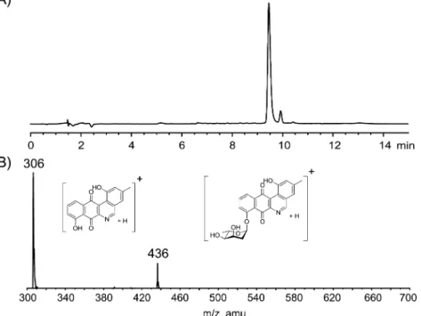
Structural Elucidation of 1. High-resolution mass spectrometry (HRMS) identified a molecular formula of C24H21NO7. LC-MS/MS analysis scanning for [M + H]+
identified an m/z of 436, with fragmentation to m/z of 306. This fragmentation pattern is typical of jadomycin-like molecules and represents the loss of L-digitoxose, identifying
a glycosylation of the unknown compound (Figure 2).11 Structural characterization was accomplished using a combination of 1H, 13C, COSY, edited-HSQC, HMBC, and
NOESY NMR spectroscopy experiments. The 1H NMR and COSY experiments confirmed the presence of theL-digitoxose
spin system (C1′ through C6′). In addition, the characteristic jadomycin A-ring (C2, C4, and C13) and D-ring (C9 through C11) spin systems were observed. An exchangeable singlet (not observable in MeOD-d4) at δH= 11.84 ppm was also present,
corresponding to the 1-OH. HMBC data provided core
Figure 2.(A) HPLC trace of 1 (tR= 9.47 min) monitored at 254 nm; (B) LC-MS/MS fragmentation of 1, showing parent ion [M + H]+and fragmentation resulting from the cleavage ofL-digitoxose [M + H − digitoxose]+(m/z 306).
Figure 3.(A) Key1H−1H COSY (bold),1H−13C HMBC (solid arrows), and1H−15N HMBC (bold arrow) correlations in 1; (B) key NOESY (dashed arrows) correlations of 1; (C) δH and δC values of H5 and C5 associated with the compounds L-digitoxosyl-dehydrorabelomycin,18 phenanthroviridin aglycone,3and 1.
connectivity typical of a jadomycin (Figure 3). No signals arising from the incorporation ofL-lysine (or any other amino
acid) were present. The key signal in the 1H NMR was a
nonexchangeable CH proton at δH= 9.42 ppm with an HSQC
correlation to a carbon at δC = 160.9 ppm. This relatively
downfield chemical shift suggested a distinct heteroaromatic chemical environment resembling that of the phenanthroviridin aglycone at C5, as opposed toL
-digitoxosyl-dehydrorabelomy-cin, which lacks the heteroaromatic ring (Figure 3). HMBC and 2D-NOESY correlations from H5 to C4 and H4, respectively, confirmed the proximity of the proton to the A-ring; HMBC analysis also identified a correlation to the C6a position of the B-ring. 1H−15N HMBC analysis established a correlation
between H5 and N6, confirming the presence of a heteroaromatic nitrogen. Glycosylation at the 8-position was confirmed by NOESY correlations between H9 of the D-ring and H1′ of theL-digitoxose moiety and an HMBC correlation
between H1′ and C8 of the D-ring (Figure 3). Stereochemistry of the sugar moiety was inferred asL-digitoxose based on past
X-ray crystallographic studies of jadomycin B.22 The bio-synthetic production of the dideoxysugar is well established for this family of molecules, and the NMR data associated with the sugar moiety match those of previously published jadomycin and jadomycin-like compounds.18 These data are consistent with our proposed structure of 1 as a functionalized benzo[b]phenanthridine framework glycosylated with L
-dig-itoxose. A full tabulated list of1H and13C chemical shift and
2D-correlations for compound 1 can be found inTable 1. Our difficulties in isolation of the jadomycin L-lysine
derivative paralleled work by Yang and co-workers, who attempted purification of the compound but were hampered by product instability and low yields that led to incomplete structural characterization.10 We propose 1 is likely a stable
degradation product of the L-lysine analogue. Isolation of 1
presented an opportunity to test the bioactivity of a unique jadomycin-like analogue and to probe the structure−activity relationship associated with the oxazolone ring system.
Cytotoxic Activity. Compound 1 was selected for evaluation against the National Cancer Institute’s (NCI) 60 DTP human tumor cell line screen. All screening was carried out according to the NCI protocol with the exception of solvent, where ethanol was substituted for dimethyl sulfoxide due to compound stability. Initial single dose screening (10 μM) identified sufficient cytotoxicity (Table S1) for further testing in a five-dose assay (10 nM to 100 μM) using log10
concentration intervals. Percent growth was plotted as a function of the concentration of 1 giving dose−response curves for each tumor cell line (Figures S3 and S4). From these curves the GI50 (growth inhibition of 50%), TGI (drug
concentration resulting in total growth inhibition), and LC50
(lethal concentration resulting in 50% tumor death) were calculated. With the exception of leukemia, compound 1 showed respectable cytotoxicity, in many cases resulting in complete or near-complete tumor death at higher concen-trations (100 μM).
Having access to cytotoxicity data for a series of jadomycin derivatives previously reported by our lab, direct comparison of the GI50, TGI, and LC50values of 1 to this group of compounds
was explored. It was discovered that despite lacking an amino acid side chain and an oxazolone ring, 1 had comparable bioactivity when compared to this small library of natural and semisynthetic jadomycins. These data are summarized inTable 2.
In an effort to probe the mechanism of action of 1, a standard COMPARE23,24 analysis screening against the NCI standard agents database using GI50, TGI, and LC50 values was
performed. The screen identified mediocre correlation (Pearson correlation coefficient (PCC) < 0.58) between 1 and the standard agents database, a library of 171 known cytotoxic compounds, when compared to GI50, TGI, and LC50
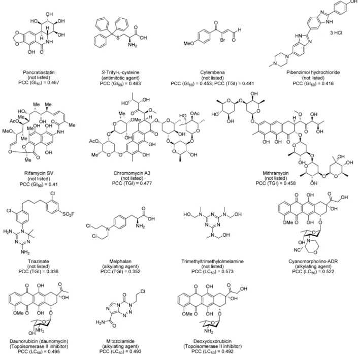
values. Results showed correlations to a wide variety of compounds including known antimitotic agents, alkylating agents, topoisomerase II inhibitors, and a series of compounds with listed unknown function (Figure 4). The highest correlation obtained was associated with trimethyltrimethylol-melamine (PCC = 0.573) while comparing LC50 data (Figure
4). This correlation, together with a GI50 correlation to
pancratiastatin (PCC = 0.467), has been previously reported for structurally similar naphthoquinone moiety containing compounds.25
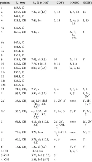
Table 1. NMR Spectroscopic Data (1H: 700 MHz and13C:
176 MHz, CD2Cl2) forL-Digitoxosyl-phenanthroviridin (1)
position δC, type δH(J in Hz)a COSY HMBC NOESY
1 155.3, C 2 123.6, CH 7.32, d (1.4) 4, 13 1, 4, 13 13 3 144.2, C 4 121.5, CH 7.46, brs 2, 13 2, 4a, 5, 13 5, 13 4a 132.8, C 5 160.9, CH 9.42, s 4a, 4, 6a, 12b 4 6a 147.4, C 7 181.5, C 7a 120.1, C 8 156.2, C 9 121.9, CH 7.63, d (8.5) 10 7a, 11 1′ 10 136.2, CH 7.76, t (8.1) 9, 11 8, 11a 11 122.7, CH 8.00, d (7.6) 10 7a, 9, 12 11a 136.3, C 12 191.1, C 12a 126.9, C 12b 120.6, C 13 21.7, CH3 2.55, s 2, 4 2, 3, 4 2, 4 1′ 95.2, CH 5.96, d (3.2) 2′ 8, 3′ 9, 2a′, 2b′ 2a′ 35.6, CH2 ax: 2.24, ddd (15.1, 3.2, 3.2)b 1′, 2b′, 3′ none 1′, 2b′, 3′, 4′ 2b′ 35.6, CH2 eq: 2.52, ddd (15.1, 3.2, 0.9)c 1′, 2a′, 3′ 3′, 4′ 1′, 2a′, 3′ 3′ 66.5, CH 4.11, dq (10.1, 3.2) 2a′, 2b′,3′-OH, 4′ none 2a′, 2b′ 4′ 4′ 72.9, CH 3.24, brm 3′, 4′-OH, 5′ none 2a′, 3′ 5′ 66.6, CH 3.79, dq (10.1, 6.2) 4′, 6′ none 6′ 6′ 18.1, CH3 1.22, d (6.2) 5′ 4′, 5′ 5′ 1-OH 11.84, brs 1, 2, 3 3′-OH 5.20, brd (10.6) 3′ 4′-OH 2.89, brd (8.7) 4′
aAll coupling constants reported were determined using the TopSpin3.2 multiplet tool software; any minor inconsistences are to be attributed to signal broadness and error associated with the measurements.bAx = axial proton at the 2′-position.cEq = equatorial proton at the 2′-position.
Journal of Natural Products Article
DOI: 10.1021/acs.jnatprod.5b00277
J. Nat. Prod. 2015, 78, 1942−1948
Correlations, although poor, between 1 and the anthracycline antibiotics daunorubicin (PCC = 0.495), deoxydoxorubicin (PCC = 0.492), and cyanomorpholino-ADR (PCC = 0.522) were observed when comparing LC50 values. These clinically
used anthracyclines exhibit potent antitumor properties. They impart cytotoxicity via intercalation into DNA, allowing the formation of a stable complex with topoisomerase II, inhibiting the enzymes ability to effectively cut DNA disrupting tumor proliferation.26,27 The jadomycin family of natural products shares structural similarities to these compounds, and jadomycin inhibition of topoisomerase II has been speculated in the past.28This may suggest a similar mode of action for 1
and other jadomycins.
When compared to the synthetic compound database (>40 000 compounds, including synthetic compounds and natural products of known structure), improved correlations between 1 and a number of jadomycins previously isolated in our lab were observed (correlations ∼0.7−0.8). Thus, it can be concluded that 1 has a similar mode of action to jadomycins incorporating amino acids through precursor-directed biosyn-thesis and that the cytotoxic effect may not be strongly dependent on the incorporated amino acid. Rather, the amino acid functionality may tune other physiochemical properties of the natural product. This discovery illustrates the opportunity to direct efforts toward modifying the sugar moiety or derivatizing the polyaromatic backbone in order to identify new analogues with improved or altered bioactivity, while concurrently varying the amino acid incorporated into the jadomycin scaffold to adjust physicochemical properties.
■
EXPERIMENTAL SECTIONGeneral Experimental Procedures.All reagents were purchased from commercial sources and used without further purification unless otherwise stated. All solvents used for compound purification were HPLC grade. Preparative TLC was carried out using 20 × 20 cm glass-backed plates (SiliCycle) layered with 1000 μm silica. Compound 1 was characterized by liquid chromatography tandem-mass spectrom-etry (LC-MS/MS), HRMS, and 1D- and 2D-NMR spectroscopy. Low-resolution LC-MS/MS spectra were obtained on an Applied Biosystems hybrid triple quadrupole linear ion trap (2000 Qtrap) mass spectrometer using an electrospray ionization (ESI+) source. This was coupled with an Agilent 1100 high-performance liquid chromatog-raphy (HPLC) instrument with a Phenomenex Kinetex 2.6 μm Hilic column (150 mm × 2.10 mm). Samples were prepared in methanol,
and 5 μL aliquots were analyzed. Elution of compounds was accomplished using an isocratic gradient of 7:3 CH3CN/2 mM ammonium acetate in water (pH 5.5) with a flow rate of 120 μL min−1 for 10 min. HRMS data were recorded on a Bruker Daltonics MicroTOF Focus mass spectrometer using an ESI+ source. Enhanced product ionization (EPI) was performed with a capillary voltage of +4500 kV, declustering potential of +80 V, and curtain gas of 10 arbitrary units. EPI scans were conducted over a range of 300−700 m/ z scanning for [M + H]+ and the appropriate jadomycin fragmentation. Scans were conducted using two steps, 300.0 to 320 amu (0.005 s) and 300.0 to 700.0 amu (0.100 s). Spectra were analyzed using Analyst software version 1.4.1 (Applied Biosystems). NMR analysis of 1 was performed on either a Bruker AV 500 MHz spectrometer (1H: 500 MHz, 13C: 125 MHz) equipped with an autotune and match (ATMA) broadband observe SmartProbe located at the Nuclear Magnetic Resonance Research Resource (NMR-3) facility (Dalhousie University) or a Bruker AV-III 700 MHz spectrometer (1H: 700 MHz, 13C: 176 MHz) equipped with an ATMA 5 mm TCI cryoprobe located at the Canadian National Research Council Institute for Marine Biosciences (NRC-IMB) in Halifax, Nova Scotia. All spectra were recorded in CD2Cl2. Chemical shifts (δ) are given in ppm and calibrated to residual solvent peaks [1H (CHDCl2): 5.32 ppm; 13C (13CD2Cl2): 54.0 ppm]. Structural characterization and signal assignments were accomplished using1H NMR chemical shifts, multiplicities, and13C NMR chemical shifts. In addition, 1H−1H correlated spectroscopy (COSY), 1H−13C hetero-nuclear single quantum coherence (HQSC) NMR, 1H−13C heteronuclear multiple bond correlation (HMBC) NMR, 1H−15N HMBC, and 2D 1H−1H nuclear Overhauser effect spectroscopy (NOESY) experiments were used in NMR analysis.
Fermentation, Extraction, and Isolation. All media was prepared with distilled water unless otherwise stated. Media utilized for this study included MYM broth [maltose 4 g/L, yeast extract 4 g/ L, malt extract 10 g/L]; MYM agar [maltose (4 g/L), yeast extract (4 g/L), malt extract (10 g/L), agar 15 (g/L), pH 7.0]; MSM media [MgSO4(0.4 g/L), MOPS (3.77 g/L), salt solution (9 mL 1% w/v NaCl, 1% w/v CaCl2), FeSO4·7H2O (4.5 mL 0.2% w/v), trace mineral solution (4.5 mL), pH 7.5]. Trace mineral solution [ZnSO4·7H2O (880 mg/L), CuSO4·5H2O (39 mg/L), MnSO4·4H2O (6.1 mg/L), H3BO3(5.7 mg/L), (NH4)6Mo7O24·4H2O (3.7 mg/L). S. venezuelae ISP5230 VS1099 was grown on MYM agar plates supplemented with apramycin (50 μg mL−1) for 1−3 weeks.29,30 Fermentations were carried out using modified conditions for jadomycin production with L-lysine (60 mM) as the sole nitrogen source.21A 1 × 1 cm lawn of S. venezuelae was used to inoculate 250 mL of MYM media (250 mL in 4 ×1 L flasks, total volume = 1 L). Fermentations were incubated at 30 °C with agitation (250 rpm) for 16−24 h. The cell suspension was centrifuged at 3750 rpm (4 °C) for 30 min. The supernatant was
Table 2. Summary of NCI 60 Cancer Cell Line Screen GI50, TGI, and LC50Values of 1 Compared to a Small Library of
Jadomycin Analoguesa
GI50(μM) TGI (μM) LC50(μM)
compounda concentration range (nM−μM)b median (range) n median (range) n median (range) n
1 10−100 2.14 (0.27−16.6) 57 5.75 (1.4−48) 56 18.2 (4.7−96) 41 jadomycin G (Scheme1) 10−100 1.68 (0.20−21.4) 54 9.6 (0.40−85) 54 38.5 (0.78−96) 42 jadomycin L (Scheme1) 10−100 1.82 (0.28−11.2) 58 4.1 (0.76−85) 51 9.3 (4.0−79) 43 jadomycin DNV (Scheme1) 3.2−32 4.79 (0.59−9.1) 59 10 (1.23−31) 57 19 (2.4−32) 42 jadomycin DNL (Scheme1) 3.2−32 3.89 (0.47−9.3) 59 8.7 (0.91−26) 56 18 (1.8−32) 44 jadomycin T (Scheme1) 10−100 1.35 (0.17−2.6) 59 3.02 (0.48−8.9) 58 6.5 (1.4−41) 48 S1 1.6−16 3.5 (0.45−16.6) 54 7.2 (1.3−14.9) 40 11.8 (7.7−16.3) 17 S3 2.5−25 6.1 (0.7−23.6) 51 11.9 (2.6−4.6) 28 14.9 (13−21.4) 10 S4 1.6−16 3.1 (0.6−10) 55 6.8 (2.3−15.1) 46 11.6 (8.2−16.3) 25 S5 1.6−16 3.7 (0.5−9.3) 54 6.6 (1.6−15.6) 42 10.9 (8.3−15) 19 S6 1.6−16 3.1 (0.5−15.6) 57 6.3 (2.1−16) 51 9.8 (1.0−16.5) 26
aCompound list includes jadomycin G,13
jadomycin L,14jadomycin DNV,11jadomycin DNL,11and jadomycin T (Scheme 1).13Compounds S1 and S3−S6 are a series of semisynthetic jadomycin triazoles (Figure S6).12bAll values are expressed as median (range), where n = the number of cancer cell lines in which GI50, TGI, or LC50were quantifiable below the maximal concentrations used in each experiment.
removed, and the cell pellet washed with 100 mL of MSM containing no amino acid. After centrifugation and removal of the supernatant, the wash was repeated. The pellet was resuspended in 100 mL of MSM without amino acid. MSM media (250 mL in 8 × 1 L flasks, total volume = 2 L, pH 7.5) containingL-lysine (final concentration 60 mM) was supplemented with glucose (final concentration 33 mM) and phosphate (final concentration 50 μM) before inoculation with the S. venezuelae ISP5230 VS1099.21The cell suspension was added to the MSM−L-lysine media to an initial OD600of 0.6. The growth was immediately ethanol shocked with 100% ethanol (30 mL L−1) and incubated at 30 °C with agitation (250 rpm) for 48 h. At 24 h, the pH of the media was adjusted back to pH 7.5 with 1 M NaOH. Bacterial cultures were monitored by absorbance at 600 nm (OD600), and colored natural product production was monitored by absorbance of clear growth media at 526 nm (Figure S1). After 48 h bacterial cells were removed via suction filtration through Whatman No. 5 filter paper, followed by 0.45 μm then 0.22 μm Millipore Durapore
membrane filters. The media was passed through a reversed-phase SiliCycle phenyl column (70 g) and washed with distilled water to remove water-soluble material. The remaining material was eluted from the column with 100% methanol and dried in vacuo. The crude mixture was dissolved in 250 mL of H2O and extracted with EtOAc (3 ×250 mL). The EtOAc fractions were dried down in vacuo. The crude ethyl acetate extract was dissolved in minimal CH2Cl2and loaded onto preparative TLC plates. The plates were developed three times with 5% methanol in CH2Cl2, and the band of interest was scraped from the glass backing and eluted with 100% methanol, yielding 16 mg of material. Further purification was accomplished by preparative TLC using a 1:1 EtOAc/CH3CN solvent system and developing the plate twice for optimal separation. The band of interest was scraped from the glass backing, eluted with 100% methanol, and dried in vacuo. Dry material was brought up in CH2Cl2, filtered to remove residual silica, and dried, yielding 8 mg of 1 (4 mg L−1) as a single diastereomer, as determined by1H NMR.
Figure 4. Top identified compounds by COMPARE analysis for each of GI50, TGI, and LC50 correlations with 1 comparing against the NCI standard agents database. Proposed functions according to the NCI Web site are given underneath each compound in parentheses. Pearson correlation coefficient (PCC) values are listed with the appropriate parameter (GI50, TGI, or LC50) in parentheses (http://dtp.nci.nih.gov/docs/
cancer/searches/standard_agent_table.html).
Journal of Natural Products Article
DOI: 10.1021/acs.jnatprod.5b00277
J. Nat. Prod. 2015, 78, 1942−1948
L-Digitoxosyl-phenanthroviridin (1): amber yellow solid; TLC Rf
0.46 (silica gel, 9:1 CH2Cl2/MeOH); HPLC tR9.47 min; 1H NMR (CD2Cl2, 700 MHz) and13C NMR (CD2Cl2, 176 MHz) seeTable 1; UV−vis (2.25 × 10⁻⁴M, MeOH) λ
max(ε) = 303 nm (3911), 369 nm (1444); LRMS (ESI+) MS/MS (436) found 436 [M + H]+, 306 [M + H − digitoxose]+; HRMS (ESI+) 458.1192 found, 458.1210 calculated for C24H21NNaO7.
■
ASSOCIATED CONTENT*
S Supporting InformationSupporting Information includes1H NMR,1H−1H COSY,13C NMR, 1H−13C edited-HSQC, 1H−13C HMBC, 1H−15N
HMBC, and 2D NOESY spectra of 1. One- and five-dose NCI 60 DTP human tumor cell line screen data of 1 are presented. Full tabulated COMPARE results and structures of compounds showing correlations to 1 are illustrated. Growth curves of S. venezuelae and media compositions are also included. The Supporting Information is available free of charge on the ACS Publications website at DOI: 10.1021/ acs.jnatprod.5b00277.
■
AUTHOR INFORMATIONCorresponding Author
*Tel: + 1 902 494 7159. E-mail:[email protected]. Notes
The authors declare no competing financial interest.
■
ACKNOWLEDGMENTSWe thank NSERC, CIHR, and CHRP for financial support. We would like to thank Dr. N. Merkley and I. Burton at the NRC-IMB for their NMR support on the 700 MHz instrument and X. Feng for acquisition of HRMS data. We would also like to thank S. Forget for her help editing the manuscript.
■
REFERENCES(1) Kharel, M. K.; Rohr, J. Curr. Opin. Chem. Biol. 2012, 16, 150− 161.
(2) Rix, U.; Zheng, J.; Remsing Rix, L. L.; Greenwell, L.; Yang, K.; Rohr, J. J. Am. Chem. Soc. 2004, 126, 4496−4497.
(3) Ayer, S. W.; McInnes, A. G.; Thibault, P.; Wang, L.; Doull, J. L.; Parnell, T.; Vining, L. C. Tetrahedron Lett. 1991, 32, 6301−6304.
(4) Doull, J. L.; Ayer, S. W.; Singh, A. K.; Thibault, P. J. Antibiot. 1993, 46, 869−871.
(5) Doull, J. L.; Singh, A. K.; Hoare, M.; Ayer, S. W. J. Ind. Microbiol. 1994, 13, 120−125.
(6) Fan, K.; Pan, G.; Peng, X.; Zheng, J.; Gao, W.; Wang, J.; Wang, W.; Li, Y.; Yang, K. Chem. Biol. 2012, 19, 1381−1390.
(7) Tibrewal, N.; Pahari, P.; Wang, G.; Kharel, M. K.; Morris, C.; Downey, T.; Hou, Y.; Bugni, T. S.; Rohr, J. J. Am. Chem. Soc. 2012, 134, 18181−18184.
(8) Yang, X.; Yu, B. Chem. - Eur. J. 2013, 19, 8431−8434. (9) Shan, M.; Sharif, E. U.; O’Doherty, G. A. Angew. Chem., Int. Ed. 2010, 49, 9492−9495.
(10) Fan, K.; Zhang, X.; Liu, H.; Han, H.; Luo, Y.; Wang, Q.; Geng, M.; Yang, K. J. Antibiot. 2012, 65, 449−452.
(11) Dupuis, S. N.; Veinot, T.; Monro, S. M. A.; Douglas, S. E.; Syvitski, R. T.; Goralski, K. B.; McFarland, S. A.; Jakeman, D. L. J. Nat. Prod. 2011, 74, 2420−2424.
(12) Dupuis, S. N.; Robertson, A. W.; Veinot, T.; Monro, S. M. A.; Douglas, S. E.; Syvitski, R. T.; Goralski, K. B.; McFarland, S. A.; Jakeman, D. L. Chem. Sci. 2012, 3, 1640−1644.
(13) Jakeman, D. L.; Bandi, S.; Graham, C. L.; Reid, T. R.; Wentzell, J. R.; Douglas, S. E. Antimicrob. Agents Chemother. 2009, 53, 1245− 1247.
(14) Jakeman, D. L.; Dupuis, S. N.; Graham, C. L. Pure Appl. Chem. 2009, 81, 1041−1049.
(15) Robertson, A. W.; Martinez-Farina, C.; Smithen, D. A.; Yin, H.; Monro, S.; Thompson, A.; Mcfarland, S. A.; Syvitski, R. T.; Jakeman, D. L. J. Am. Chem. Soc. 2015, 137, 3271−3275.
(16) Borissow, C. N.; Graham, C. L.; Syvitski, R. T.; Reid, T. R.; Blay, J.; Jakeman, D. L. ChemBioChem 2007, 8, 1198−1203.
(17) Jakeman, D. L.; Borissow, C. N.; Reid, T. R.; Graham, C. L.; Timmons, S. C.; Syvitski, R. T. Chem. Commun. 2006, 35, 3738−3740. (18) Rix, U.; Wang, C. C.; Chen, Y. H.; Lipata, F. M.; Rix, L. L. R.; Greenwell, L. M.; Vining, L. C.; Yang, K. Q.; Rohr, J. ChemBioChem 2005, 6, 838−845.
(19) Jakeman, D. L.; Farrell, S.; Young, W.; Doucet, R. J.; Timmons, S. C. Bioorg. Med. Chem. Lett. 2005, 15, 1447−1449.
(20) Martinez-Farina, C. F.; Robertson, A. W.; Yin, H.; Monro, S. M. A.; McFarland, S. A.; Syvitski, R. T.; Jakeman, D. L. J. Nat. Prod. 2015, 78, 1208−1214.
(21) Jakeman, D. L.; Graham, C. L.; Young, W.; Vining, L. C. J. Ind. Microbiol. Biotechnol. 2006, 33, 767−772.
(22) Wang, L.; White, R. L.; Vining, L. C. Microbiology 2002, 148, 1091−1103.
(23) Bai, R. L.; Paull, K. D.; Herald, C. L.; Malspeis, L.; Pettit, G. R.; Hamel, E. J. Biol. Chem. 1991, 266, 15882−15889.
(24) Paull, K. D.; Lin, C. M.; Malspeis, L.; Hamel, E. Cancer Res. 1992, 52, 3892−3900.
(25) Atamanyuk, D.; Zimenkovsky, B.; Atamanyuk, V.; Nektegayev, I.; Lesyk, R. Sci. Pharm. 2013, 81, 423−436.
(26) Gewirtz, D. Biochem. Pharmacol. 1999, 57, 727−741.
(27) Kathiravan, M. K.; Khilare, M. M.; Nikoomanesh, K.; Chothe, A. S.; Jain, K. S. J. Enzyme Inhib. Med. Chem. 2013, 28, 419−435.
(28) Monro, S. M. A.; Cottreau, K. M.; Spencer, C.; Wentzell, J. R.; Graham, C. L.; Borissow, C. N.; Jakeman, D. L.; McFarland, S. A. Bioorg. Med. Chem. 2011, 19, 3357−3360.
(29) Wang, L. Genes for Jadomycin B Biosynthesis and Regulation in Streptomyces venezuelae ISP5320. Ph.D. thesis, Dalhousie University, Halifax, Nova Scotia, Canada, 2002.