A DIRECTED EVOLUTION APPROACH TO ENGINEERING
RECOMBINANT PROTEIN PRODUCTION IN S. CEREVISIAE
By James A. Rakestraw
B.S. ChE The Georgia Institute of Technology, 2000
Submitted to the Department of Biological Engineering in Partial Fulfillment of the Requirements for the Degree of
OF: TEC IES
LIBRARIES
DOCTOR OF PHILOSOPHY In Biological Engineering
At the
Massachusetts Institute of Technology May 2006
C 2006 Massachusetts Institute of Technology All rights reserved
Signature of Author
7
James A. RakestrawDepartment of Biological Engineering May 2006 Certified by
-/ K. Dane Wittrup
J.R. Mares Professor of Chemical Engineering and Bioengineering Thesis advisor
Certified by I
Chris A. Kaiser
Pr e ent of Biology, Department Head
Thesis Committee Member
Certified by
Peter K. Sorger
Professor, MIT Department of Biology Thesis Committee Chair
A Directed Evolution Approach to Increasing Recombinant Protein Secretion in S.
cerevisiae
By James A. Rakestraw
Submitted to the Department of Biological Engineering on May 17, 2006 in Partial Fulfillment of the Requirements for the
Degree of Doctor of Philosophy in Biological Engineering
ABSTRACT
The continued success of protein therapeutics has put a strain on industry's ability to meet the large demand. Creating a more productive expression host for the
manufacture of these proteins is a potential solution. Although heterologous proteins are frequently made in organisms as disparate as E. coli and bovines, the single-celled
organism S. cerevisiae has emerged as a well-qualified candidate due to its approachable genetic and fermentation attributes as well as its ability to stably fold disulfide bonded and multi domain proteins. Because S. cerevisiae screens for enhanced protein secretion have traditionally utilized low-throughput and often plate-based methods, a high-throughput, liquid phase assay could offer a real advantage in secretory selection. In this thesis, yeast surface display is investigated as a potential proxy for
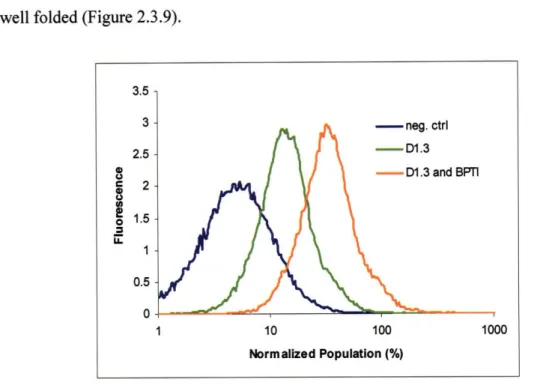
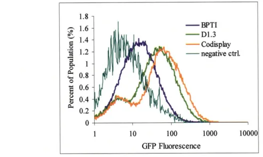
heterologous protein secretion. Although ultimately unsuitable as a screening proxy, the surface display experiments did show a novel method of improving protein secretion by co-expressing a more stably folded protein with the protein of interest. In these studies the secretion of an scFv-Aga2p fusion was stimulated 10-fold by the concomitant surface expression of BPTI. BPTI surface expression also stimulated the secretion of secreted
scFv three-fold suggesting a niche for protein coexpression as well as secretion by way of Aga2p fusions.
A new screening method was developed that involves the capture of secreted protein on the surface of the cell where it can be labeled and sorted by FACS. This new method was verified to achieve thirty-five fold enrichment per pass for a three-fold
enhanced protein secretor making it easily suitable for screening. The new screening methodology, the Cell Surface Secretion Assay (CeSSA), was also modeled and verified with time course data that enabled optimization of sort parameters and predicted sort
outcomes based on user-derived selection parameters. The CeSSA was used to screen a library of mutant yeast alpha mating factor leader sequences for improved secretion of the scFv 4m5.3. The improved leaders imparted up to a twenty-fold improvement in
scFv secretion per cell and up to thirty-fold improvement after expression tuning. These engineered leader sequences also conferred improved secretion on other scFv's and proteins including whole IgG. Moreover, the leader sequence mutants give indications of where the important residues in secretory leaders lie and the aberrations in protein traffic that result in reduced secretion.
Acknowledgements
I would like to first and foremost thank my advisor Dane Wittrup who, in addition
to being a brilliant and creative scientist and mentor, has shown me how to perform
research and interact with colleagues fairly, ethically, and with the right priorities. It's no
wonder that he has created such a terrific group of people at MIT. I would also like to
thank the many people I have worked with over the years. Specifically, I would like to
give special thanks to my undergraduate research assistants Charlyn Lu, Hana Oh, and
Angelin Baskaran who were a lot of fun to work with. I would also like to thank
Christilyn Graff whose debilitating calf injury provided with me with a personal guide to
some basic lab procedures, Katarina Midelfort and Jason Burbank who came with a lot of
Dane at Illinois stories and then Pete Heinzelman who provided me with plenty of MIT
stories of my own as well. Jeffrey Swers, and Andy Yeung allowed me to bug them with
questions and requests for reagents, and Bala Rao was quite inspirational with his
musings on "life and science." I would also like to thank my good friend Dave Colby
who helped me to see my work from a different point of view, Stefan Zajic who has been
a great officemate through the years, and Shanshan Howland, Greg Thurber, Steve
Sazinksy, and Ben Hackel for picking up the pieces behind me. I would especially like to
thank Annie Gai and Mike Schmidt for their help in the construction of some of the
prepro vectors, and most especially Andrea Piatesi for his help with the creation and
screening of the prepro library and also Margie Ackerman who taught me the true power
of a GROP. And of course most most especially I would like to thank Ginger Chao who
for three years has been a special form of personal support. Also, much thanks to the flow lab team of Mike "bald Mike" Doire, Mike "long-haired Mike" Jennings, Yong "my
Yong "my name was Mike before I got here" Xie, Michelle "my name's not Mike but it's
close" Perry, and Glenn Paradis who must know more than any other person about the
intricacies of modem flow cytometry (except for you Dane.) I'd like to say thank you to
my committee members Drs. Peter Sorger and Chris Kaiser in the Biology Department
for their valuable advice and for teaching me the right way intimidate a young graduate
student as well as Andrea Vala who was my go-to girl on when I had yeast questions. In
addition, I've had three great roommates for the past five years in Trent Yang, Will
Kuhlman, and John Fiorenza with whom I've swapped jaded stories of success and
failure.
I've had great sources of funding from the DuPont/MIT Alliance and the National
Institute of Health without which this project would never have gotten off the ground.
Lastly, I would like to thank those people from my family, to my teachers, to my friends
who have contributed to my academic well-being and my mental stability by always
For my family, Jim, Sandy, and Molly who have given me unconditional love and support through these often trying years, and for my grandfather Jack who has shown me
Table of Contents
Chapter 1: Introduction and Background 1
1.1 Recombinant Protein Production 2
1.2 The S. cerevisiae Secretory Pathway 4
1.2.1 Secretory Leader Sequence and ER Translocation 6
1.2.2 Protein Folding in the ER 11
1.2.3 The Late Secretory Pathway 12
1.3 Protein Trafficking and Secretory Bottlenecks 13
1.4 Gene Expression in S. cerevisiae 17
1.5 A Directed Evolution Approach 19
1.6 Flow Cytometry and FACS 21
1.7 Thesis Overview 23
1.8 Works Cited 25
Chapter 2: Yeast Surface Display and Simultaneous Expression 30
2.1 Introduction 30
2.2 Experimental Protocol 34
2.3 Results 40
2.3.1 Yeast Surface Display and Secretion 40 2.3.2 Co-expression ofD1.3 and BPTI 42 2.3.3 Co-expression and the Unfolded Protein Response 51
2.3.4 Secretory Co-expression 55
2.3.5 Surface Display and Mutagenic Libraries 58
2.4 Conclusions 61
2.5 Works Cited 66
Chapter 3: Cell Surface Secretion Assay 69
3.1 Introduction 69
3.2 Experimental Protocol 71
3.3 Results 76
3.3.1 CeSSA Development 76
3.3.2 CeSSA Generality 80
3.3.4 CeSSA Modeling 86
3.3.5 CeSSA Time Course 91
3.4 Conclusions 96
3.5 Works Cited 99
Chapter 4: Directed Evolution of Alpha Mating Factor for
Improved Secretion 100
4.1 Introduction 100
4.2 Experimental Protocol 104
4.3 Results 110
4.3.1 Alpha Factor Prepro Library Screening 110
4.3.2 Isolated Clones 112
4.3.3 Mutant Leaders and the
Secretion of Other (non-4m5.3) Proteins 127 4.3.4 Further Expression Improvement 129
4.4 Conclusions 134
4.5 Works Cited 141
Appendix 1: Protocols 143
Appendix 2: Plasmid Maps, Schematics, and PCR Primer Sets 149
Chapter 1: Introduction and Background
Recent advances in human genomics and proteomics have given science an
opportunity to better identify the causes and treatments for a vast array of diseases.
These advancements have been accompanied by an augmented ability to treat these
disorders using human proteins in place of traditional small molecules. Since the Elli
Lilly Corporation first made recombinant insulin in the early eighties, biotech companies
have staked their futures on their ability to solve ailments ranging from autoimmune
diseases, to metabolic disorders, to cancer using recombinant protein technology and the
industrial production of actual human proteins. Of course due to practical and ethical
considerations, it is impossible to make these proteins on any reasonable scale using
human hosts. Consequently, science and industry have turned to a variety of other
organisms to make protein for them. The work presented here utilizes the baker's yeast
Saccharomyces cerevisiae as an organism suitable for heterologous protein expression.
These studies include a novel method of improving the secretion of a single-chain
antibody using the co-expression of a more stable heterologous protein, the development
of a high-throughput assay capable of screening large libraries for mutations conferring
elevated levels of recombinant protein secretion, and finish with the development and
isolation of an improved protein leader sequence that, in combination with expression
tuning and additional host mutations, stimulates a thirty-fold increase in the production of
a single-chain antibody. The capability to produce more heterologous protein from an
expression host such as S. cerevisiae could help curb the current shortage in protein therapeutic supply.
1.1 Recombinant Protein Production
Technological advances in drug discovery as well as the sequencing of the human
genome have provided a significant boost in the need for the production of therapeutic
proteins. In fact, it has been estimated that the worldwide protein therapeutic market will
reach $32 billion by 2005 and $71 billion by 2008 [1, 2] with 140 FDA approved
biologics and another 500 in clinical trials as of 2004 [3]. Most therapeutic protein
production is centered on the development of recombinant proteins (non-human and
human protein altered for improved efficacy) and recombinant monoclonal antibodies for
the treatment of cancer, neurological disorders, AIDS, and heart disease [4]. These
treatments either replace native proteins that are dysfunctional, poorly expressed, or
completely missing in a patient or interfere with processes involved in the progression of
a disease.
Almost as varied as the types of heterologous proteins being manufactured are the
types of expression hosts available for their production. Expression hosts range from
simple, prokaryotic organisms such as E. coli to large multi-system mammals and plants.
Each system has its requisite list of advantages and drawbacks typically involving an
inverse relationship between ease of production and protein fidelity. Microbial systems
involving organisms such as coli and S. cerevisiae tend to be highly fermentable,
relatively inexpensive to use, and genetically pliable yet are unsuitable for the production
of large, complex, and glycosylated proteins. Mammalian systems on the other hand can
produce complex proteins yet are difficult to generate, relatively expensive to maintain, and create potential for toxic and infectious contaminants [5]. A summary of the benefits
and drawbacks of four groups of expression hosts is given in Table 1.1 (condensed from Kamath (2005).)
Table 1.I Aspects of Heterologous Protein Production Hosts.
Microbial Mammalian Plants Animals
Expression System Bacteria, Animal and Plant cells, Transgenics
Yeast Human Transgenics
Cell Lines
Protein Levels High Medium High High
Cycle Time Short Short Seasonal Long
Complexity No Sometimes Yes Yes
(glycosylation)
Example Proteins mAb, Glycoproteins, mAb, vaccines, IgG, blood
enzymes vaccines enzymes proteins
At the present time, however, the demand for protein therapeutics far outpaces the production capacity. Furthermore, issues with transgenic manufacturing cGMP, cell culture intellectual property, expanded pharmaceutical indications, and high dosage requirements threatens to worsen the problem [6]. Considering all of these potential
impediments to adequate market delivery, a more productive heterologous expression vehicle would be beneficial in helping protein supply keep pace with demand.
The yeast Saccharomyces cerevisiae is an organism well-suited for heterologous protein expression because of its eukaryotic secretory processing machinery, simple high-density growth characteristics, short generation times, and easily alterable genetics. However, although heterologous proteins secreted by S. cerevisiae are usually stably folded and secreted into the supernatant, proteins are typically expressed at lower levels relative to prokaryotic systems [7-11]. Improving the capacity of S. cerevisiae to secrete recombinant protein could be a promising way of meeting market demand.
1.2
The S. cerevisiae Secretory Pathway
Although there are a variety of organisms suitable for heterologous protein
production, the yeast S. cerevisiae is ideal for production of simple, non-glycosylated
proteins because, unlike prokaryotic systems, it is capable of folding multi-domain
proteins including those that require disulfide bonds and then secreting the product into
the supernatant where it can be readily collected and purified. However, like prokaryotic
systems, S. cerevisiae is a single-celled organism able to be grown in dense-culture
fermentations utilizing simple media. S. cerevisiae and its relative P. pastoris are
currently used in the industrial production of insulin, Hepatitis B Surface Antigen,
Granulocyte Colony Stimulating Factor, Platelet-Derived Growth Factor, glucagon,
angiostatin, endostatin, and human serum albumin demonstrating its suitability for the
production of many important therapeutic proteins.
Eukaryotic secretory pathways are largely conserved albeit in different
complexities across phyla and rely on hierarchical progression of secreted protein
through distinct organelle environments. This traversal relies on both specific as well as
quite general protein/protein and protein/carbohydrate interactions. After transcription of
the expressed gene in the nucleus, the mRNA is exported into the cytosol where it begins
to be translated into protein. Signal sequences on the N-terminus of the nascent
polypeptide either directs the ribosomal complex to continue translation in the cytosol or
to migrate to a ribosomal receptor on the endoplasmic reticulum membrane to continue
translation concomitant with protein extrusion into the ER lumen [12]. Most of the
folding is performed in the ER lumen where folding chaperones assist in translocation,
The ER also possesses a quality control apparatus that ensures that only well-folded
protein is exported into later stages of the pathway as misfolded proteins are returned to
the cytosol for proteosomal degradation in a process termed ER associated degradation
(ERAD) [20-23]. This quality checkpoint is a distinct advantage of eukaryotic
expression over bacterial-based systems. After the protein has reached a stable
conformation in the ER, it is exported to the Golgi apparatus where it may be modified
by carbohydrate alterations or proteolysis. From the Golgi, the protein can either be directed to the surface or to the vacuole, which serves as a degradation organelle among
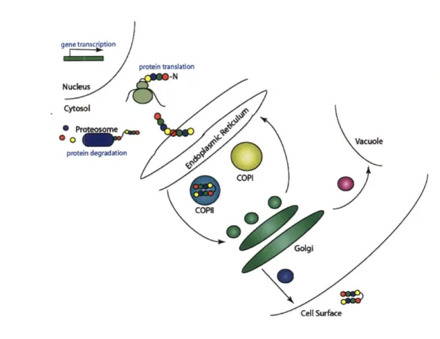
other things (secretory pathway summarized in Figure 1.2.1) [24-29]. As we shall see,
improving recombinant protein secretion often means exerting influence over protein fate
C
protein degradation
COPI
Cell Surface
Figure 1.2.1 The Yeast Secretory Pathway. mRNA transcripts are translated on ribosomes, and the nascent polypeptide is translocated into the endoplasmic reticulum. Proteins are either folded correctly or retrotranslocated back into the cytosol for degradation. Correctly folded proteins are trafficked to the Golgi via COPII vesicles while ER resident proteins and misfolded secretory proteins are shuttled back in COPI vesicles. From the late Golgi, protein can either be trafficked to the vacuole or the surface.
1.2.1 Secretory Leader Sequence and ER Translocation
All secretion-directed protein must contain an N-terminal sequence that "informs" the cell that the gene product is to be processed utilizing the secretory pathway. This sequence is called the secretory leader or the "pre" sequence. Pre sequences can vary widely in individual amino acid composition but do share some common themes among
eukaryotes. Typically, leader sequences contain a net positive charge between +1 and +3
toward their N-terminus. The charged sequence is then followed by a hydrophobic span,
and the leader ends with a more polar motif just before the non-polar signal-peptidase
(Sec 1 ip) cleavage site [12]. These motifs are required for proper processing and are,
therefore, sensitive to mutations affecting charge and hydrophobicity [30]. Although
translated protein must ultimately make its way to the ER, a couple of different pathways
for doing so are utilized in yeast, and the mechanism for ER entry is chiefly determined
by leader sequence [31]. In one pathway, proteins are completely translated before being
shuttled to the translocon complex on the ER membrane in a schema termed
posttranslational translocation. The yeast proteins alpha mating factor, the focus of
Chapter 4, and invertase are trafficked in this way [32, 33]. However, during cytosolic
translation nascent proteins exhibit exposed hydrophobic patches that are prone to
aggregation if not protected. The yeast protein Ssalp conceals hydrophobic motifs on the
polypeptide as well as chaperones the fully translated product to the translocon pore.
Chaperone activity requires ATP and is strongly enhanced by the presence of the ATPase
Ydjlp co-chaperone: a homolog to the DnaJ co-chaperones in coli [12, 34-37]. In fact it
is thought that Ydjlp activity is responsible for Ssalp dissociation that must occur before
translocation restarts [34, 38].
An alternative ER entry route involves simultaneous protein translocation and
translation in a process termed co-translational translocation. Although cerevisiae uses
both translocation pathways, mammalian cells use the co-translational translocation pathway exclusively with the exception of the synthesis of some small peptides.
direct the polypeptide/ribosome complex to a ribosome receptor (arguably p 180p, Sec6lp
itself, or a combination of both) near the translocon pore [12, 39, 40]. It is thought that
SRP binding stalls translation sufficiently to allow the complex to diffuse to the
Sec6lp/Sec63p complex where the SRP dissociates and translation continues [41].
Translational schemes are outlined in Figure 1.2.2.
ERklme
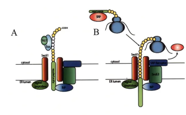
Figure 1.2.2 Translocation into the Endoplasmic Reticulum. Ssalp and the ATPase stimulating Ydj 1 bind hydrophobic patches on post-translationally translocated protein. The entire translation is fed through the Sec61p translocon pore through a process of diffusion and ATP-driven BiP binding. Signal peptidase cleaves off the signal peptide (A). In co-translational translocation, the signal recognition particle (SRP) binds the signal sequence and guides the ribosomal complex to the SRP receptor where SRP dissociates. Sec62 binds the signal sequence and guides it into the translocon pore (Sec62 action not shown) where it is extruded by the translational activity of the ribosome. BiP shields hydrophobic patches and signal peptidase cleaves the signal peptide.
c >)
The translocon pore exists as a complex of proteins that serves to engage the
polypeptide and then guide it into the ER lumen [42, 43]. Although the Sec61lp protein
has been shown to be the actual translocon pore [44, 45], Sec62p has also been shown to
be important as a potential signal sequence binder, and Sec63p may posses some
DnaJ-like co-chaperone activity in conjunction with the ER lumenal chaperone Kar2 [44, 46].
In post-translationally translocated proteins, the polypeptide is extruded by a "Brownian
ratchet" mechanism that relies on the random diffusion of the polypeptide into the lumen
where the binding of Kar2 inhibits retrograde motion [47]. This mechanism would
explain the importance of Kar2's association with the Sec63 subunit of the translocation
complex. In co-translational translocation, protein extrusion is powered by ribosomal
translation itself [48]. The signal peptide sequence is translocated as a hairpin loop and
then cleaved by signal peptidase (Sec 1lp), [12].
After the signal pre sequence is cleaved by signal peptidase, the remainder of the
leader, the "pro" region, helps direct the secretion of the protein through the rest of the
secretory pathway. Although a great many pro sequences exist in yeast, the most studied
by far is the pro region of the yeast alpha mating factor (MFalp). MFalp is a sixty-four
residue peptide that consists of three N-linked glycosylation sites provided by Asn-X-Thr
motifs. Although unglycosylated synthetic prepro leader sequences are able to direct the
secretion of heterologous protein, the glycosylation sites in MFa Ip are important to its
function as a secretory leader [49, 50]. Deletion of the glycosylation sites reduces
secretory competence and results in the intracellular accumulation of secretory protein
intermediate as unglycosylated alpha factor leader has been shown to be a substrate for
required before ER to Golgi transport in mating factor secretion [52]. Glycosylation sites
may influence protein folding by interacting with the calnexin/calreticulin glycoprotein
regulatory folding cycle [53]. Further studies with MFalp indicate that it is relatively
tolerant of in-frame amino acid insertions; however, in-frame deletions negatively impact
alpha factor secretion [52].
In addition to aspects of glycosylation, the pro region influences other stages of
secretory processing. ER translocation of insulin is dependent on the pro region even
with an intact pre sequence [54]. Furthermore, the pro sequence seems to be important
for the packaging of secretory protein into COPII vesicles for ER to Golgi transport.
Specifically, mutations in the 139, L42, and V52 residues significantly impact COPII
packaging in vitro and negatively impact MFalp processing in vivo [55]. Interestingly
synthetic prepro sequences have been designed that improve insulin precursor secretion
and are characterized by increased residence time in the ER suggesting that the pro
sequence may influence trafficking kinetics to effect a better folded protein [56]. Perhaps
one of the most important functions of the pro region is to stabilize the protein so that it is
not degraded. It has been shown that cleavage of the MFa p sequence leading the
secretion of an insulin-like protein influences whether the protein is directed to the
surface or to the vacuole. Thus, the presence of the pro region seems to have a stabilizing
effect on the protein allowing it to be directed to the surface. When the pro region is
cleaved, the protein is directed to the vacuole through one of many vacuolar sorting
1.2.2 Protein Folding in the ER
After translocation into the ER, the nascent protein interacts with a variety of
folding chaperones to ensure a well-folded final conformation. The most prominent
chaperone is the heat shock protein Kar2, the yeast homolog of mammalian
immunoglobulin binding protein (BiP). Kar2 contains a protein binding domain as well
as an ATPase domain. As the N-terminal ATP-binding domain cycles between ATP and
ADP, the substrate binding domain changes affinity for hydrophobic patches on unfolded
protein [13, 17, 18]. In this manner, the Kar2 protein can transiently interact with the
folding polypeptide keeping it from aggregating until the peptide is fully folded and the
Kar2 binding domains are hidden. In addition to its role in protein translocation and
folding, Kar2 is an intimate player in the unfolded protein response (UPR) that will be
discussed in detail in Chapter 2.
Another well-studied ER chaperone is the protein chiefly responsible for yeast's
ability to perform oxidative protein folding, protein disulfide isomerase (PDI). Like
Kar2, PDI is essential to yeast viability but also serves in promoting the formation and
isomerization of disulfide bonds [57]. PDI's oxidizing potential comes immediately from
the ER membrane protein Erolp but is thought to come ultimately from molecular
oxygen in a manner mediated by free flavin adenine dinucleotide (FAD) levels [14-16,
58]. As will be discussed later, overexpression of both Kar2 and PDI has been shown to
improve the secretion of recombinant protein [59-61].
Calnexin and calreticulin are two carbohydrate binding lectins that assist in
folding glycoproteins such as MFalp [62] as well as in protein degradation [51, 63]. Both chaperones bind to oligosaccharides containing terminal glucose residues on
N-linked carbohydrates which are transiently expressed during protein folding [64, 65].
This binding specificity allows them to work in concert with enzymes affecting
glycosylation to achieve a properly folded product. The enzyme mannosidase II cleaves
the terminal glucose on the carbohydrate causing calreticulin/calnexin to dissociate. This
release gives the protein a chance to fold properly. If the improper conformation is
reached, then glucosyltransferase, an enzyme with affinity for unfolded protein only,
reattaches the glucose subunit allowing the chaperone to bind it once again [19, 53]. In
this manner a nascent protein can make multiple attempts at correctly folding.
Carbohydrates on terminally misfolded proteins are cleaved in another place that causes
the protein to interact with the lectin ER-degradation enhancing mannosidase-like protein
(EDEM) that directs the protein to degradation in the proteosome [66, 67].
1.2.3 The Late Secretory Pathway
The two theories of protein exit from the ER involve the retention of misfolded or
resident ER protein in the face Golgi-directed bulk flow or the active sorting of proteins
into COPII vesicles destined for export. It seems that there are data to support both
theories [68] [55]. Protein can progress through the Golgi cisternae in both anterograde
and retrograde directions. ER resident protein that has escaped into the Golgi is retrieved
via its HDEL (KDEL in mammalian cells) C-terminal retrieval sequence and is packed
into COPI vesicles to be shuttled back to the ER. It has been shown that misfolded
proteins in the Golgi can be recycled back to the ER via BiP's retrieval sequence to be
refolded [69, 70]. This feature is important as the Golgi contains no folding chaperones
whether to send protein to the surface or to the vacuole for degradation. The sorting
receptors responsible for this decision are the family of vacuolar sorting proteins (VPS).
The tendency for a protein to be sorted to the vacuole by a VPS appears to be dependent
on protein stability as well as a positive sorting signal (particularly for hydrolases that are
normally vacuole resident proteins) [24]. In addition to surface directed traffic, proteins
can be sorted to the vacuole via clathrin-coated pits [25], through an endosomal
intermediate [26-28] or to a late endosome/multi-vesicular body [24]. It has been shown
that for some yeast proteins ubiquination is required for vacuole sorting by way of an
endosomal intermediate [71]. It has also been shown that insulin-like protein can be
sorted to the vacuole by way of the endosome through interaction with VPS receptors
[29].
1.3 Protein Trafficking and Secretory Bottlenecks
With a clear understanding of secretory pathway fundamentals and trafficking
steps, more information about secretory impediments and trafficking rates can be derived.
We can get a feeling of the size of the flux through the translational, secretory, and
degradation arms of the secretory pathway by looking at pulse-chase experiments. When
radioactive alpha-amylase is chased through the secretory pathway, one finds that only
two percent of the radioactive protein that had been synthesized during the pulse is
ultimately secreted. Meanwhile, the amount of intracellularly retained radioactive protein
gradually declines from 50% of the original retention at ten minutes into the chase down
to 23% thirty minutes into the chase during which time no increase in extracellular
occurring in the cell that are limiting the amount of protein able to reach the surface
(Figure 1.3.1A). This degradation could be occurring in the ER or vacuole and may be
due to protein mistrafficking or protein instability and misfolding.
Protein molecular weights as well as glycosylation patterns in cell lysate are
indicative of the progress the protein has made through the secretory pathway. For an
example where lysate is used to measure rates of protein maturation, one can look at
Heim's work examining the processing of radioactively labeled alpha mating factor
prepro led human insulin-like growth factor in a yeast strain deficient in vacuolar
proteases [73]. Thirty seconds after the radioactive pulse, high molecular weight species
of IGFI begin to appear in the intracellular lysate. These species are prepro IGFI
representing singly, doubly, and triply glycosylated forms proven by their sensitivity to
the deglycosylating enzyme EndoH. It is important to note that no unglycosylated
MFapp IGFI appears in the lysate at any time point during the chase suggesting that
translation, translocation, and glycosylation rates are too fast to allow significant
accumulation of MFapp IGFI in an unglycosylated form. At the two-minute time point,
significant amounts of lower molecular weight unglycosylated IGFI begin to accumulate
intracellularly. This form is Kex2 cleaved mature IGFI that has traversed at least as far
as the late Golgi. However, despite the early accumulation of mature protein, it is not
until thirty minutes after the pulse that protein begins to appear in the supernatant. From
these data it is apparent that MFapp IGFI is efficiently translocated, glycosylated and
folded. It is also clear that ER export and trafficking to the late Golgi is also relatively
rapid. However, once the protein has reached its mature form, a processing bottleneck keeps it inside the cell and out of the supernatant. From what is known about post-Golgi
secretory trafficking, one might hypothesize that mature IGFI is being routed to the
vacuole where a pep4 deletion in the host slows its degradation instead of the surface. A
schematic of IGFI trafficking fitting this hypothesis is given in Figure 1.3.1B. To
improve the secretion of IGFI in yeast cells, one would have to find a way to reroute
mature insulin to the surface. Indeed, Zhang and colleagues did exactly that when they
found that deletions in the late Golgi localized vacuolar sorting proteins VPS8, 35, 13, 4,
and 6 caused MFapp led IGFI precursor to be diverted to the surface instead of the
vacuole (2001). By removing a rate-limiting bottleneck (a counterproductive sorting
step), the rate of IGFI secretion was enhanced.
Other studies have found that vacuolar degradation is important in trafficking of
other proteins. The trafficking of destabilized mutants of BPTI are unaffected by
mutations in ERAD-associated proteins [74] but are instead trafficked to the vacuole
[75]. From these results it is apparent that some protein is able to escape the primary
quality control checkpoint in the ER only to be sorted from the Golgi for vacuolar
degradation. Proteins may be sorted due to instabilities caused by Golgi processing.
Movement through the Golgi body is concomitant with protein concentration, organelle
acidification, and extensive alteration in glycosylation patterns [76, 77]. Perhaps these
modifications destabilize the protein and cause it to fail quality control in the late Golgi.
If this scenario is accurate, then perhaps altering genes governing organelle acidification,
concentrative protein sorting, or glycosylation would be another method of improving
gene transc~ption
V
A
protein translation secret[0
ERADB
gene transcriptionI
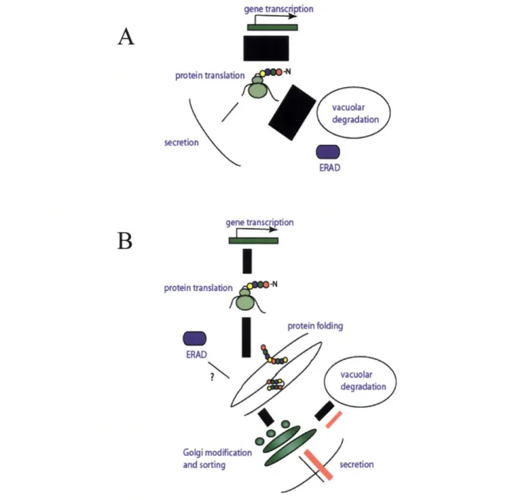
protein translatio n ~N ERAD Golgi modifi and sortingFigure 1.3.1 Secretory Bottlenecks. Pulse-chase experiments of a-amylase secretion indicate that only -2% of synthesized protein is secreted suggesting that the vast majority is degraded by ERAD or the vacuole (A). The bar width indicates the size of protein flux. A more detailed pulse-chase of MFapp IGFI indicates that vacuole directed protein trafficking causes a reduction in secretion of IGFI (black lines); furthermore, yeast mutations compromising the vacuolar sorting proteins responsible for this step as well as a deletion of Kex2 successfully reroute insulin to the surface (red lines, B).
It should be pointed out at this point that different proteins can have different
secretory bottlenecks, and there is sometimes more than one significant bottleneck in a
pathway for a single protein. For example, is seems scFv secretion rates are limited by
folding in the ER. Overexpression of proteins involved in ER folding, PDI and Kar2,
increase the amount of scFv secreted into the supernatant suggesting that formation of
disulfide bonds as well as aggregation or translocation issues may impose secretory
bottlenecks [61]. On the other hand, mRNA transcripts limit the secretory processing of
wild-type BPTI. Consequently, BPTI secretion is improved by increasing the copy
number of the gene [78]. It is these types of studies that highlight the nuances involved
in improving heterologous protein secretion. Furthermore, these studies also illustrate the
need for a comprehensive method of examining all of the potential bottlenecks for a
protein of interest in order to relieve the secretory bottleneck most pertinent to the
protein.
1.4 Gene Expression in S. cerevisiae
Genes to be expressed in S. cerevisiae are typically introduced into yeast on
low-copy (1 to 3 copies per cell) centromeric (CEN) plasmids, high low-copy 2pm plasmids, or
through integration into the chromosome via homologous recombination. The studies
outlined here mostly utilize CEN plasmids as well as a few examples of integration.
Heterologous genes are usually expressed from a strong promoter in order to maximize
the amount of mRNA that can be translated into protein. Promoters such as the
cytochrome c (CYC) or glycerol phosphate dehydrogenase (GPD) promoters can be constitutively activated, and other promoters can be specifically activated usually through
the introduction of a nutrient such as copper or galactose into the media. The promoter
used for these studies is the Gall-10 promoter normally used for galactose metabolism.
The Gall-10 promoter governs the divergent transcription of two genes (Gall and
Gall0). Transcription is stimulated by the binding of Gal4p to the upstream activating
sequence inside the Gal promoter. However, under conditions where galactose is not the
primary carbon source, Gal80p inhibits Gal4p activity. When galactose is added to the
culture, it activates the transcription of Gal3p that in turn binds and sequesters Gal80p
causing it to dissociate from Gal4p leaving Gal4p to activate the transcription of the Gall
and Gall0O gene products. Transcription of Gal3p is turned off in the presence of glucose.
This regulatory loop enables a gene inserted in place of the Gall gene to be strongly
transcribed when galactose is added to the media but be suppressed when grown in
another carbon source particularly glucose (Figure 1.4) [79]. In fact cell growth in
galactose stimulates the production of mRNA transcribed from the Gall-10 promoter
Glucose I
Gal UAS
Gal UAS
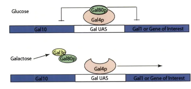
Figure 1.4 The Gall-10 Regulatory Operon. Gal80p causes a repression in gene expression by binding the transcription activating protein Gal4p. Galactose relieves this repression by stimulating the production of Gal3p, which binds Gal80p leaving Gal4p free to stimulate transcription of a downstream gene.
1.5 A Directed Evolution Approach
Many approaches to improving recombinant protein secretion in yeast have been
attempted with some degree of success. Most of these successes rely on the perturbation
of a secretory pathway participant through overexpression [59-61, 72] or deletion [29, 81,
82]. However, it is clear from previous work that large libraries are needed in order to
robustly isolate higher secreting clones. Moreover, different types of secreted proteins
require the modification of different secretory pathway participants. Proteins in the ER,
Golgi, transport vesicles, and nucleus have all been found to improve protein secretion
when modified (Table 1.II). A screening philosophy that embraces and even integrates the large diversity of potential solutions would be ideal for improving heterologous
protein secretion. With these themes in mind, a model approach to secretory maturation
i
Galactose "C
is directed evolution. Directed evolution, frequently utilized in protein engineering, relies
on the generation of diversity from a previously homologous population followed by
iterative rounds of selection in order to isolate clones that are best fit for the selection
criteria. As applied to secretory improvement, directed evolution would mean the
generation of a library of host or protein mutants and then the selection of mutants that
exhibit elevated levels of secretion. This process could be performed over and over even
employing different types of libraries until the best, or at least a sufficient, solution to the
secretory problem is found (Figure 1.5).
0o
Original Strain°°
0
I
Introduction of Diversity SelectionFigure 1.5 Directed Evolution of a Diversified Library. A homologous population of cells undergoes diversification through the introduction of host or protein mutations. In this case, the higher secretors are selected and the process is repeated until the library has enriched sufficiently. This method may incorporate many rounds of selection utilizing different types of libraries.
olated
1.6 Flow Cytometry and FACS
One of the problems associated with isolating mutations for secretion is the rather
unwieldiness of the screening process. In order to screen large libraries, time consuming
and inefficient assays such as Western blots and halo assays have been employed. These
assays frequently require the screens to be performed on plates, which is an environment
far removed from the fermentation conditions experienced in industrial production.
Furthermore, the library size becomes limited to the amount of clones one is able to put
into the assay. Table 1.II summarizes a few previous strategies for improving
heterologous protein secretion in yeast and outlines the methods used as well as the
results.
Table 1.II Selected Studies on Improving Heterologous Protein Secretion in Yeast
4x to 10x 4x 5x to 50x Redirected from total degradation 3x in K. lactis Overexpression of PDI, BiP (ER) Overexpression of Sso 1 (transport vesicles) Deletion of PMR1 (Golgi) Deletion of VPS genes (Golgi) Deletion of SELl
(unknown)
Rational Suppression Screen Suppression Screen Western blot Plate screeningAs can be inferred from Table 1.II, the ability to screen large numbers of clones in
a high throughput manner would be a real advantage to the field. A solution to that
problem is to utilize fluorescence activated cell sorting (FACS) as a selection tool. Flow cytometry relies on the differentiation of phenotype through the association of fluorescent labels and cells. Cells are labeled with a fluorescent dye and immersed in droplets of
scFv a-amylase Various Proinsulin Human Growth Hormone [61] [72]
[81]
[29] [82]saline solution that have been charged by a piezoelectric tip on a vibrating nozzle. A laser
excites the cell-associated fluorophores, and the resulting photons are absorbed by a
series of detectors each calibrated for a specific wavelength of light. The cell's
fluorescent properties are detected and analyzed, and cells meeting the fluorescence
criteria are kept by way of electromagnetic deflection into a sample collection tube. Cells
that do not meet the criteria pass into a waste stream (Figure 1.6). This method is very
efficient relative to traditional screening methods in that it can analyze and sort up to one
hundred million cells an hour. Consequently, a couple of seconds of flow time can
reproduce a screen that might take months using other techniques. Furthermore, cell
fluorescence is quantifiable thus presenting a numerical relationship between clone and
Detector 1
Droplet/Cell I
Laser
0
Waste
Figure 1.6 Fluorescence Activated Cell Sorting. Charged cells are dispersed in droplets then travel through a laser that excites cell-associated fluorophores. The resulting photons are detected and analyzed such that a comparison between the cellular properties and the sort criteria can be made. Cells meeting the criteria are sorted by a deflection plate into a sample collection tube. Other cells are discarded.
1.7 Thesis Overview
Having established the need for a high-throughput method to select S. cerevisiae
hosts with enhanced recombinant protein secretion capability, it is the goal of this thesis to devise and validate a flow cytometric method for selection and to successfully
demonstrate a directed evolutionary approach resulting in the generation of a hyper-Selected
secreting clone or construct. In the second chapter, yeast surface display is explored as a
potential screening proxy. Furthermore, experiments will show that co-expressing two
heterologous proteins simultaneously improves the expression of both and results in the
dramatic secretory improvement of a single-chain antibody. Chapter 3 describes a yeast
screening assay centered on using actual secreted protein rather than protein fusions to
generate a link between secretory phenotype and host genotype. This method will be
validated and modeled so that it may be efficiently used for the selection of mutagenic
libraries. Chapter 4 will apply this screening system to the directed evolution of a
secretory leader peptide and demonstrate that this approach can be a successful way to
generate hyper-secreting constructs from a large library. The isolated leader sequences
will be used alone and in combination with other strategies to enhance the secretion of a
1.8 Works Cited
1. Roner, L., Proteins in Peril? 2002, Eye for Pharma.
2. Rajan, M., Worldwide Production for Protein Drugs to Reach Nearly $71 Billion by 2008. 2003, Business Communications Company, Inc.
3. Gerngross, T.U., Advances in the production of human therapeutic proteins in
yeasts andfilamentous fungi. Nat Biotechnol, 2004. 22(11): p. 1409-14.
4. Das, R., Proteins and Antibodies make Advances as Therapeutic Products. American Biotechnology Laboratory, 2000(February 2000).
5. Kamath, L., Keeping up with Protein Demand, in Drug Discovery and
Development. 2006, Reed Business Information.
6. AviGenics, I., Production Capacity Needs. 2003, AviGenics, Inc.
7. Harrison, J.S. and E. Keshavarz-Moore, Production of antibody fragments in
Escherichia coli. Ann N Y Acad Sci, 1996. 782: p. 143-58.
8. Olmos-Soto, J. and R. Contreras-Flores, Genetic system constructed to
overproduce and secrete proinsulin in Bacillus subtilis. Appl Microbiol
Biotechnol, 2003. 62(4): p. 369-73.
9. Palva, I., Molecular cloning of alpha-amylase gene from Bacillus
amyloliquefaciens and its expression in B. subtilis. Gene, 1982. 19(1): p. 81-7.
10. Pluckthun, A., Escherichia coli producing recombinant antibodies. Bioprocess Technol, 1994. 19: p. 233-52.
11. Wu, S.C., et al., Functional production and characterization ofafibrin-specific single-chain antibody fragment from Bacillus subtilis: effects of molecular chaperones and a wall-bound protease on antibody fragment production. Appl
Environ Microbiol, 2002. 68(7): p. 3261-9.
12. Brodsky, J.L., Targeting to and Translocation across the Endoplasmic Reticulum
Membrane, in Protein Targeting and Translocation, D.A. Phoenix, Editor. 1998,
Princeton University Press: Princeton, NJ. p. 169-191.
13. McKay, D.B., Structure and mechanism of 70-kDa heat-shock-related proteins.
Adv Protein Chem, 1993. 44: p. 67-98.
14. Frand, A.R., J.W. Cuozzo, and C.A. Kaiser, Pathways for protein disulphide bond
formation. Trends Cell Biol, 2000. 10(5): p. 203-10.
15. Frand, A.R. and C.A. Kaiser, The ERO1 gene ofyeast is requiredfor oxidation of
protein dithiols in the endoplasmic reticulum. Mol Cell, 1998. 1(2): p. 161-70.
16. Frand, A.R. and C.A. Kaiser, Erolp oxidizes protein disulfide isomerase in a
pathway for disulfide bond formation in the endoplasmic reticulum. Mol Cell,
1999. 4(4): p. 469-77.
17. Flynn, G.C., T.G. Chappell, and J.E. Rothman, Peptide binding and release by
proteins implicated as catalysts ofprotein assembly. Science, 1989. 245(4916): p.
385-90.
18. Blond-Elguindi, S., et al., Peptide-dependent stimulation of the ATPase activity of
the molecular chaperone BiP is the result of conversion of oligomers to active monomers. J Biol Chem, 1993. 268(17): p. 12730-5.
19. Parodi, A.J., Protein glucosylation and its role in protein folding. Annu Rev Biochem, 2000. 69: p. 69-93.
20. Cox, J.S. and P. Walter, A novel mechanism for regulating activity of a
transcription factor that controls the unfolded protein response. Cell, 1996. 87(3):
p. 391-404.
21. Ng, D.T., E.D. Spear, and P. Walter, The unfolded protein response regulates
multiple aspects of secretory and membrane protein biogenesis and endoplasmic reticulum quality control. J Cell Biol, 2000. 150(1): p. 77-88.
22. Valkonen, M., M. Penttila, and M. Saloheimo, Effects of inactivation and
constitutive expression of the unfolded- protein response pathway on protein production in the yeast Saccharomyces cerevisiae. Appl Environ Microbiol, 2003.
69(4): p. 2065-72.
23. Werner, E.D., J.L. Brodsky, and A.A. McCracken, Proteasome-dependent
endoplasmic reticulum-associated protein degradation: an unconventional route to afamiliar fate. Proc Natl Acad Sci U S A, 1996. 93(24): p. 13797-801.
24. Bowers, K. and T.H. Stevens, Protein transport from the late Golgi to the vacuole in the yeast Saccharomyces cerevisiae. Biochim Biophys Acta, 2005. 1744(3): p.
438-54.
25. Cowles, C.R., et al., Novel Golgi to vacuole delivery pathway in yeast:
identification of a sorting determinant and required transport component. Embo
J, 1997. 16(10): p. 2769-82.
26. Bryant, N.J. and T.H. Stevens, Vacuole biogenesis in Saccharomyces cerevisiae:
protein transportpathways to the yeast vacuole. Microbiol Mol Biol Rev, 1998.
62(1): p. 230-47.
27. Harsay, E. and A. Bretscher, Parallel secretory pathways to the cell surface in
yeast. J Cell Biol, 1995. 131(2): p. 297-310.
28. Harsay, E. and R. Schekman, A subset ofyeast vacuolar protein sorting mutants
is blocked in one branch of the exocytic pathway. J Cell Biol, 2002. 156(2): p.
271-85.
29. Zhang, B., et al., Intracellular retention of newly synthesized insulin in yeast is
caused by endoproteolytic processing in the Golgi complex. J Cell Biol, 2001.
153(6): p. 1187-98.
30. Rapoport, T.A., B. Jungnickel, and U. Kutay, Protein transport across the
eukaryotic endoplasmic reticulum and bacterial inner membranes. Annu Rev
Biochem, 1996. 65: p. 271-303.
31. Ng, D.T., J.D. Brown, and P. Walter, Signal sequences specify the targeting route
to the endoplasmic reticulum membrane. J Cell Biol, 1996. 134(2): p. 269-78.
32. Lodish, H., Berk, A., Zipursky, S.L., Matsudaira, P., Baltimore, D., Darnell,
Molecular Cell Biology, ed. S. Tenney. 2000, New York, NY: W.H. Freeman and
Co.
33. Arnold, C.E., et al., Leader peptide efficiency correlates with signal recognition
particle dependence in Saccharomyces cerevisiae. Biotechnol Bioeng, 1998.
59(3): p. 286-93.
34. Cyr, D.M., X. Lu, and M.G. Douglas, Regulation ofHsp70 function by a
eukaryotic DnaJ homolog. J Biol Chem, 1992. 267(29): p. 20927-31.
35. Caplan, A.J., D.M. Cyr, and M.G. Douglas, YDJlpfacilitates polypeptide
translocation across different intracellular membranes by a conserved mechanism. Cell, 1992. 71(7): p. 1143-55.
36. Cheetham, M.E. and A.J. Caplan, Structure, function and evolution of DnaJ:
conservation and adaptation of chaperone function. Cell Stress Chaperones,
1998. 3(1): p. 28-36.
37. Szyperski, T., et al., NMR structure determination of the Escherichia coli DnaJ
molecular chaperone: secondary structure and backbone fold of the N-terminal region (residues 2-108) containing the highly conserved J domain. Proc Natl
Acad Sci U S A, 1994. 91(24): p. 11343-7.
38. Chirico, W.J., Dissociation of complexes between 70 kDa stress proteins and
presecretory proteins is facilitated by a cytosolic factor. Biochem Biophys Res
Commun, 1992. 189(2): p. 1150-6.
39. Collins, P.G. and R. Gilmore, Ribosome binding to the endoplasmic reticulum: a
180-kD protein identified by crosslinking to membrane-bound ribosomes is not required for ribosome binding activity. J Cell Biol, 1991. 114(4): p. 639-49.
40. Prinz, A., E. Hartmann, and K.U. Kalies, Sec61lp is the main ribosome receptor in
the endoplasmic reticulum ofSaccharomyces cerevisiae. Biol Chem, 2000.
381(9-10): p. 1025-9.
41. Walter, P. and A.E. Johnson, Signal sequence recognition and protein targeting to
the endoplasmic reticulum membrane. Annu Rev Cell Biol, 1994. 10: p. 87-119.
42. Panzner, S., et al., Posttranslational protein transport in yeast reconstituted with a purified complex of Sec proteins and Kar2p. Cell, 1995. 81(4): p. 561-70.
43. Deshaies, R.J., et al., Assembly ofyeast Sec proteins involved in translocation into
the endoplasmic reticulum into a membrane-bound multisubunit complex. Nature,
1991. 349(6312): p. 806-8.
44. Musch, A., M. Wiedmann, and T.A. Rapoport, Yeast Sec proteins interact with
polypeptides traversing the endoplasmic reticulum membrane. Cell, 1992. 69(2):
p. 343-52.
45. Sanders, S.L., et al., Sec61p and BiP directly facilitate polypeptide translocation
into the ER. Cell, 1992. 69(2): p. 353-65.
46. Brodsky, J.L. and R. Schekman, A Sec63p-BiP complex from yeast is requiredfor
protein translocation in a reconstituted proteoliposome. J Cell Biol, 1993. 123(6
Pt 1): p. 1355-63.
47. Matlack, K.E., et al., BiP acts as a molecular ratchet during posttranslational
transport ofprepro-alpha factor across the ER membrane. Cell, 1999. 97(5): p.
553-64.
48. Beckmann, R., et al., Alignment of conduits for the nascent polypeptide chain in
the ribosome-Sec61l complex. Science, 1997. 278(5346): p. 2123-6.
49. Kjeldsen, T., et al., Prepro-leaders lacking N-linked glycosylation for secretory
expression in the yeast Saccharomyces cerevisiae. Protein Expr Purif, 1998.
14(3): p. 309-16.
50. Kjeldsen, T., et al., alpha-Factor pro-peptide N-linked oligosaccharidesfacilitate secretion of the insulin precursor in Saccharomyces cerevisiae. Biotechnol Appl
Biochem, 1998. 27 ( Pt 2): p. 109-15.
51. McCracken, A.A. and J.L. Brodsky, Assembly ofER-associatedprotein
degradation in vitro: dependence on cytosol, calnexin, and ATP. J Cell Biol,
52. Caplan, S., et al., Glycosylation and structure of the yeast MF alpha 1
alpha-factor precursor is important for efficient transport through the secretory pathway. J Bacteriol, 1991. 173(2): p. 627-35.
53. Ellgaard, L. and A. Helenius, Quality control in the endoplasmic reticulum. Nat Rev Mol Cell Biol, 2003. 4(3): p. 181-91.
54. Chaudhuri, B., K. Steube, and C. Stephan, The pro-region of the yeast
prepro-alpha-factor is essential for membrane translocation of human insulin-like growth factor 1 in vivo. Eur J Biochem, 1992. 206(3): p. 793-800.
55. Otte, S. and C. Barlowe, Sorting signals can direct receptor-mediated export of
soluble proteins into COPIIvesicles. Nat Cell Biol, 2004. 6(12): p. 1189-94.
56. Kjeldsen, T., et al., Synthetic leaders with potential BiP binding mediate
high-yield secretion of correctly folded insulin precursors from Saccharomyces cerevisiae. Protein Expr Purif, 1997. 9(3): p. 331-6.
57. LaMantia, M.L. and W.J. Lennarz, The essential function ofyeast protein
disulfide isomerase does not reside in its isomerase activity. Cell, 1993. 74(5): p.
899-908.
58. Tu, B.P. and J.S. Weissman, The FAD- and 0(2)-dependent reaction cycle of
Erol-mediated oxidative protein folding in the endoplasmic reticulum. Mol Cell,
2002. 10(5): p. 983-94.
59. Hayano, T., M. Hirose, and M. Kikuchi, Protein disulfide isomerase mutant
lacking its isomerase activity accelerates protein folding in the cell. FEBS Lett,
1995. 377(3): p. 505-11.
60. Robinson, A.S., V. Hines, and K.D. Wittrup, Protein disulfide isomerase
overexpression increases secretion offoreign proteins in Saccharomyces cerevisiae. Biotechnology (N Y), 1994. 12(4): p. 381-4.
61. Shusta, E.V., et al., Increasing the secretory capacity of Saccharomyces
cerevisiae for production of single-chain antibody fragments. Nat Biotechnol,
1998. 16(8): p. 773-7.
62. Ou, W.J., et al., Association offolding intermediates of glycoproteins with
calnexin during protein maturation. Nature, 1993. 364(6440): p. 771-6.
63. Liu, Y., et al., Oligosaccharide modification in the early secretory pathway
directs the selection of a misfolded glycoprotein for degradation by the proteasome. J Biol Chem, 1999. 274(9): p. 5861-7.
64. Ware, F.E., et al., The molecular chaperone calnexin binds GlclMan9GlcNAc2
oligosaccharide as an initial step in recognizing unfolded glycoproteins. J Biol
Chem, 1995. 270(9): p. 4697-704.
65. Hebert, D.N., B. Foellmer, and A. Helenius, Glucose trimming and
reglucosylation determine glycoprotein association with calnexin in the endoplasmic reticulum. Cell, 1995. 81(3): p. 425-33.
66. Jakob, C.A., et al., Htmlp, a mannosidase-like protein, is involved in glycoprotein
degradation in yeast. EMBO Rep, 2001. 2(5): p. 423-30.
67. Hosokawa, N., et al., A novel ER alpha-mannosidase-like protein accelerates ER-associated degradation. EMBO Rep, 2001. 2(5): p. 415-22.
68. Pfeffer, S.R. and J.E. Rothman, Biosynthetic protein transport and sorting by the
69. Yamamoto, K., et al., The KDEL receptor mediates a retrieval mechanism that
contributes to quality control at the endoplasmic reticulum. Embo J, 2001.
20(12): p. 3082-91.
70. Hammond, C. and A. Helenius, Quality control in the secretory pathway. Curr Opin Cell Biol, 1995. 7(4): p. 523-9.
71. Rubio-Texeira, M. and C.A. Kaiser, Amino Acids Regulate Retrieval of the Yeast
General Amino Acid Permease from the Vacuolar Targeting Pathway. Mol Biol
Cell, 2006.
72. Ruohonen, L., et al., Enhancement ofprotein secretion in Saccharomyces
cerevisiae by overproduction of Sso protein, a late-acting component of the secretory machinery. Yeast, 1997. 13(4): p. 337-51.
73. Steube, K., et al., Alpha-factor-leader-directed secretion of recombinant human-insulin-like growth factor Ifrom Saccharomyces cerevisiae. Precursor formation and processing in the yeast secretory pathway. Eur J Biochem, 1991. 198(3): p.
651-7.
74. Kowalski, J.M., et al., Protein folding stability can determine the efficiency of
escape from endoplasmic reticulum quality control. J Biol Chem, 1998. 273(31):
p. 19453-8.
75. Coughlan, C.M., et al., Degradation of mutated bovine pancreatic trypsin
inhibitor in the yeast vacuole suggests post-endoplasmic reticulum protein quality control. J Biol Chem, 2004. 279(15): p. 15289-97.
76. Helenius, A., T. Marquardt, and I. Braakman, The endoplasmic reticulum as a
protein-folding compartment. Trends Cell Biol, 1992. 2(8): p. 227-31.
77. Trombetta, E.S. and A.J. Parodi, Quality control and protein folding in the
secretory pathway. Annu Rev Cell Dev Biol, 2003. 19: p. 649-76.
78. Parekh, R.N. and K.D. Wittrup, Expression level tuning for optimal heterologous
protein secretion in Saccharomyces cerevisiae. Biotechnol Prog, 1997. 13(2): p.
117-22.
79. Hawkins, K.M. and C.D. Smolke, The regulatory roles of the galactose permease
and kinase in the induction response of the GAL network in Saccharomyces cerevisiae. J Biol Chem, 2006.
80. Ideker, T., et al., Integrated genomic andproteomic analyses of a systematically
perturbed metabolic network. Science, 2001. 292(5518): p. 929-34.
81. Rudolph, H.K., et al., The yeast secretory pathway is perturbed by mutations in
PMR1, a member of a Ca2+ ATPase family. Cell, 1989. 58(1): p. 133-45.
82. Bartkeviciute, D. and K. Sasnauskas, Studies ofyeast Kluyveromyces lactis
mutations conferring super-secretion of recombinant proteins. Yeast, 2003. 20(1):
Chapter 2: Yeast Surface Display and Simultaneous
Expression
2.1 Introduction
A screening proxy for protein secretion selections should take full advantage of the yeast secretory pathway just as a true secreted protein would. In yeast,
secretion-directed proteins are translocated into the ER through the Sec61/Sec63 complex.
Translocation can be performed either post-translationally or co-translationally although
the latter relies on the interaction with the signal recognition particle (SRP) for shuttling
to the ER membrane and is used exclusively in mammalian cells [1-3]. After
translocation into the ER lumen, the nascent proteins interact with a variety of folding
chaperones that assist the protein in reaching a state suitable for export from the ER. The
most prominent of the folding chaperones is immunoglobulin binding protein (BiP, Kar2
in yeast) that not only inhibits protein aggregation but is also thought to assist in protein
translocation as well as the unfolded protein response (UPR). In addition chaperones
such as protein disulfide isomerase (PDI) participate in the formation of disulfide bonds
between cystein residues, and carbohydrate-binding lectins such as calnexin and
calreticulin are also used to ensure proper folding and glycosylation [4]. Historically,
overexpression of these proteins has been a productive method for improving the
secretion of some heterologous proteins such as single-chain antibodies (scFv) and
human lysozyme [5-7]. One of the attractive features of expression in yeast is that they
have their own internalized protein quality control. Protein that does not fold properly is
degraded by a 26S proteosome. This collective process is termed ER associated
degradation (ERAD) and is intricately related to the secretory health of the cell [8-10].
Secretory stress is often concomitant with high-level expression of recombinant
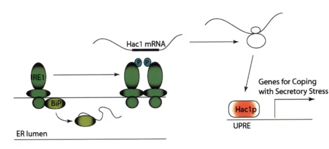
protein and triggers a mechanism known as the unfolded protein response (UPR) [11,
12]. This response is mediated through the ER membrane localized kinase inositol
requiring protein (IRE1). Although normally associated with BiP, IRE1 homodimerizes
and transphosphorylates when BiP is recruited away by high levels of unfolded protein.
IRE1 phosphorylation activates the endonucleolytic activity of the dimer causing it to
splice an intron out of the mRNA for the transcription factor Hac 1. Spliced Hac 1 is then
translated and goes on to stimulate the transcription of numerous genes involved in
helping the cell cope with secretory stress (Figure 2.1.1) [13]. These upregulated genes
code for proteins involved in membrane proliferation, protein folding, and protein
degradation [10, 14, 15]. UPR manipulation has even been used to increase the secretion
Hacl mRNA
Genes for Coping
with Secretory Stress
(
UPRE ER lumen
Figure 2.1.1-The Yeast Unfolded Protein Response. The UPR is instigated by the endonuclease activity of the ER membrane protein IRE 1. IRE1 is normally associated with the soluble ER protein BiP until BiP is recruited away to help stabilize high levels of unfolded protein. This migration allows IRE to homodimerize and transphosphorylate stimulating its endonucleolytic activity. The activated IRE1 dimer splices an intron out of Hac 1 mRNA allowing active Hac Ip transcription factor to be translated. Hac lp then goes on to stimulate the expression of a variety of genes involved in helping the cell cope with ER stress.
Proteins that have passed ER quality control are packaged into COPII vesicles and
shuttled to the Golgi. The Golgi serves as the final protein quality checkpoint as well as a
location for further protein modifications that typically consist of glycosylation and
peptidase activity. It has also been shown that improperly folded proteins can be
retrieved from the Golgi and transported back to the ER. This late quality control may be mediated by BiP acting as an adapter complexing with both the unfolded protein and the
KDEL ER-retention receptor [17-19]. Vacuole targeted and misfolded proteins are sorted to the vacuole by a family of vacuolar sorting proteins (VPS) [20]. Proteins can be sorted directly to the vacuole via clathrin-coated vesicles in a pathway termed the alkaline
1
1
I
phosphatase pathway (ALP) [21], through an endosomal intermediate [22-24], or directly
to a multi-vesicular body (MVB) in the CPY pathway [25]. In addition proteins can be
sorted directly from the trans-Golgi to the surface. The factors that govern this sorting
decision are still being worked out, but it appears that a positive sorting signal on the
secreted proteins as well as varying levels of protein ubiquitination may play a role [25,
26]. Some heterologous proteins such as insulin are sorted through the endosomal
pathway, and deletions of various VPS's involved in endosomal sorting have increased
surface directed traffic of this protein [27]. In addition, it has been shown that protein
stability may contribute to the propensity of a protein to be sorted to the vacuole.
It is clear that cells process different heterologous proteins in different ways. In
the following experiments, we dissect the trafficking of two different, simultaneously
expressed heterologous proteins: bovine pancreatic trypsin inhibitor, which can be
secreted at levels up to 180 mg/L [28] and D1.3, which is a member of a class of proteins
termed single-chain antibodies (scFv) and are relatively poorly secreted at 10-20 mg/L
[7]. These studies show that co-expression has strong effects on protein trafficking
particularly in the late secretory pathway. It appears that BPTI and D 1.3 compete for the
same retentive sorting protein resulting in a significant improvement in the secretion of
the less stable D1.3 and a more subtle improvement in the surface expression of BPTI.
These results demonstrate a novel method for improving the secretion of a heterologous
protein with a potential improvement anywhere from three to ten-fold depending on the