HAL Id: inserm-02877776
https://www.hal.inserm.fr/inserm-02877776
Submitted on 16 Nov 2020
HAL is a multi-disciplinary open access
archive for the deposit and dissemination of
sci-entific research documents, whether they are
pub-lished or not. The documents may come from
teaching and research institutions in France or
abroad, or from public or private research centers.
L’archive ouverte pluridisciplinaire HAL, est
destinée au dépôt et à la diffusion de documents
scientifiques de niveau recherche, publiés ou non,
émanant des établissements d’enseignement et de
recherche français ou étrangers, des laboratoires
publics ou privés.
Tyrosine conjugation methods for protein labelling
Dimitri Alvarez Dorta, David Deniaud, Mathieu Mével, Sébastien Gouin
To cite this version:
Dimitri Alvarez Dorta, David Deniaud, Mathieu Mével, Sébastien Gouin. Tyrosine conjugation
meth-ods for protein labelling. Chemistry - A European Journal, Wiley-VCH Verlag, 2020, Online ahead
of print. �10.1002/chem.202001992�. �inserm-02877776�
Tyrosine-click chemistry for
protein labelling
Dimitri Alvarez Dorta,
[a]David Deniaud,
[a]Mathieu
Mével,
[b]Sébastien G. Gouin*
[a]Dedicated to the memory of Carlos F. Barbas III
[a] Title(s), Initial(s), Surname(s) of Author(s) including Corresponding Author(s)
Department Institution Address 1 E-mail:
[b] Title(s), Initial(s), Surname(s) of Author(s) Department
Institution Address 2
Supporting information for this article is given via a link at the end of the document.((Please delete this text if not appropriate))
Abstract: Over the last two decades, the development of chemical
biology and the need for more defined protein conjugates have fostered active research on new bioconjugation techniques. In particular, a wide range of biorthogonal click strategies have been reported to functionalize the phenol side chain of tyrosines (Y). Y occur at medium frequency and are partially buried at the protein surface, offering interesting opportunities for site-selective labelling of the most reactive residues. Y-targeting has proved effective for designing a wide range of important biomolecules including antibody-drug conjugates, fluorescent or radioactive protein probes, glycovaccines, protein aggregates and PEG-conjugates. Innovative methods have also been reported for site-specific labelling with ligand-directed anchors and for specific affinity capture of proteins. This review will present and discuss these promising alternatives to the conventional labelling of the nucleophilic lysine and cysteine residues.
1. Introduction
Site-specific chemical modification of proteins is becoming increasingly important in research and industry for monitoring cellular events or designing protein therapeutics. An extreme level of specificity can be achieved by genetic code expansion techniques where the translational machinery is reprogrammed with unnatural amino acids bearing a biorthogonal handle.1–4
Such genetic engineering must, however, be carried out in a specialized laboratory and the chemical post-translational modification of native protein is still a highly complementary approach, although its effectiveness is continually improving. Historically, the chemical modifications of proteins nearly exclusively focused on the abundant lysine (Lys) and relatively rare cysteine5 (Cys) residues, while other amino acids (AAs)
where much less exploited.6
Classical and recently improved bioconjugation methods include reductive aminations, and amide, urea and thiourea formation with solvent-exposed Lys residues, while Cys anchors are generally functionalized by alkylations,7,8 metal assisted
arylation,9,10 disulfide exchange, or addition to a maleimide
Michael acceptor. These different methods generally provide a high level of chemoselectivity for one type of AA. For therapeutic applications, a higher level of control may be required, as single-site protein modification is preferable to avoid a heterogeneous mixture of protein conjugates that would potentially show different pharmacodynamic profiles and therapeutic indices. The goal is more easily reached when the highly nucleophilic Cys, with low abundance, is targeted, but promising protocols have also been recently developed for the kinetically-controlled labelling of the
most reactive Lys residues.11–14 Indeed, Lys reactivity is strongly
affected by the neighbouring AAs with regard to their accessibility and pKa value.15
The growing interest in chemical biology and high demand for new bioconjugates has driven the development of new conjugation methods for selective anchoring on other AAs. Promising methods have been actively developed for the selective bioconjugation of less nucleophilic AAs such as arginine,16
tryptophan,17 and methionine,18,19 with a specific focus on tyrosine
(Y). Y is a versatile AA due to the presence of the phenolic moiety. The aromatic side chain is hydrophobic and mainly involved in π-stacking interactions or cation-π interactions. However, due to the presence of the hydroxyl group, it can also be involved in hydrogen bonding, and due to its redox potential, can form tyrosyl radicals enabling electron transfers.20,21 Y occurs at medium
frequency on the protein surface and the amphiphilic phenol ring is generally partially or fully buried in the protein surface. Y on proteins therefore have varying levels of accessibility and reactivity, which could be exploited for site-selective modification. Furthermore, Y is neutral over a wide range of pH and its modification does not alter the overall charge of the protein. Y residues experience highly diverse biological modifications such as nitration, oxidation, cross-linking, AMPylation, halogenation or glycosylation, which are involved in important physiological processes or triggered by certain inflammatory or neurodegenerative diseases.22 Thus, the specific chemical
labelling of Y may also offer a unique opportunity to deepen our understanding of the relevance of these post-translational modifications. The aim of this article is to review the promising chemical methods for Y labelling published over the last 15 years (Figure 1) and their implementation. Recently published labelling methods on non-natural Y analogues (such as aryl halides),23 or
highly reactive ortho-quinone, were deliberately omitted.24,25
2. Cross-linking via catalytic tyrosine
mono-electronic oxidation
Aggregative processes leading to multiprotein complexes mediate a host of binding events and catalytic processes. The formation of covalently cross-linked protein is also largely exploited in the development of biomimetic materials such as hydrogels, in the food industry for enhanced storage or improved organoleptic properties, and in textile manufacturing for the treatment of protein fibres. The formation of protein-protein covalent bonds can be performed chemically using a bifunctional cross-linker such as glutaraldehyde, or with an enzyme turning surface-exposed amino acids into reactive species.26 Y is an AA
of choice for protein cross-linking because oxidoreductases such as peroxidases27 or laccases28 catalyse electron abstraction from
the phenol ring, leading to a reactive radical that dimerizes with a tyrosyl radical from a second protein (Figure 1). If the two enzymes have broad substrate selectivities, allowing protein multimerization on a wide range of targets, the cross-linking process may fail with polypeptides bearing Y with low accessibility.
[a] D. Alvarez Dorta, D. Deniaud, M. Mével, S. G. Gouin Université de Nantes, CNRS, CEISAM UMR, 6230 F-44000, Nantes, France
E-mail: [email protected]
[b] M. Mével,
Université de Nantes, CHU de Nantes, INSERM UMR 1089 F-44200, Nantes, France
Figure 1. Enzymatically-promoted Y cross-linking.
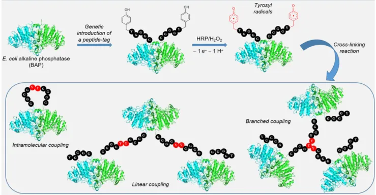
Conversely, if several Y are surface-exposed, a complex cross-linked mixture may be formed. A better control of protein polymerization can be achieved by the genetic incorporation of peptide tags containing a Y residue. Kamiya and co-workers genetically introduced a GGGGY or GGYYY tag at the
C-terminus of the model protein Escherichia coli alkaline phosphatase (BAP). The Y in tags are much more available than Y trapped in a folded protein. After horseradish peroxidase (HRP)-promoted oxidation of phenolic moiety, tyrosyl radicals react to form linear or branched cross-linked proteins, with different levels of polymerization (Figure 2). The authors showed that the catalytic activity of these polymers is not affected by the radical polymerization. Site-specificity was demonstrated because peroxidase (HRP)-mediated Y cross-linking was shown to occur exclusively with the tagged BAP. Wild-type BAP, or BAP after tag removal by enzymatic digestion, showed no reactivity.29
Figure 2. Y cross-linking reaction promoted by HRP in genetically modified alkaline phosphatase BAP. Y displayed on the penta-peptide tag (GGGGY) are oxidized into tyrosyl radicals which cross-couple to yield different polymeric arrengements of BAP.
Protein cross-linking was also successfully achieved using a photogenerated oxidant.30 The photolysis of a ruthenium(II)
tris-bipyridyldication [Ru(II)bpy3]2+ in the presence of
ammonium sulfate results in the production of Ru(III), a strong one-electron oxidant, and of the sulfate radical, proposed to be good hydrogen abstracting agents. These products lead to Y dimerization following the proposed mechanism presented in Figure 3.
The formation of protein multimers was observed after rapid (<1s) flash light irradiation. Long-wavelength light irradiation is particularly convenient as few biomolecules absorb light outside of the UV region. Notably, photoinduced protein cross-linking was also successfully mediated with palladium porphyrins though a similar mechanism.31
Chemical Y oxidation was also shown to be a convenient method to functionalize peptides, proteins or viral capsids.32,33
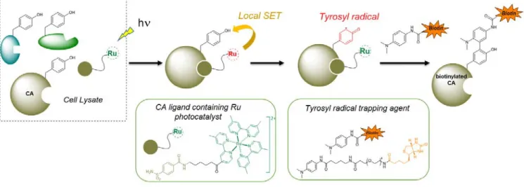
Nakamura and co-workers demonstrated the interest of using such a photocatalyst for the selective labelling of a protein of interest (carbonic anhydrase, CA) in cell lysate.34 A
benzensulfonamide ligand of CA was covalently attached to a [Ru(II)bpy3]2+ derivative and incubated in a lysate of
erythrocytes (Figure 4). After light irradiation and tyrosyl radical formation, the proteins were labelled with a
biotin-functionalized tyrosyl radical-trapping agent (N,N-dimethyl-1,4-phenylenediamine). A high level of labelling selectivity was observed on SDS-PAGE with a unique band at 29 kDa corresponding to CA, confirming the affinity-guided activation of Y. In a subsequent report, the photocatalyst-proximity dependence of the reactive substrate was studied and the concept was extended to another class of Y-reactive species (urazoles).35
Figure 4. Specific carbonic anhydrase (CA) labeling through affinity-guided activation of Y. Irradiation of the CA-ligand complex generates a tyrosyl radical reacting with a biotine-labelled radical-trapping agent.
3. Three component Mannich-type Y
conjugation
The three-component Mannich-type reaction was recognized in the early 1950s as a tool for protein modification and cross-linking.36 Aminomethylol derivatives obtained after
formaldehyde addition to proteins amino groups (lysine) were shown to induce peptides and proteins cross-linking after coupling with a variety of AAs such as asparagine, glutamine, tryptophan, and Y. The reaction mechanism starts by imine condensation of a primary aromatic amine with formaldehyde. Then, the phenol ring is deprotonated (Betti reaction) to undergo an electrophilic aromatic substitution with the iminium ion.
Figure 5: Mechanism of the three component Mannich-type Y conjugation
Francis’ research group developed this non-specific Mannich-type reaction for selective Y labelling.37 Electron-rich anilines
combined with formaldehyde were shown to form imines that selectively react with accessible Y under mild conditions and relatively low pH (6.5). The authors efficiently labelled chymotrypsinogen A, lysosyme, RNase and myoglobin using an aniline bearing a fluorescent chromophore and a set of aldehydes (Figure 6). The chemoselectivity for a specific Y of chymotrypsinogen (Y 146) was demonstrated by tryptic digestion and MALDI-MS experiments.
Figure 6. Modification of chymotrypsinogen A by a Mannich type reaction
In a second paper, the group extended the substrate scope of the reaction to short peptide sequences.38 Anilines containing
peptides at either the C- or N-termini were designed by solid phase synthetic routes. The local Y environment was shown to
significantly impact the level of reactivity and the eGFP protein, which had several surface-exposed Y, demonstrated much higher levels of reactivity compared with myoglobin.
4. Y-conjugation via sulfur fluoride
exchange chemistry (SuFEx)
SuFEx has become an increasingly popular click chemistry method since the potential of sulfur(VI) fluorides was reintroduced by Sharpless and co-workers in 2014.39,40 SuFEx
occurs when the fluoride atom is displaced from the sulfur by O- or N- nucleophiles to form stable conjugates. This sulfuryl chemistry can be sluggish with electrophiles such as arylsulfonyl fluorides (Ar-SO2-F) and arylfluorosulfate
(ArO-SO2-F) that are kinetically stable to hydrolysis and resistant to
oxidation and reduction. This specific reactivity pattern has been successfully exploited in chemical biology and medicinal chemistry for click protein labelling,41–43 after reaction with the
side chain of nucleophilic AAs. Although SuFEx may be performed on a potentially wide range of AAS, chemoselectivity
can only be achieved by fine-tuning the experimental procedure or in a context-specific environment.44
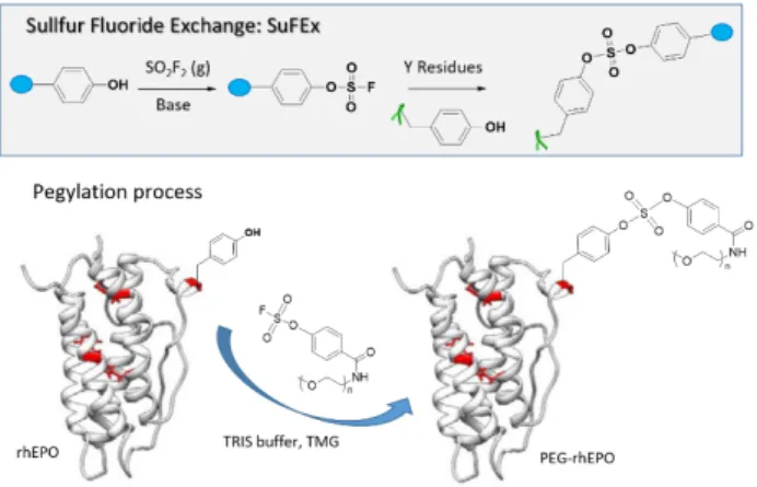
Recently, Kim and co-workers showed that Y model nucleophile p-cresol reacted with arylfluorosulfate in DMSO in the presence of the specific base tetramethylguanidine.45 In the
same conditions, n-butylamine, propanethiol, methanol, N-propylguanidine and 3-methylindole, as representative models of the nucleophilic AAs Lys, Cys, Ser, Arg andTrp, respectively, failed to provide the expected product. Based on these results, the authors hypothesized that Y can chemoselectively participate in a SuFEx reaction. A cell penetrating peptide (TAT 47-57) possessing one Y at the N-terminus was labelled with a rhodamine dye connected to the arylsulfonate anchor. After incubation of the conjugate with HeLa, an efficient cell permeation was observed by confocal laser scanning microscopy. The procedure also proved effective for the PEGylation of recombinant human erythropoietin (Figure 7).
Figure 7. SuFEx reaction with a fluorosulfate derivative to graft PEG molecules on proteins.
SuFEx is also a relevant strategy for the design of covalent inhibitors. Arylfluorosulfates are less reactive than the better-known aryl-sulfonyl fluorides and are therefore more suited for site-specific conjugation with Y. The former are virtually stable to nucleophilic substitution, except if a protein-binding site provides the means to activate the reactivity. This was exemplified by Kelly and co-workers who discovered that members of the intracellular lipid binding protein (iLBP) family, delivering ligands to nuclear hormone receptors, are selectively labelled by biphenylic fluorosulfates both in vitro and in vivo.46 Such simple hydrophobic probes covalently
modify cellular retinoic acid binding protein 2 (CRABP2) with low background labelling of the human proteome. Crystal structure of the CRABP2-conjugate and binding site mutagenesis provide hints about the ligand binding and fluorosulfate activation. The conserved Y 134 residue in the fatty acid binding site was shown to be bound to the sulfur atom. The specific reactivity of the Y 134 phenol could be explained by its low pKa~7.6, perturbated by two proximal Arg residues. Thus, it seemed that both the non-covalent binding-equilibrium of the probe with CRABP2 increasing the local concentration, and the presence of Arg residues lowering the pKa value contributed to the chemoselective Y 134 modification. Deliberate targeting of a specific Y residue from a protein binding site by SuFEx may also be expected. Jones and co-workers designed covalent inhibitors of the mRNA-decapping scavenger enzyme DcpS.44 Based on the already reported
X-ray structures of DcpS bound to diaminoquinazoline (DAQ) ligands, the authors designed DAQ containing aryl sulfonyl fluoride anchors, ortho- (SF-o1) meta-(SF-m1) or para- (SF-p1) substituted (Figure 8).47 X-ray crystallographic structures
confirmed the expected reactivity of the three isomers towards the proximal Y 113 and Y 143. As expected, SF-o1 and SF-m1 reacted with Y 113, whereas SF-p1 reacted with Y 143. These studies illustrate the precise level of selectivity that can be achieved by SuFEx when the experimental conditions are fine-tuned, despite the reactivity of sulfur(VI) fluorides for a broad range of nucleophiles.
Figure 8. Ortho- (SF-o1) and para- (SF-p1) isomers of diaminoquinazoline inhibitors of a mRNA-decapping scavenger enzyme (DcpS) were shown to specifically react with Y113 and Y143 buried in the binding site, respectively. Adapted with permission from ACS Chem. Biol. 2015, 10, 1094-1098, copyright 2015, American Chemical Society.
5. Transition metal complexes for Y
conjugation
Some transition metal catalysts possess sufficient functional group tolerance to perform chemoselective reactions in aqueous buffers. Iridium catalysts were shown to promote reductive alkylation of aldehydes on protein lysine residues and N-terminii.48 A selective method for the modification of Trp
residue using rhodium carbenoid species was also reported by Francis and co-workers.49 The same research group
developed π-allylpalladium complexes for Y conjugation (Figure 9).50
Figure 9 π-allylpalladium complexes for Y bioconjugation.
Rhodamine-labelled allylic acetate exposed to palladium acetate and triphenylphosphine tris-sulfonate (TPPTS) at pH 8.5-9 successfully derivatized the accessible Y of chymotrypsinogen A, H-Ras, bacteriophage MS2 and α-chymotrypsin. This procedure was also implemented for grafting a novel polarity-sensitive probe on Y108 of bovine Cu/Zn superoxide dismutase.51 The probe efficiently recorded
the local polarity change around the Y108 domain during acid or heat denaturation of the protein. This approach might provide a general way to measure local environment changes in Y proximity.
A particularly attractive feature of the π-allylpalladium complexes method is the possibility to switch the allylic acetate for a more hydrophilic leaving group.50 The relevance of this
“solubility switching” strategy was demonstrated by the design of synthetic lipoproteins obtained from a hydrophobic C17 chain solubilized in a 95:5 H2O:DMSO mixture, thanks to a
hydrophilic taurine carbamate moiety (Figure 10). Thus, a highly hydrophobic moiety can be solubilized thanks to the hydrophilic carrier, which improves access to the artificial lipoprotein.
Figure 10 π-allylpalladium complexes to graft lipophilic molecules on proteins. Palladium-cleavable taurine enhanced the water solubility of a C15 lipid allowing chymotrypsinogen A modification.
Transition metals not only catalyse Y conjugation but may also be directly involved in the formation of stable complexes with Y. A first evidence of a chemoselective formation of a π6-Y
complex was provided by rhodium on G-protein coupled receptor (GPCRs) peptides.52 In this study, [Cp*Rh(H
2O) 3
]-(OTf)2 reacted with the small peptides enkephalin, neurotensin
and octreotide complexes to form Cp*Rh-Y peptides in water, at room temperature and pH 5-6. This labelling technique, using preformed reactive complexes, was only explored with unfunctionalized Rh ligands. Ball and co-workers significantly extended the scope of organometallic Y labelling when they reported a coupling method using a simple Rh salt (RhCl3) and
aryl boronic acids incorporating an affinity handle or a fluorescent dye (Figure 11).53 The in situ formation of a π6-Y
complex was shown to be effective on a wide range of peptides and proteins and to be stable towards a variety of biologically relevant reagents excepted with the nucleophilic redox mediators dithiothreitol (DTT) and H2O2. The dose-dependent
complex degradation may actually offer interesting opportunities for a controlled released of the arylboronate substituent.
Figure 11 Formation of rhodium complexes to anchor fluorophores on proteins. Bar graph representing fluorescence detection for modification of selected proteins with NBD-functionalized boronate. Adapted with permission from Angew. Chem. Int. Ed. 2018, 57, 2827-2830, copyright 2018, Wiley.
6. diazonium coupling reaction
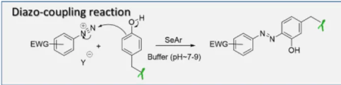
The multimodal reactivity of aryldiazonium salts can be exploited to modify a wide range of amino acids,54 including Y.
Aryldiazonium salts, generally prepared from the corresponding anilines by action of a nitrite in a strong acid medium, can establish an azo linkage with the phenol side ring of Y by an electrophilic aromatic substitution (Figure 12). This diazo-coupling reaction is significantly more efficient between aryldiazonium cations with electron withdrawing substituents,
and an electron-rich aromatic partner-like phenolic ring, under basic or neutral conditions. Non-activated aryldiazonium species may also react with phenol residues, but a higher pH is required to form the more reactive phenolate species.
Figure12 Mechanism of the diazo-coupling reaction
The potential of Y modification by diazonium coupling was shown by the modification of the interior surface of the viral capsids of bacteriophage MS2.55 This first example used a
rather laborious procedure, in four steps, to graft Y under mild conditions. This strategy was significantly simplified by the same authors for the surface modification of tobacco mosaic virus (TMV).56 Y 139 from the capsid was selectively
functionalized using diazonium salts from p-aminoacetophenone. The ketone introduced at the protein surface was used as a functional handle to introduce alkoxyamines bearing PEGs or a biotin moiety (Figure 13).
Figure 13 Surface modification of the Tobacco Mosaic Virus. Y139 was modified by diazo-coupling reaction then functionalized through oxime bond formation.
This two-step procedure was efficiently applied for the conjugation of broadly neutralizing antibodies to the HIV-1 drug aplaviroc,57 the design of magnetic resonance agents from
capsid shells,58 or the labelling of luciferase truncated variants
for bioluminescence based detection applications.59 These
successful Y modifications were achieved with arydiazonium reagents bearing a formyl group for oxime ligation, which also proved essential for the stability of the diazonium ion. Although direct peptide and protein ligation with aryldiazonium salts bearing PEGs, radionuclide complexes, or grafted on polymethacrylate polymers has been reported,60,61 examples
remains scarce and one-step Y coupling by the diazo procedure seems to be limited to a restricted number of conjugates. Indeed, a major limitation of diazo coupling is the general instability of diazonium reagents that are prepared in situ just prior to use and in strongly acidic conditions while the protein coupling is performed at mildly basic pH. The Barbas’ research group showed that formylbenzene diazonium hexafluorophosphate (FBDP), a bench stable crystalline solid prepared on a multigram scale, was an efficient staple for peptide, protein, antibody and HeLa cell surface labelling.62 A
stable diazonium analogue, but with an alkyne group instead of an aldehyde was also successfully obtained from commercial 4-ethynylaniline (Figure 14).63 After diazo coupling,
a fluorescent dye and mPEGs armed with an azido-group could be grafted by copper-catalysed azide-alkyne cyclization (CuAAC) at model proteins or the TMV surface. Combining diazo-coupling with CuAAC may extend the potential of the two-step Y targeting due to the highly biorthogonal alkyne-azide coupling.64
Figure14. Y modification with alkynyl-armed bench stables diazonium salts
Another possible way to counteract the instability of diazonium salts is to use of a chemical function masking the aryldiazonium cations. Jewett and co-workers showed that triazabutadiene scaffolds can be prepared by reacting functionalized aryl azides with N-heterocyclic carbenes (Figure 14). This chemical moiety can generate aryldiazonium salt after protonation in a pH-dependant manner.65,66 This strategy
proved effective for BSA conjugation in acidic buffer (pH 4).
As previously discussed, electron-withdrawing groups in para position improve diazonium salt electrophily and reactivity. However, lowering the reactivity of aryldiazonium salts would be profitable for controlled site-specific Y conjugations. Fridkin and Gilon described the possibility of forming cyclic azo-bridged peptides (derived from RGD, GnRH, Tuftsin, VIP and SV40 NLS) by an intramolecular azo-coupling reaction between a Y and a phenylalanine residue (amino-Phe). They showed that diazophenylalanine derivatives (diazo-Phe) have a moderate reactivity with Y due to the electron donating effect of the CH2 group in the para position of the diazo group.67
Xia and co-workers also studied the proximity-induced cyclization of a synthetic peptide with a hairpin structure confining an N-terminal Y and C terminal amino-Phe in close spatial proximity (Figure 16). 68 Mass spectroscopy analysis
and UV-vis spectroscopy confirmed the formation of the expected cyclic peptide after converting amino-Phe to diazo-Phe. When the two reactive groups diazo-Phe and Y were mixed in solution at micromolar concentration, no azo coupling occurred, confirming the proximity-driven reaction observed on the peptide.
Figure 16 Proximity-driven cyclization: Schematic description of a diazo-coupling reaction forming a macrocyclic azo-bridged peptide.
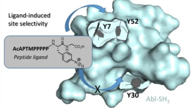
This method was also exploited by the authors for the intermolecular covalent labelling of an SH3 protein with a diazonium derivative of the polyproline peptide APTMPPPPP, displaying a micromolar affinity for Abl-SH3. Single mutants Abl-SH3Y7A and Abl-SH3Y52A and the corresponding double
mutant showed low and no reactivity with the peptide ligand respectively. These results suggest a ligand-directed reactivity for Y7 and Y52 in closer binding-site proximity compared with Y30 (Figure 17).68
Figure 17 Proximity-driven intermolecular covalent labelling of an SH3 protein with a diazonium ligand peptide.
Beside Y, Xia’s research group also discovered that diazo-Phe species react with the imidazole of histidine (His).68 His
coupling observed therein, and double modifications of Y or His peptide residues observed with a nitroazobenzene tag,69
suggest a rather low specificity of the reaction between the two AAs, which may limit the broad applicability of the reaction for site-specific coupling. Wuest and co-workers also studied the chemoselectivity of the diazo-coupling reaction by reacting radiometal-diazo-Phe species with an equimolar mixture of L-tyrosine and L-histidine. Radio-HPLC analysis, confirmed a better diazo-coupling with Y at Ph 9, while a preferential coupling to His was observed at neutral pH (pH=7).61. The
genetic incorporation of more reactive handles than Y or His towards diazo-coupling has been shown to efficiently circumvent these selectivity issues. The electron-rich 2-naphtol analogue of Y (NpOH) was genetically incorporated into proteins in E. coli by Tsao and co-workers (Figure 18).70 This
more reactive group was selectively targeted with diazotized anilines bearing an electron donating group for site-specific PEGylation. Combining the presence of a more reactive NpOH group on protein with deactivated diazo -species proved a particularly efficient way to improve site-selective targeting.
Figure 18. Selective diazo-coupling reaction for genetically introduced 2-naphtol analogue of Y.
Genetically encoded 5-hydroxyindoles (5-HTP) were also shown to exhibit high reactivity toward aromatic diazonium ions, as demonstrated by the group of Chatterjee.71 In a model
reaction, 4-nitrobenzendiazonium was found to react 4500-fold faster with 5-HTP relative to Y (k2 = 63000 vs 14.2 M-1s-1). This
large shift in reactivity does not guarantee a complete inertness of the diazo -species and analogues of the less reactive 4-carbobenzendiazonium (k2 = 193 M-1s-1 with 5-HTP) were
selected to label the engineered protein models of the study.
7. Reactions with triazolinediones
The preparation and reactivity of triazolinediones was recently reviewed by Du Prez and co-workers,72 and will not be covered
here.
Cyclic diazodicarboxamides are among the most promising and exemplary Y anchors. These silent urazoles are readily activated to highly reactive triazolinediones (TAD) with oxidizers such as NBS or dibromo-dimethylhydantoin. Although TAD grafting to Y is generally described in literature as an ene-like reaction, an SEAr mechanism (Figure 19) seems
observed after addition of bases and with recently published quantum chemical approaches.73
Figure 19. Plausible mechanisms for triazolinedione (TAD) addition to Y.
The versatility of cyclic diazodicarboxamides for Y -bioconjugation was first reported ten years ago by the research Barbas’ research group at the Scripps research institute.74
Since then, published examples of successful phenyltriazolinedione (PTAD) conjugations have flourished, not only in the field of chemical biology but also in material chemistry. Original applications in the latter field include i) tailoring of electrode surfaces,75 ii) formation of bottlebrush
polymers,76 iii) improvement of polymer tensile strength by
bis-TAD reticulation, iv) design of dynamic polymer systems where reversible PTAD adducts are able to translick,77 and v) the
design of shape-memory polyurethane networks using thermoreversible PTAD chemistry.78 Although of high interest
and relevant for development in material sciences, this section will only discuss representative examples of PTAD for bioconjugations.
In their initial report,74 Barbas and co-workers showed that
PTAD could selectively label the N-acyl methyl amide of Y among mixtures of other amino acid amides in phosphate buffer/acetonitrile, opening an interesting perspective for protein labelling. The newly formed C-N bond between p-cresol, as a model phenol, and PTAD was stable in strongly acidic and basic conditions (10% HCl or 10% NaOH in MeOH) for 24H at room temperature or high temperature (120°C) for 1H. This suggests that the PTAD linkage is more robust than classical thiol-maleimide conjugate, as latter observed by the authors in human blood plasma (Figure 20).79
Thus, the PTAD conjugate remained stable for more than 7 days while thiol-maleimide completely degraded after one hour. Interestingly, BSA labelling with a PTAD-functionalized rhodamine dye was effective over a wide range of pH (2-10) with 54% labelling at pH=2 and up to 98% yields at pH 10.
Figure 20. Stability of PTAD and thiol-maleimide conjugate in human plasma. Reprinted with permission from Bioconjugate Chem. 2013, 24, 520-530, Copyright 2013, American Chemical Society.
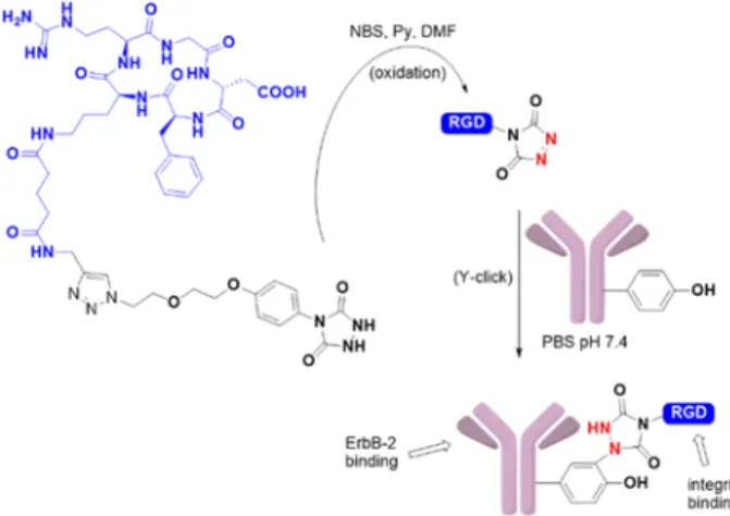
The authors also demonstrated the compatibility of the bioconjugation method on peptides and model proteins such as chymotrypsinogen A or myoglobin and on the therapeutic antibody Herceptin (Figure 21).74 After urazole oxidation of a
conjugated RGD peptide in organic media, the reactive PTAD derivative was added to a PBS solution of Herceptin antibody. The antibody conjugate was studied by ELISA and shown to conserve its ability to bind to the growth factor receptor 2 ErbB-2 while the RGD tag allowed αvβ3 integrin binding.
Figure 21. PTAD anchors to functionalize Herceptin antibody with cyclic RGD
The PTAD anchor also proved efficient for the design of well-defined glycoconjugate vaccines. Berti and co-workers in Novartis developed a two-step approach for the design of a vaccine against Candida albicans.80 The protein carrier, a
non-toxic mutant diphtheria toxin (CRM197) was first armed with an
alkyne group by the Y coupling. The carbohydrate antigen, a synthetic hexameric β-(1,3)-glucan bearing an azido-group, was subsequently grafted by CuAAC onto CRM197. The
in an immunization study in mice. This two-step method was, however, ineffective for grafting polysaccharides of higher molecular weight as later shown by Adamo and co-workers.81
This goal was achieved using the strain-promoted azide-alkyne cyclization (SPAAC) instead of the conventional CuAAc. This modified two-step conjugation enabled the conjugation of a complex and negatively charged streptococcal polysaccharide to CRM197. The glycoconjugate proved effective in protecting
new-born mice against Streptococcus agalactiae infections following vaccination of the mothers.82
Although very promising for a wide range of applications, Y-bioconjugation with PTAD shows inherent limitations and more research is needed to extend its potential scope. PTAD are unstable in physiological conditions and have to be prepared immediately before use by chemical oxidation of a phenylurazole precursor. The use of a chemical oxidant to form the reactive PTAD intermediate may not be compatible with substrates with low oxidation potentials. Depending on the Y accessibility and reagent stoichiometry, two molecules of PTAD may be grafted to a single Y phenol ring (Figure 22). PTAD derivatives were also shown to be stable in acetonitrile but a rapidly accelerated decomposition was observed after water addition.80 In water mixtures, an isocyanate
decomposition product of the PTAD may be formed, resulting in the unwanted formation of thiourea bonds on the lysine amino groups of the targeted peptides or proteins. To avoid this side-reaction, PTAD coupling should be performed in Tris buffer (containing a primary amine group) or in a mixture of buffers with Tris to scavenge the isocyanate side-product.79,80,83 This procedure was however not always
efficient at suppressing the unspecific labelling of amino -groups on more challenging targets.84
Figure 22. Possible unwanted reactions during PTAD anchoring
Nakamura and co-workers showed that Y-bioconjugation with luminol derivatives was more selective than PTAD due to the absence of lysine modification.84 Luminol is used in forensic
investigations, wherein traces of blood (hemin) in the presence of hydrogen peroxide (H2O2) catalyses luminol oxidation
leading to the chemiluminescence reaction. This oxidation protocol was adapted with N-methylluminol derivatives for Y-coupling. BSA, streptavidin and CA were efficiently labeled using 10µM hemin and 1 mM H2O2. Lysine coupling was never
observed, but cysteine residues were shown to be partially oxidized on angiotensin II variants, and double modifications were observed at the most accessible Y residue of BSA. The oxidation procedure using excess H2O2 may also partially
damage the protein structures as suggested by anti-tubulin antibody which showed slightly decreased antigen selectivity after modification.
In a subsequent paper, the authors described a drastically improved procedure using horseradish peroxidase (HRP) instead of hemin.85 HRP showed excellent catalytic activity,
and almost total conversion of angiotensin II into the expected Y-clicked adduct was observed at only 0.1% mol HRP while 10% mol hemin was required for a similar conversion yield (Figure 23). Importantly, and in contrast to hemin, side reactions such as Y cross-coupling involving an oxidative radical process or cysteine oxidation were not observed when HRP was used on model substrates or BSA. Thus N-luminol derivatives are promising precursors of Y chemical anchors.
Figure 23. Angiotensin II (sequence DRVIHPF)-luminol adduct is obtained by in situ oxidative activation of luminol anchors. Authors observed a higher catalytic activity using HRP protein.
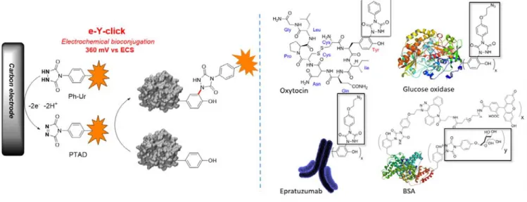
As excess of chemical oxidant may be problematic with sensitive protein targets and substrates, we recently explored the possibility to activate Y anchors electrochemically (Figure 24).86 Instead of using a chemical oxidant, the reactive PTAD
anchors were generated on demand and in situ from the corresponding Ph-Ur species by applying an electrochemical activation potential of 0.36V. At this value, the electroactive amino-acids from proteins (L_Tyr, L-Phe, L-Trp or L-Hist) were inert and unique Y- labeling was observed on model peptides and proteins such as oxytocin, angiotensin 2, BSA or epratuzumab. Although the reactions were conducted in pure PBS buffer without Tris scavenger, we never observed lysine modifications as a side reaction.
Figure 24. Electrochemically-promoted tyrosine click reaction (e-Y-click). After chemoselective electroxidation of dormant Ph-Ur species at the surface of a carbon electrode, the protein of interest is labelled in situ and in high conversion yield.
Furthermore, enzyme activity was conserved after applying the e-Y-click labelling protocol, as shown on the glucose oxidase bearing the redox active coenzyme FAD buried in the active centre. The conserved activity may be explained by a lower diffusion coefficient of proteins at the electrode surface due to higher molecular weight. The e-Y-click protocol is a traceless procedure for activating Y anchors at electrode surfaces without damaging the protein structures or activity.
Recently, a second report of Y electrochemical bioconjugation was published by Lei and co-workers. The authors demonstrated the selective incorporation of a phenothiazine motif on Y of polypeptides, insulin and myoglobin.87 This
second published example confirms the interesting potential of electrochemistry for protein bioconjugations.
8. Summary and Outlook
Y -bioconjugation for native protein labelling has focus a specific interest in academic research over the last two decades. This has yielded a broad range of new Y-labelling strategies developed by different research groups highlighted in the present research review. All these strategies are of specific interest but also have inherent limitations. In consequence, they should be selected with care depending on the main purpose of the research. Proteins with solvent-exposed Y can easily be covalently aggregated by the formation of Y radicals with oxidoreductase enzymes or light harvesting ruthenium complexes. This method is, however, less suited for anchoring a chemical pharmacophore or probes onto the protein. Such coupling may be successfully achieved by electrophilic aromatic substitutions with small arenediazonium salts or ene-like addition with PTAD derivatives, as shown by the numerous successful examples published on proteins or viral capsides. These two strategies have recently been technically improved with the development of bench-stable arenediazonium salts, N-methylluminol derivatives limiting PTAD cross-reactivity, or soft electrochemical instead of chemical Ph-Ur activation. For site-specific Y modifications, such as binding site labelling with a functionalized ligand, the reactivity of the chemical anchors selected should actually be lowered. This allows key-lock interaction to dramatically enhance the concentration of the anchor in the local environment of the targeted proteins. Normally broad coupling reactions such as SuFEx may become very selective for a specific Y by ligand-directed and reactivity-lowered Y-click.
To conclude, Y-click chemistry emerges as a particularly promising approach for designing medically relevant bioconjugates such as antibody-drug conjugates or glycovaccines, or new biological tools for drug-target identification or protein binding site tagging. We foresee that future success in developing more efficient protocols will further expand the scope of Y-click bioconjugations.
Dimitri Alvarez-Dorta received his PhD degree at La Laguna University in 2010 (Tenerife, Spain). He worked under the guidance of Professor E. Suarez in the field of photo-radicalary glycochemistry. After a post-doctoral position in 2011 at the IIQ (Seville, Spain), he moved in 2013 to the CEISAM laboratory (Nantes, France) to work in the area of glycochemistry and protein bioconjugations.
David Deniaud received his PhD (1996) in hybrid organic/inorganic materials from de Institute Jean Rouxel, France. After that, he joined the D. Mansuy research group at the University Paris Descartes as a postdoctoral researcher. In 2000 he became associate professor and the full professor in 2012 in the University of Nantes, and leads his research in the Institute Ceisam in the field of Organic chemistry. His current
research topics are at the interface of chemistry and biology, developing ligands and chemical tools for application in gene therapy.
Mathieu Mével received his PhD degree from Université de Bretagne Occidentale in 2007 (Brest – France). He then worked as a post-doctoral fellow at Imperial college (University of London) in 2008 and at Institut du Thorax (Nantes) from 2008-2014. Since 2014, he has worked as a researcher at INSERM UMR1089 – Translational gene therapy for genetic diseases and CEISAM CNRS UMR6230. His research interests are focused at the
interface of chemistry and vectorology, on the development of viral and non-viral vectors forgene delivery.
Dr. Sébastien G. Gouin studied organic chemistry at the University of Nantes (France) where he received his PhD in 2003. After postdoctoral training with Prof. Paul V. Murphy at University College Dublin (Ireland), he was appointed as a CNRS research associate at the University of Amiens (2005) and CNRS research director at the University of Nantes (2018). The present research activities of his group
“glycochemistry and bioconjugates”in the laboratory CEISAM, are focused on the development of glycoconjugates targeting pathogenic lectins and glycosidases.
Acknowledgement
This work was carried out with financial support from the Centre National de la Recherche Scientifique (CNRS) and the Ministère de l’Enseignement Supérieur et de la Recherche in France.
Keywords: bioconjugates • tyrosine • site-specificity • bioorthogonality • click chemistry
1 C. H. Kim, J. Y. Axup and P. G. Schultz, Curr. Opin. Chem. Biol., 2013, 17, 412–419.
2 N. Wang, B. Yang, C. Fu, H. Zhu, F. Zheng, T. Kobayashi, J. Liu, S. Li, C. Ma, P. G. Wang, Q. Wang and L. Wang, J. Am. Chem. Soc., 2018, 140, 4995–4999.
3 J. E. Lopez, S. E. Haynes, J. D. Majmudar, B. R. Martin and C. A. Fierke, J. Am. Chem. Soc., 2017, 139, 16222–16227.
4 B. Bhushan, Y. A. Lin, M. Bak, A. Phanumartwiwath, N. Yang, M. K. Bilyard, T. Tanaka, K. L. Hudson, L. Lercher, M. Stegmann, S. Mohammed and B. G. Davis, J. Am. Chem. Soc., 2018, 140, 14599– 14603.
5 J. M. Chalker, G. J. L. Bernardes, Y. A. Lin and B. G. Davis, Chem. -
Asian J., 2009, 4, 630–640.
6 E. M. Sletten and C. R. Bertozzi, Angew. Chem. Int. Ed., 2009, 48, 6974–6998.
7 M.-A. Kasper, A. Stengl, P. Ochtrop, M. Gerlach, T. Stoschek, D. Schumacher, J. Helma, M. Penkert, E. Krause, H. Leonhardt and C. P. R. Hackenberger, Angew. Chem. Int. Ed., 2019, 58, 11631–11636. 8 N. Martínez-Sáez, S. Sun, D. Oldrini, P. Sormanni, O. Boutureira, F. Carboni, I. Compañón, M. J. Deery, M. Vendruscolo, F. Corzana, R. Adamo and G. J. L. Bernardes, Angew. Chem. Int. Ed., 2017, 56, 14963–14967.
9 M. S. Messina, J. M. Stauber, M. A. Waddington, A. L. Rheingold, H. D. Maynard and A. M. Spokoyny, J. Am. Chem. Soc., 2018, 140, 7065–7069.
10 J. Willwacher, R. Raj, S. Mohammed and B. G. Davis, J. Am. Chem.
Soc., 2016, 138, 8678–8681.
11 X. Chen, K. Muthoosamy, A. Pfisterer, B. Neumann and T. Weil,
Bioconjug. Chem., 2012, 23, 500–508.
12 M. J. Matos, B. L. Oliveira, N. Martínez-Sáez, A. Guerreiro, P. M. S. D. Cal, J. Bertoldo, M. Maneiro, E. Perkins, J. Howard, M. J. Deery, J. M. Chalker, F. Corzana, G. Jiménez-Osés and G. J. L. Bernardes,
J. Am. Chem. Soc., 2018, 140, 4004–4017.
13 S. Asano, J. T. Patterson, T. Gaj and C. F. Barbas, Angew. Chem.
Int. Ed., 2014, 53, 11783–11786.
14 S. M. Hacker, K. M. Backus, M. R. Lazear, S. Forli, B. E. Correia and B. F. Cravatt, Nat. Chem., 2017, 9, 1181–1190.
15 D. G. Isom, C. A. Castañeda, B. R. Cannon and B. G.-M. E, Proc.
Natl. Acad. Sci., 2011, 108, 5260–5265.
16 I. Dovgan, S. Erb, S. Hessmann, S. Ursuegui, C. Michel, C. Muller, G. Chaubet, S. Cianférani and A. Wagner, Org. Biomol. Chem., 2018, 16, 1305–1311.
17 Y. Seki, T. Ishiyama, D. Sasaki, J. Abe, Y. Sohma, K. Oisaki and M. Kanai, J. Am. Chem. Soc., 2016, 138, 10798–10801.
18 S. Lin, X. Yang, S. Jia, A. M. Weeks, M. Hornsby, P. S. Lee, R. V. Nichiporuk, A. T. Iavarone, J. A. Wells, F. D. Toste and C. J. Chang,
Science, 2017, 355, 597–602.
19 A. H. Christian, S. Jia, W. Cao, P. Zhang, A. T. Meza, M. S. Sigman, C. J. Chang and F. D. Toste, J. Am. Chem. Soc., 2019, 141, 12657– 12662.
20 H. Dau, I. Zaharieva and M. Haumann, Curr. Opin. Chem. Biol., 2012, 16, 3–10.
21 B. J. Reeder, M. Grey, R.-L. Silaghi-Dumitrescu, D. A. Svistunenko, L. Bülow, C. E. Cooper and M. T. Wilson, J. Biol. Chem., 2008, 283, 30780–30787.
22 L. H. Jones, A. Narayanan and E. C. Hett, Mol. Biosyst., 2014, 10, 952–969.
23 D. Montoir, M. Amoura, Z. E. A. Ababsa, T. M. Vishwanatha, E. Yen-Pon, V. Robert, M. Beltramo, V. Piller, M. Alami, V. Aucagne and S. Messaoudi, Chem. Sci., 2018, 9, 8753–8759.
24 J. J. Bruins, B. Albada and F. van Delft, Chem. – Eur. J., 2018, 24, 4749–4756.
25 A. M. ElSohly and M. B. Francis, Acc. Chem. Res., 2015, 48, 1971– 1978.
26 T. Heck, G. Faccio, M. Richter and L. Thöny-Meyer, Appl. Microbiol.
Biotechnol., 2013, 97, 461–475.
27 J. Gross, 234, 5.
28 N. Santhanam, J. M. Vivanco, S. R. Decker and K. F. Reardon,
Trends Biotechnol., 2011, 29, 480–489.
29 K. Minamihata, M. Goto and N. Kamiya, Bioconjug. Chem., 2011, 22, 74–81.
30 D. A. Fancy and T. Kodadek, Proc. Natl. Acad. Sci., 1999, 96, 6020– 6024.
31 K. Kim, D. A. Fancy, D. Carney and T. Kodadek, J. Am. Chem. Soc., 1999, 121, 11896–11897.
32 S. Meunier, E. Strable and M. G. Finn, Chem. Biol., 2004, 11, 319– 326.
33 K. L. Seim, A. C. Obermeyer and M. B. Francis, J. Am. Chem. Soc., 2011, 133, 16970–16976.
34 S. Sato and H. Nakamura, Angew. Chem. Int. Ed., 2013, 52, 8681– 8684.
35 S. Sato, K. Hatano, M. Tsushima and H. Nakamura, Chem. Commun., 2018, 54, 5871–5874.
36 H. Fraenkel-Conrat and H. S. Olcott, J. Biol. Chem., 1948, 174, 827– 843.
37 N. S. Joshi, L. R. Whitaker and M. B. Francis, J. Am. Chem. Soc., 2004, 126, 15942–15943.
38 D. W. Romanini and M. B. Francis, Bioconjug. Chem., 2008, 19, 153– 157.
39 J. Dong, L. Krasnova, M. G. Finn and K. B. Sharpless, Angew. Chem.
Int. Ed., 2014, 53, 9430–9448.
40 J. Dong, L. Krasnova, M. G. Finn and K. B. Sharpless, Angew. Chem., 2014, 126, 9584–9603.
41 A. Marra, J. Dong, T. Ma, S. Giuntini, E. Crescenzo, L. Cerofolini, M. Martinucci, C. Luchinat, M. Fragai, C. Nativi and A. Dondoni, Chem.
– Eur. J., 2018, 24, 18981–18987.
42 N. N. Gushwa, S. Kang, J. Chen and J. Taunton, J. Am. Chem. Soc., 2012, 134, 20214–20217.
43 N. P. Grimster, S. Connelly, A. Baranczak, J. Dong, L. B. Krasnova, K. B. Sharpless, E. T. Powers, I. A. Wilson and J. W. Kelly, J. Am.
Chem. Soc., 2013, 135, 5656–5668.
44 O. O. Fadeyi, L. R. Hoth, C. Choi, X. Feng, A. Gopalsamy, E. C. Hett, R. E. Kyne, R. P. Robinson and L. H. Jones, ACS Chem. Biol., 2017, 12, 2015–2020.
45 E. J. Choi, D. Jung, J.-S. Kim, Y. Lee and B. M. Kim, Chem. – Eur. J., 2018, 24, 10948–10952.
46 W. Chen, J. Dong, L. Plate, D. E. Mortenson, G. J. Brighty, S. Li, Y. Liu, A. Galmozzi, P. S. Lee, J. J. Hulce, B. F. Cravatt, E. Saez, E. T. Powers, I. A. Wilson, K. B. Sharpless and J. W. Kelly, J. Am. Chem.
Soc., 2016, 138, 7353–7364.
47 E. C. Hett, H. Xu, K. F. Geoghegan, A. Gopalsamy, R. E. Kyne, C. A. Menard, A. Narayanan, M. D. Parikh, S. Liu, L. Roberts, R. P. Robinson, M. A. Tones and L. H. Jones, ACS Chem. Biol., 2015, 10, 1094–1098.
48 J. M. McFarland and M. B. Francis, J. Am. Chem. Soc., 2005, 127, 13490–13491.
49 J. M. Antos and M. B. Francis, J. Am. Chem. Soc., 2004, 126, 10256– 10257.
50 S. D. Tilley and M. B. Francis, J. Am. Chem. Soc., 2006, 128, 1080– 1081.
52 H. B. Albada, F. Wieberneit, I. Dijkgraaf, J. H. Harvey, J. L. Whistler, R. Stoll, N. Metzler-Nolte and R. H. Fish, J. Am. Chem. Soc., 2012, 134, 10321–10324.
53 J. Ohata, M. K. Miller, C. M. Mountain, F. Vohidov and Z. T. Ball,
Angew. Chem. Int. Ed., 2018, 57, 2827–2830.
54 S. Sengupta and S. Chandrasekaran, Org. Biomol. Chem., 2019, 17, 8308–8329.
55 J. M. Hooker, E. W. Kovacs and M. B. Francis, J. Am. Chem. Soc., 2004, 126, 3718–3719.
56 T. L. Schlick, Z. Ding, E. W. Kovacs and M. B. Francis, J. Am. Chem.
Soc., 2005, 127, 3718–3723.
57 J. Gavrilyuk, H. Ban, H. Uehara, S. J. Sirk, K. Saye-Francisco, A. Cuevas, E. Zablowsky, A. Oza, M. S. Seaman, D. R. Burton and C. F. Barbas, J. Virol., 2013, 87, 4985–4993.
58 J. M. Hooker, A. Datta, M. Botta, K. N. Raymond and M. B. Francis,
Nano Lett., 2007, 7, 2207–2210.
59 E. A. Hunt, A. Moutsiopoulou, S. Ioannou, K. Ahern, K. Woodward, E. Dikici, S. Daunert and S. K. Deo, Sci. Rep., 2016, 6, 26814. 60 M. W. Jones, G. Mantovani, C. A. Blindauer, S. M. Ryan, X. Wang, D.
J. Brayden and D. M. Haddleton, J. Am. Chem. Soc., 2012, 134, 7406–7413.
61 S. Leier, S. Richter, R. Bergmann, M. Wuest and F. Wuest, ACS
Omega, 2019, 4, 22101–22107.
62 J. Gavrilyuk, H. Ban, M. Nagano, W. Hakamata and C. F. Barbas,
Bioconjug. Chem., 2012, 23, 2321–2328.
63 J. Zhang, D. Ma, D. Du, Z. Xi and L. Yi, Org Biomol Chem, 2014, 12, 9528–9531.
64 Miffy. H. Y. Cheng, H. Savoie, F. Bryden and Ross. W. Boyle,
Photochem. Photobiol. Sci., 2017, 16, 1260–1267.
65 F. W. Kimani and J. C. Jewett, Angew. Chem. Int. Ed., 2015, 54, 4051–4054.
66 B. M. Cornali, F. W. Kimani and J. C. Jewett, Org. Lett., 2016, 18, 4948–4950.
67 G. Fridkin, C. Gilon and C. Gilon, 8.
68 F. Huang, Y. Nie, F. Ye, M. Zhang and J. Xia, Bioconjug. Chem., 2015, 26, 1613–1622.
69 J. R. Aponte, L. Vasicek, J. Swaminathan, H. Xu, M. C. Koag, S. Lee and J. S. Brodbelt, Anal. Chem., 2014, 86, 6237–6244.
70 S. Chen and M.-L. Tsao, Bioconjug. Chem., 2013, 24, 1645–1649. 71 P. S. Addy, S. B. Erickson, J. S. Italia and A. Chatterjee, J. Am. Chem.
Soc., 2017, 139, 11670–11673.
72 K. De Bruycker, S. Billiet, H. A. Houck, S. Chattopadhyay, J. M. Winne and F. E. Du Prez, Chem. Rev., 2016, 116, 3919–3974. 73 D. Kaiser, J. M. Winne, M. E. Ortiz-Soto, J. Seibel, T. A. Le and B.
Engels, J. Org. Chem., , DOI:10.1021/acs.joc.8b01445.
74 H. Ban, J. Gavrilyuk and Barbas Carlos F., J. Am. Chem. Soc., 2010, 132, 1523–1525.
75 W. Laure, K. De Bruycker, P. Espeel, D. Fournier, P. Woisel, F. E. Du Prez and J. Lyskawa, Langmuir, 2018, 34, 2397–2402.
76 L. Xiao, Y. Chen and K. Zhang, Macromolecules, 2016, 49, 4452– 4461.
77 S. Billiet, K. De Bruycker, F. Driessen, H. Goossens, V. Van Speybroeck, J. M. Winne and F. E. Du Prez, Nat. Chem., 2014, 6, 815–821.
78 N. Van Herck and F. E. Du Prez, Macromolecules, 2018, 51, 3405– 3414.
79 H. Ban, M. Nagano, J. Gavrilyuk, W. Hakamata, T. Inokuma and C. F. Barbas, Bioconjug. Chem., 2013, 24, 520–532.
80 Q.-Y. Hu, M. Allan, R. Adamo, D. Quinn, H. Zhai, G. Wu, K. Clark, J. Zhou, S. Ortiz, B. Wang, E. Danieli, S. Crotti, M. Tontini, G. Brogioni and F. Berti, Chem. Sci., 2013, 4, 3827.
81 A. Nilo, M. Allan, B. Brogioni, D. Proietti, V. Cattaneo, S. Crotti, S. Sokup, H. Zhai, I. Margarit, F. Berti, Q.-Y. Hu and R. Adamo,
Bioconjug. Chem., 2014, 25, 2105–2111.
82 A. Nilo, L. Morelli, I. Passalacqua, B. Brogioni, M. Allan, F. Carboni, A. Pezzicoli, F. Zerbini, D. Maione, M. Fabbrini, M. R. Romano, Q.-Y. Hu, I. Margarit, F. Berti and R. Adamo, ACS Chem. Biol., 2015, 10, 1737–1746.
83 C. M. Madl and S. C. Heilshorn, Bioconjug. Chem., , DOI:10.1021/acs.bioconjchem.6b00720.
84 S. Sato, K. Nakamura and H. Nakamura, ACS Chem. Biol., 2015, 10, 2633–2640.
85 S. Sato, K. Nakamura and H. Nakamura, ChemBioChem, 2017, 18, 475–478.
86 D. Alvarez-Dorta, C. Thobie-Gautier, M. Croyal, M. Bouzelha, M. Mével, D. Deniaud, M. Boujtita and S. G. Gouin, J. Am. Chem. Soc., 2018, 140, 17120–17126.
87 C. Song, K. Liu, Z. Wang, B. Ding, S. Wang, Y. Weng, C.-W. Chiang and A. Lei, Chem. Sci., 2019, 10, 7982–7987.