HAL Id: inserm-02285428
https://www.hal.inserm.fr/inserm-02285428
Submitted on 12 Sep 2019
HAL is a multi-disciplinary open access
archive for the deposit and dissemination of
sci-entific research documents, whether they are
pub-lished or not. The documents may come from
teaching and research institutions in France or
abroad, or from public or private research centers.
L’archive ouverte pluridisciplinaire HAL, est
destinée au dépôt et à la diffusion de documents
scientifiques de niveau recherche, publiés ou non,
émanant des établissements d’enseignement et de
recherche français ou étrangers, des laboratoires
publics ou privés.
Fulminant arterial vasculitis as an unusual complication
of disseminated staphylococcal disease due to the
emerging CC1 methicillin-susceptible Staphylococcus
aureus clone: a case report
Charles Vidal, Florence Moulin, Xavier Nassif, Louise Galmiche, Delphine
Borgel, Alain Charbit, Capucine Picard, Jean-Paul Mira, Olivier Lortholary,
Anne Jamet, et al.
To cite this version:
Charles Vidal, Florence Moulin, Xavier Nassif, Louise Galmiche, Delphine Borgel, et al.. Fulminant
arterial vasculitis as an unusual complication of disseminated staphylococcal disease due to the
emerg-ing CC1 methicillin-susceptible Staphylococcus aureus clone: a case report. BMC Infectious Diseases,
BioMed Central, 2019, 19 (1), pp.302. �10.1186/s12879-019-3933-3�. �inserm-02285428�
C A S E R E P O R T
Open Access
Fulminant arterial vasculitis as an unusual
complication of disseminated staphylococcal
disease due to the emerging CC1
methicillin-susceptible Staphylococcus aureus
clone: a case report
Charles Vidal
1, Florence Moulin
2, Xavier Nassif
1, Louise Galmiche
3, Delphine Borgel
4, Alain Charbit
5,
Capucine Picard
6,7, Jean-Paul Mira
8,9, Olivier Lortholary
10, Anne Jamet
1,5and Julie Toubiana
9,11*Abstract
Background: Staphylococcus aureus has emerged as a leading cause of invasive severe diseases with a high rate of morbidity and mortality worldwide. The wide spectrum of clinical manifestations and outcome observed in
staphylococcal illness may be a consequence of both microbial factors and variability of the host immune response. Case presentation: A 14-years old child developed limb ischemia with gangrene following S. aureus bloodstream infection. Histopathology revealed medium-sized arterial vasculitis. The causing strain belonged to the emerging clone CC1-MSSA and numerous pathogenesis-related genes were identified. Patient’s genotyping revealed
functional variants associated with severe infections. A combination of virulence and host factors might explain this unique severe form of staphylococcal disease.
Conclusion: A combination of virulence and genetic host factors might explain this unique severe form of staphylococcal disease.
Keywords: S. aureus, Sepsis, Vasculitis, Virulence, Genetic susceptibility, Polymorphism, Case report Background
Staphylococcus aureus is a major human pathogen and a global healthcare issue. Humans are a natural reservoir of S. aureus, which can occasionally cause diseases that range in severity from minor skin infections to severe cases of pneumonia, bacteremia and septic shock [1]. The severity and outcome of the infection relies on bac-terial virulence, asS. aureus is known to have a wide ar-senal of components that contribute to the pathogenesis of infection. S. aureus is the cause of septic shock through cell wall components eliciting production of
inflammatory cytokines through TLR2 pathway activa-tion in innate immune cells [2]. S. aureus also contains several toxins that are able to potentiate host-inflammatory response, target and injure leukocytes and tissues, and inhibit bacterial clearance [3]. Finally,S. aureus avidly adheres to endothelial cells and platelet-fibrin thrombi involved in the physiopathology of endocarditis or thrombophlebitis [4]. Recently, the role of genetic factors of the host has been extensively studied, revealing their in-fluence on the susceptibility to or the severity of sepsis. Rare monogenic inborn errors of immunity were found to predispose to severe infectious diseases, such as mutations that impair NF-κB responses [5]. In parallel, common vari-ants of genes involved in innate immune response, inflam-mation and coagulation during sepsis were found to be associated with severity of bacterial infections [6, 7]. We report here a severe and atypical phenotype of infection
© The Author(s). 2019 Open Access This article is distributed under the terms of the Creative Commons Attribution 4.0 International License (http://creativecommons.org/licenses/by/4.0/), which permits unrestricted use, distribution, and reproduction in any medium, provided you give appropriate credit to the original author(s) and the source, provide a link to the Creative Commons license, and indicate if changes were made. The Creative Commons Public Domain Dedication waiver (http://creativecommons.org/publicdomain/zero/1.0/) applies to the data made available in this article, unless otherwise stated.
* Correspondence:julie.toubiana@aphp.fr
9Department of Infection, Immunity and Inflammation, Institut Cochin,
INSERM U1016, Paris, EU, France
11Department of General Pediatrics and Pediatric Infectious Diseases, Necker
Enfants-Malades Hospital, APHP, Paris Descartes University, 149 rue de Sèvres, 75015 Paris, EU, France
due to S. aureus. The young patient developed medium-sized arterial infectious vasculitis that led to ischemia and gangrene. We intended to decipher pathogen and host fac-tors that could explain this dramatic presentation.
Case presentation
Clinical case
The patient was a 14-year-old male child without notable past medical history and no recent travel. On admission, he had fever, lethargy and diarrhea, and physical examin-ation revealed fever (39 °C), tachycardia, blood pressure 65/35 mmHg and poor peripheral perfusion. Several boils were observed on the right elbow. He had subnormal white blood cell count 2.8 × 10⁹/ L with low lymphocyte count (13%), a moderate thrombocytopenia and raised CRP (347 mg/L) and procalcitonin plasma levels (279 ng/ mL). The diagnosis of septic shock with a possible associ-ated toxic mechanism was retained and intravenous (i.v.) cefotaxime and clindamycin were started with concomi-tant volume expansion. In the Intensive Care Unit, the pa-tient was intubated as he became confused, needed norepinephrine, inotropic support by epinephrine, and re-quired hemofiltration. Blood cultures showed methicillin susceptibleS. aureus (MSSA) and treatment was switched to i.v. cloxacillin, clindamycin, and gentamicin. Comple-mentary explorations revealed multiple septic pulmonary abscesses, an abscess of the left occipital lobe, and the ab-sence of endocarditis. In view of septic emboli, clindamy-cin was switched to fosfomyclindamy-cin in order to have a better cerebral diffusion. The patient then developed ischemia of the four limbs without any purpuric lesion or signs of dis-seminated intravascular coagulation, which unfortunately rapidly progressed to dry gangrene of the left toes and of the right leg requiring amputation at day 11 after admis-sion. The clinical status of the patient improved slowly. Catecholamines were stopped at day 6, the patient got extubated at day 11, and he recovered an efficient renal function 5 weeks after his admission. He had no serious neurological injury and a control cerebral MRI was nor-mal three months later.
Histopathology, biological and genetic investigations
Our initial hypothesis underlying this atypical phenotype was the association of a vasospasm due to high doses of va-sopressors and microcirculation disorders due to sepsis. We therefore analyzed the amputated specimen by light microscopy. Samples of left and right amputations were for-malin fixed, paraffin embedded, and cut into 3μm-thick sections. Sections were stained with Hematoxylin and Eosin, Gram, Periodic Acid Schiff and Grocott staining. The histopathologic analysis revealed perivascular infiltrates of mononuclear cells and neutrophils with arterial involve-ment (medium-sized vessels) without any capillary lesion (Fig.1a and b). Furthermore, the arterial endothelium was
destroyed and the walls were invaded by Gram-positive cocci with subsequent thrombosis (Fig.1c and d).
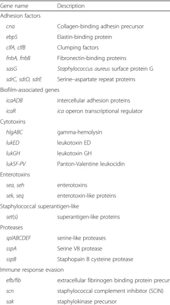
In order to decipher mechanisms underlying these clinical and histopathological findings, pathogen viru-lence and host factors were analyzed. A whole-genome shotgun library was prepared with Nextera XT Kit (Illumina, San Diego, CA, USA) and se-quenced on the MiSeq Illumina sequencing platform (2 × 150 bp paired-end reads). All reads were screened by mapping to known alleles of virulence genes using the Short Read Sequence Typing for Bacterial Patho-gens program (SRST2) [8]. Virulence gene allele se-quences were retrieved from the virulence factor database (VFDB, http://www.mgc.ac.cn/VFs/). SRST2 was also used to define the sequence type of the strain. Antibiotic-resistance predictions were per-formed directly from raw reads using “Mykrobe pre-dictor S. aureus” tool [9]. All generated sequences are available at NCBI’s BioProject database under acces-sion number PRJNA315766 (http://www.ncbi.nlm.nih. gov/bioproject/PRJNA315766). The sequenced S. aur-eus isolate was a methicillin-susceptible strain harbor-ing a staphylococcal cassette chromosome (SCC). The patient’s strain genome encoded numerous pathogenesis-related genes. The virulence gene equip-ment of the strain notably included several cytotoxin genes encoding the gamma-hemolysin and the Panton-Valentine (LukSF-PV), LukED and LukGH leukotoxins. It also included genes involved in adhe-sion and biofilm formation, genes encoding various enterotoxins (A, H, K, and Q) and superantigens, exoproteases and immune response evasion proteins (Table 1). The patient’s strain belonged to the clonal
complex CC1-MSSA-SCCfus [PVL+].
Then, thrombophilic factors and immunity of the pa-tient were examined. Initial low circulating protein an-tithrombin, C and S returned to normal after one month, and other factors associated with hereditary thrombophilia including activated protein C resistance, lupus anticoagulant and prothrombin G20210A muta-tion, were absent. Lymphocyte subsets of the patient were determined by routine flow cytometry, serum levels of the IgM, IgA, IgG, and IgG subclasses were assessed by standard nephelometry techniques, and complement was measured via enzyme-linked immuno-sorbent assay (ELISA). Activation of complement path-ways was normal as well as B, T and NK lymphocyte cell count and immunoglobulin levels. Then, activation of cells in whole-blood of the patients were determined after granulocyte isolation by Ficoll density gradient centrifugation. Cells were activated with Toll like recep-tors agonists, stained with anti-CD62L-FITC, and ana-lyzed by flow cytometry. In vitro stimulation of the patient’s peripheral blood mononuclear cells by LPS
showed a standard production of cytokines IL-6 and IL-10 by ELISA.
Finally, functional common polymorphisms associated with severe infections and septic shock (PAI-1, rs1799768; MIF, rs755622; ACE rs17326674 IL6, rs1800795; TNF rs1800750, rs1800629, rs361525, and rs909253; TLR2, rs5743708 and rs5743704, TLR4, rs4986790; TLR5, rs5744168; IRAK1, rs1059703; IKB, rs2233406 and rs3138053; FCgammaRIIA, rs1801274 and CFH, rs1065489) [6, 7] were genotyped using Taq-Man Single Nucleotide Polymorphism Genotyping Assays as previously described [7]. Genomic DNA was extracted from mononuclear cells using MagnaPure Compact automate (Roche Diagnostics®). The SNP geno-typing revealed that the patient was homozygous for four potential deleterious variants of IRAK1, MIF, ACE, andPAI-I (Table2).
Discussion and conclusions
To our knowledge, it is the first reported case of vascu-litis occurring during S. aureus disseminated infection affecting the medium-sized arteries without any capillary injury. Few cases of extremity necrosis induced by S. aureus-producing superantigens have been reported in both adults and children but necrosis was associated
with purpura fulminans caused by lesions of the capillar-ies and subsequent thrombosis [10, 11]. Most of these patients had a fatal outcome due to septic shock or dis-played gangrene of the distal extremities. This uncom-mon phenotype could be the consequence of the interaction of a virulent pathogen and a susceptible host. Recently, an emergence of CC1-MSSA has been re-ported [12, 13]. However, to our knowledge, no severe cases due to CC1-MSSA clone had been reported so far. The PVL toxin and the four other enterotoxins isolated from our patient are likely to be responsible for a massive cytokine release with subsequent toxic shock syndrome and transient alteration of the immune system [14]. Staphylococcal superantigens could also be respon-sible for an inflammatory vessel vasculitis. However, the association between superantigen production and poor prognosis of MSSA infection is still under debate [15].S. aureus is also known to interact with the endovascular system through the expression of numerous adhesins leading to specific infection such as infective endocardi-tis, suppurative thrombophlebiendocardi-tis, or vascular graft infec-tion [4].ClfB, cna and sea genes that encode respectively clumping factor B, collagen adhesin and enterotoxin A, were present in the patient strain. However, the venular endothelium seems to be the predominant target for S.
Fig. 1 Histopathology of the amputation specimen. a Coexistence of normal (star) and pathologic medium-sized arteries (arrow) in necrotic and ischemic fibro-adipose tissue. Focal infiltration by inflammatory cells is noted. b and c Pathologic artery occluded by fibrinous thrombus (arrow). Arterial wall is replaced by cellular debris without residual endothelium. Surrounding tissues are necrotic and ischemic. d Gram staining shows bacterial colonization of arterial wall. Numerous bacteria (arrow) are found around the lumen
aureus binding. We only provided here a list of virulence genes present in the strain, but variations in the expression of these genes or in protein production could be respon-sible for the dramatic clinical presentation and the extreme prothrombotic phenotype observed in our patient [16].
Associated with virulent factors, our data argue for dir-ect role of unusual immune and coagulation system re-sponses of the host. The expression of adhesion molecules on endothelial cell wall is up-regulated during sepsis and might help colonization by the pathogen [4]. A hyperin-flammatory state associated with dysfunction of the endo-thelium in sepsis can lead to the disturbance of the coagulation balance and subsequent thrombosis [2, 17]. This may be facilitated by the presence of functional vari-ants of genes involved in inflammatory response [6]. In-deed, our patient was homozygous for 4 polymorphisms associated with higher susceptibility and/or severity of
severe sepsis:IRAK1, ACE, PAI-1 and MIF genes [6,7,18,
19]. In particular, PAI-1 (gene encoding for plasminogen activator inhibitor-1) variant is associated with decreased fi-brinolysis and a higher risk of amputation of the extremities in septic shock [6, 18]. Even if the final effect of combin-ation of these four potentially deleterious polymorphisms is unknown, it might explain in part the increased inflamma-tory and pro-coagulant state, and the severity of the staphylococcal infection in our patient.
In conclusion, disseminated S. aureus infection, apart from septic shock, could lead to arterial vasculitis and arterial thrombosis, with severe consequences such as limb amputation. The combination of pathogen viru-lence and genetic variability of the host response prob-ably explain the dramatic severity of this infection. The investigation of predisposing factors might help for future tailor-made adjunctive therapy in sepsis.
Abbreviations
ACE:Angiotensin I-converting enzyme; CRP: C-reactive protein; ELISA: Enzyme-liked immunosorbent assay; IRAK: Interleukin-1 receptor-associated kinase 1; MIF: Macrophage inhibition inhibitory factor;
MRI: Magnetic resonance imaging; PAI-I: Plasminogen activator inhibitor type I; PVL: Panton-Valentine leukocidin; SCC: Staphylococcal cassette
chromosome; SNP: Single polymorphism nucleotid
Table 1 Significant virulence genes identified by whole-genome sequencing of the clinical isolate
Gene name Description
Adhesion factors
cna Collagen-binding adhesin precursor
ebpS Elastin-binding protein
clfA, clfB Clumping factors
fnbA, fnbB Fibronectin-binding proteins
sasG Staphylococcus aureus surface protein G
sdrC, sdrD, sdrE Serine–aspartate repeat proteins Biofilm-associated genes
icaADB intercellular adhesion proteins
icaR ica operon transcriptional regulator
Cytotoxins
hlgABC gamma-hemolysin
lukED leukotoxin ED
lukGH leukotoxin GH
lukSF-PV Panton-Valentine leukocidin
Enterotoxins
sea, seh enterotoxins
sek, seq enterotoxin-like proteins Staphylococcal superantigen-like
set(s) superantigen-like proteins
Proteases
splABCDEF serine-like proteases
sspA Serine V8 protease
sspB Staphopain B cysteine protease
Immune response evasion
efb/fib extracellular fibrinogen binding protein precursor
scn staphylococcal complement inhibitor (SCIN)
sak staphylokinase precursor
Table 2 Genotype findings of the patient at all loci tested. Homozygous potential deleterious variants were observed for four genes: IRAK1, MIF, ACE involved in inflammation and PAI-1 in thrombotic events
Gene name Ref WT/WT WT/M M/M
PAI-1 rs1799768 x MIF rs755622 x ACE rs17326674 x IL-6 rs1800795 x TNF rs1800750 x rs1800629 x rs361525 x rs909253 x TLR2 rs5743708 x TLR2 rs5743704 x TLR4 rs4986790 x TLR5 rs5744168 x IRAK1 rs1059703 x IKB rs2233406 x rs3138053 x FcgammaRIIA rs1801274 x CFH rs1065489 x
PAI-1 Plasminogen activator inhibitor 1, MIF Macrophage migration inhibitor factor, ACE Angiotensin converting enzyme, IL-6 Interleukin 6, TNF Tumor Necrosis Factor, TLR Toll like receptor, IRAK1 Interleukin-1 receptor associated kinase, IΚB Inhibitory protein of nuclear factor-κB, FCgammaRIIA Fc receptor for IgG, CFH Complement factor H, WT Wild type concerns the frequent allele, M Mutation concerns the variant rare allele, Ref Reference number for the studied SNP
Acknowledgements None.
Funding
There is no financial support for the study to declare.
Availability of data and materials
Most of the data generated and/or analyzed are available in the current study. If necessary, all genomic data could be available from the corresponding author on reasonable request.
Authors’ contributions
CV and JT wrote the report. CV, FM, OL and JT managed the patients. LG interpreted histopathologic slides. CV and LG created the figures. XN, AJ, DB, CP, AC and JPM carried out laboratory testing and interpreted the data. All authors were involved in the writing and/or revision of the manuscript. All authors have read and approved the manuscript.
Ethics approval Not applicable.
Consent for publication
Written consents from the patient and his parents were obtained for publication of this case report, images and all information contained in it.
Competing interests
The authors declare that they have no competing interests.
Publisher’s Note
Springer Nature remains neutral with regard to jurisdictional claims in published maps and institutional affiliations.
Author details
1Department of Microbiology, Necker Enfants-malades hospital, APHP, Paris
Descartes University, Paris, EU, France.2Department of Pediatric Intensive
Care Unit, Necker Enfants-Malades Hospital, APHP, Paris Descartes University, Paris, EU, France.3Pathology Department, Necker Enfants-Malades Hospital,
APHP, Paris Descartes University, Paris, EU, France.4Department of
Hematology, Necker Enfants-Malades Hospital, APHP, Paris Descartes University, Paris, EU, France.5Necker-Enfants-Malades Institute, INSERM U1151; CNRS UMR8253, Paris, France.6Center for the Study of Primary
Immunodeficiencies, Necker Enfants Malades Hospital, APHP, Paris Descartes University, Paris, EU, France.7IHU Imagine, Laboratory of Human Genetics of
Infectious Diseases, INSERM U1163, Paris, EU, France.8Medical Intensive Care Unit, Cochin Hospital, AP-HP, Paris Descartes University, Paris, EU, France.
9Department of Infection, Immunity and Inflammation, Institut Cochin,
INSERM U1016, Paris, EU, France.10Department of Infectious Diseases and
Tropical Medicine, Necker Enfants-Malades Hospital, Necker-Pasteur Infectious Diseases Center, Université Paris Descartes, IHU Imagine, Paris, EU, France.
11Department of General Pediatrics and Pediatric Infectious Diseases, Necker
Enfants-Malades Hospital, APHP, Paris Descartes University, 149 rue de Sèvres, 75015 Paris, EU, France.
Received: 4 November 2018 Accepted: 24 March 2019
References
1. Lowy FD. Staphylococcus aureus infections. N Engl J Med. 1998;339(8):520–32. 2. Powers ME, Bubeck Wardenburg J. Igniting the fire: Staphylococcus aureus
virulence factors in the pathogenesis of sepsis. PLoS Pathog. 2014;10(2): e1003871.
3. Yoong P, Torres VJ. The effects of Staphylococcus aureus leukotoxins on the host: cell lysis and beyond. Curr Opin Microbiol. 2013;16(1):63–9.
4. Chavakis T, Wiechmann K, Preissner KT, Herrmann M. Staphylococcus aureus interactions with the endothelium: the role of bacterial "secretable expanded repertoire adhesive molecules" (SERAM) in disturbing host defense systems. Thromb Haemost. 2005;94(2):278–85.
5. Casanova JL. Severe infectious diseases of childhood as monogenic inborn errors of immunity. Proc Natl Acad Sci U S A. 2015;112(51):E7128–37. 6. Arcaroli J, Fessler MB, Abraham E. Genetic polymorphisms and sepsis. Shock.
2005;24(4):300–12.
7. Toubiana J, Courtine E, Pene F, Viallon V, Asfar P, Daubin C, et al. IRAK1 functional genetic variant affects severity of septic shock. Crit Care Med. 2010;38(12):2287–94.
8. Inouye M, Dashnow H, Raven LA, Schultz MB, Pope BJ, Tomita T, et al. SRST2: rapid genomic surveillance for public health and hospital microbiology labs. Genome Med. 2014;6(11):90.
9. Bradley P, Gordon NC, Walker TM, Dunn L, Heys S, Huang B, et al. Rapid antibiotic-resistance predictions from genome sequence data for Staphylococcus aureus and mycobacterium tuberculosis. Nat Commun. 2015;6:10063.
10. Fitzgerald CJ, Pranikoff TV, Ross GA, Mou S, Givner LB, Shetty AK. Purpura fulminans caused by community-associated methicillin-resistant staphylococcus aureus. Am J Emerg Med. 2012;30(6):1013 e1011–4. 11. Kravitz GR, Dries DJ, Peterson ML, Schlievert PM. Purpura fulminans due to
Staphylococcus aureus. Clin Infect Dis. 2005;40(7):941–7.
12. Baines SL, Howden BP, Heffernan H, Stinear TP, Carter GP, Seemann T, et al. Rapid emergence and evolution of Staphylococcus aureus clones harboring fusC-containing staphylococcal cassette chromosome elements. Antimicrob Agents Chemother. 2016;60(4):2359–65.
13. Bourigault C, Corvec S, Brulet V, Robert PY, Mounoury O, Goubin C, et al. Outbreak of skin infections due to Panton-valentine Leukocidin-positive methicillin-susceptible Staphylococcus aureus in a French prison in 2010-2011. PLoS Curr. 2014;6.
14. Spaulding AR, Salgado-Pabon W, Kohler PL, Horswill AR, Leung DY, Schlievert PM. Staphylococcal and streptococcal superantigen exotoxins. Clin Microbiol Rev. 2013;26(3):422–47.
15. van Timmeren MM, Heeringa P, Kallenberg CG. Infectious triggers for vasculitis. Curr Opin Rheumatol. 2014;26(4):416–23.
16. Jenkins A, Diep BA, Mai TT, Vo NH, Warrener P, Suzich J, et al. Differential expression and roles of Staphylococcus aureus virulence determinants during colonization and disease. MBio. 2015;6(1):e02272–14.
17. Angus DC, van der Poll T. Severe sepsis and septic shock. N Engl J Med. 2013;369(21):2063.
18. Garcia-Segarra G, Espinosa G, Tassies D, Oriola J, Aibar J, Bove A, et al. Increased mortality in septic shock with the 4G/4G genotype of plasminogen activator inhibitor 1 in patients of white descent. Intensive Care Med. 2007;33(8):1354–62.
19. Savva A, Brouwer MC, Roger T, Valls Seron M, Le Roy D, Ferwerda B, et al. Functional polymorphisms of macrophage migration inhibitory factor as predictors of morbidity and mortality of pneumococcal meningitis. Proc Natl Acad Sci U S A. 2016;113(13):3597–602.