HAL Id: hal-02947289
https://hal.inria.fr/hal-02947289v2
Submitted on 8 Feb 2021
HAL is a multi-disciplinary open access
archive for the deposit and dissemination of
sci-entific research documents, whether they are
pub-lished or not. The documents may come from
teaching and research institutions in France or
abroad, or from public or private research centers.
L’archive ouverte pluridisciplinaire HAL, est
destinée au dépôt et à la diffusion de documents
scientifiques de niveau recherche, publiés ou non,
émanant des établissements d’enseignement et de
recherche français ou étrangers, des laboratoires
publics ou privés.
cardiology
Jorge Corral-Acero, Francesca Margara, Maciej Marciniak, Cristobal Rodero,
Filip Loncaric, Yingjing Feng, Andrew Gilbert, Joao Fernandes, Hassaan
Bukhari, Ali Wajdan, et al.
To cite this version:
Jorge Corral-Acero, Francesca Margara, Maciej Marciniak, Cristobal Rodero, Filip Loncaric, et al..
The ’Digital Twin’ to enable the vision of precision cardiology. European Heart Journal, Oxford
University Press (OUP): Policy B, 2020, 41 (48), pp.4556-4564. �10.1093/eurheartj/ehaa159�.
�hal-02947289v2�
The ‘Digital Twin’ to enable the vision of
precision cardiology
Jorge Corral-Acero
1, Francesca Margara
2, Maciej Marciniak
3,
Cristobal Rodero
3, Filip Loncaric
4, Yingjing Feng
5,6, Andrew Gilbert
7,
Joao F. Fernandes
3, Hassaan A. Bukhari
6,8, Ali Wajdan
9,
Manuel Villegas Martinez
9, Mariana Sousa Santos
10, Mehrdad Shamohammdi
11,
Hongxing Luo
11, Philip Westphal
12, Paul Leeson
13, Paolo DiAchille
14,
Viatcheslav Gurev
14, Manuel Mayr
15, Liesbet Geris
16, Pras Pathmanathan
17,
Tina Morrison
17, Richard Cornelussen
12, Frits Prinzen
11, Tammo Delhaas
11,
Ada Doltra
4, Marta Sitges
4,18, Edward J. Vigmond
5,6, Ernesto Zacur
1,
Vicente Grau
1, Blanca Rodriguez
2, Espen W. Remme
9, Steven Niederer
3,
Peter Mortier
10, Kristin McLeod
7, Mark Potse
5,6,19, Esther Pueyo
8,20,
Alfonso Bueno-Orovio
2, and Pablo Lamata
3*
1
Department of Engineering Science, University of Oxford, Oxford, UK;2
Department of Computer Science, British Heart Foundation Centre of Research Excellence, University of Oxford, Oxford, UK;3Department of Biomedical Engineering, Division of Imaging Sciences and Biomedical Engineering, King’s College London, London, UK;4Institut Clı´nic Cardiovascular, Hospital Clı´nic, Universitat de Barcelona, Institut d’Investigacions Biome`diques August Pi i Sunyer (IDIBAPS), Barcelona, Spain;5
IHU Liryc, Electrophysiology and Heart Modeling Institute, fondation Bordeaux Universite´, Pessac-Bordeaux F-33600, France;6IMB, UMR 5251, University of Bordeaux, Talence F-33400, France;7GE Vingmed Ultrasound AS, Horton, Norway;8
Arago´n Institute of Engineering Research, Universidad de Zaragoza, IIS Arago´n, Zaragoza, Spain;9
The Intervention Centre, Oslo University Hospital, Rikshospitalet, Oslo, Norway;10FEops NV, Ghent, Belgium;11CARIM School for Cardiovascular Diseases, Maastricht University, Maastricht, The Netherlands;
12
Medtronic PLC, Bakken Research Center, Maastricht, the Netherlands;13
Radcliffe Department of Medicine, Division of Cardiovascular Medicine, Oxford Cardiovascular Clinical Research Facility, John Radcliffe Hospital, University of Oxford, Oxford, UK;14Healthcare and Life Sciences Research, IBM T.J. Watson Research Center, Yorktown Heights, NY, USA;15
King’s British Heart Foundation Centre, King’s College London, London, UK;16
Virtual Physiological Human Institute, Leuven, Belgium;17
Center for Devices and Radiological Health, U.S. Food and Drug Administration, Silver Spring, MD, USA;18CIBERCV, Instituto de Salud Carlos III, (CB16/11/00354), CERCA Programme/Generalitat de, Catalunya, Spain;19
Inria Bordeaux Sud-Ouest, CARMEN team, Talence F-33400, France; and20
CIBER in Bioengineering, Biomaterials and Nanomedicine (CIBER-BBN), Madrid, Spain
Received 6 September 2019; revised 29 November 2019; editorial decision 16 February 2020; accepted 24 February 2020; online publish-ahead-of-print 4 March 2020
Providing therapies tailored to each patient is the vision of precision medicine, enabled by the increasing ability to capture extensive data about individual patients. In this position paper, we argue that the second enabling pillar towards this vision is the increasing power of computers and algorithms to learn, reason, and build the ‘digital twin’ of a patient. Computational models are boosting the capacity to draw diagnosis and prognosis, and future treatments will be tailored not only to current health status and data, but also to an accurate projection of the pathways to restore health by model predictions. The early steps of the digital twin in the area of cardiovascular medi-cine are reviewed in this article, together with a discussion of the challenges and opportunities ahead. We emphasize the synergies be-tween mechanistic and statistical models in accelerating cardiovascular research and enabling the vision of precision medicine.
... Keywords Precision medicine
•
Digital twin•
Computational modelling•
Artificial intelligence* Corresponding author. Tel: (þ44) 20 784 89563, Email:pablo.lamata@kcl.ac.uk
VCThe Author(s) 2020. Published by Oxford University Press on behalf of the European Society of Cardiology.
This is an Open Access article distributed under the terms of the Creative Commons Attribution License (http://creativecommons.org/licenses/by/4.0/), which permits unrestricted reuse, distribution, and reproduction in any medium, provided the original work is properly cited.
European Heart Journal (2020) 41, 4556–4564
STATE OF THE ART REVIEW
doi:10.1093/eurheartj/ehaa159Disease management
..
..
..
..
..
..
..
..
..
..
..
..
..
..
..
..
..
..
..
..
..
..
..
..
..
..
..
..
..
..
..
..
..
..
..
..
..
..
..
..
..
..
..
..
..
..
..
..
..
..
..
..
..
..
..
..
..
..
..
..
..
..
..
..
..
..
..
..
..
..
..
..
..
..
..
..
..
..
..
..
..
..
..
..
..
Introduction
Providing therapies that are tailored to each patient, and that maxi-mize the efficacy and efficiency of our healthcare system, is the broad goal of precision medicine. The main shift from current clinical prac-tice is to take inter-individual variability into greater account. This exciting vision has been championed by the -omics revolution, i.e., the increasing ability to capture extensive data about the patho-physiology of the patient.1,2This -omics approach has already deliv-ered great achievements, especially in the management of specific cancer conditions.3Nevertheless, the initial conception of precision medicine has already been criticized for being too centred in genom-ics and failing to address challenges of clinical management.4The con-cept is thus gradually widening, shifting from the original gene-centric perspective to the wide spectrum of lifestyle, environment, and biol-ogy data.5,6
In this context, we argue that the definition of optimal therapy options requires a mechanistic understanding that links all levels from genetic and molecular traces to the pathophysiology, lifestyle and en-vironment of the patient, and back. Precision medicine requires, not only better and more detailed data, but also the increasing ability of computers to analyse, integrate, and exploit these data, and to con-struct the ‘digital twin’ of a patient. In health care, the ‘digital twin’ denotes the vision of a comprehensive, virtual tool that integrates co-herently and dynamically the clinical data acquired over time for an individual using mechanistic and statistical models.7This borrows but expands the concept of ‘digital twin’ used in engineering industries, where in silico representations of a physical system, such as an engine or a wind farm, are used to optimize design or control processes, with a real-time connection between the physical system and the model.8
This position paper claims that precision cardiology will be deliv-ered in a synergetic fashion that combines induction, by using statis-tical models learnt from data, and deduction, through mechanistic modelling and simulation integrating multiscale knowledge and data.9 These are the two pillars of the digital twin (Figure1). We review the state of the art of the interplay between such models that supports this vision, considering that there are already excellent independent review papers in the fields of statistical14–16 and mechanistic17,18 models for cardiovascular medicine.
Mechanistic models encapsulate our knowledge of physiology and the fundamental laws of physics and chemistry. They provide a frame-work to integrate and augment experimental and clinical data, ena-bling the identification of mechanisms and/or the prediction of outcomes, even under unseen scenarios without the need for retraining.19Examples of such mechanistic models are the bidomain equations for cardiac electrophysiology20 or the Navier–Stokes equations for coronary blood flow.21In a complementary manner, statistical models encapsulate the knowledge and relations induced from the data. They allow the extraction and optimal combination of individualized biomarkers with mathematical rules. Examples of stat-istical models applied to computational cardiology are random for-ests for assessment of heart failure severity22or Gaussian processes to capture heart rate variability.23
There are clinical needs that can be solved with a single modelling approach. But both mechanistic and statistical models have
limitations that can be addressed by combining them. Mechanistic models are constrained by their premises (assumptions and princi-ples), while statistical models are constrained by the observations available (the amount and diversity of data). A mechanistic model may be a good choice when a good understanding of the system is available. A statistical model, on the other hand, can serve to find pre-dictive relations even when the underlying mechanisms are poorly understood or are too complex to be modelled mechanistically. The rest of the article describes the synergies between mechanistic and statistic models (see Figure2for an overview), motivated by actual clinical problems and needs, with specific representative components of the digital twin.Supplementary material onlinereviews the model synergies for exploiting and integrating clinical data.
Mechanistic and statistical model
synergy for improving clinical
decisions
Technical, ethical, and financial constraints limit the data acquisition needed to assist clinical decision-making.14,15Synergy between mech-anistic and statistical models has shown value in aiding diagnosis, treatment, and prognosis evaluation. A fully developed digital twin will combine population and individual representations to optimally inform clinical decisions (Figure3).
Model synergy in aiding diagnosis
Models can pinpoint the most valuable piece of diagnostic data. An example is the simulation study that revealed that fibrosis and other pulmonary vein properties may better characterize susceptibility to atrial fibrillation.24Models can also reliably infer biomarkers that can-not be directly measured or that require invasive procedures. For in-stance, the combination of cardiovascular imaging and computational fluid dynamics enables non-invasive characterizations of flow fields and the calculation of diagnostic metrics in the domains of coronary artery disease, aortic aneurysm, aortic dissection, valve prostheses, and stent design.25–29
The key to guide diagnosis is the personalization of a mechanistic model to the actual health status of the patient as captured in avail-able clinical data. In this personalization process, statistical models en-able robust and reproducible analysis of clinical data and infer missing parameters. An example of this synergy is the assessment of left ven-tricular myocardial stiffness and decaying diastolic active tension by fitting mechanical models to pressure data and images during dia-stole.30,31Another example is the non-invasive computation of pres-sure drops in flow obstructions,32,33such as aortic stenosis or aortic coarctation, which has been proven more accurate than methods recommended in clinical guidelines.34Models have also been used to derive fractional flow reserve from computed tomography (CT) to non-invasively identify ischaemia in patients with suspected coronary artery disease, avoiding invasive catheterized procedures.29,35–37
Some diagnostic medical devices based on personalized mechanis-tic models have already reached their industrial translation and clinic-al adoption. HeartFlow FFRCT Analysis (HeartFlow, USA) and CardioInsight (Medtronic, USA) use patient-specific mechanistic
..
..
..
..
..
..
..
..
..
..
..
..
..
..
..
..
..
..
..
..
..
..
..
..
..
..
..
..
..
..
models to non-invasively calculate clinically relevant diagnostic indexes and have received clearance from the USA Food and Drug Administration (FDA).38HeartFlow predicts fractional flow reserve by means of a personalized 3D model of blood flow in the coronary arteries.36In the CardioInsight mapping system, the electrical activity on the heart surface is recovered from body surface potentials using a personalized model of the patient’s heart and torso.39Model synergy in guiding treatments
A digital twin may indicate whether a medical device or pharmaceut-ical treatment is appropriate for a patient by simulating device re-sponse or dosage effects before a specific therapy is selected.
The benefits of cardiac resynchronization therapy (CRT) have been demonstrated in patients with prolonged QRS duration. However, uncertainty remains in patients with more intermediate electrocardiogram (ECG) criteria.40To guide decision-making in this ‘grey zone’, approaches using mechanistic modelling have investigated the role of different aetiologies of mechanical discoordination in CRT response.10 For example, a novel radial strain-based metric was defined based on simulations of the human heart and circulation to
differentiate patterns of mechanical discoordination, suggesting that the response to CRT could be predicted from the presence of non-electrical substrates.11 Statistical methods were used to verify these findings in a clinical cohort, and the novel index remained useful in predicting response in the clinical ‘grey zone’, creating the oppor-tunity to improve patient selection in the group with intermediate ECG.
Another example is the improvements in ablation guidance of infarct-related ventricular tachycardia, where the accurate identifica-tion of patient-specific optimal targets is provided before the clinical procedure.41 Mechanistic models can propose novel electro-anatomical mapping indices to locate critical sites of re-entry forma-tion in scar-related arrhythmias, aid acquisiforma-tion and quantitative inter-pretation of electrophysiological data, and optimize future clinical use.42
The industrial translation and clinical adoption of models for guid-ing treatment are exemplified by the optimal plannguid-ing of valve pros-thesis with the HEARTguideTMplatform (FEops nv, Belgium), or by the platform to guide ventricular tachycardia ablations (inHeart, France).
Figure 1 The two pillars of the digital twin, mechanistic and statistical models, illustrating its construction and four examples of use: a1,10a2,11 b1,12b2.13
4558
J. Corral-Acero et al...
..
..
..
..
..
..
..
..
..
..
..
..
..
..
..
..
..
..
..
..
..
..
..
..
..
..
..
..
..
..
..
..
..
..
..
..
..
..
..
..
..
..
..
..
..
..
..
..
..
..
..
..
..
..
..
..
..
..
..
..
.
Model synergy in evaluating prognosis
While statistical modelling allows for categorizing patients based on the probability of various outcomes, mechanistic modelling provides more insights to support or reject the categorization.
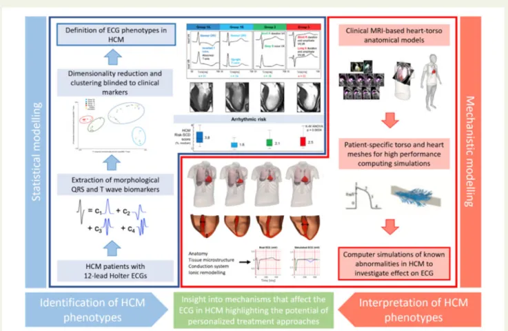
For example, model synergies represent an exciting approach to interpret structure–function relationships and improve risk predic-tion in inherited disease condipredic-tions, such as hypertrophic cardiomy-opathy (HCM). Relationships among specific ECG changes, ventricle morphologies, and sudden cardiac death have been inferred from observations.43,44 However, the complex process of translating underlying heterogeneous substrates in HCM to ECG findings is still poorly understood, and there exists a ‘grey zone’ of clinical decision-making in the low-risk patient subgroups, specifically when deciding on restriction of involvement in professional sports.45In this context, by using methods of statistical inference and mathematical modelling (see Figure4), HCM patients were categorized into phenogroups based on ECG biomarkers extracted from 24-h ECG recordings,48 and the aetiology of each ECG phenogroup linked with different underlying substrates, suggesting ion-channel and conduction system abnormalities.46,47The results directly highlighted the potential of personalized anti-arrhythmic approaches in the treatment of HCM patients, and addressed the low-risk patients, showing that a normal ECG might indeed be the discriminatory factor signalling minimal ionic remodelling, fibrosis, disarray, and ischaemia in these ‘grey zone’ patients.
Models have also been used in the prediction of arrhythmic events in post-myocardial infarction, outperforming existing clinical metrics including ejection fraction.49When the amount of data is not suffi-cient to inform state-of-the-art machine learning methods, statistical methods can still prove useful. An example is the use of principal component analysis to account for right ventricular motion in pre-dicting survival in pulmonary hypertension,50or to identify signatures of anatomical remodelling that predict a patient’s prognosis following CRT implantation.51
While statistical models allow predictions, mechanistic models pro-vide the underlying explanations. Understanding the actual meaning of the selected features improves the plausibility of findings and increases their credibility. For both approaches, quantifying uncertainty of pre-diction can help identify cases that may require further review, while building trust in cases where models are shown to be robust.52,53
Mechanistic and statistical model
synergy to accelerate evidence
generation
While digital twin technologies in cardiology show promising re-search results, only a small number of models have reached clinic-al translation. The difficulties encountered include the need to increase validation, lack of clinical interpretability, and potentially obscure model failures.54Therefore, solid evidence for the gener-alization of preliminary findings and efficient testing strategies are needed. Even when these barriers are overcome, rigid assessment of algorithmic performance and quality control from regulatory bodies can slow down the adoption. In this context, model syn-ergy can be used to accelerate the integration of novel technolo-gies into clinical practice by increasing clinical interpretability, validating generality of findings, and accelerating regulatory deci-sion-making.
Model validation towards generality of
findings
The goal after validating an initial concept is to extend it to a more general patient cohort, with less controlled characteristics. The prob-lem of sampling bias, based on both intrinsic (physiological) and
ex-trinsic (environmental) demographic heterogeneity of the
population, becomes relevant when implementing solutions for broader patient cohorts.55,56Consequently, models (as clinical guide-lines) may need recalibrations when used on populations from differ-ent countries or ethnicities, or even from differdiffer-ent cdiffer-entres in the same country. In recent years, only 6% of artificial intelligence algo-rithms had external evaluation performed (note this is beyond the minimum requirement of using the learning, validation, and testing partitions of the data), and none adopted the three design criteria of a robust validation: diagnostic cohort design, the inclusion of multiple institutions, and prospective data collection.57The quality of datasets also needs to be thoroughly validated to avoid possible biases before the models developed from them can be integrated in clinical deci-sion-making.58
To address this issue, an increasing number of institutions are cre-ating initiatives for data-sharing platforms, aiming at reusing existing
Figure 2Conceptual summary of the main benefits of digital twin technologies.
Figure 3Envisioned clinical workflow using the fully developed digital twin concept. Population data, collected from preceding patients and study cohorts, are used to create and validate statistical and mechanistic models, as well as to create a population-based digital twin (green). Novel patient data are analysed with the help of the existing models and integrated to form the patient’s digital twin (purple). The comparison and interaction be-tween digital twins give valuable insight (phenotyping, risk assessment, prediction of disease development. . .) that is clinically interpreted and com-bined with traditional data to aid in the process of clinical decision-making. The digital twin develops in line with the patient’s condition—adjusting and improving in accordance with the follow-up data. Resulting outcomes are supplemented to shape population data and refine the follow-up data.
Figure 4Synergy between mechanistic and statistical models in the definition of electrocardiogram (ECG) biomarkers for the management of hypertrophic cardiomyopathy.47,48
4560
J. Corral-Acero et al...
..
..
..
..
..
..
..
..
..
..
..
..
..
..
..
..
..
..
..
..
..
..
..
..
..
..
..
..
..
..
..
..
..
..
..
..
..
..
..
..
..
..
..
..
..
..
..
..
..
..
..
..
..
..
..
..
..
..
..
..
..
..
..
..
..
..
..
..
..
..
..
..
..
..
..
..
..
..
..
..
..
..
..
..
..
.
datasets and verifying published research works.59 Governments, regulatory agencies, and philanthropic funders are promoting the open science culture, enforcing publishing patient-level data by means of compliance to product launching, funding application, and journal publishing.60Another approach to improve the generality of data is the gener-ation of synthetic cases of a representative wider populgener-ation. The core idea is to expand the average mechanistic model to obtain pop-ulations of models, all of them parameterized within the range of physiological variability obtained by experimental protocols.61,62 Such an approach, which allows investigating many more scenarios than possible experimental acquisitions, is not only able to evaluate the impact of physiological variability but to explain the mechanisms underpinning inter-individual variability in therapy response (e.g. ad-verse drug reactions), and to identify sub-populations at higher risk.63,64Statistical shape modelling techniques can represent inter-patient anatomical variability for a cohort, and be used in combination of mechanistic models for clinical decision support systems.65
As in traditional scientific research, mechanistic and statistical mod-els are complementary tools to verify the findings derived from one another. Finding a mechanistic explanation of an inductive inference from statistical models increases its plausibility, such as the redistribu-tion of work in the left bundle branch block to explain the remodel-ling pattern that predicts response to CRT.51 Equivalently, data computed from mechanistic models need to be scrutinized quantitatively as it was done in the comparison of clinical and simula-tion groups to validate a model for acute normovolaemic haemodilution.66
An important final remark is that randomized control trials will al-ways be needed to establish evidence that can never be obtained from large observational databases.67
Models as critical tools for accelerating
regulatory decision-making
Clinical decisions are built on evidence from bench to bedside. Regulatory decisions, on the contrary, are often based on heteroge-neous, limited, or completely absent human data, as in the case of ap-proval for first-in-human clinical trials. In this regard, the results of computational models can now be accepted for some regulatory submissions.68,69 Digital evidence obtained using computer simula-tions can be used for safety of therapy prior to first-in-human use, or under scenarios not ethically possible in human.38 Computational models have an increasingly important role in the overall product life cycle management, proving useful in the processes of design opti-mization for development and testing, supplemental non-clinical test-ing, and post-market design changes and failure assessment.27
The development process for medical devices involves manufac-turing and testing samples under a wide range of scenarios, which is often time-consuming and financially overwhelming. Moreover, pre-clinical testing conditions are often very simplified with respect to the actual patient environment. Statistical and mechanistic models syner-gistically offer to streamline this process, where statistical models can be used to collect a representative virtual patient cohort, and mech-anistic models can then be used to simulate the device behaviour under defined scenarios. In this way, new devices can be tested in a representative virtual patient population, thereby decreasing the
risk before moving to an actual clinical trial. An example is HEARTguideTM(FEops nv, Belgium), where device–patient interac-tions after transcatheter aortic valve implantation can be predicted.25 The augmentation of clinical trial design with virtual patients is also an evolving idea.70–72This would overcome limitations of current empirical trials, where patients burdened with comorbidities or com-plex treatment regimens are often excluded from the trials, and enrolled individuals are handled under reductionist approaches, assuming they share a common phenotype. Such approaches often fail to capture differences in response to treatment.70Alternatively, computational evidence can inform collection of novel evidence from clinical trials,13,38where models can improve patient selection by derived biomarkers and predictions. This offers an opportunity to answer questions traditionally restricted by financial or ethical con-siderations, and to investigate therapy efficacy in more clinically rele-vant cases. Computational modelling can also facilitate safe methods to explore treatment effects in sub-populations clinically more com-plex to address, such as patients with rare diseases or paediatric cohorts, and therefore may allow for insights not possible in the cur-rent clinical trial practice.
One of the first examples in which digital evidence (i.e. an in silico trial) replaced any additional clinical evidence was in the approval of the Advisa MRI SureScan pacemaker (Medtronic, Inc.).73 Another powerful example is a computer simulator of type 1 diabetes melli-tus,74which was accepted by the FDA as a substitute to animal trials for the pre-clinical testing of control strategies in artificial pancreas studies. Later, an investigational device exemption (i.e. the approval needed to initiate a clinical study), issued solely on the basis of model-ling testing, was granted by the FDA for a closed-loop control clinical trial of the safety and effectiveness of the proposed artificial pancreas algorithm.
In the context of drug safety and efficacy assessment, an unmet need is filling the gaps between animal translation or in vitro prepara-tions and prediction of the human response. Mechanistic models may assist in scaling observations into humans.75This is, for example, the goal of the CIPA initiative,69sponsored by the FDA among others, aiming at facilitating the adoption of a new paradigm for assessment of potential risk of clinical Torsades de Pointes, where mechanistic models of human electrophysiology will play a crucial role. This is reinforced by a recent study in which human in silico trials outper-formed animal models in predicting clinical pro-arrhythmic cardio-toxicity, so they might be soon integrated into existing drug safety assessment pipelines.63
Finally, after a product is launched, mechanistic models can be still used for post-market re-evaluation and failure assessment in order to identify any potential underlying problems. This creates a valuable op-portunity for simulations to evaluate any design changes planned for next-generation productions, ultimately closing the product life cycle loop, and demonstrating the ubiquitous presence and utility of statis-tical and mechanistic models in the future of medical product regulation.
Discussion
The digital twin, i.e., the dynamic integration and augmentation of pa-tient data using mechanistic and statistical models, is the actual
..
..
..
..
..
..
..
..
..
..
..
..
..
..
..
..
..
..
..
..
..
..
..
..
..
..
..
..
..
..
..
..
..
..
..
..
..
..
..
..
..
..
..
..
..
..
..
..
..
..
..
.
pathway towards the vision of precision medicine. Simple and frag-mented components of the digital twin are already used in clinical practice: a decision tree in a clinical guideline encapsulates the best-documented evidence that is based in statistical and mechanistic insights. The digital twin will gradually include tailored computer-enabled decision points, and create the transition from healthcare systems founded on describing disease to healthcare systems focused on predicting response, and thus shifting treatment selection from being based on the state of the patient today to optimizing the state of the patient tomorrow.Envisioned impact and timeline
The digital twin provides a pathway to map current patient observa-tions into a predictive framework, combining inductive and deductive reasoning. Early components of the digital twin are already making a clinical impact. In a generic clinical workflow divided in the stages of data acquisition, diagnosis, and therapy planning, computational mod-els can provide value in the three stages, see Figure5. To improve data acquisition techniques, there are already statistical models to automate the image analysis tasks.16To provide better diagnosis, a virtual fractional flow reserve can replace an invasive catheter,29,37or the body surface recordings can be mapped to the surface of the heart.39With regards to therapy planning, a virtual deployment of the valve replacement25,76or a roadmap to guide ablation proce-dures77,78represents existing techniques (statistical and mechanistic) that have been implemented into the clinical workflow. These solu-tions have thus met regulatory approval, where they are referred to as ‘software as a medical device’, and where guidelines from the International Medical Device Regulators Forum are accepted by the EU and the USA.
A digital twin will follow the life journey of each person and har-ness both data collected by wearable sensors and lifestyle informa-tion that patients may register, shifting the clinical approach towards preventive healthcare. A notable challenge is the integration of these
data with healthcare organizations, where security and confidentiality of the sensitive information remain paramount.
The currently still fragmented and incipient concept of the digital twin will be gradually crystallized and adopted during the next 5–10 years. The holistic integration of a Digital Twin is the aspiration that will be reached through two complementary and synergetic pathways: the first is the refinement of key deci-sion points in the management of cardiac disease, driven by per-sonalized mechanistic models that are informed by key pieces of patient’s data; and the second is the disease-centred optimization of the patient’s lifetime journey through the healthcare system, driven by statistical models being informed by the electronic health record of a large population.
On the actual implementation of the digital twin, we envision that the evolution will be towards a gradually better inter-operability of current health information systems, leading to a distributed location of the information. Digital twin users will mainly be citizens and physi-cians, with different interfaces that retrieve the relevant data and trig-ger the analysis capabilities hosted in the local device or remote cloud resources. The analysis may also require specialized skills that may be delivered by industry, or even by computational cardiologists in-side healthcare organizations.
Organizational and societal challenges
ahead
Access to data is the main challenge in both the development and the clinical translation of the digital twin, caused by infrastructural, regula-tory, and societal reasons. Information systems and electronic health records are fragmented, highly heterogeneous and difficult to inter-operate. Information is often contained in unstructured format, and its extraction requires either manual work or further research efforts of automation through natural language processing technologies.79 Simulations may also require specialized skills and supercomputers.
Figure 5The vision of a personalized in silico cardiology, where the digital twin informs all the stages through the clinical workflow. Models are used (i) to optimize data acquisition and the information extracted from it, (ii) to evaluate current health status and inform diagnosis and risk stratifica-tion, and (iii) to optimize clinical devices and drug selection to deliver a personalized therapy.
4562
J. Corral-Acero et al...
..
..
..
..
..
..
..
..
..
..
..
..
..
..
..
..
..
..
..
..
..
..
..
..
..
..
..
..
..
..
..
..
..
..
..
..
..
..
..
..
..
..
..
..
..
..
..
In this context, provision of digital twin technologies may be enabled by cloud infrastructures (e.g. HeartFlow FFRCTAnalysis).Consent and confidentiality are key ingredients to address the so-cietal concerns when handling the personal data needed to develop and validate digital twin technologies. The EU General Data Protection Regulation (GDPR) has imposed new legal requirements, such as the right to withdraw consent and the right to be forgotten, causing controversy about the cost and feasibility of its enforce-ment.80Any digital twin solution that holds enough information to identify a patient needs to carefully watch these requirements, that also apply to retrospective data and safety backups.
Potential professional, cultural, and
ethical issues
As more clinical tasks are performed by models, the fear of replace-ment of physicians by machines may arise. In some scenarios, machines may match or even outperform physicians.81In other scen-arios, human experts, by not practising on the easy problems solved by the machine, may lose the skills that may still be needed when dealing with difficult cases.
The second professional barrier is the mistrust that originates from a ‘black box’, where predictions derived by algorithms are not matched with a plausible explanation. Generation of evidence is one clear way to generate trust. Another solution is to use methods to il-lustrate the logic inside the box, including clustering and association techniques,82which may help to identify the causes and mechanisms.
From the patient’s perspective, personalization creates the oppor-tunity of more involvement in healthcare decisions. Patients will be empowered to better manage their disease using the digital twin to gain information about their current and predicted state, and poten-tially to adopt optimized lifestyle suggestions. A well-informed patient
shall have more efficient discussions with physicians, and consent and decide faster on diagnostic or treatment procedures.
Finally, on the ethical side, there is a risk of models to create or ex-acerbate existing racial or societal biases in healthcare systems: if a group is misrepresented in the data used to train models, that group may receive a sub-optimal treatment.83
Recommendations
The pathway to accelerate the clinical impact with digital twin tech-nologies is to generate trust among researchers, clinicians, and society.
Research communities shall avoid inflating expectations. Claims about generality and potential impact should be based on rigorous methodology, with external cohorts to demonstrate the validity of inferences, and with the quantification of the un-certainty of predictions.84Any model is a simplified representa-tion of the reality, with a limited scope and dependence on assumptions made. The opportunity is an adequate handling of these limitations, with models able to identify data inconsisten-cies, and with data used to constrain and verify the model assumptions.85
As an emerging field, the digital twin needs guidelines, gold-stand-ards, and benchmark tests.86,87Scientific organizations and regulatory bodies have released guidelines that can be used to establish the level of rigour needed for computational modelling.27Such guidelines and standards are useful tools as they allow regulators to judge computa-tional evidence and industry to understand regulatory requirements for computational models, leveraging a substantial part of the risk and uncertainty associated to the development of these new technolo-gies. They can even increase and facilitate their translational impact, as the quality and robustness of the models and their reporting will
Take home figureThe cardiovascular digital twin that will deliver the vision of precision medicine by the synergetic combination of computer-enhanced induction (using statistical models learnt from data) and deduction (mechanistic modelling and simulation integrating multi-scale knowledge).
..
..
..
..
..
..
..
..
..
..
..
..
..
..
..
..
..
..
..
..
..
..
..
..
..
..
..
..
..
..
..
..
..
..
..
..
..
..
..
..
..
..
..
..
..
..
..
..
..
..
..
..
..
..
..
..
..
..
..
..
..
..
..
..
..
..
..
..
..
..
..
..
..
..
..
..
..
..
..
..
..
..
..
..
..
.
increase by adhering to such guidance during model development. Further effort is needed to widen the scope of these first multi-stakeholder consensuses involving industry, academia, and reg-ulators. Current initiatives that develop visions, technologies, or infra-structure relevant to the ‘digital twin’ community are Elixir (https:// elixir-europe.org/), FAIRDOM (https://fair-dom.org/), and EOSC (https://ec.europa.eu/research/openscience/index.cfm? pg=open-sci ence-cloud).The education of citizens, care providers, physicians, and research-ers in the uses and possibilities of digital twin technologies is key for its adoption and acceptance. University education systems should also allow for the exchange of knowledge at the earliest stages of the career: medical students should have some computational training, just as engineers in biomedical industry should be trained in cardi-ology during their studies.88And postgraduate training programmes should bridge remaining cultural and language gaps between disci-plines, such as our Personalised In-silico Cardiology EU funded Innovative Training Network (https://picnet.eu).
Conclusion
Precision cardiology will be delivered, not only by data, but also by the inductive and deductive reasoning built in the digital twin of each patient. Treatment and prevention of cardiovascular disease will be based on accurate predictions of both the underlying causes of dis-ease and the pathways to sustain or restore health. These predictions will be provided and validated by the synergistic interplay between mechanistic and statistical models. The early steps towards this vision have been taken, and the next ones depend on the coordinated drive from scientific, clinical, industrial, and regulatory stakeholders in order to build the evidence and tackle the organizational and societal challenges ahead.
Supplementary material
Supplementary materialis available at European Heart Journal online.
Conflict of interest: none declared.
Funding
This work was supported by the EU’s Horizon 2020 Marie Sklodowska-Curie ITN Projects (g.a. 764738 and 766082), the EU’s Horizon 2020 re-search and innovation programme (g.a. 675451 and 823712), the Wellcome/EPSRC Centre for Medical Engineering (WT 203148/Z/16/Z), the National Research Agency (ANR) (g.a. ANR-10-IAHU-04), the NC3RS (NC/P001076/1) and the British Heart Foundation (RE/13/2/ 30182, RE/13/1/30181, TG/17/3/33406, PG/16/75/32383, FS/17/22/ 32644, CH/16/3/21406, RG/16/14/32397). E.Pueyo holds an ERC Starting Grant (g.a. 638284). B. Rodriguez and P.Lamata hold Wellcome Trust Senior Research Fellowships (214290/Z/18/Z, 209450/Z/17/Z).
References
1. Antman EM, Loscalzo J. Precision medicine in cardiology. Nat Rev Cardiol 2016; 13:591–602.
2. Trayanova N. From genetics to smart watches: developments in precision cardi-ology. Nat Rev Cardiol 2019;16:72–73.
3. Collins FS, Varmus H. A new initiative on precision medicine. N Engl J Med 2015; 372:793–795.
4. Joyner MJ, Paneth N. Promises, promises, and precision medicine. J Clin Invest 2019;129:946–948.
5. Khoury MJ. Precision medicine vs preventive medicine. JAMA 2019;321:406. 6. Noble D. Evolution beyond neo-Darwinism: a new conceptual framework. J Exp
Biol 2015;218:7–13.
7. Alber M, Buganza Tepole A, Cannon WR, De S, Dura-Bernal S, Garikipati K, Karniadakis G, Lytton WW, Perdikaris P, Petzold L, Kuhl E. Integrating machine learning and multiscale modeling—perspectives, challenges, and opportunities in the biological, biomedical, and behavioral sciences. NPJ Digit Med 2019;2:115. 8. Tao F, Cheng J, Qi Q, Zhang M, Zhang H, Sui F. Digital twin-driven product
de-sign, manufacturing and service with big data. Int J Adv Manuf Technol 2018;94: 3563–3576.
9. Lamata P. Teaching cardiovascular medicine to machines. Cardiovasc Res 2018; 114:e62–e64.
10. Niederer SA, Plank G, Chinchapatnam P, Ginks M, Lamata P, Rhode KS, Rinaldi CA, Razavi R, Smith NP. Length-dependent tension in the failing heart and the ef-ficacy of cardiac resynchronization therapy. Cardiovasc Res 2011;89:336–343. 11. Lumens J, Tayal B, Walmsley J, Delgado-Montero A, Huntjens PR, Schwartzman
D, Althouse AD, Delhaas T, Prinzen FW, Gorcsan J. Differentiating electromech-anical from non-electrical substrates of mechelectromech-anical discoordination to identify responders to cardiac resynchronization therapy. Circ Cardiovasc Imaging 2015;8: e003744.
12. Corral Acero J, Zacur E, Xu H, Ariga R, Bueno-Orovio A, Lamata P, Grau V. SMOD - Data Augmentation Based on Statistical Models of Deformation to Enhance Segmentation in 2D Cine Cardiac MRI. FIMH 2019: Functional Imaging and Modeling of the Heart - pp. 361–369. doi: 10.1007/978-3-030-21949-9_39. 13. Cikes M, Sanchez Martinez S, Claggett B, Solomon SD, Bijnens B.
Machine-learn-ing integration of complex echocardiographic patterns and clinical parameters from cohorts and trials. Eur Heart J 2019;40: doi: 10.1093/eurheartj/ehz745.0147. 14. Shameer K, Johnson KW, Glicksberg BS, Dudley JT, Sengupta PP. Machine learning in cardiovascular medicine: are we there yet? Heart 2018;104:1156–1164. 15. Rumsfeld JS, Joynt KE, Maddox TM. Big data analytics to improve cardiovascular
care: promise and challenges. Nat Rev Cardiol 2016;13:350–359.
16. Dey D, Slomka PJ, Leeson P, Comaniciu D, Shrestha S, Sengupta PP, Marwick TH. Artificial intelligence in cardiovascular imaging: JACC state-of-the-art review. J Am Coll Cardiol 2019;73:1317–1335.
17. Niederer SA, Lumens J, Trayanova NA. Computational models in cardiology. Nat Rev Cardiol 2019;16:100–111.
18. Johnson KW, Shameer K, Glicksberg BS, Readhead B, Sengupta PP, Bjo¨rkegren JLM, Kovacic JC, Dudley JT. Enabling precision cardiology through multiscale biol-ogy and systems medicine. JACC Basic Transl Sci 2017;2:311–327.
19. Davies MR, Wang K, Mirams GR, Caruso A, Noble D, Walz A, Lave´ T, Schuler F, Singer T, Polonchuk L. Recent developments in using mechanistic cardiac model-ling for drug safety evaluation. Drug Discov Today 2016;21:924–938.
20. Tung L. A bi-domain model for describing ischemic myocardial D-C potentials. 1978. https://dspace.mit.edu/handle/1721.1/16177(29 February 2020).
21. Sherwin SJ, Formaggia L, Peiro´ J, Franke V. Computational modelling of 1D blood flow with variable mechanical properties and its application to the simulation of wave propagation in the human arterial system. Int J Numer Methods Fluids 2003; 43:673–700.
22. Guidi G, Pettenati MC, Miniati R, Iadanza E. Random forest for automatic assess-ment of heart failure severity in a telemonitoring scenario. In: 2013 35th Annual International Conference of the IEEE Engineering in Medicine and Biology Society (EMBC). IEEE, pp. 3230–3233.
23. Stegle O, Fallert SV, MacKay DJ, Brage S. Gaussian process robust regression for noisy heart rate data. IEEE Trans Biomed Eng 2008;55:2143–2151.
24. Roney CH, Bayer JD, Cochet H, Meo M, Dubois R, Jaı¨s P, Vigmond EJ. Variability in pulmonary vein electrophysiology and fibrosis determines arrhythmia suscepti-bility and dynamics. PLoS Comput Biol 2018;14:e1006166.
25. de Jaegere P, De Santis G, Rodriguez-Olivares R, Bosmans J, Bruining N, Dezutter T, Rahhab Z, El Faquir N, Collas V, Bosmans B, Verhegghe B, Ren C, Geleinse M, Schultz C, van Mieghem N, De Beule M, Mortier P. Patient-specific computer modeling to predict aortic regurgitation after transcatheter aortic valve replacement. JACC Cardiovasc Interv 2016;9:508–512.
26. Gray RA, Pathmanathan P. Patient-specific cardiovascular computational model-ing: diversity of personalization and challenges. J Cardiovasc Transl Res 2018;11: 80–88.
27. Morrison TM, Dreher ML, Nagaraja S, Angelone LM, Kainz W. The role of com-putational modeling and simulation in the total product life cycle of peripheral vascular devices. J Med Device 2017;11:024503.
28. Dillon-Murphy D, Noorani A, Nordsletten D, Figueroa CA. Multi-modality image-based computational analysis of haemodynamics in aortic dissection. Biomech Model Mechanobiol 2016;15:857–876.
29. Morris PD, van de Vosse FN, Lawford PV, Hose DR, Gunn JP. “Virtual” (com-puted) fractional flow reserve. JACC Cardiovasc Interv 2015;8:1009–1017.
4564
J. Corral-Acero et al...
..
..
..
..
..
..
..
..
..
..
..
..
..
..
..
..
..
..
..
..
..
..
..
..
..
..
..
..
..
..
..
..
..
..
..
..
..
..
..
..
..
..
..
..
..
..
..
..
..
..
..
..
..
..
..
..
..
..
..
..
..
..
..
..
..
..
..
..
..
..
..
..
..
..
..
..
..
..
..
..
..
..
..
..
.
30. Xi J, Lamata P, Niederer S, Land S, Shi W, Zhuang X, Ourselin S, Duckett SG,Shetty AK, Rinaldi CA, Rueckert D, Razavi R, Smith NP. The estimation of patient-specific cardiac diastolic functions from clinical measurements. Med Image Anal 2013;17:133–146.
31. Wang ZJ, Wang VY, Bradley CP, Nash MP, Young AA, Cao JJ. Left ventricular diastolic myocardial stiffness and end-diastolic myofibre stress in human heart failure using personalised biomechanical analysis. J Cardiovasc Transl Res 2018;11: 346–356.
32. Krittian SBS, Lamata P, Michler C, Nordsletten DA, Bock J, Bradley CP, Pitcher A, Kilner PJ, Markl M, Smith NP. A finite-element approach to the direct compu-tation of relative cardiovascular pressure from time-resolved MR velocity data. Med Image Anal 2012;16:1029–1037.
33. Donati F, Figueroa CA, Smith NP, Lamata P, Nordsletten DA. Non-invasive pres-sure difference estimation from PC-MRI using the work-energy equation. Med Image Anal 2015;26:159–172.
34. Donati F, Myerson S, Bissell MM, Smith NP, Neubauer S, Monaghan MJ, Nordsletten DA, Lamata P. Beyond Bernoulli: improving the accuracy and preci-sion of non-invasive estimation of peak pressure drops. Circ Cardiovasc Imaging 2017;10:e005207.
35. Nørgaard BL, Leipsic J, Gaur S, Seneviratne S, Ko BS, Ito H, Jensen JM, Mauri L, De Bruyne B, Bezerra H, Osawa K, Marwan M, Naber C, Erglis A, Park S-J, Christiansen EH, Kaltoft A, Lassen JF, Bøtker HE, Achenbach S. Diagnostic per-formance of noninvasive fractional flow reserve derived from coronary com-puted tomography angiography in suspected coronary artery disease. J Am Coll Cardiol 2014;63:1145–1155.
36. Min JK, Taylor CA, Achenbach S, Koo BK, Leipsic J, Nørgaard BL, Pijls NJ, De Bruyne B. Noninvasive fractional flow reserve derived from coronary CT angiog-raphy. JACC Cardiovasc Imaging 2015;8:1209–1222.
37. Rajani R, Modi B, Ntalas I, Curzen N. Non-invasive fractional flow reserve using computed tomographic angiography: where are we now and where are we going? Heart 2017;103:1216–1222.
38. Morrison TM, Pathmanathan P, Adwan M, Margerrison E. Advancing regulatory science with computational modeling for medical devices at the FDA’s Office of Science and Engineering Laboratories. Front Med 2018;5: doi: 10.3389/ fmed.2018.00241.
39. Haissaguerre M, Hocini M, Shah AJ, Derval N, Sacher F, Jais P, Dubois R. Noninvasive panoramic mapping of human atrial fibrillation mechanisms: a feasi-bility report noninvasive panoramic mapping of human atrial fibrillation mecha-nisms. Introduction. J Cardiovasc Electrophysiol 2013;24:711–717.
40. Daubert C, Behar N, Martins RP, Mabo P, Leclercq C,. Avoiding non-responders to cardiac resynchronization therapy: a practical guide. Eur Heart J 2016;38: 1463–1472.
41. Prakosa A, Arevalo HJ, Deng D, Boyle PM, Nikolov PP, Ashikaga H, Blauer JJE, Ghafoori E, Park CJ, Blake RC, Han FT, MacLeod RS, Halperin HR, Callans DJ, Ranjan R, Chrispin J, Nazarian S, Trayanova NA. Personalized virtual-heart tech-nology for guiding the ablation of infarct-related ventricular tachycardia. Nat Biomed Eng 2018;2:732–740.
42. Hill YR, Child N, Hanson B, Wallman M, Coronel R, Plank G, Rinaldi CA, Gill J, Smith NP, Taggart P, Bishop MJ. Investigating a novel activation-repolarisation time metric to predict localised Vulnerability to reentry using computational modelling. PLoS One 2016;11: doi: 10.1371/journal.pone.0149342.
43. Alfonso F, Nihoyannopoulos P, Stewart J, Dickie S, Lemery R, McKenna WJ. Clinical significance of giant negative T waves in hypertrophic cardiomyopathy. J Am Coll Cardiol 1990;15:965–971.
44. Pelliccia A, Di Paolo FM, Quattrini FM, Basso C, Culasso F, Popoli G, De Luca R, Spataro A, Biffi A, Thiene G, Maron BJ. Outcomes in athletes with marked ECG repolarization abnormalities. N Engl J Med 2008;358:152–161.
45. Pelliccia A, Corrado D, Bjørnstad HH, Panhuyzen-Goedkoop N, Urhausen A, Carre F, Anastasakis A, Vanhees L, Arbustini E, Priori S. Recommendations for participation in competitive sport and leisure-time physical activity in individuals with cardiomyopathies, myocarditis and pericarditis. Eur J Prev Cardiol 2006;13: 876–885.
46. Passini E, Minchole´ A, Coppini R, Cerbai E, Rodriguez B, Severi S, Bueno-Orovio A. Mechanisms of pro-arrhythmic abnormalities in ventricular repolarisation and anti-arrhythmic therapies in human hypertrophic cardiomyopathy. J Mol Cell Cardiol 2016;96:72–81.
47. Lyon A, Bueno-Orovio A, Zacur E, Ariga R, Grau V, Neubauer S, Watkins H, Rodriguez B, Minchole´ A. Electrocardiogram phenotypes in hypertrophic cardio-myopathy caused by distinct mechanisms: apico-basal repolarization gradients vs. Purkinje-myocardial coupling abnormalities. Europace 2018;20: III102–III112.
48. Lyon A, Ariga R, Minchole´ A, Mahmod M, Ormondroyd E, Laguna P, de Freitas N, Neubauer S, Watkins H, Rodriguez B. Distinct ECG phenotypes identified in hypertrophic cardiomyopathy using machine learning associate with arrhythmic risk markers. Front Physiol 2018;9:213.
49. Arevalo HJ, Vadakkumpadan F, Guallar E, Jebb A, Malamas P, Wu KC, Trayanova NA. Arrhythmia risk stratification of patients after myocardial infarction using personalized heart models. Nat Commun 2016;7: doi: 10.1038/ncomms11437. 50. Dawes TJW, de Marvao A, Shi W, Fletcher T, Watson GMJ, Wharton J, Rhodes
CJ, Howard LSGE, Gibbs JSR, Rueckert D, Cook SA, Wilkins MR, O’Regan DP. Machine learning of three-dimensional right ventricular motion enables outcome prediction in pulmonary hypertension: a cardiac MR imaging study. Radiology 2017;283:381–390.
51. Warriner DR, Jackson T, Zacur E, Sammut E, Sheridan P, Hose DR, Lawford P, Razavi R, Niederer SA, Rinaldi CA, Lamata P. An asymmetric wall-thickening pat-tern predicts response to cardiac resynchronization therapy. JACC Cardiovasc Imaging 2018;11:1545–1546.
52. Mirams GR, Pathmanathan P, Gray RA, Challenor P, Clayton RH. Uncertainty and variability in computational and mathematical models of cardiac physiology. J Physiol 2016;594:6833–6847.
53. Pathmanathan P, Gray RA. Ensuring reliability of safety-critical clinical applications of computational cardiac models. Front Physiol 2013;4:358.
54. Winslow RL, Trayanova N, Geman D, Miller MI. Computational medicine: trans-lating models to clinical care. Sci Transl Med 2012;4:158rv11–158rv11. 55. Kurokawa J, Kodama M, Clancy CE, Furukawa T. Sex hormonal regulation of
car-diac ion channels in drug-induced QT syndromes. Pharmacol Ther 2016;168: 23–28.
56. Niemeijer MN, van den Berg ME, Deckers JW, Aarnoudse ALHJ, Hofman A, Franco OH, Uitterlinden AG, Rijnbeek PR, Eijgelsheim M, Stricker BH. ABCB1 gene variants, digoxin and risk of sudden cardiac death in a general population. Heart 2015;101:1973–1979.
57. Kim DW, Jang HY, Kim KW, Shin Y, Park SH. Design characteristics of studies reporting the performance of artificial intelligence algorithms for diagnostic ana-lysis of medical images: results from recently published papers. Korean J Radiol 2019;20:405.
58. Chang KC, Dutta S, Mirams GR, Beattie KA, Sheng J, Tran PN, Wu M, Wu WW, Colatsky T, Strauss DG, Li Z. Uncertainty quantification reveals the importance of data variability and experimental design considerations for in silico proarrhyth-mia risk assessment. Front Physiol 2017;8:917.
59. Dey P, Ross JS, Ritchie JD, Desai NR, Bhavnani SP, Krumholz HM. Data sharing and cardiology. J Am Coll Cardiol 2017;70:3018–3025.
60. Schiltz M. Science without publication paywalls: cOAlition S for the realisation of full and immediate open access. PLoS Med 2018;15:e1002663.
61. Britton OJ, Bueno-Orovio A, Van Ammel K, Lu HR, Towart R, Gallacher DJ, Rodriguez B. Experimentally calibrated population of models predicts and explains intersubject variability in cardiac cellular electrophysiology. Proc Natl Acad Sci USA 2013;110:E2098–E2105.
62. Sa´nchez C, Bueno-Orovio A, Wettwer E, Loose S, Simon J, Ravens U, Pueyo E, Rodriguez B. Inter-subject variability in human atrial action potential in sinus rhythm versus chronic atrial fibrillation. PLoS One 2014;9:e105897.
63. Passini E, Britton OJ, Lu HR, Rohrbacher J, Hermans AN, Gallacher DJ, Greig RJH, Bueno-Orovio A, Rodriguez. Human in silico drug trials demonstrate higher accuracy than animal models in predicting clinical pro-arrhythmic cardiotoxicity. Front Physiol 2017;8: doi: 10.3389/fphys.2017.00668.
64. Sa´nchez C, Bueno-Orovio A, Pueyo E, Rodrı´guez B. Atrial fibrillation dynamics and ionic block effects in six heterogeneous human 3D virtual atria with distinct repolarization dynamics. Front Bioeng Biotechnol 2017;5:29.
65. Liang L, Liu M, Martin C, Elefteriades JA, Sun W. A machine learning approach to investigate the relationship between shape features and numerically predicted risk of ascending aortic aneurysm. Biomech Model Mechanobiol 2017;16: 1519–1533.
66. Sims CR, Delima LR, Calimaran A, Hester R, Pruett WA. Validating the physio-logic model HumMod as a substitute for clinical trials involving acute normovole-mic hemodilution. Anesth Analg 2018;126:93–101.
67. Herna´n MA, Robins JM. Using big data to emulate a target trial when a random-ized trial is not available: table 1. Am J Epidemiol 2016;183:758–764.
68. Colatsky T, Fermini B, Gintant G, Pierson JB, Sager P, Sekino Y, Strauss DG, Stockbridge N. The Comprehensive in Vitro Proarrhythmia Assay (CiPA) initia-tive—update on progress. J Pharmacol Toxicol Methods 2016;81:15–20. 69. Cavero I, Holzgrefe H. CiPA: ongoing testing, future qualification procedures,
and pending issues. J Pharmacol Toxicol Methods 2015;76:27–37.
70. Pappalardo F, Russo G, Tshinanu FM, Viceconti M. In silico clinical trials: concepts and early adoptions. Brief Bioinform 2019:20:1699–1708.
71. Viceconti M, Henney A, Morley-Fletcher E. In silico clinical trials: how computer simulation will transform the biomedical industry. Int J Clin Trials 2016; 3:37.
72. Haddad T, Himes A, Thompson L, Irony T, Nair R; MDIC Computer Modeling and Simulation Working Group Participants. Incorporation of stochastic engin-eering models as prior information in Bayesian medical device trials. J Biopharm Stat 2017;27:1089–1103.
..
..
..
..
..
..
..
..
..
..
..
..
..
..
..
..
..
..
..
..
..
..
..
..
..
..
..
..
..
..
..
..
..
..
73. Faris O, Shuren J. An FDA viewpoint on unique considerations formedical-device clinical trials. N Engl J Med 2017;376:1350–1357.
74. Kovatchev BP, Breton M, Man CD, Cobelli C. In silico preclinical trials: a proof of concept in closed-loop control of type 1 diabetes. J Diabetes Sci Technol 2009;3: 44–55.
75. Zemzemi N, Bernabeu MO, Saiz J, Cooper J, Pathmanathan P, Mirams GR, Pitt-Francis J, Rodriguez B. Computational assessment of drug-induced effects on the electrocardiogram: from ion channel to body surface potentials. Br J Pharmacol 2013;168:718–733.
76. Rocatello G, El Faquir N, De Santis G, Iannaccone F, Bosmans J, De Backer O, Sondergaard L, Segers P, De Beule M, de Jaegere P, Mortier P. Patient-specific computer simulation to elucidate the role of contact pressure in the develop-ment of new conduction abnormalities after catheter-based implantation of a self-expanding aortic valve. Circ Cardiovasc Interv 2018;11:e005344.
77. Andreu D, Ortiz-Pe´rez JT, Ferna´ndez-Armenta J, Guiu E, Acosta J, Prat-Gonza´lez S, De Caralt TM, Perea RJ, Garrido C, Mont L, Brugada J, Berruezo A. 3D delayed-enhanced magnetic resonance sequences improve conducting channel delineation prior to ventricular tachycardia ablation. Europace 2015;17:938–945. 78. Cedilnik N, Duchateau J, Dubois R, Sacher F, Jaı¨s P, Cochet H,Sermesant M. Fast
personalized electrophysiological models from computed tomography images for ventricular tachycardia ablation planning. Europace 2018;20:iii94–iii101. 79. Kreimeyer K, Foster M, Pandey A, Arya N, Halford G, Jones SF, Forshee R,
Walderhaug M, Botsis T. Natural language processing systems for capturing and standardizing unstructured clinical information: a systematic review. J Biomed Inform 2017;73:14–29.
80. Politou E, Alepis E, Patsakis C. Forgetting personal data and revoking consent under the GDPR: challenges and proposed solutions. J Cybersecurity 2018;4: doi: 10.1093/cybsec/tyy001.
81. Darcy AM, Louie AK, Roberts LW. Machine learning and the profession of medi-cine. JAMA 2016;315:551.
82. Esfandiari N, Babavalian MR, Moghadam A-ME, Tabar VK. Expert systems with applications knowledge discovery in medicine: current issue and future trend. Expert Syst Appl 2014;41:4434–4463.
83. Nordling L. A fairer way forward for AI in health care. Nature 2019;573: S103–S105.
84. Pathmanathan P, Cordeiro JM, Gray RA. Comprehensive uncertainty quantifica-tion and sensitivity analysis for cardiac acquantifica-tion potential models. Front Physiol 2019; 10:721.
85. Miotto R, Li L, Kidd BA, Dudley JT. Deep patient: an unsupervised representation to predict the future of patients from the electronic health records. Sci Rep 2016;6:26094.
86. Land S, Gurev V, Arens S, Augustin CM, Baron L, Blake R, Bradley C, Castro S, Crozier A, Favino M, Fastl TE, Fritz T, Gao H, Gizzi A, Griffith BE, Hurtado DE, Krause R, Luo X, Nash MP, Pezzuto S, Plank G, Rossi S, Ruprecht D, Seemann G, Smith NP, Sundnes J, Rice JJ, Trayanova N, Wang D, Jenny Wang Z, Niederer SA. Verification of car-diac mechanics software: benchmark problems and solutions for testing active and passive material behaviour. Proc R Soc A Math Phys Eng Sci 2015;471:20150641.
87. Niederer SA, Kerfoot E, Benson AP, Bernabeu MO, Bernus O, Bradley C, Cherry EM, Clayton R, Fenton FH, Garny A, Heidenreich E, Land S, Maleckar M, Pathmanathan P, Plank G, Rodrı´guez JF, Roy I, Sachse FB, Seemann G, Skavhaug O, Smith NP. Verification of cardiac tissue electrophysiology simulators using an N-version benchmark. Philos Trans R Soc A Math Phys Eng Sci 2011;369: 4331–4351.
88. Eden C, Johnson KW, Gottesman O, Bottinger EP, Abul-Husn NS. Medical stu-dent preparedness for an era of personalized medicine: findings from one US medical school. Per Med 2016;13:129–141.