HAL Id: hal-03025102
https://hal-amu.archives-ouvertes.fr/hal-03025102
Submitted on 17 Dec 2020HAL is a multi-disciplinary open access
archive for the deposit and dissemination of sci-entific research documents, whether they are pub-lished or not. The documents may come from teaching and research institutions in France or abroad, or from public or private research centers.
L’archive ouverte pluridisciplinaire HAL, est destinée au dépôt et à la diffusion de documents scientifiques de niveau recherche, publiés ou non, émanant des établissements d’enseignement et de recherche français ou étrangers, des laboratoires publics ou privés.
peritoneal macrophages
C. Bain, D. Gibson, N. Steers, K. Boufea, P. Louwe, C. Doherty, V.
González-Huici, R. Gentek, M. Magalhaes-Pinto, T. Shaw, et al.
To cite this version:
C. Bain, D. Gibson, N. Steers, K. Boufea, P. Louwe, et al.. Rate of replenishment and microen-vironment contribute to the sexually dimorphic phenotype and function of peritoneal macrophages. Science Immunology, American Association for the Advancement of Science, 2020, 5 (48), pp.eabc4466. �10.1126/sciimmunol.abc4466�. �hal-03025102�
Origin and microenvironment contribute to the sexually dimorphic
1
phenotype and function of peritoneal macrophages.
2
3
Calum C. Bain1*, Douglas A. Gibson1, Nicholas Steers2, Katarina Boufea3,Pieter A. Louwe1, Catherine
4
Docherty1, Victor Huici3, Rebecca Gentek4, Marlene Magalhaes-Pinto5, Marc Bajenoff4, Cecile
5
Benezech5, David Dockrell1, Philippa TK Saunders1, Nizar Batada3, Stephen J Jenkins1*
6
7
1 University of Edinburgh Centre for Inflammation Research, Queens Medical Research Institute,
8
Edinburgh, UK
9
2 Columbia University Medical Centre, Columbia University, New York, USA
10
3 Institute for Genetics and Molecular Medicine, University of Edinburgh, Edinburgh, EH4 2XU, UK
11
4 Centre d'Immunologie de Marseille-Luminy, Aix Marseille Université UM2, INSERM, U1104,
12
CNRS UMR7280, 13288 Marseille, France
13
5 Centre for Cardiovascular Sciences, University of Edinburgh, Edinburgh, UK.
14
15
* Correspondence to: calum.bain@ed.ac.uk, stephen.jenkins@ed.ac.uk
Abstract
1
2
Macrophages reside in the body cavities where they maintain serosal homeostasis and provide immune
3
surveillance. Peritoneal macrophages are implicated in the aetiology of pathologies including
4
peritonitis, endometriosis and metastatic cancer thus understanding the factors that govern their
5
behaviour is vital. Using a combination of fate mapping techniques, we have investigated the impact of
6
sex and age on murine peritoneal macrophage differentiation, turnover and function. We demonstrate
7
that the sexually dimorphic replenishment of peritoneal macrophages from the bone marrow, which is
8
high in males and very low in females, is driven by changes in the local microenvironment that arise
9
upon sexual maturation. Population and single cell RNAseq revealed striking dimorphisms in gene
10
expression between male and female peritoneal macrophages that was in part explained by differences
11
in composition of these populations. By estimating the time of residency of different subsets within the
12
cavity and assessing development of dimorphisms with age and in monocytopenic Ccr2–/– mice, we
13
demonstrate that key sex-dependent features of peritoneal macrophages are a function of the differential
14
rate of replenishment from the bone marrow while others are reliant on local microenvironment signals.
15
Importantly, we demonstrate that the dimorphic turnover of peritoneal macrophages contributes to
16
differences in the ability to protect against pneumococcal peritonitis between the sexes. These data
17
highlight the importance of considering both sex and age in susceptibility to inflammatory and
18
infectious disease.19
20
21
22
Introduction
1
2
Macrophages are present in every tissue of the body, where they provide immune protection
3
and orchestrate tissue repair following insult or injury. Peritoneal macrophages are arguably
4
the most studied population of macrophages in the body, having been used extensively as a
5
convenient source of macrophages for ex vivo analyses for decades. Despite this, the
6
heterogeneity of peritoneal macrophages and much of the biology that governs their
7
development, differentiation and function remains unclear. Macrophages in the peritoneal
8
cavity are programmed for ‘silent’ clearance of apoptotic cells, maintenance of innate B1 cells
9
through secretion of CXCL13, and for immune surveillance of the cavity and neighbouring
10
viscera
1-4. However, they are also implicated in many pathologies, including peritonitis,
11
endometriosis, post-surgical adhesions, pancreatitis and metastatic cancer
5-15, although the
12
exact role(s) they play in these processes is not fully understood.
13
Under physiological conditions, at least two macrophage populations are present in the
14
murine peritoneal cavity, with those expressing high levels of F4/80, CD11b and CD102
15
outnumbering their F4/80
loMHCII
+counterparts by approximately 10-fold. F4/80
hiCD102
+16
macrophages (sometimes referred to as ‘large’ peritoneal macrophages
16) rely on the
17
transcription factors C/EBPb and GATA6 for their differentiation and survival
17-20, with the
18
latter under the control of retinoic acid proposed to derive, in part, from the omentum
19. In
19
contrast, F4/80
loMHCII
+macrophages (sometimes referred to as ‘small’ peritoneal
20
macrophages) rely on IRF4 for their differentiation and can be further defined by their
21
expression of CD226 and the immunomodulatory molecule RELMa
21,22. Notably, recent
22
studies employing lineage tracing techniques have established that F4/80
loMHCII
+23
macrophages arise postnatally, are short-lived and replaced by Ly6C
hiclassical monocytes in
24
a CCR2-dependent manner
20-23. In contrast, F4/80
hiCD102
+macrophages are longer-lived
25
cells that originally derive from embryonic sources, but are subsequently replaced by cells of
26
haematopoietic stem cell (HSC) origin
21,24. Importantly, we have recently shown that unlike
27
resident macrophages in numerous other tissues the turnover of peritoneal F4/80
hiCD102
+28
macrophages from the bone marrow is highly sex-dependent, with high and low rates in male
29
and female mice respectively
21. We have also shown that long-lived macrophages can be
30
identified by their expression of the phagocytic receptor, Tim4, whereas most recent
31
descendants of BM-derived cells amongst the F4/80
hiCD102
+macrophage compartment are
32
Tim4
–21. Indeed Tim4 expression has been shown to a feature of long-lived macrophages in
33
other tissues
25-29. However, it remains unclear if further heterogeneity exists amongst these
broadly-defined populations and if sexually-dimorphic turnover influences the composition
1
and function of the F4/80
hiCD102
+macrophage population in other ways.
2
Sex is a variable often overlooked in immunological research
30despite strong sex
3
biases in many pathologies including autoimmune disorders and infection susceptibility
31.
4
Notably, sex dimorphisms in the immune system are present a diverse range of species from
5
insects, bird, lizards and mammals
31, demonstrating this is an evolutionary conserved
6
phenomenon. It is therefore essential to understand how intrinsic factors such as sex control
7
the behaviour of innate immune effector cells. Specifically, sex has been proposed to affect
8
macrophage behaviour, such as influencing the differentiation of brain microglia
32-34and sex
9
hormones appear able to directly regulate gene expression
35and proliferation
36of
10
macrophages. While previous studies have considered the effects of sex on peritoneal
11
macrophage behaviour, many of these have focussed on in vitro functional assessments using
12
macrophages elicited by injection of an irritant or inflammatory agent
37, or have not
13
appreciated the complexity of the peritoneal macrophage compartment
36,38.
14
Here we have used a combination of fate-mapping techniques together with
population-15
level and single cell RNA sequencing (scRNAseq) to dissect the role of sex in the composition,
16
environmental imprinting and function of peritoneal macrophages. We show that the
17
F4/80
hiCD102
+macrophage population is heterogeneous and that dimorphic turnover is
18
associated with divergence in the heterogeneity of this compartment with age. Specifically, we
19
demonstrate that the sexual dimorphism in replenishment from the bone marrow and phenotype
20
arise following sexual maturation. Furthermore, we provide examples of transcriptional and
21
functional dimorphisms that arise due to sex differences in turnover versus those arising
22
directly from sex differences in the peritoneal microenvironment. Importantly, we identify the
23
C-type lectin receptor CD209b (also known as Specific ICAM3-grabbing nonintegrin-related
24
1; SIGN-R1) as a marker whose expression is determined by replenishment that becomes
25
increasingly dimorphic with age, and show that sex-dependent resistance to pneumococcal
26
peritonitis arises, in part, due to dimorphic expression of CD209b.
27
28
Results
29
Environment drives sexual dimorphism in macrophage replenishment in the peritoneal cavity
30
We first set out to determine if the dimorphic effects in peritoneal macrophage replenishment were due
31
to the peritoneal environment or to cell-intrinsic differences in the ability of male and female monocytes
32
to generate F4/80hiCD102+ macrophages at this site. To this end, we generated sex-mismatched,
tissue-33
protected bone marrow (BM) chimeric mice to measure the turnover of peritoneal F4/80hiCD102+
34
macrophages from the BM (Figure 1a) and assess the role of sex in this process. Wild type (CD45.1/.2+)
35
mice were irradiated, with all but the head and upper torso protected with lead to prevent direct exposure
to ionising radiation (Figure 1b), before being reconstituted with sex-matched (female > female, male
1
> male) or sex-mismatched (female > male, male > female) BM. Following at least 8 weeks
2
reconstitution, the non-host chimerism was measured in peritoneal macrophages. Consistent with our
3
previous work 21, only low levels of non-host chimerism could be detected amongst peritoneal
4
F4/80hiCD102+ macrophages from female > female BM chimeric mice, whereas high levels were
5
detected in their male > male counterparts (Figure 1c&d), confirming marked sex dimorphism in
6
macrophage turnover. Importantly, this dimorphism was specific to F4/80hiCD102+ peritoneal
7
macrophages, as all other leukocyte subsets showed identical replenishment in male and female BM
8
chimeric mice (Supplementary Figure 1a). Strikingly, F4/80hiCD102+ peritoneal macrophages from
9
sex mismatched (female > male) chimeras had similar levels of chimerism to male > male chimeras
10
(Figure 1c&d), demonstrating that female and male monocytes have equal ability to generate
11
F4/80hiCD102+ macrophages in the male peritoneal cavity. Female recipients rejected male BM and
12
thus chimerism in this group could not be determined.
13
The omentum has been implicated in the differentiation of F4/80hi macrophages in the
14
peritoneal cavity, potentially acting as site of macrophage maturation 19,39,40. Indeed, CD102+
15
macrophages that co-express GATA6, can be detected amongst omental isolates 19, together with
16
CD102–MHCII+ macrophages and a population of Ly6C+ CD11b+ cells similar to monocytes (Figure
17
1e and Supplementary Figure 1b-c). To determine if the dimorphic replenishment of peritoneal
18
F4/80hiCD102+ macrophages arises in the omentum, we assessed non-host chimerism in the
19
macrophage populations within this site. While this showed clear differences in the turnover of
CD102-20
defined macrophage populations from BM, with higher replenishment in the CD102– fraction, no sex
21
dimorphism was detected in any monocyte/macrophage population within the omentum (Figure 1f).
22
Furthermore, the chimerism of omental and peritoneal CD102+ macrophages in male recipients was
23
identical, rather than showing the gradation that would have been expected if omental macrophages
24
were intermediate precursors between monocytes and cavity CD102+ cells (Supplementary Figure
25
1d). Thus, the sexual dimorphism in peritoneal F4/80hiCD102+ macrophage replenishment is driven by
26
factors present in the local environment.
27
28
Sexual dimorphism in peritoneal macrophage replenishment occurs following sexual maturity
29
To extend these findings and to assess macrophage turnover at different stages of maturity, we next
30
used a genetic fate mapping approach. Adoptive transfer experiments suggest F4/80loMHCII+
31
macrophages in the peritoneal macrophage compartment act, in part, as precursors of F4/80hiCD102+
32
macrophages 20 and we have recently shown that this differentiation can be mapped by exploiting their
33
expression of CD11c 21. Thus, in CD11cCre.Rosa26LSL-eYFP mice (Figure 2a), in whom active or historic
34
expression of CD11c leads to irreversible labelling with eYFP, labelled cells accumulate with age in
35
the F4/80hiCD102+ macrophage compartment, despite these cells themselves not actively expressing
36
CD11c 21. We therefore used CD11cCre.Rosa26LSL-eYFP mice to compare the rate of eYFP+ cell
37
accumulation in peritoneal F4/80hiCD102+ macrophages from male and female mice. In
juvenile/prepubescent mice (4 weeks of age), the extent of eYFP labelling was relatively similar
1
between male and female peritoneal F4/80hiCD102+ macrophages and indeed was marginally higher in
2
female mice (Figure 2b). By 16 weeks of age, the frequency of eYFP+ cells amongst F4/80hiCD102+
3
macrophages had increased in both male and female mice compared with their 4-week-old counterparts.
4
However, although there was no difference in CD11c protein expression by male and female
5
F4/80hiCD102+ macrophages (Figure 2c), significantly higher levels of eYFP labelling were detected
6
amongst male peritoneal macrophages, consistent with more rapid accumulation of newly differentiated
7
macrophages in male mice (Figure 2b). Consistent with our previous findings made using
tissue-8
protected BM chimeras21, the sexual dimorphism in eYFP labelling was not detected in F4/80hiCD102+
9
macrophages from the pleural cavity (Figure 2d), where both male and female pleural cells exhibited
10
high levels of labelling that were equivalent to those seen in the male peritoneal cavity by 16 weeks .
11
Collectively, these data confirm that the peritoneal environment controls macrophage turnover and
12
suggest that dimorphisms arise in this site following sexual maturity.
13
14
Ovariectomy leads to increased macrophage replenishment
15
The onset of sexually dimorphic turnover of peritoneal macrophages following sexual maturation and
16
the uniquely slow replenishment of female peritoneal macrophages suggested that factors involved in
17
female reproductive function may drive this dimorphism. Therefore, we next assessed macrophage
18
turnover in females after ovariectomy (OVX). Thus, female > female tissue protected BM chimeric
19
were generated and after 8 weeks reconstitution, both ovaries were surgically removed (bilateral OVX),
20
before measuring non-host chimerism after another 8 weeks. To account for the potential effects of
21
surgery on macrophage replenishment, BM chimeric mice receiving sham surgery or unilateral OVX
22
were used as controls, together with unmanipulated BM chimeric mice. As expected, the cessation of
23
ovarian estradiol production caused by bilateral OVX led to complete atrophy of the uterine horn; this
24
was not seen in mice with unilateral OVX, or in other control groups (Figure 3a). Bilateral OVX had
25
no effect on the numbers of F4/80hiCD102+ and CD102–MHCII+ macrophages in the peritoneal cavity
26
when compared with the control groups (Figure 3b). Although bilateral OVX led to increased
27
proportions and absolute numbers of eosinophils, these differences did not attain statistical significance
28
and the opposite pattern was found with B1 cells (Supplementary Figure 2). Importantly and in
29
striking contrast to the very low levels of chimerism (~1%) detected in unmanipulated control chimeras
30
(Figure 3c), sham surgery and unilateral OVX led to significant increases the level of chimerism
31
compared with unmanipulated chimeric mice, demonstrating that minimally-invasive laparotomy itself
32
appears to have long term effects on the dynamics of peritoneal macrophages in female mice.
33
Nevertheless, complete removal of the ovaries further elevated macrophage turnover, with chimerism
34
reaching approximately 12%. No difference was found between the chimerism seen after sham surgery
35
with or without unilateral OVX, indicating that the OVX procedure itself does not exaggerate the effects
36
of laparotomy and that it is the compete loss of ovarian function that underlies the further elevation in
37
macrophage turnover that results from bilateral OVX. Consistent with these results, significantly more
Tim4–CD102+ macrophages were present in the cavity of mice that received bilateral OVX than any
1
other group (Figure 3d), further supporting the idea of elevated macrophage replenishment from BM.
2
Notably, no differences in chimerism or in Tim4-defined subsets could be detected amongst
3
F4/80hiCD102+ macrophages from the pleural cavity, again confirming that the effect of surgery and
4
ovariectomy on macrophage turnover are specific to the peritoneal cavity (Figure 3b, c).
5
Estrogens are the prototypical female sex steroid hormones which are ablated by OVX. To
6
assess if estrogen influences macrophage replacement, we repeated the OVX experiment with an
7
additional group of bilateral OVX mice receiving exogenous estradiol (E2). However, while this
8
treatment reversed OVX-mediated atrophy of the uterine horns and peritoneal eosinophilia, it had no
9
effect on the heightened rate of replenishment of F4/80hiCD102+ peritoneal macrophages in OVX mice,
10
suggesting that estradiol is not directly responsible for generating the sex dimorphism in peritoneal
11
macrophage turnover (Figure 3e). As males exhibit much greater levels of adipose tissue in the
12
peritoneal cavity (Supplementary Figure 3) and a common feature of ovariectomy/oophorectomy in
13
mice and humans is increased adiposity 41,42, something we also noted in our experiments (data not
14
shown), we combined a high fat diet (HFD) with our BM chimeric system to reveal if changes in
15
adiposity affect replenishment of peritoneal F4/80hiCD102+ macrophage. However, replenishment was
16
not affected by diet in either males or females, despite the expected increase in body weight and adipose
17
tissue seen in mice on an HFD (Supplementary Figure 3). Hence, sexual maturation controls
18
dimorphic turnover in the peritoneal cavity through a mechanism controlled at least in part by the female
19
reproductive system, but independently of estrogen and local fat deposition.
20
21
22
Sex determines the transcriptional signature of peritoneal macrophages
23
The difference in macrophage replenishment prompted us to assess the wider effects of sex on the
24
imprinting of peritoneal macrophage identity and function. We therefore first performed
population-25
level RNASeq on peritoneal F4/80hiCD102+ macrophages FACS-purified from unmanipulated 10-12
26
week old male and female mice (Supplementary Figure 4). To limit potential confounding effects of
27
estrous cycle, the stage of each female mouse was confirmed by vaginal cytology and samples pooled
28
to include cells from all stages of the cycle. Furthermore, to limit the effects of circadian influence,
29
mice in each biological replicate were euthanised at the same time each day. Unbiased clustering was
30
then used to group the populations based on sex, with sex explaining 81% of the variance within the
31
datasets (Figure 4a) and differential gene expression analysis revealed that 486 mRNA transcripts were
32
differentially expressed (>1.5fold) between female and male peritoneal CD102+ macrophages (Figure
33
4b & Supplementary Table 1). Analysis of the 148 mRNA transcripts more highly expressed in female
34
peritoneal macrophages revealed that a large proportion was associated with immune function,
35
including the C-type lectin receptors Clec4g, Cd209a and Cd209b, the complement components C4b,
36
C1qa, and C3 the immunoregulatory cytokine Tgfb2, the B cell chemoattractant Cxcl13, and as expected
37
the phagocytic receptor Timd4 (Figure 4c & Supplementary Table 1). Consistent with this, ‘immune
response’ and ‘immune system processes’ were among the top pathways identified by gene-set
1
enrichment analysis in genes up-regulated in female cells (Supplementary Table 2). Transcripts for
2
the apolipoproteins Apoe, Saa2, Saa3 and Apoc1 were also expressed more highly in female cells.
3
Notably, in contrast to previous work that assessed basal gene expression by total peritoneal cells across
4
the sexes 38, we did not detect any dimorphism in expression of toll-like receptors (TLRs), the TLR
5
adaptor molecule MyD88 or CD14 (Supplementary Figure 5). Moreover, the dimorphic cassette of
6
genes we identified is distinct from that recently shown to be sexually dimorphic in microglia
7
(Supplementary Figure 5).
8
We used flow cytometry to confirm higher expression of Cd209b, Cxcl13, and Apoc1 by female
9
macrophages, as these were the most differentially expressed non-X-linked genes with mapped read
10
counts greater than 10. This analysis revealed unexpected heterogeneity within resident peritoneal
11
macrophages. For instance, only a proportion of male and female CD102+ macrophages expressed
12
CD209b, although the frequency of these was greater in females than in males (35% and 20%
13
respectively). Moreover, CD209b was expressed at a higher level on a per cell basis by female CD102+
14
macrophages compared with their male counterparts (Figure 4c), a finding consistent across different
15
strains, including Rag1–/– mice, and mice from different housing environments (Supplementary Figure
16
6). Due to the unavailability of commercial antibodies for CXCL13 and ApoC1, we used PrimeFlow
17
technology to measure CXCL13 and ApoC1 mRNA at a single cell level using flow cytometry. Again,
18
this revealed that a greater proportion of female CD102+ macrophages expressed mRNA for CXCL13
19
and ApoC1 than their male counterparts, and CXCL13 mRNA was also higher on a per cell basis in
20
female cells (Figure 4d, e). In contrast, PrimeFlow measurement of mRNA for ApoE, the most highly
21
expressed of all differentially expressed genes by female cells by RNAseq, revealed that all peritoneal
22
macrophages expressed ApoE irrespective of sex, but that expression was higher in female cells on a
23
per cell basis. Hence, the transcriptional differences seen at population level appear to result from
24
differential gene expression at a single cell level but also from different frequencies of gene-expressing
25
cells amongst the CD102+ population.
26
The majority of genes more highly expressed by male peritoneal CD102+ macrophages were
27
associated with cell cycle, including Cdk1, E2f2, and Mki67 (Figure 4b & Supplementary Table 1).
28
Pathway analysis also revealed that at least 162 of the 338 genes differentially up-regulated in male
29
CD102+ macrophages were associated with proliferation, and cell cycle-related processes predominated
30
among the significantly enriched pathways (Supplementary Table 2). Short-term BrdU pulse-chase
31
experiments confirmed that male CD102+ macrophages have elevated levels of in situ proliferation
32
compared with their female counterparts (Figure 4g). These analyses also identified that Retlna, which
33
encodes the immunomodulatory cytokine RELMa and is expressed specifically by those resident
34
peritoneal macrophages that are most recently-derived from monocytes 21, was differentially expressed
35
between sexes, with higher expression by male cells at both the mRNA and protein level (Figure 4h).
36
Of note, a number of genes previously reported to distinguish long-lived,
embryonically-37
derived macrophages from those of recent BM origin in the lung and liver were more highly expressed
in females. These included receptors involved in phagocytosis and immunity (i.e. Timd4, Colec12, and
1
Cd209 family members), Apoc1, as well as the bone morphogenic receptor Bmpr1a (Supplementary
2
Table 1). Lowering the stringency of selection of differentially-expressed genes identified additional
3
genes within the female-specific cluster that have been associated with embryonically-derived or
long-4
lived macrophages, including Marco and Cd163 that also encode phagocytic receptors (data not shown).
5
Furthermore, to discern systemic from local effects of sex, we compared gene expression by CD102+
6
macrophages from female peritoneal cavity to pleural CD102+ macrophages from both sexes. This
7
analysis identified a module of 18 genes that was uniquely upregulated by female peritoneal
8
macrophages, and that included Apoc1, Cd209b, and Colec12, as well as Saa3, C4b, and Tgfb2
9
(Supplementary Table 3). Conversely, the 86 genes uniquely downregulated by female peritoneal
10
macrophages compared with the other CD102+ populations were highly enriched for cell cycle related
11
genes and pathways (Supplementary Table 3&4). Thus, the more limited proliferative activity of
12
female peritoneal macrophages and their expression of numerous immune-related genes appear either
13
related to their slower replenishment from the bone-marrow or regulated directly by the unique signals
14
present within the female peritoneal microenvironment.
15
16
scRNAseq analysis reveals dimorphic macrophage heterogeneity
17
We next applied single cell RNA sequencing (scRNAseq) to determine whether the transcriptional
18
differences seen in our population-level data were the result of gene differences at a single cell level or
19
if dimorphism was a reflection of differential subset composition between the sexes. A broad approach
20
was used to capture all CD11b+ cells depleted of granulocytes and B1 B cells to allow both CD102–
21
F4/80loMHCII+ macrophages and resident CD102+ cells to be examined. These cells were
FACS-22
purified from age-matched 12-week-old male and female mice and droplet-based scRNASeq performed
23
using the 10X Genomics platform. 10,000 sorted cells of each sex were sequenced and following quality
24
control, analysis was performed on 4341 and 2564 cells from female and male respectively.
25
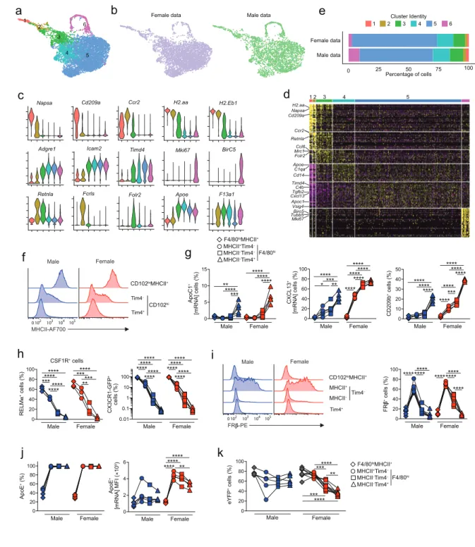
Uniform Manifold Approximation and Projection (UMAP) dimensionality reduction analysis
26
revealed 6 clusters that were present in both male and female cells (Figure 5a, b). Given that the starting
27
population of CD11b+ cells is known to be phenotypically heterogeneous, containing resident
28
CD102+F4/80hi macrophages, CD102–F4/80lo MHCII+ macrophages and CD11c+MHCII+ cDC2 20-23,
29
we first used a panel of known markers to validate subset identity (Figure 5b). 3 clusters of resident
30
macrophages (3-5) could be identified on the basis of their high expression of Adgre1 (F4/80) and Icam2
31
(CD102). As expected, these were clearly distinct from short-lived CD102–F4/80loMHCII+
32
macrophages and cDC2, which were found in clusters 1 and 2 respectively, and expressed Ccr2 (Figure
33
5c, d) 21. However, CD102–F4/80loMHCII+ macrophages and cDC2 could be distinguished from one
34
another on the basis of expression of the DC markers Cd209a and Napsa 27,43, and of Retnla and Fclrs,
35
which we and others have shown to be signature markers of cavity CD102–F4/80loMHCII+ macrophages
36
21,22,44. Cluster 6 was defined by genes associated with cell cycle, such as Mki67 and Birc5, suggesting
37
this cluster represents proliferating cells. In both sexes, the majority of cells was in cluster 5 (Figure
5e), which was characterised by markers of resident F4/80hiCD102+ macrophages including Icam2,
1
Prg4 and Tgfb2 (Figure 5c, d & Supplementary Table 5) that form part of the core peritoneal
2
macrophage-specific transcriptional signature 45; cluster 5 cells also expressed markers of long-lived
3
macrophages, including Timd4 and Apoc1, confirming the findings above. Although the cells in cluster
4
3 expressed Icam2, they also expressed a number of genes that were highly expressed by the CD102–
5
F4/80loMHCII+ macrophages in cluster 2, such as Retnla, H2.Aa and Ccr2, suggesting a common origin
6
of these clusters, or a close relationship between them. This analysis also identified genes uniquely
7
expressed by cluster 3, including Folr2, which encodes the beta subunit of the folate receptor (FRb).
8
Although cluster 4 showed a distinct pattern of gene expression, such as high expression of Apoe, it
9
also shared features with cluster 3 and cluster 5, suggesting it may contain differentiation intermediates.
10
Consistent with our earlier analysis, we found that the Timd4-expressing cluster 5 was more abundant
11
amongst female cells, whereas more male cells were found within clusters 1, 2, 3 and 6 (Figure 5c).
12
We next used flow cytometry to determine if we could validate the additional heterogeneity
13
uncovered by our scRNAseq analysis. Given that MHCII-associated genes appeared to define
14
heterogeneity amongst Tim4–CD102+ macrophages (i.e. clusters 3 & 4), we assessed expression of
15
MHCII by Tim4-defined subsets of CD102+ macrophages. This confirmed that a proportion of Tim4–
16
CD102+ macrophages expressed MHCII, albeit at lower levels than CD102–F4/80loMHCII+
17
macrophages, whereas Tim4+ macrophages had negligible MHCII expression (Figure 5f). Thus, we
18
used a combination of Tim4 and MHCII to identify macrophage subsets and assessed expression of
19
other subset defining markers from the scRNAseq analysis. Consistent with the analysis above, we
20
found that ApoC1 expression was essentially exclusive to Tim4+MHCII– macrophages (Figure 5g),
21
whereas both MHCII-defined Tim4– macrophages lacked ApoC1 expression. Expression of CXCL13
22
was also highest amongst Tim4+MHCII– cells, although interestingly, the proportion of CXCL13+ cells
23
increased progressively from Tim4–MHCII+ to Tim4–MHCII– to Tim4+MHCII– CD102+ macrophages
24
(Figure 5g). Although not identified as a cluster defining gene in our scRNAseq analysis due to low
25
coverage, we found that CD209b displayed the same pattern of expression as CXCL13 (Figure 5g).
26
Consistent with the idea that they may derive from CD102–F4/80loMHCII+ macrophages, the expression
27
of RELMa and CX3CR1 was highest on Tim4–MHCII+ macrophages and was essentially absent from
28
Tim4+MHCII– CD102+ macrophages. The majority of Tim4–MHCII+ macrophages expressed high
29
levels of FRb, whereas all other populations had little or no expression, consistent with our scRNAseq
30
analysis. While Apoe was proposed to define cluster 4 in our scRNAseq analysis, consistent with our
31
analysis above, we found it was expressed by all CD102+ macrophages, although, in females, most
32
highly expressed by Tim4–MHCII+ and the level decreased progressively to Tim4+MHCII– CD102+
33
macrophages. Finally, we returned to using CD11cCre.Rosa26LSL-eYFP mice to assess if
MHCII/Tim4-34
defined subsets showed differential levels of replenishment. Notably, we found that MHCII-expressing
35
Tim4–CD102+ macrophages showed equivalent labelling to CD102–F4/80loMHCII+ macrophages in
36
female CD11cCre.Rosa26LSL-eYFP mice, indicative of more recent derivation from CD102–
37
F4/80loMHCII+ cells. Consistent with their intermediate transcriptional profile, Tim4–CD102+
macrophages that had lost MHCII expression showed intermediate labelling when compared with their
1
MHCII+Tim4– and Tim4+ counterparts. No difference in eYFP labelling between Tim4-defined subsets
2
was noted in male mice, consistent with more rapid replenishment of all subsets of macrophages in this
3
environment.
4
Collectively these data show that excluding proliferating cells, resident peritoneal macrophages
5
comprise three main clusters, with Tim4– macrophages displaying an intermediate phenotype compared
6
with F4/80loMHCII+ macrophages and Tim4+ macrophages.
7
8
Differential replenishment and environmental signals drive the dimorphic features of peritoneal
9
macrophages
10
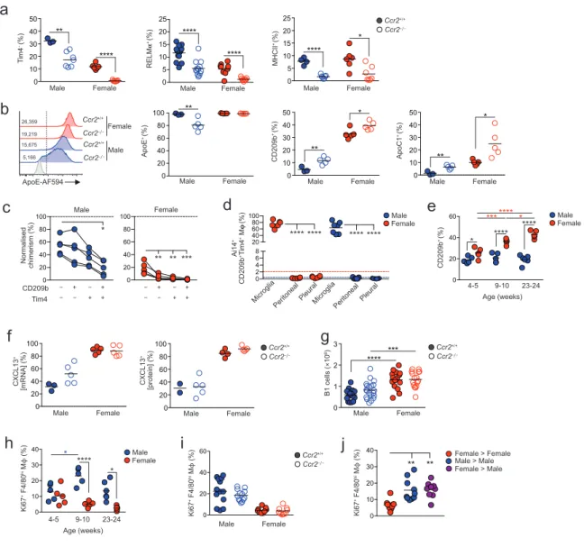
To dissect the dimorphic features of CD102+ macrophages that could be related to longevity from those
11
more directly controlled by dimorphic environmental signals, we next assessed expression of these in
12
Ccr2–/– mice in whom macrophage replenishment is markedly reduced due to severe monocytopenia
13
46,47. Strikingly, the frequency of Tim4– macrophages, as well as those expressing RELMa, FRb or
14
MHCII were markedly reduced in Ccr2–/– mice compared with Ccr2+/+mice irrespective of sex,
15
confirming these cells to be recently derived from monocytes (Figure 6a & Supplementary Figure
16
7). In males, CCR2 deficiency also led to reduced expression of ApoE and emergence of an ApoE–
17
subset of CD102+ macrophages (Figure 6b). In contrast, a higher proportion of CD102+ macrophages
18
in Ccr2–/–- mice expressed CD209b and ApoC1, markers that are characteristic of the Tim4+MHCII–
19
subset, suggesting these markers may be expressed selectively by long-lived macrophages (Figure 6b).
20
Consistent with this, Tim4+ macrophages expressing CD209b displayed the lowest level of replacement
21
by donor cells in BM chimeras when compared with all other CD209b/Tim4-defined macrophages,
22
even in male mice where overall replenishment from the bone marrow is markedly higher (Figure 6c).
23
The low levels of replacement of peritoneal CD209b+Tim4+ macrophages does not reflect derivation
24
from yolk sac progenitors, as, unlike microglia in the brain, these cells are not labelled in male or female
25
Cdh5Cre-ERT2.RosaLSL-tdT mice, which allow tracing of cells arising from yolk sac haematopoiesis 48.
26
Similar results were obtained with CD209b+Tim4+ macrophages in the pleural cavity (Figure 6d).
27
Hence, despite being long-lived, CD209b+Tim4+ macrophages derive from conventional
28
haematopoiesis in both sexes. Importantly, temporal analysis revealed that while little difference in
29
abundance of CD209b-expressing CD102+ macrophages was seen in pre-pubescent (4-5-week-old)
30
male and female mice, these cells accumulated progressively in the cavity of female mice following
31
sexual maturation. This did not occur in male mice, consistent with their higher rate of replenishment
32
from the bone marrow and indicating that acquisition of CD209b expression appears to be associated
33
with time-of-residency in the female cavity (Figure 6e).
34
Not all dimorphic features of peritoneal CD102+ macrophageswere influenced by their rate of
35
replenishment. For instance, the intrinsically higher expression of CXCL13 by female macrophages
36
was not altered by CCR2 deficiency (Figure 6f). In parallel, although we confirmed previous findings
37
of a clear dimorphism in the numbers of B1 cells between adult male and female mice 15 and this
developed gradually following sexual maturation (Supplementary Figure 8), this phenomenon
1
remained in Ccr2–/– mice (Figure 6g). Similarly, while the higher levels of proliferation by male
2
CD102+ macrophages developed following sexual maturation (Figure 6h), this was unaffected by
3
CCR2 deficiency (Figure 6i). This evidence that certain dimorphic features are driven by
4
environmental factors, independent of cell replenishment was supported further by the fact that
5
macrophages derived from female BM in the cavity of chimeric male showed levels of proliferation
6
that were identical to those of male BM derived macrophages in the male cavity and were higher than
7
those of female BM derived macrophages in female cavity (Figure 6i). Thus, the differential
8
proliferation of female and male macrophages is not due to cell-intrinsic differences in their
9
proliferative activity.
10
Taken together these data demonstrate that both local imprinting and differential turnover
11
contribute to the sexual dimorphisms seen in peritoneal macrophages.
12
13
14
Differential CD209b expression confers an advantage on female macrophages in the setting of
15
pneumococcal peritonitis
16
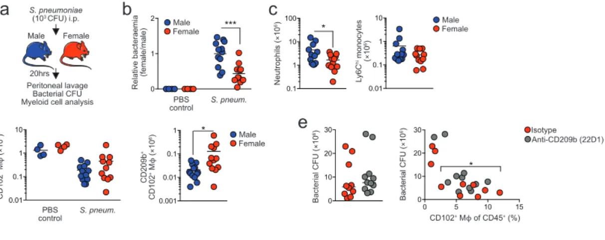
We postulated that differential expression of key pattern recognition receptors such as CD209b might
17
endow female macrophages with an enhanced ability to deal with bacterial infection. To test this idea,
18
we examined the acute peritonitis caused by the gram-positive bacterium Streptococcus pneumoniae
19
(Figure 7a), a localised model of infection in which resident macrophages, and in particular CD209b,
20
are indispensable for protective immunity 49,50 whereas recruitment of neutrophils is not required 49and
21
hence avoids any confounding effects of systemic sex-dependent effects on innate immune responses
22
that have been reported previously 51. As CD209b is expressed exclusively by CD102+ macrophages in
23
the peritoneal cavity (Supplementary Figure 6), this model allowed us to directly assess the
24
importance of differential CD209b expression by CD102+ macrophages in bacterial elimination.
25
Strikingly, females showed enhanced capability to control S. pneumoniae infection, with significantly
26
lower levels of bacteria in peritoneal fluid of female mice compared with their male counterparts 20hrs
27
after inoculation (Figure 7b). Fewer neutrophils and Ly6Chi monocytes were present in the female
28
cavity compared with male mice (Figure 7c), consistent with a model in which resident macrophages
29
control infection 49. In contrast, while the well-documented macrophage ‘disappearance reaction’ 52
30
occurred in both male and female mice after infection (Figure 7d), significantly higher numbers of
31
CD209b-expressing macrophages persisted in the female cavity (Figure 7d). Administration of an
anti-32
CD209b blocking antibody (22D1) 53 led to increased levels of bacteraemia in female mice, although
33
this did not attain statistical significance due to variance in bacterial counts in some mice in whom the
34
macrophage ‘disappearance reaction’ was more pronounced (Figure 7e).
35
Thus, dimorphic expression of key immune receptors and molecules leads to differential ability
36
to handle local bacterial infection.
37
38
Discussion
1
Understanding the extrinsic and intrinsic factors that govern tissue macrophage differentiation
2
is a key goal in the field of macrophage biology. Here we reveal a striking effect of sex on the
3
phenotypic and transcriptional identity of resident peritoneal macrophages and demonstrate that this
4
contributes to the sex-dependent resistance of mice to bacterial peritonitis. Moreover, we show that this
5
arises through a combination of dimorphic microenvironmental signals and sex-dependent differences
6
in the rate of macrophage renewal from the bone marrow.
7
Using classical defining markers such as F4/80, CD11b and CD102, we found peritoneal
8
macrophages from male and female mice to be phenotypically identical. Furthermore, while some
9
studies have reported that the number of peritoneal macrophages is greater in females 38,51, we did not
10
routinely detect significant differences in the number of peritoneal macrophages between the sexes.
11
However, mRNA sequencing revealed marked dimorphism in the transcriptional fingerprint of resident
12
peritoneal macrophages under homeostatic conditions. Importantly, this showed that female CD102+
13
macrophages express higher levels of genes associated with lipid uptake and transport as well as
14
immune defence/response, including those encoding complement components, the chemoattractant
15
CXCL13, and numerous receptors involved in recognition and uptake of pathogens and apoptotic cells.
16
In contrast, the signature of male peritoneal macrophages was dominated by cell cycle associated genes
17
consistent with their elevated levels of proliferation, a dimorphism we have reported previously 21.
18
Although others have reported dimorphic expression of TLRs and CD14 by peritoneal macrophages,
19
no consistent pattern was observed in these studies 37,38 and we found no significant difference in mRNA
20
transcripts of the adaptor protein MyD88 or any TLRs, consistent with more recent analysis of surface
21
protein expression 51. We have also found no sex differences in CD14 expression at the gene or protein
22
level. These discrepancies may relate to differences in the nature of the cells being analysed, with one
23
study using peritoneal macrophages elicited by incomplete Freund’s adjuvant 37 and the other assessing
24
gene expression by total peritoneal cells 38. In contrast, our analyses involved minimal handling and
25
used rigorously characterized resident macrophages.
26
Our further analyses revealed that many of the sexually dimorphic features of macrophages
27
arose following sexual maturation, including the higher expression of CD209b and Tim4 by female
28
macrophages. Furthermore, the enhanced accumulation of peritoneal B1 cells found in females was also
29
age-dependent, suggesting that the higher levels of CXCL13 production by female macrophages may
30
also be driven by sexual maturation. Dimorphic differences in the turnover of CD102+ resident
31
peritoneal macrophages also appeared to largely arise following sexual maturation. Notably, the
32
kinetics of labelling in CD11ccre.Rosa26LSL-eYFP mice and a significant reversal in the autonomy of
33
female peritoneal macrophages in BM chimeras following ovariectomy suggest this aspect of
34
dimorphism in normal mice reflects a switch from replenishment to self-maintenance in females. These
35
results are consistent with recent monocyte fate mapping using Ms4a3Cre.Rosa26LSL-tdTomato mice
36
showing that while monocytes contribute to the maintenance of peritoneal macrophages during the
37
perinatal and adolescent period, this process wanes during adulthood in female mice (54 & F. Ginhoux,
personal communication). Hence, sexual maturation leads to dimorphic changes in replenishment,
1
proliferation and gene expression by peritoneal macrophages, at least some of which are driven by the
2
female peritoneal environment.
3
Despite a significant degree of transcriptional difference at the population level, single cell
4
mRNA sequencing showed that male and female resident macrophages encompassed very similar
5
transcriptionally-defined clusters of cells. However, the relative abundance of these clusters differed
6
between sexes. In this regard, CD102+ peritoneal macrophages could be divided into three predominant
7
transcriptionally-distinct clusters. Of these, the cells in cluster 3 expressed CD102 together with
8
MHCII, RELMa and CX3CR1, all of which are key markers of F4/80loMHCII+ peritoneal
9
macrophages, suggesting that cluster 3 may be recently derived from the F4/80loMHCII+ macrophage
10
population that is derived from blood monocytes in adult mice 21-23. As cells in cluster 4 shared features
11
with both the MHCII-defined cluster 3 and the dominant cluster 5 population of Tim4+ macrophages,
12
these may represent a further intermediate differentiation state. Consistent with this idea, in female mice
13
Tim4–MHCII– CD102+ macrophages, which likely represent those in cluster 4, displayed an
14
intermediate degree of replenishment from the bone marrow in our fate mapping studies between that
15
of the rapidly replenished Tim4–MHCII+ CD102+ macrophages and slowly replenished Tim4+MHCII–
16
cells. Furthermore, all Tim4– cells were largely ablated in female Ccr2–/– mice. A linear-developmental
17
relationship that culminates at cluster 5 would be consistent with the greater abundance of cells in this
18
cluster in females, given the slower entry of bone-marrow-derived cells into the female CD102+
19
macrophage pool. However, it seems unlikely that such a linear developmental relationship between
20
clusters exists in males, as Tim4 and MHCII defined subsets were found to be replenished at similarly
21
high rates. Hence, what dictates cluster identity in males remains unclear.
22
Given that the rate of replenishment from BM was markedly different between the sexes, this
23
raised the possibility that transcriptional differences could reflect different ontogenies of male and
24
female peritoneal macrophages. Indeed, a number of the genes we found to be expressed more highly
25
by female peritoneal macrophages, including Colec12, Cd163, Bmpr1a, Cdc42bpa, Timd4, Apoc1, and
26
members of the Cd209 family have been reported to be expressed by embryonically, but not
monocyte-27
derived macrophages in other tissues 25,55. This could reflect an intrinsic property of their embryonic
28
origin, or that such cells are likely to have resided in the tissue for a long period. However the fact that
29
a proportion of BM-derived peritoneal macrophages can acquire the expression of at least some of these
30
“embryonic” signature markers (e.g. Tim4, CD209b) in the setting of tissue-protected BM chimeras,
31
suggests that this is more likely related to their time-of-residency rather than rigid differences related
32
to origin. Consistent with this, co-expression of Tim4 and CD209b identifies the longest-lived
33
macrophages in the peritoneal cavity irrespective of sex. The concept that macrophages require
34
prolonged residence within the tissue to acquire their characteristic features is consistent with work
35
from the Guilliams lab showing that acquisition of Tim4 expression by monocyte-derived cells that
36
engraft in the liver following deletion of endogenous Kupffer cells increases with time 25. Notably, of
37
the two populations of peritoneal macrophages that have been described in ascites fluid from patients
with decompensated cirrhosis, the subset that aligns with mouse resident F4/80hi macrophages exhibits
1
significantly higher expression of TIMD4, CD209, COLEC12, CD163 and APOC1 56, suggesting these
2
may represent phylogenetically conserved markers of long-lived macrophages.
3
We also identified features that mark newly differentiated CD102+ macrophages and are
4
inversely related to time-of-residency, such as ApoE. This finding is consistent with repopulation
5
studies showing that microglia of monocyte origin express higher levels of ApoE 57-59. While the
6
association between ApoE and recent arrival seems to be at odds with the higher level of ApoE
7
expression by female peritoneal macrophages, ApoE expression may also be controlled partly by
8
estrogen, as the Apoe gene contains an estrogen response element, and its expression is reduced in
9
inflammatory peritoneal macrophages by macrophage-specific deletion of the estrogen receptor alpha
10
35. Understanding how ApoE expression is regulated by tissue-residency and hormonal control may be
11
important in many diseases in which is it is a known genetic risk factor and that exhibit strong
sex-12
biases in risk, such as Alzheimer’s and cardiovascular disease 60, but also in healthy aging where APOE
13
variants are among the strongest predictors of human longevity 61.
14
Importantly, we found the dimorphism in proliferation and CXCL13 expression by peritoneal
15
macrophages to be regulated independently of macrophage replenishment kinetics, consistent with
16
previous data showing that the proliferative capacity of macrophages is determined by signals in the
17
local microenvironment rather than their origin 25,62. Although these dimorphisms only developed
18
following sexual maturation, it seems unlikely that estradiol levels are responsible for the lower
19
proliferation of female peritoneal macrophages, as estradiol is reported to increase rather than inhibit
20
proliferation of these cells 36. Furthermore, exogenous estradiol did not rescue the elevated turnover of
21
female macrophages we found in ovariectomised mice. Similarly, exogenous estradiol does not
22
influence CXCL13 expression by peritoneal macrophage 36, nor did it rescue the loss of B1 cells that
23
occurred after ovariectomy (data not shown). While estradiol has been reported to upregulate the B1
24
cell regulators Tnfsf13b, Tnfsf13, and Il10 15by female peritoneal macrophages in vitro, none of these
25
genes were differentially expressed by macrophages in our study. Moreover, genes highly expressed by
26
male peritoneal macrophages, such as Arg1 and Chil3, are increased by estradiol 36. Although
27
expression of receptors for progesterone and androgens did not differ between the sexes, we cannot rule
28
out a role for these steroids in generating sex dimorphisms. Thus, the exact local factor(s) driving the
29
sex dimorphisms identified here remain to be elucidated. Interestingly, pathway analysis of our
30
transcriptomic data identified ‘Interferon Gamma Response’ and ‘Interferon Alpha Response’ as gene
31
sets enriched within female macrophages, and interferons are known to be hormonally regulated 63.
32
Whether interferon receptor signalling plays a role in dimorphic characteristics of peritoneal
33
macrophages is the focus of ongoing work.
34
The incidence and severity of sepsis and post-surgical infections are profoundly lower in
35
women than men 64,but the mechanisms underlying these differences remain unclear. Our finding that
36
female mice are more resistant to S. pneumoniae peritonitis is consistent with previous work on group
37
B streptococcal peritonitis 38. However, while others attributed this to other elements of innate immune
responses, such as neutrophil recruitment 51, our data suggest that the resistance of females is at least,
1
in part, due to differences in resident peritoneal macrophages, such as elevated expression of CD209b.
2
Dimorphic expression of CXCL13 may also contribute, as it plays a central role in recruiting B1 cells
3
that produce the natural IgM 2 that protects against multiple forms of infectious peritonitis 65,66. As
4
activation of complement is essential for innate resistance to against S.pneumoniae }65 and both CD209b
5
and natural IgM can activate the classical pathway of complement fixation during S.pneumoniae
6
infection 65-67, peritoneal macrophages may play several, overlapping roles in protective immunity
7
against this infection. Indeed C1q, C3, and C4b, as well as Cfb, which encodes factor B and is essential
8
for the alternative pathway of complement fixation during S.pneumoniae infection, were also all
9
expressed more highly by female peritoneal macrophages. We propose that this heightened barrier
10
function in the female peritoneum may have evolved to mitigate the risk of sexually transmitted
11
infection disseminating from the lower female reproductive tract 68 or to protect against puerperal
12
peritonitis. Our findings also have wider implications for understanding peritoneal macrophage
13
behaviour following a local mechanical or inflammatory insult, when tissue resident macrophages may
14
be replaced by monocyte-derived cells that may require prolonged residence in the tissue before
15
acquiring the full profile of resident macrophages with protective functions 55,69-71. The potential risks
16
in this process have been highlighted in the context of viral meningitis, where a failure of newly elicited
17
macrophages to rapidly acquire CD209b expression led to impaired neutrophil recruitment to
18
subsequent intra-cranial immune challenge 72, and could explain why animals exposed to sterile
19
peritoneal inflammation are more susceptible to S. pneumoniae peritonitis for at least several months
20
73.
21
Our studies highlight the importance of taking age and sex into account when understanding
22
the peritoneal response to disease and implicate time-of-residency as an underlying determinant of
23
resident macrophage function. Further work is needed to understand the molecular processes that
24
underlie the requirement for time-of-residency on expression of these genes and to identify the local
25
signals that govern this process. Beyond the cavity, our findings also have wider implications for the
26
molecular mechanisms that drive dimorphic production of natural IgG by peritoneal B1 cells that
27
provides women and infants with heightened resistance to blood-borne bacterial infections, particularly
28
as these antibodies are lost in the absence of peritoneal macrophages 15.