REPORTS
Induction of p53 Expression
in Skin by Radiotherapy
and UV Radiation:
a Randomized Study
Fredrik Ponten, Henrik Lindman,
Asa Bostrom, Berit Berne,
Jonas Bergh
Background: p53 protein plays an
im-portant role in the response to DNA
damage, and radiotherapy can cause
radiation dermatitis. p53 and p21 levels
increase in vitro when DNA is damaged
by UVA, UVB, or
␥-radiation. To
de-termine whether this response occurs
in human skin and predicts the level of
radiation dermatitis, we investigated
levels of p53 and p21 in skin exposed to
different types of radiation as part of a
randomized study of women with
breast cancer to evaluate topical
ste-roid or emollient cream treatments for
radiation dermatitis of their irradiated
b r e a s t . M e t h o d s : A f t e r s u r g e r y
but before receiving tangential 5-mV
photo-beam radiotherapy (2 Gy and
54 Gy) to the affected breast
paren-chyma, multiple areas on the backs of
50 women were irradiated with UVA
and other areas were irradiated with
UVB. Skin biopsy samples were taken
from areas of normal unirradiated skin
and all irradiated areas, and p53 and
p21 were detected
immunohistochemi-cally. All statistical tests are two-sided.
Results: In skin irradiated with UVA
or UVB, medians of 4.4% (range =
0%–40.5%) or 45.5% (range = 5.3%–
74.6%) p53-positive keratinocytes,
respectively, were observed.
Radio-therapy produced medians of 31.0%
(range = 0%–79.3%)
p53-immunoreac-tive cells after 2 Gy of radiation and
83.2% (range = 37.6%–95.2%) after 54
Gy of radiation. Despite large
interin-dividual differences in p53 response,
comparable increases in epidermal p53
response were independent of the type
of radiation. A correlation between p53
and p21 was also evident (r
s= .78). In
breast skin, there was no association
between the p53 response and the
de-gree of erythema (a measure of
radia-tion dermatitis) and no statistically
sig-nificant difference between treatment
arms and p21/p53 responses.
Conclu-sions: Individual responses to
radia-tion-induced DNA damage varied widely
and may be independent of the type of
radiation. The epidermal p53 response
does not predict the degree of radiation
dermatitis. [J Natl Cancer Inst 2001;
93:128–33]
p53 and p21, its downstream-effector
protein, are sensitive indicators of DNA
damage. In normal human epidermis,
there are two patterns of p53
immunore-activity, patch like and diffuse (1–3). The
patch-like pattern represents a cluster of
keratinocytes with a mutated p53 gene
(4–6), and the diffuse pattern is found
in the epidermis after DNA damage has
triggered a reactive accumulation of p53
protein (7,8). p53 accumulates rapidly
in the human epidermis after a single
physiologically active dose of UV
radia-tion. Topical sun-protection lotion applied
before exposure to UV radiation can, in
part, block the epidermal p53 response
(1,9) and, in animal models, the number
of induced p53 mutations are reduced
(10).
UV radiation generates DNA
photo-products, such as pyrimidine dimers and
6-4 photoproducts (11,12). Ionizing
irra-diation produces double- and
single-strand DNA breaks. Cells respond to
DNA photoproducts and DNA breaks by
accumulation of functionally active p53
protein, a key event in response to cellular
stress. Although the initial events and
pathways involved are not completely
un-derstood, increased protein stability
re-sulting in a prolonged half-life appears to
be an important mechanism (13). One
pathway downstream of p53 is mediated
through p21, a general cyclin-dependent
kinase inhibitor. The p21 pathway leads
to cell cycle arrest at the G
1restriction
point caused by decreased
phosphoryla-tion of the retinoblastoma protein (14). If
DNA damage is more severe, p53 is
in-volved in the pathway leading to
apop-totic cell death (15–18). The p53 response
is thus a normal defense mechanism,
pro-tecting an organism from acquiring clones
of mutant cells (19–21). In vitro, a
cellu-lar p53 response has been observed after
␥-irradiation (18,22), but, to our
knowl-edge, this response has not been studied in
vivo in human skin. That the cellular p53
response can be observed in vivo in
ani-mal models has been shown previously
(23–25).
Unexplained large individual
varia-tions of human skin to UV radiation have
been observed in the p53 response (1) and
the DNA repair reaction (26). An acute
clinical side effect of radiotherapy is
ra-diation dermatitis (27), and large
differ-ences in the degree of radiation dermatitis
have also been noted among individuals
after radiotherapy (28). However, it has
not been determined whether the
re-sponses to UV and ionizing radiation are
related. We address this issue by
measur-ing changes in the levels of p53 and p21
and assessing erythema in the same
pa-tient after exposure to three types of
ra-diation, UVA, UVB, and 5-mV
photon-beam radiation. The study also assessed
the protective effect of a topically applied
steroid on radiation dermatitis, where a
major conclusion was that corticosteroid
cream reduced acute radiation dermatitis
(Bostrom A, Lindman H, Swartling C,
Berne B, Bergh J: manuscript submitted
for publication). In this study, we
inves-tigate interindividual variations in the
re-sponses of p53 and p21 to DNA damage;
analyze the relationships of p53 and p21
with UVA, UVB, and high-voltage
pho-ton-beam irradiation; and determine
whether p53 or p21 would be a good
marker to predict clinical radiation
der-matitis.
P
ATIENTS ANDM
ETHODSThis study was a part of a double-blind random-ized study that also evaluated the effects of a potent topical corticosteroid application (mometasone fu-roate) and placebo (emollient cream) on radiation dermatitis. The patients received oral and written information before inclusion in the study.
Affiliations of authors: F. Ponten (Department of
Genetics and Pathology), H. Lindman (Department of Oncology), A. Bostrom, B. Berne (Department of Dermatology), University Hospital, Uppsala, Swe-den; J. Bergh, Department of Oncology, Karolinska Hospital, Stockholm, Sweden.
Correspondence to: Fredrik Ponten, M.D., Ph.D.,
Department of Genetics and Pathology, University Hospital, S-751 85 Uppsala, Sweden (e-mail: Fredrik.Ponten@genpat.uu.se).
See “Notes” following “References.”
Patients
Fifty women with breast cancer between the ages of 47 and 77 years (mean⳱ 59.5 years) entered the study, which was approved by the ethical committee with jurisdiction for Uppsala University Hospital, Sweden. Patients with known cutaneous or severe systemic disease were excluded. All patients had been treated by sector resection of a morphologi-cally verified invasive breast cancer without lymph node metastasis. No concomitant systemic antican-cer treatment was administrated. No patient had re-cently been in the sun or in a tanning bed before entering the study. All patients were Caucasian and had the following skin types, according to Fitzpa-trick (29): 32 patients had skin type 3, 15 had skin type 2, and three had skin type 4. Patients were instructed to apply the “blinded” cream to the irdiated breast twice a week from the start of the ra-diotherapy until they had received a total radiation dose of 24 Gy and then to apply it once a day until 3 weeks after radiotherapy was completed.
Irradiation
Skin on the back of all patients was UV irradiated. UVB was administered to nine areas (each 7 mm in diameter) in a stepwise fashion (300–1500 J/m2)
with a monochromator (Applied Photophysics, Leatherhead, Surrey, U.K.) emitting a narrow band at 313 nm (±4–6 nm) through a 1-m liquid light guide with a 7-mm aperture at an irradiance of 15– 17 mW/cm2. UVA was administered with an UVASUN
lamp (Muthzas Co., Munich, Germany) equipped with a UVA filter (330–450 nm) emitting with an irradiance of 86 mW/cm2. UVA was delivered to
four areas of skin, each 50 × 50 mm. Each area received a dose of 10, 20, 40, or 80 J/cm2.
Radio-therapy was started after the UV tests were com-pleted. All patients received tangential 5-mV pho-ton-beam radiotherapy to the breast parenchyma, at 2 Gy per visit from a Philips SL 75–5 linear accel-erator (Philips Inc., Crawley, U.K.) as described pre-viously (30), for 5 days per week until a total dose of 54 Gy was reached.
Evaluation of Erythema and
Skin Sampling
Degree of UV-induced erythema was determined from readings 24 hours after irradiation. The follow-ing scale was used: 0 ⳱ no erythema; 1 ⳱ just perceptible erythema (the minimal erythema dose); 2⳱ mild erythema; 3 ⳱ marked erythema; 4 ⳱ marked erythema and slight edema; 5⳱ marked erythema and strong edema; and 6⳱ bullous reac-tion. In addition, the degree of pigmentation after UVA irradiation was assessed. For UVB, the test sites that most closely corresponded to a site receiv-ing twice the minimal erythema dose were subjected to skin biopsy 24 hours after irradiation. For UVA, biopsy sites received 40 or 80 J/cm2 (14 patients
received 40 J/cm2 because of a previous medical
history of high sensitivity to UV radiation). Biopsy samples of normal nonirradiated and irradiated back skin were taken at the same time, 24 hours after UV irradiation.
Three breast skin biopsy samples were taken 1 cm below the areola of the photon-beam-irradiated breast. The first sample was taken before the start of radiotherapy, the second was taken 20–30 hours af-ter the first 2-Gy dose of photon-beam radiation, and
the third was taken immediately after radiotherapy was completed (total radiation dose⳱ 54 or 56 Gy). Erythema was evaluated, with a reflectance spectro-photometer, four times during radiotherapy (after 24, 34, 44, and 54 Gy) and 3 weeks after treatment was completed. The mean of these data, including data from the five irradiated areas of the breast, was calculated for each patient and used as an objective erythema score. Reciprocal areas from the nonirra-diated breast were used as normal control site.
Skin Biopsy Samples and
Immunohistochemistry
The protocol above resulted in six biopsy samples (3-mm in diameter) per patient. Ten of these samples were technically insufficient, leaving a total of 290 interpretable punch biopsy samples. The 3-mm punch biopsy samples were immediately fixed in neutral-buffered formalin. After 24–48 hours, the tissue was embedded in paraffin and 4-m-thick sections were cut. Immunohistochemistry was per-formed essentially as described previously (31). Briefly, sections were cooked in a 750-W micro-wave oven (two 5-minute periods in 10 mM citric buffer [pH 6.0]) and stained with avidin–biotin– coupled immunoperoxidase. An automated stan-dardized procedure (Ventana Medical Systems Inc, Tucson, AZ) was used with primary antibodies D-07 (code M7001; dilution 1 : 200; DAKO A/S, Glos-trup, Denmark), which reacts with both wild-type and mutant human p53 protein, and WAF-1 (Ab-1, dilution 1 : 40; Oncogene Research Products from CALBIOCHEM, Cambridge, MA), which reacts with human p21 protein. Sections were counter-stained with Mayer’s hematoxylin.
Scoring of p53 and p21
Immunoreactivity
Sections of immunostained skin were evaluated under the microscope by one person (Dr. Christina Nyberg, University Hospital, Uppsala, Sweden). All epidermal keratinocyte nuclei from four randomly chosen high-power fields, covering more than 50% of the total epidermal length in each biopsy sample, were evaluated and counted. In anti-p53-stained sections, the difference between nonimmunoreactive and immunoreactive keratinocyte nuclei was clear-cut, and cells were scored as positive or negative. Anti-p21-stained sections showed gradients of reac-tivity, rendering binary assessment impossible. Consequently, the proportion of immunohistochem-ically positive cells was estimated on the following scale of 1–4: 1⳱ less than 25% immunoreactive keratinocytes; 2 ⳱ 25% to 50% immunoreactive keratinocytes; 3 ⳱ 50% to 75% immunoreactive keratinocytes; and 4⳱ more than 75% immunore-active keratinocytes. This modified scale was moti-vated by a diffuse reaction pattern for the p21 anti-body. The intensity of the p21 immunoreaction was determined as weak, moderate, or strong.
Methods for Statistical Analysis
This study did not have the statistical power to investigate possible differences in local and sys-temic relapse frequencies. It would, of course, have been ideal to have a larger study population to be able to investigate whether the p53 activation pattern in the normal skin of a cancer patient is associated
with the p53 response status in the same patient’s tumor cells.
Nonparametric methods were used for the statis-tical analysis. The Spearman’s rank correlation test was used to calculate potential correlations. The cor-responding correlation coefficient is denoted rs in
the text. Because multiple correlation analyses were performed, only P values of less than .01 were con-sidered to be statistically significant. For compari-son of two groups, the Mann–Whitney U test was used. All statistical tests are two-sided.
R
ESULTSThe average number of cells counted
per anti-p53-stained biopsy sample was
528. Skin type did not influence p53
im-munoreactivity in nonirradiated or
irradi-ated skin. Table 1 shows a summary of
the mean percentage of p53-positive cells
and p21 score. Fig. 1 shows p53
immu-noreactivity in the following six groups:
1) normal back skin, 2) normal breast
skin, 3) UVA-irradiated back skin, 4)
UVB-irradiated back skin, 5) 2 Gy of
photon-beam-irradiated breast skin, and
6) 54 Gy of photon-beam-irradiated
breast skin. Table 2 shows correlations in
p53 immunoreactivity between the
differ-ent groups.
Nonirradiated Skin
In general, breast skin was slightly
thinner than back skin. The number of
ke-ratinocytes, however, did not differ
statis-tically significantly between back (mean
⳱ 618 keratinocytes; range ⳱ 427–936
keratinocytes) and breast (mean
⳱ 583
keratinocytes; range
⳱ 384–938
kera-tinocytes) skin. A few scattered
p53-positive keratinocytes were present in 41
of the 50 biopsy samples from normal
breast (Fig. 2, a) and 43 of the 49 from
back skin (Fig. 2, b). p53-positive
kera-tinocytes were 2.3% of cells (median
⳱
1.2%; range
⳱ 0%–31.1%) in breast skin
and 1.8% (median
⳱ 1.3%; range ⳱ 0%–
8.9%) in back skin. In one patient (No.
30), 31.1% of nuclei of the unirradiated
breast skin were p53 positive. Only a few
keratinocytes showed weak p21 staining.
Photon-Beam Radiotherapy
Compared with nonirradiated skin, no
changes were detected by light
micros-copy in skin biopsy samples taken after
the patient had received 2 Gy of
photon-beam radiation. However, severe
morpho-logic changes, similar to what has been
described previously (32), were observed
(Fig. 2, d) in biopsy samples taken after
radiotherapy was completed (total dose
⳱ 54 Gy). The epidermis in these
samples was thin, containing only one to
three layers of keratinocytes, and the
ke-ratinocytes were enlarged with cytologic
atypia. The dermis had dilated capillaries
and a partly perivascular chronic
inflam-matory infiltrate.
Immunohistochemical-ly, 30.1% (median
⳱ 31.0%; range ⳱
0%–79.3%) of the keratinocytes were
positive for p53 after 2 Gy of irradiation
(Fig. 2, c). There were fewer than 25%
p21-positive cells (score
⳱ 1.1), and the
intensity of p21 staining was weak. The
most widespread, intense, and uniform
staining for p53 and p21 was observed in
skin that had received 54 Gy of
irradia-tion. After radiotherapy was completed,
80.1% (median
⳱ 83.2%; range ⳱
37.6%–95.2%) of keratinocytes were p53
positive (Fig. 2, d). In most cases, 75% or
more of the keratinocytes (score
⳱ 3.4)
showed a moderate-to-strong intensity of
p21 staining. Objective measurements of
erythema varied between scores of 2.3
and 11.2 (mean score
⳱ 6.4).
UVA Irradiation
No morphologic changes were found
in skin after UVA irradiation compared
with nonirradiated skin. In UVA-irradiated
skin, 7.4% (median
⳱ 4.4%; range ⳱
0%–40.5%) of the keratinocytes
overex-pressed p53 (Fig. 2, e). p53
immunoreac-tivity did not differ statistically
signifi-cantly between patients irradiated with 40
J/cm
2and patients irradiated with 80
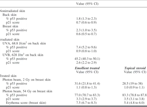
Table 1. p53- and p21-immunoreactive cells in normal and irradiated skin*
Value (95% CI) Nonirradiated skin Back skin % p53 positive 1.8 (1.3 to 2.3) p21 score 0.7 (0.6 to 0.9) Breast skin % p53 positive 2.3 (1.0 to 3.5) p21 score 0.6 (0.5 to 0.7) Irradiated skin
UVA, 68.8 J/cm2on back skin
% p53 positive 7.4 (5.2 to 9.6) p21 score 0.9 (0.8 to 1.0) UVB, 628 J/m2on back skin
% p53 positive 45.2 (40.3 to 50.1) p21 score 2.6 (2.2 to 2.9) Emollient treated Value (95% CI) Topical steroid Value (95% CI) Treated skin
Photon beam, 2 Gy on breast skin
% p53 positive 31.6 (21.8 to 41.4) 28.5 (19 to 38) p21 score 1.1 (0.8 to 1.3) 1.0 (0.9 to 1.1) Photon beam, 54 Gy on breast skin
% p53 positive 77.0 (70.7 to 83.3) 83.3 (78.8 to 87.8) p21 score 3.3 (2.9 to 3.7) 3.5 (3.1 to 3.8) Erythema score (breast skin) 7.5 (6.7 to 8.3) 5.4 (4.8 to 6.0) *For normal and irradiated skin, the mean percentage (including 95% confidence interval [CI]) of p53-immunoreactive cells is shown. The mean score (including 95% CI) for the number of p21-positive cells is also shown (scores: 1 ⳱ <25%; 2 ⳱ 25%–50%; 3 ⳱ >50%–75%; and 4 ⳱ >75% immunoreactive keratinocytes). The location for the skin biopsy sample and the mean administered UVA and UVB dose are shown. Mean values for breast skin treated with photon-beam radiation are split in the two treatment groups (topical steroid cream and emollient cream). There was a marked interindividual heterogeneity in the number of p53-positive cells in the various irradiated and nonirradiated samples. Erythema scores were lower in patients treated with topical steroids than in those treated with only moisturizer, but no statistically significant difference was observed in p53 immunoreactivity after completed radiotherapy (54 Gy) in these groups. No statistically significant difference was found between use of topical steroids and moisturizer with respect to the p53/p21 response of epidermal keratinocytes to high-voltage photon-beam radiation (2 Gy, P⳱ .59; 54 Gy, P⳱ .23). All statistical tests were two-sided. A supplementary table containing an overview of the data from the 50 patients in this study can be found as an on-line supplement on the Journal’s web site <http://www.jnci.oupjournals.org>.
Fig. 1. p53 immunoreactivity in the
following six groups: normal back skin, normal breast skin, and UVA-, UVB-, 2 Gy of photon-beam-, and 54 Gy of photon-beam-irradiated skin.
Bars represent the range within the
different groups as indicated. The
box plots for 2 Gy and 54 Gy of
ra-diation have been split by topical treatment.
J/cm
2. Fewer than 25% of keratinocytes
were p21 immunoreactive (mean score
⳱
0.9), and the intensity of p21 staining was
weak.
UVB Irradiation
The administered dose of UVB (300–
1500 J/m
2) depended on the patient’s
sen-sitivity to UVB (i.e., the minimal
erythe-ma dose), which did not correlate directly
with the patient’s skin type (r
s⳱ .21).
There was no clear correlation between
the physical UVB dose and the number of
p53-positive cells observed (r
s⳱ .28).
Morphologic changes (i.e., epidermal
edema, sunburned cells, and dermal
in-flammation) were observed in
UVB-irradiated skin compared with
nonirradi-ated skin, 45.2% (median
⳱ 45.5%;
range
⳱ 5.3%–74.6%) of keratinocytes
were p53 positive (Fig. 2, f), and
in-creased p21 expression with a
moderate-to-strong intensity was found in
approxi-mately 50% of the cells (score
⳱ 2.6).
We have not, in our initial aims,
ana-lyzed the topographic distribution of p53
immunoreactivity in different layers of
the epidermis. However, the five cases
with strongest p53 reaction to UVA and
to 2 Gy, as well as the five with the
weak-est reaction, were reviewed to examine
eventual differences in expression
pat-terns. We found that UVA resulted in p53
immunoreactivity within all epidermal
layers with relatively more positivity
within the basal cell layer when compared
with UVB. The expression pattern for 2
Gy and UVA showed a similar pattern,
i.e., positivity within all nucleated levels
of epidermis with a tendency to a relative
increase in basal cells. In skin irradiated
with 54 Gy, the epidermis was severely
altered without differences in p53
immu-noreactivity in basal or suprabasal cells.
Similar to what is known for UV
ra-diation, ionizing radiation did not lead to
p53 immunoreactivity in mesenchymal
cells of the dermis. Only rare p53-positive
dermal cells were seen after 54 Gy of
ra-diation.
D
ISCUSSIONThe major finding in this study was the
close correlation between p53 expression
after photon-beam radiotherapy and UV
irradiation, especially with UVB
irradia-tion (r
s⳱ .61). The large number of
pa-tients (50 papa-tients) and multiple biopsy
sites (six sites) also permitted an
over-view of interindividual variations in
nor-mal skin and sensitivity to different sources
of radiation. The long-term goal with this
project was to gain insight into the
differ-ences in radiation sensitivity in normal
tissues so that therapies can be tailored
specifically for each individual and also
to understand the value of topical steroid
for protection of radiation dermatitis.
After UV radiation-induced DNA
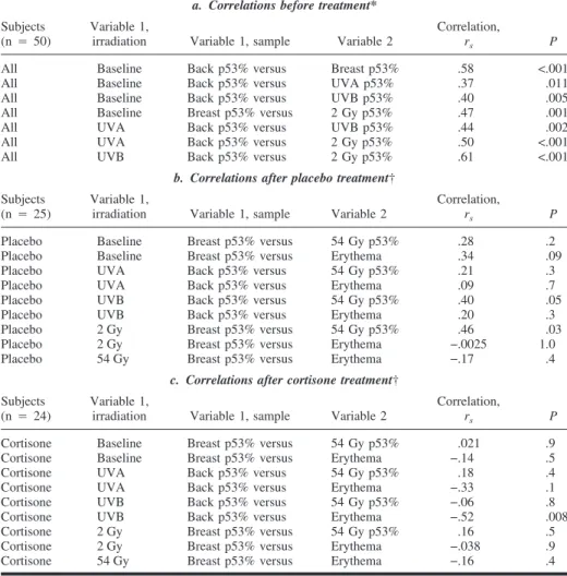
Table 2. Correlations
a. Correlations before treatment*
Subjects (n⳱ 50)
Variable 1,
irradiation Variable 1, sample Variable 2
Correlation,
rs P
All Baseline Back p53% versus Breast p53% .58 <.001 All Baseline Back p53% versus UVA p53% .37 .011 All Baseline Back p53% versus UVB p53% .40 .005 All Baseline Breast p53% versus 2 Gy p53% .47 .001 All UVA Back p53% versus UVB p53% .44 .002 All UVA Back p53% versus 2 Gy p53% .50 <.001 All UVB Back p53% versus 2 Gy p53% .61 <.001
b. Correlations after placebo treatment†
Subjects (n⳱ 25)
Variable 1,
irradiation Variable 1, sample Variable 2
Correlation,
rs P
Placebo Baseline Breast p53% versus 54 Gy p53% .28 .2 Placebo Baseline Breast p53% versus Erythema .34 .09 Placebo UVA Back p53% versus 54 Gy p53% .21 .3 Placebo UVA Back p53% versus Erythema .09 .7 Placebo UVB Back p53% versus 54 Gy p53% .40 .05 Placebo UVB Back p53% versus Erythema .20 .3 Placebo 2 Gy Breast p53% versus 54 Gy p53% .46 .03 Placebo 2 Gy Breast p53% versus Erythema −.0025 1.0 Placebo 54 Gy Breast p53% versus Erythema −.17 .4
c. Correlations after cortisone treatment†
Subjects (n⳱ 24)
Variable 1,
irradiation Variable 1, sample Variable 2
Correlation,
rs P
Cortisone Baseline Breast p53% versus 54 Gy p53% .021 .9 Cortisone Baseline Breast p53% versus Erythema −.14 .5 Cortisone UVA Back p53% versus 54 Gy p53% .18 .4 Cortisone UVA Back p53% versus Erythema −.33 .1 Cortisone UVB Back p53% versus 54 Gy p53% −.06 .8 Cortisone UVB Back p53% versus Erythema −.52 .008 Cortisone 2 Gy Breast p53% versus 54 Gy p53% .16 .5 Cortisone 2 Gy Breast p53% versus Erythema −.038 .9 Cortisone 54 Gy Breast p53% versus Erythema −.16 .4 *a: Nonparametric correlations of p53 immunoreactivity before radiotherapy and after 2 Gy of 5 mV photon-beam radiotherapy to the breast. Data from all 50 patients were used for this calculation. Both Spearman’s rank coefficient of correlation (rs) and the corresponding P value are shown; all statistical tests
are two-sided. In all studied groups, there was a statistically significant correlation between p53 and p21 immunoreactivity. When we compared all skin samples (n⳱ 290), a very good correlation between p53 and p21 was seen (rs⳱ .78; P<.001). There was a strong association between the number of p21-immunoreactive
cells and the intensity of the p21 reaction. Within-individual p53 immunoreactivity correlated irrespective of whether samples were from nonirradiated, UVA-irradiated, UVB-irradiated, or 2-Gy-irradiated skin. In nonirradiated skin, the number of p53-positive back skin cells correlated with the number of p53-positive cells in breast skin (rs⳱ .58; P<.001). There was also a positive correlation among nonirradiated skin and
skin irradiated with UVA, UVB, and 2-Gy photon-beam radiation. There was a clear and statistically significant correlation between p53 immunoreactivity in UVA-irradiated skin and UVB-irradiated skin (rs⳱
.44; P⳱ .002) and between UVA-irradiated skin and skin that had received 2 Gy of photon-beam irradiation (rs⳱ .50; P<.001). The strongest correlation was between p53 immunoreactivity in UVB-irradiated skin and
2-Gy photon-beam irradiated skin (rs⳱ .61; P<.001). In samples taken from skin irradiated with 54 Gy of
photon-beam irradiation, p53 and p21 immunoreactivities were consistently stronger and were not statisti-cally significantly correlated with immunoreactivities in other samples.
†b and c: Nonparametric correlations for both breast erythema and p53 immunoreactivity after radio-therapy was completed in the group of 25 emollient-treated patients and in the topical steroid group of 24 patients. Analyzing the steroid-treated groups and non-steroid-treated patient groups separately could only identify a weak correlation in the p53 reactions after 54 Gy of photon-beam radiation compared with after 2 Gy of radiation and UVB in the non-steroid-treated group. The average objective erythema of the breast showed no correlation to p53 or p21 immunoreactivity in any calculation, except for the negative correlation to UVB in the steroid-treated group. Because the group of emollient-treated patients showed an opposite but very weak correlation, the analysis of UVB versus erythema for all patients did not reveal any correlation (rs⳱ −.18; P ⳱ .2).
damage (1,8), p53 and p21 proteins have
been detected in higher than normal levels
in the epidermis, and we have
demon-strated individual differences in the
ex-pression of p53 protein after exposure to
sunlight (9). Interindividual differences in
the radiosensitivity of human skin have
been detected with clinical end points
(28), and the effects of ionizing radiation
on human skin have also been studied, in
part (33). However, to our knowledge, the
expression of p53 and p21 during and
af-ter radiotherapy has not been analyzed.
We observed that, after 2 Gy of
pho-ton-beam radiation, the p53 response in
epidermis showed the same large
interin-dividual variation as UV-irradiated skin.
Aside from skin repeatedly subjected to
5-mV photon-beam irradiation (total dose
⳱ 54 Gy), where most (80%) of the
ke-ratinocytes were p53 positive, UVB
irra-diation provoked the strongest response;
approximately half (45%) of the
epider-mal cells were strongly immunoreactive
for p53. In addition, we observed that
only 2 Gy of photon-beam irradiation
caused a substantial p53 response (30%)
and that UVA irradiation only caused a
relatively mild response (7%). The p53
responses induced by different types of
radiation might be caused by variations in
DNA damage associated with
wavelength-related differences in absorption.
Alterna-tively, different types of radiation could
trigger different p53 activation pathways,
in a manner independent of the degree of
DNA damage.
It should be noted that a patient with a
strong reaction to UVA will tend to react
strongly to UVB and photon-beam (2 Gy)
irradiation and vice versa. Although the
correlation is strongest between UVB and
photon-beam (2 Gy) irradiation (r
s⳱
.61), there appears to be a general
intrin-sic variation in the general p53
respon-siveness. The molecular reasons for this
interindividual variation are unclear;
however, large interindividual differences
have also been shown in repair of
cy-clobutane pyrimidine dimers and 6-4
photoproducts in human skin exposed to
solar-simulating radiation (26). There
does not appear to be a simple association
of skin type and p53 or p21 response. All
patients received the same dose of
radio-therapy (2 Gy), and the p53 response did
not correlate with skin type (r
s⳱ .12).
The doses of UVA and UVB radiation
that were given were determined because
of different clinical responses and did not
clearly correlate with the intensity of the
p53 response (UVA r
s⳱ .11; UVB r
s⳱
.28). Samples from UVB-irradiated skin
were taken from areas subjected to double
the minimal erythema dose, which
re-sulted in a “just perceptible erythema”
(2). UV radiation causes thymine dimers
and 6-4 photoproducts in DNA; thus,
DNA has been proposed as the
chromo-phore for erythema (34). However, there
is no simple association between the UV
radiation-induced increased levels of p53
in epidermis and clinical erythema (35).
Skin irradiated with a total of 54 Gy was
severely damaged and had strong p53 and
p21 responses. Eventually, individual
variations are probably masked by the
se-vere damage caused by large amounts of
ionizing radiation. The p21 response, in
general, mirrored the p53 response, in
agreement with the hypothesis that, after
DNA damage, p53 induces p21. In
gen-eral, when there was a strong p53
re-sponse (UVB and especially 54 Gy of
photon-beam radiation), there was a strong
p21 response. However, skin that had
re-ceived only 2 Gy of photon-beam radiation
had a weaker p21 response than would be
expected from the p53 response. The
ex-planation for this observation is unclear.
In this study, we show that a few
scat-tered p53-positive cells (approximately
2%) are present in normal, previously
sun-exposed back skin and nonexposed
breast skin. Although some patients may
have an early history of sun exposure to
the breasts, we assume that breast skin of
most women in the age group studied
(47–77 years old) has not been
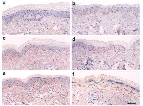
exten-Fig. 2. Immunohistochemically
stained sections of skin, displaying the various layers of the epidermis and the upper part of underlying der-mis. The p53 antibody from one pa-tient who showed a strong p53 re-sponse was used. Immunoreactive keratinocyte nuclei are brown, and nonimmunoreactive nuclei are stained blue with hematoxylin. Nor-mal, nonirradiated breast skin (a) and back skin (b) have a few scattered keratinocytes with various levels of p53 immunoreactivity. Twenty-four hours after 2 Gy of high-voltage pho-ton-beam irradiation, an increased number of p53 immunoreactive cells are seen in the epidermis (c). After radiotherapy was completed (54 Gy), the epidermis is severely damaged, and keratinocytes show strong p53 immunoreactivity (d). Twenty-four hours after UV irradiation, epidermal keratinocytes show an elevated p53 response (e). UVA⳱ 80 J/cm2. (f)
UVB⳱ 600 J/m2. Scale bar for all
sively exposed to the sun. The
p53-positive cells have not been characterized.
These cells could represent single cells
that have acquired a p53 mutation, not
sufficient to cause clonal expansion,
which is so commonly found in
chroni-cally sun-exposed skin (4,5,36). The use
of laser microdissection, gene
amplifica-tion, and DNA sequencing of single cells
(37) would be an important tool to
spe-cifically analyze these cells.
In summary, large interindividual
dif-ferences were observed in the
radiation-induced p53 response in human skin that
is independent of the type of radiation
used. The p53 response did not reflect the
individual radiation erythema. The
bio-logic importance of being a weak or a
strong p53 responder is not known. The
mechanisms regulating the increase in
p53 in vivo in response to cellular damage
and the role of p53-positive cells in normal
nonirradiated skin need additional study.
R
EFERENCES(1) Ponten F, Berne B, Ren ZP, Nister M, Ponten
J. Ultraviolet light induces expression of p53 and p21 in human skin: effect of sunscreen and constitutive p21 expression in skin append-ages. J Invest Dermatol 1995;105:402–6.
(2) Ren ZP, Ponten F, Nister M, Ponten J. Two
distinct p53 immunohistochemical patterns in human squamous-cell skin cancer, precursors and normal epidermis. Int J Cancer 1996;66:174–9.
(3) Ziegler A, Jonason AS, Leffell DJ, Simon JA,
Sharma HW, Kimmelman J, et al. Sunburn and p53 in the onset of skin cancer. Nature 1994; 372:773–6.
(4) Jonason AS, Kunala S, Price GJ, Restifo RJ,
Spinelli HM, Persing JA, et al. Frequent clones of p53-mutated keratinocytes in normal human skin. Proc Natl Acad Sci U S A 1996;93:14025–9.
(5) Ponten F, Berg C, Ahmadian A, Ren ZP, Nister
M, Lundeberg J, et al. Molecular pathology in basal cell cancer with p53 as a genetic marker. Oncogene 1997;15:1059–67.
(6) Ren ZP, Ahmadian A, Ponten F, Nister M,
Berg C, Lundeberg J, et al. Benign clonal ke-ratinocyte patches with p53 mutations show no genetic link to synchronous squamous cell pre-cancer or pre-cancer in human skin. Am J Pathol 1997;150:1791–803.
(7) Campbell C, Quinn AG, Angus B, Farr PM,
Rees JL. Wavelength specific patterns of p53 induction in human skin following exposure to UV radiation. Cancer Res 1993;53:2697–9.
(8) Hall PA, McKee PH, Menage HD, Dover R,
Lane DP. High levels of p53 protein in UV-irradiated normal human skin. Oncogene 1993; 8:203–7.
(9) Berne B, Ponten J, Ponten F. Decreased p53
expression in chronically sun-exposed human skin after topical photoprotection. Photoderma-tol Photoimmunol Photomed 1998;14:148–53.
(10) Ananthaswamy HN, Loughlin SM, Cox P,
Evans RL, Ullrich SE, Kripke ML. Sunlight and skin cancer: inhibition of p53 mutations in
UV-irradiated mouse skin by sunscreens. Nat Med 1997;3:510–4.
(11) Chadwick CA, Potten CS, Nikaido O,
Matsu-naga T, Proby C, Young AR. The detection of cyclobutane thymine dimers, (6-4) photole-sions and the Dewar photoisomers in sections of UV-irradiated human skin using specific an-tibodies, and the demonstration of depth pen-etration effects. J Photochem Photobiol B 1995;28:163–70.
(12) Young AR, Chadwick CA, Harrison GI, Hawk
JL, Nikaido O, Potten CS. The in situ repair kinetics of epidermal thymine dimers and 6-4 photoproducts in human skin types I and II. J Invest Dermatol 1996;106:1307–13.
(13) Liu M, Dhanwada KR, Birt DF, Hecht S,
Pel-ling JC. Increase in p53 protein half-life in mouse keratinocytes following UV-B irradia-tion. Carcinogenesis 1994;15:1089–92.
(14) el-Deiry WS, Tokino T, Velculescu VE, Levy
DB, Parsons R, Trent JM, et al. WAF1, a po-tential mediator of p53 tumor suppression. Cell 1993;75:817–25.
(15) Harris C. Structure and function of the p53
tumor suppressor gene: clues for rational can-cer therapeutic strategies. J Natl Cancan-cer Inst 1996;16:1442–55.
(16) Brown J, Wouters BG. Apoptosis, p53, and
tumor cell sensitivity to anticancer agents. Cancer Res 1999;59:1391–9.
(17) Brash DE. Cellular proofreading. Nat Med
1996;2:525–6.
(18) Oda K, Arakawa H, Tanaka T, Matsuda K,
Tanikawa C, Mori T, et al. p53AIP1, a poten-tial mediator of p53-dependent apoptosis, and its regulation by Ser-46-phosphorylated p53. Cell 2000;102:849–62.
(19) Brash DE, Ziegler A, Jonason AS, Simon JA,
Kunala S, Leffell DJ. Sunlight and sunburn in human skin cancer: p53, apoptosis, and tumor promotion. J Investig Dermatol Symp Proc 1996;1:136–42.
(20) Ko LJ, Prives C. p53: puzzle and paradigm.
Genes Dev 1996;10:1054–72.
(21) Levine AJ. p53, the cellular gatekeeper for
growth and division. Cell 1997;88:323–31.
(22) Kastan MB, Onyekwere O, Sidransky D,
Vo-gelstein B, Craig RW. Participation of p53 pro-tein in the cellular response to DNA damage. Cancer Res 1991;51(23 Pt 1):6304–11.
(23) Gottlieb E, Haffner R, King A, Asher G, Gruss
P, Lonai P, et al. Transgenic mouse model for studying the transcriptional activity of the p53 protein: age- and tissue-dependent changes in radiation-induced activation during embryo-genesis. EMBO J 1997;16:1381–90.
(24) Komarova EA, Chernov MV, Franks R, Wang
K, Armin G, Zelnick CR, et al. Transgenic mice with p53-responsive lacZ: p53 activity varies dramatically during normal development and determines radiation and drug sensitivity in
vivo. EMBO J 1997;16:1391–400.
(25) Midgley CA, Owens B, Briscoe CV, Thomas
DB, Lane DP, Hall PA. Coupling between gamma irradiation, p53 induction and the apoptotic response depends upon cell type in
vivo. J Cell Sci 1995;108(Pt 5):1843–8. (26) Bykov VJ, Sheehan JM, Hemminki K, Young
AR. In situ repair of cyclobutane pyrimidine dimers and 6-4 photoproducts in human skin
exposed to solar simulating radiation. J Invest Dermatol 1999;112:326–31.
(27) Spittle MT. Radiotherapy and reactions to
ion-izing radiation. In: Champion RH, Burton JL, Ebling FJ, editors. Textbook of dermatology. Oxford (U.K.): Blackwell; 1992. p. 3089.
(28) Tucker SL, Turesson I, Thames HD. Evidence
for individual differences in the radiosensitiv-ity of human skin. Eur J Cancer 1992;28A: 1783–91.
(29) Fitzpatrick TB. The validity and practicality of
sun-reactive skin types I through VI. Arch Der-matol 1988;124:869–71.
(30) Liljegren G, Holmberg L, Adami HO,
West-man G, GraffWest-man S, Bergh J. Sector resection with or without postoperative radiotherapy for stage I breast cancer: five-year results of a ran-domized trial. Uppsala–Orebro Breast Cancer Study Group. J Natl Cancer Inst 1994;86:717–22.
(31) Williams C, Ponten F, Ahmadian A, Ren ZP,
Ling G, Rollman O, et al. Clones of normal keratinocytes and a variety of simultaneously present epidermal neoplastic lesions contain a multitude of p53 gene mutations in a xero-derma pigmentosum patient. Cancer Res 1998; 58:2449–55.
(32) Lever WF, Schaumberg-Lever G.
Histopathol-ogy of the skin. 6thed. Philadelphia (PA): J. B.
Lippincott; 1983.
(33) Nyman J, Turesson I. Basal cell density in
hu-man skin for various fractionation schedules in radiotherapy. Radiother Oncol 1994;33:117–24.
(34) Young AR, Chadwick CA, Harrison GI,
Ni-kaido O, Ramsden J, Potten CS. The similarity of action spectra for thymine dimers in human epidermis and erythema suggests that DNA is the chromophore for erythema. J Invest Der-matol 1998;111:982–8.
(35) Healy E, Reynolds NJ, Smith MD, Campbell
C, Farr PM, Rees JL. Dissociation of erythema and p53 protein expression in human skin fol-lowing UVB irradiation, and induction of p53 protein and mRNA following application of skin irritants. J Invest Dermatol 1994;103:493–9.
(36) Ren ZP, Hedrum A, Ponten F, Nister M,
Ah-madian A, Lundeberg J, et al. Human epider-mal cancer and accompanying precursors have identical p53 mutations different from p53 mu-tations in adjacent areas of clonally expanded non-neoplastic keratinocytes. Oncogene 1996; 12:765–73.
(37) Ponten F, Williams C, Ling G, Afshin A,
Nis-ter M, Lundeberg J, et al. Genomic analysis of single cells from human basal cell using laser-assisted capture microscopy. Mutat Res 1997; 382:45–55.
N
OTESSupported by grants from the Swedish Cancer So-ciety, the Lions Cancer Research Foundation, the Ju-bilee Foundation, the Torsten and Ragnar So¨derbergs Foundation, and Stockholm’s Cancer Foundation.
We thank Dr. Christina Nyberg who has per-formed meticulous microscopy of all p53- and p21-stained slides. The late professor Jan Ponten is greatly acknowledged for his stringent comments and suggestions on the project and on the earlier versions of this manuscript.
Manuscript received June 5, 2000; revised No-vember 1, 2000; accepted NoNo-vember 14, 2000.
![[PDF] Cours vba Access avec explications et exemples pour reviser ensemble | Cours access](data:image/gif;base64,R0lGODlhAQABAIAAAP///wAAACH5BAEAAAAALAAAAAABAAEAAAICRAEAOw==)