528 Letters to the Editor
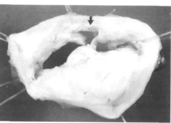
Figure 1 Excised mitral valve specimen. View is from the atrial side. Note the large tear in the posterolateral leaflet
111.
commissures has been documented to be the mechanism by which mitral valve area increases following balloon valvuloplasty. This same mechanism is responsible for mild increases in mitral regurgitation141.
Outcome following balloon valvuloplasty has been related to the BVR score'31. Only one study has been able to demonstrate a correlation between valvular morphology and the development of mitral regurgitation151. From an echocardiographic point of view the patient described had a favourable valve for mitral com-missurotomy. The severe mitral regur-gitation that resulted was due to non-commissural tearing of the pos-terolateral leaflet. This complication is in keeping with the findings of Essop'41, who speculates that 'ex-cessively pliable valves may constitute a risk factor for leaflet rupture and abrupt mitral regurgitation'. Though outcome was better for patients with a lower BVR score in the NHLBI study131, valve morphology did not predict the development of mitral regurgitation in this or other studies'41. T. G. HENNESSY H. A. MCCANN D. D. SUGRUE Department of Clinical Cardiology,
Mater Misericordiae Hospital, Dublin, Ireland References
[1] Herrmann HC, Lima JA. Feldman T et al. Mechanisms and outcome of severe mitral regurgitation after Inoue balloon valvuloplasty. J Am Coll Cardiol 1993; 22: 783-9.
[2] Wilkins GT, Weyman AE, Abascal VM el al. Percutaneous balloon dilatation
of the mitral valve: an analysis of echocardiographic variables related to outcome and the mechanism of dilation. Br Heart J 1988; 60: 299-308. [3] Multicenter experience with balloon mitral commissurotomy The NHBLI balloon valvuloplasty registry report on immediate and 30 day follow up results. Circulation 1992; 85. 448-61.
[4] Essop MR, Wisenbaugh T, Skoularigis J, Middlemost S, Sareh P. Mitral re-gurgitation following mitral balloon valvotomy. Circulation 1991; 84: 1669-79.
[5] Nobuyoshi M, Hamaski N, Kimura T et al. Indications, complications, and short term clinical outcome of percu-taneous transvenous mitral com-missurotomy. Circulation 1989; 80: 782-92.
Late in-stent restenosis in coronary arteries and in grafts
Intracoronary stents were at first limited to the treatment of restenosis, angioplasty-related acute coronary closure and graft lesions. Their use has greatly widened to include new onset lesions in native vessels, following two recent trials showing less restenosis in patients treated with stents compared to conventional angioplasty'1-21. No long-term prospective data are avail-able on stenting. One report suggests that the time course of restenosis after intracoronary stenting is similar to that after conventional angioplasty131, while another hypothesizes that stent-ing may only be a mechanical way of delaying restenosis process'41. Thus, we reviewed the incidence of late (defined as >6 months) in-stent restenosis in our early series of 131 Wallstents implanted between 1986 and 1989.
We report on four male patients aged 50 to 70 who underwent conventional balloon angioplasty for stenosis >50% measured by quanti-tative coronary angiographic analysis. Stents were implanted for restenosis in one native right coronary artery, for angioplasty-induced acute closure in one venous graft and one native right coronary artery, and for suboptimal results after angioplasty in a venous graft. A total of six self-expandable Wallstents (Schneider AG, Zurich, Switzerland) were implanted using the standard technique'51. All patients were treated with anticoagulation for 6 months and antiplatelet therapy. Six months following the procedure, a control coronary angiogram was per-formed. From then onwards, repeat angiograms were dictated by each patient's individual clinical outcome.
Quantitative coronary angio-graphic analysis was performed and in-stent restenosis was defined as a reduction of the vessel diameter to < 50% located at the very same site as the original lesion.
Clinically, patients remained well during the first 6 months follow-ing stent implantation. There was no angiographic restenosis at 6 months (Fig. 1, Table 1). In two patients, further follow-up angiograms were obtained. One patient complained of increasing atypical chest pain 18 months after stent implantation and a coronary angiogram showed no changes in the coronary status, in par-ticular there was no in-stent restenosis (Table 1). The second patient devel-oped recurrent angina 38 months after stent implantation and underwent suc-cessful coronary artery bypass surgery for a proximal left anterior descending artery lesion; there was no in-stent restenosis (Fig. 1, Table 1).
All four patients presented with recurrent angina pectoris 43 and 87 months after stent implantation and late in-stent restenosis was diag-nosed (Fig. 1, Tale 1). There was also progression of coronary artery disease (significant stenosis >50%) within the same vessel in two patients.
These late restenosis were successfully treated with conventional balloon angioplasty in three patients and with coronary artery bypass grafting in one.
Following intracoronary stent-ing. as after conventional angioplasty, a 6-month period is considered to be critical for restenosis. Its rate varies between 20 and 40% depending on many factors such as definition of Eur Heart J. Vol. 18. March 1997
Letters to the Editor 529
Figure 1 Angiographic images of
the right coronary artery in patient 1: (a) stenosis before stent implan-tation; (b) immediate result after stenting; (c) 6 months follow-up angiogram showing no in-stent re-stenosis; (d) in-stent restenosis 7 years and 3 months after implan-tation; (e) immediate result after in-stent conventional balloon angioplasty.
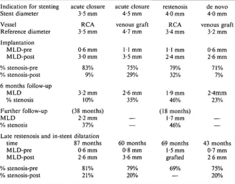
Table 1 Angiographic data in four patients developing very late re-stenosis within Wallstents
Indication for stenting Stent diameter Vessel Reference diameter Implantation MLD-pre MLD-post % stenosis-pre % stenosis-post 6 months follow-up MLD % stenosis Further follow-up MLD % stenosis acute closure 3-5 mm RCA 3-5 mm 0 6 mm 3 0 mm 83% 9% 3-2 mm 10% (38 months) 2-2 mm 37% Late restenosis and in-stent dilatation
time 87 months MLD-pre 0-6 mm MLD-post 2-6 mm % stenosis-pre % stenosis-post 81% 21% acute closure 4-5 mm venous graft 4-7 mm 11 mm 3-5 mm 75% 29% 2-6 mm 35% — 60 months 0-8 mm 3-6 mm 79% 20% restenosis 4 0 mm RCA 3-4 mm 11 mm 2-4 mm 79% 32% 1-9 mm 46% (18 months) 1-7 mm 46% 69 months 1-5 mm grafted 69% de novo 4-0 mm venous graft 3-2 mm 0 6 mm 2-6 mm 71% 7% 2-4mm 23% — 43 months 0-7 mm 2 6 mm 75% 20% RCA = right coronary artery; MLD = mean lumen diameter; pre = before procedure; post = after procedure.
restenosis, indication for stenting, cohort of patients, anticoagulation regimen etc. Assessment of coronary lumen dimension demonstrates peak changes at 3 months and, in most cases, remains stable after 6 months'31. Restenosis is believed to be very rare later'1"31. However, some isolated reports suggest that in-stent re-stenosis can occur late'6', particularly in grafts. The theory that stenting, through
its double action of creating a larger immediate vessel diameter and of inhibiting elastic recoil, may only be a way of delaying intimal hyperplasia — the characteristic feature of restenosis — as opposed to avoiding it, has also recently been put forward'41.
In our series of 131 Wallstents implanted in 105 patients, 100% 6-months clinical and angiographic follow-up was obtained. At late
follow-up 89 (range 55 to 104) months after stenting, 20 (19%) patients (including four non-cardiac deaths) were lost to follow-up, and clinical follow-up was obtained on the 85 (81%) remaining patients. There were 9 (9%) stent related deaths. Eight patients (8%) had had a subacute thrombosis and 15 (14%), a restenosis at 6 months. Of the 53 remaining patients, 34 (representing 64% of the eligible patients), including the four patients reported here, underwent a late angiogram. This control was re-fused by the other 19 patients or their referring doctors, because of lack of symptoms. Hence in our group of 105 patients, four symptomatic late in-stent restenoses have occurred, amounting to 4% of the initial group. Because of the long delay between stenting and restenosis, cor-onary artery disease progression could be advocated as opposed to true re-stenosis. Great care was taken to en-sure that restenosis was located at the site originally dilated. Moreover, pro-gression of coronary artery disease was noted in two of the four cases outside the stent and was treated as separate lesions.
One potential explanation for these very late in-stent restenoses would be a reaction to stent breakage due to shear wear and tear over years. However this remains pure speculation as we do not have any pathology specimen.
In conclusion, very late-in-stent restenosis has to be expected. It is encouraging to estimate its incidence at a low figure, probably comparable
530 Letters to the Editor
to that after conventional balloon angioplasty. N. M. G. DEBBAS U. SIGWART E. EECKHOUT P. VOGT J. C. STAUFFER L. K.APPENBERGER J. J. GOY Division of Cardiology, Centre Hospitalier Universitaire Vaudois,
Lausanne, Switzerland References
[1] Serruys PW, de Jaegere P, Kiemeneij F et al. A comparison of balloon expand-able stent implantation with balloon angioplasty in patients with coronary artery disease. N Engl J Med 1994; 331: 489-95.
[2] Fischman DL, Leon MB, Bairn DS et al. A randomized comparison of coronary stent placement and balloon angioplasty in the treatment of cor-onary artery disease. N Engl J Med
1994; 331: 496-501.
[3] Kimura T, Nosaka H, Yokoi H, Iwabuchi M, Nobuyoshi M. Serial angiographic follow-up after Palmaz-Schatz stent implantation: comparison with conventional angioplasty. J Am Coll Cardiol 1993; 21: 1557-63.
[4] Topol E. Caveats about elective cor-onary stenting. N Engl J Med 1994; 539^1.
[5] Sigwart U, Puel J, Mirkowitch V, Joffre F, Kappenberger I. Intravascular stents to prevent occlusion and restenosis after transluminal angioplasty. N Engl J Med 1987; 316: 701-6.
[6] Eeckhout E, Goy JJ, Stauffer JC, Vogt P, Kappenberger L. Endoluminal stent-ing of narrowed saphenous vein grafts: long term clinical and angiographical follow-up. Cathet Cardiovasc Diagn 1994; 32: 139-46.
The mechanism of catecholaminergic polymorphic ventricular tachycardia may be triggered activity due to delayed afterdepolarization
Bi-directional or polymorphic ven-tricular tachycardia (VT) is an uncom-mon disorder whose aetiology and mechanism is unknown. There are a few reports on patients with bidirec-tional or polymorphic VT in the absence of structural heart disease. Among them, Leenhardt et al. have reported cases of catecholaminergic polymorphic VT[1).
We also had a case of cat-echolaminergic polymorphic VT. A 14-year-old Japanese male was referred to us for the evaluation of ventricular tachyarrhythmias. Since the age of 5 years, he had experienced several episodes of syncope due to ventricular tachyarrhythmia associ-ated with exercise or an increase in physical activity. His family history revealed no evidence of syncope or of sudden death. Physical examination revealed a slow pulse rate, and a car-diac pansystolic murmur (Levein 2/6) was audible at the apex of the heart. Results of biochemical tests were nor-mal. The resting 12-lead ECG showed sinus bradycardia with a rate of 42 beats, min"1, a normal QT interval with QTc 0-41 s. Chest X-rays showed a cardiothoracic index of 0-48. Echocardiography revealed mild mitral valve regurgitation, but good ventricular function. Cardiac cath-eterization revealed normal coronary arteries. Despite the presence of mild mitral valve regurgitation, we were convinced that structural heart disease was absent. In this case, bidirectional or polymorphic VT was reproducibly induced by humoral (exercise, iso-proterenol injection) or neurogenic (stress, emotion) sympathetic
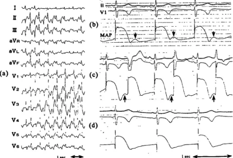
stimu-Figure 1 (a) Twelve-lead electrocardiogram during bidirectional VT. There is an
alternating pattern of right and left bundle branch block (R/LBBB), or an alternating left and right axis deviation with an RBBB or LBBB pattern. Thus, QRS pattern varied, (b) Recordings of MAP at right ventricular inflow. After bidirectional VT induced spontaneously by emotion disappeared, humps are observed during phase 4 (arrows) (c) During the infusion of isoproterenol, the humps gradually increased (arrows), (d) After the injection of propranolol, the humps gradually reduced and finally disappeared.