AJH 1988; 1:245-248
Effect of Calcium-Channel Blockade on the
Aldosterone Response to Sodium Depletion
and Potassium Loading in Man
Laurent Favre, Anne Riondel, and Michel B. Vallotton
Angiotensin H (Ang II) and potassium (K+) increase
aldosterone (Aldo) production in vitro via Ca1*-dependent
mechanisms. To determine the effects of Ca2+ antagonism
in vivo, we examined the influence of nifedipine on the Aldo response to Na+ depletion and K+ loading in 11
healthy subjects. On the fifth day of a low-Na+/high-K+
diet (10 mmol Na+/100 mmol K+) the subjects were
randomly given either nifedipine 30 mg po or placebo, and on the sixth day they received the alternative drug. KCl in 5% glucose was infused on days 5 and 6 from 10:00 to 12:00 AM(0.6 mmol/kg over 2 hours). Dexa-methasone was given to suppress adrenal corticotrophic hormone. Plasma renin activity (PRA) and plasma Aldo were determined every 20 minutes. Nifedipine induced a rise in heart rate at 60 minutes but did not change blood pressure. During KCl/glucose infusions, plasma glucose
increased significantly, but plasma K+ remained stable.
PRA, but not baseline plasma Aldo, was stimulated by nifedipine. KCl provoked a significant and similar Aldo rise (P < .01) under placebo and nifedipine. Baseline Aldo/PRA ratio was reduced under nifedipine when
compared to placebo (P < .01), whereas during KCl infusions this ratio was similarly elevated under placebo and nifedipine. We conclude that acute inhibition of slow Ca2+ channels does not interfere with K+-induced Aldo
secretion in man, suggesting that adaptive mechanisms operate in vivo. Am J Hypertens 1 9 8 8 ; 1 : 2 4 5 - 2 4 8
KEY WORDS: Aldosterone response, calcium-channel
blockade, nifedipine, potassium loading, sodium depletion.
P
otassium (K+) and angiotensin II (Ang II) stimulate aldosterone (Aldo) production via calcium (Ca2 +)-dependent mechanisms. Earlier studies
using isolated adrenal glomerulosa cells have shown that inhibition of C a2 + uptake blocked the steroi
dogenic response to both K+ and Ang I I .1"3 Recent work
from this laboratory4 indicates that both Ang II and K+
increase intracellular C a2 + ( [ C a2 +] i ) , but they do so by
different mechanisms: Whereas C a2 + antagonists can
From the Division of Endocrinology, University Hospital, Geneva, Switzerland.
This study was supported by a grant from the Swiss National Science Foundation No. 3. 9 1 4 - 0 . 8 3 .
Address correspondence and reprint requests to Michael B. Vallot ton, Division of Endocrinology, University Hospital, C H - 1 2 1 1 G e neva 4/Switzerland.
totally block the K+-induced [ C a2 +] i rise resulting from
the opening of voltage-dependent C a2 + channels and
the subsequent Aldo production, they do not suppress the receptor-mediated [ C a2 +] i rise induced by Ang II,
though they reduce the steroidogenesis slightly. An inhibition of Aldo response to Ang II by acute administration of Ca2 +-antagonists has been observed
in human studies,5"7 but the effect of calcium antago
nism on K+-induced Aldo stimulation has not yet been
examined. The present study was therefore designed to compare the influence of nifedipine on the Aldo re sponse to N a+ depletion and K+ loading in normal man.
The experimental protocol we used raised a further working hypothesis regarding the mechanism by which K+-induced Aldo stimulation may involve insulin ac
tion. Our results suggest that intracellular K+ may also
influence Aldo secretion via a Ca2 +-independent mech
anism.
246 F A V R E ET A L . AJH-JULY 1988-VOL 1, NO. 3, PART 1
M E T H O D S
Eleven healthy male subjects aged 23 to 27 years volun teered to participate in the study. They gave their in formed consent to the experimental protocol, which has been approved by the Ethical Committee of the Depart ment of Medicine.
The subjects were kept for 6 days on a strict low-Na+
(15 mmol) and high-K+ (100 mmol) daily intake. On
day 5 the subjects were randomly given either nifedi pine (20 mg at 7:00 AM and 10 mg at 10:00 AM po) or a placebo. On day 6 they received the alternative drug. Dexamethasone (1 mg) was taken at midnight on days 4 and 5 to suppress adrenal corticotrophic hormone (ACTH). On the fifth and the sixth days two similar KCl-infusion tests were performed from 10:00 AM to 12:00 noon. After fasting overnight, the subjects re mained supine from 7:00 AM to 12:00 noon. KCl diluted in 5 % glucose (50 mmol KC1/L) was infused over 2 hours (0.6 mmol/kg) through a large humeral vein.
Blood was drawn every 20 minutes during the KCl infusion for determination of plasma N a+ and K+,
plasma renin activity (PRA) and Aldo. Plasma glucose and plasma Cortisol were measured at 60 and 120 min utes. Plasma insulin was also determined in five sub jects. Blood pressure and heart rate were checked regu larly during the tests. Urinary N a+ and K+ were
measured in 24-hour urine on the fifth day of the diet and in a urine spot on the sixth day.
N a+ and K+ were determined by flame photometry
and plasma glucose by the glucose-oxydase method. PRA, plasma Cortisol and insulin, and urinary Aldo were determined by radioimmunoassay (RIA). Plasma aldos terone was measured by RIA using a commercial kit (Coat-A-Count-Diagnostic Products Corp). The limit of detection was 1.5 n g / 1 0 0 mL, and the intra-assay coef ficient of variation was 5 . 1 % .
Statistical analysis was performed by paired t test, the subjects being their own controls. Results are expressed as the mean ± SEM.
R E S U L T S
On the fifth day of l o w - N a+/ h i g h - K+ diet, 24-hour uri
nary excretion was 19.8 ± 2.1 mmol for N a+ and
109.9 ± 9.0 mmol for K+. Urinary excretion of Aldo was
42.0 ± 2 . 7 / / g / 2 4 hours. In the sixth day urine spot N a+
was 12.0 ± 2.2 m m o l / L and K+, 77.0 ± 2.7 mmol/L.
Mean body weight was 68.7 ± 3.3 kg on the fifth day and 68.5 ± 3.2 kg on the sixth day (difference not sig nificant [NS]).
Before KCl infusion blood pressure was 114 ± 2 / 69 ± 2 mm Hg on placebo and 115 ± 2 / 6 9 ± 1 mm Hg on nifedipine (NS). Heart rate was 61 ± 2 and 66 ± 3 beats/minute, respectively (NS). At the end of the KCl infusion, blood pressure was 1 1 3 ± 2 / 7 1 ± 2 mm Hg on placebo and 115 ± 2 / 6 9 ± 2 mm Hg on nifedipine
(NS), and heart rate was 60 ± 2 and 65 ± 3 beats/min-ute, respectively (NS). However at 60 minutes, heart rate was significantly higher on nifedipine (75 ± 4) than on placebo (60 ± 2, Ρ < .01).
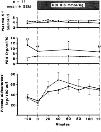
The mean amount of KCl infused over 2 hours (0.6 mmol/kg) was 41.1 ± 1 . 7 mmol. Despite this large load, plasma K+ did not change significantly during the
2-hour infusion in either test (Figure 1). Plasma N a+
remained constant. The infused volume was 831 ± 42 mL over 2 hours, providing a glucose load of 41.6 ± 2.1 g. On both placebo and nifedipine days, plasma glu cose and plasma insulin increased significantly to a similar extent during K C l - 5 % glucose infusion with a peak at 60 minutes.
As shown in Figure 1, baseline PRA increased by N a+
depletion was further stimulated by nifedipine treat ment, whereas plasma Aldo levels were similar on pla cebo and nifedipine days. Consequently the Aldo/PRA ratio was significantly higher on placebo (5.6 ± 0.6 at time 0) than on nifedipine (3.9 ± 0.5, Ρ < .01). During KCl infusion, despite the absence of change in plasma K+ concentration, there was a marked and highly signif
icant rise in plasma Aldo, reaching a peak at 40 minutes on placebo (P < .01 vs baseline) and at 60 minutes on
ι 1 ι ι ι ι ι I
- 2 0 0 2 0 4 0 6 0 8 0 1 0 0 1 2 0 Minutes
ο ο P l a c e b o · · Nifedipine
FIGURE 1. Effect of a 2-hour KCl IV infusion (0.6 mmol/kg in 5% glucose) on plasma potassium concentration, plasma renin activity and plasma aldosterone levels, in 11 healthy subjects during sodium restriction. See text for statistics.
AJH-JULY 1988-VOL 1, NO. 3, PART 1 NIFEDIPINE A N D A L D O S T E R O N E 247
nifedipine (P < .001 vs baseline); peak values were not significantly different on placebo day (41.2 ± 10.0 n g / dL) and on nifedipine day (34.8 ± 3.8 ng/dL). The Aldo/PRA ratio during KCl infusion was not signifi cantly different on placebo and on nifedipine. Plasma
Cortisol levels remained suppressed during the tests
(1.7 ± 0.4 /zg/dL on placebo and 1.3 ± 0.3 on nifedi-pine/NS).
D I S C U S S I O N
In vitro studies using adrenal glomerulosa cells indicate that although Ang II and K+ both stimulate Aldo secre
tion by increasing [Ca2 - 1^, only the K+-mediated stimula
tion, acting through voltage-dependent C a2 + channels,
is specifically blocked by C a2 + antagonists.4 Slow C a2 +
-channel blockers can also decrease Ang II-induced Aldo production, but their effect is less marked than on KCl-induced steroidogenesis. This lesser effect is a con sequence of a lower basal intracellular C a2 + concentra
tion resulting from depletion of [ C a2 +] i stores by these
drugs and preventing attainment of the critical thresh old for triggering steroidogenesis. From these in vitro findings, it might therefore be inferred that acute inhibi tion of C a2 + channels would interfere with K+-induced
Aldo stimulation. In a sheep whose adrenal gland was transplanted to its neck, the local infusion of C a2 + antag
onists reversed the Aldo response to K+ but did not
affect Aldo secretion induced by N a+ depletion.8'9
To assess the effect of K+ on Aldo secretion, KCl
was infused acutely after stimulation of the r e n i n -angiotensin system as in previous studies.1 0 , 1 1 It is
known that in normal subjects a clear Aldo response to K+ occurs only in the presence of high concentrations of
Ang I I .1 0 This fact emphasizes the important interde
pendence of Ang II and K+.1 2 KCl was diluted in glucose
to prevent a local venous toxic reaction. Although plasma K+ did not rise during the KCl-glucose infusion,
there is strong evidence that Aldo stimulation was di rectly induced by K+, as previously reported.1 0 Glucose
ingestion acutely lowers plasma concentrations of K+
and Aldo,1 1 whereas plasma Aldo levels rise during
KCl - glucose infusion, despite an unchanged K+ plasma
concentration,1 0 as confirmed in the present study.
Whether K+ enters the cell in order to stimulate steroi
dogenesis is still conjectural, as opposite conclusions have been drawn from in vitro or in vivo studies using direct or indirect estimation of intracellular K+ con
t e n t .1 0 1 1 , 1 3 Insulin stimulates K+ cellular uptake by the
liver and muscle tissues,14 but it is not known whether
tissues like the adrenal cortex are also involved. These findings suggest that either membrane depolarization occurs without any apparent change in K+ extracellular
concentration (which is unlikely, as high concentration is required in vitro to depolarize glomerulosa cells4), or
that the signal may result from transmembrane K+
fluxes leading to a higher intracellular K+ concentration.
If these processes are mediated by the C a2 + fluxes dem
onstrated in vitro,4 they would also be blocked in vivo
by C a2 + antagonists. This was not the case in this study
because, contrary to our expectation, nifedipine was un able to prevent the K+-induced rise in Aldo concentra
tion, the peak value of which during KCl infusion being similar on placebo and nifedipine treatment. Similar re sults showing no interference by nifedipine with Aldo secretion have been reported recently in hypertensive patients given an acute KCl infusion.1 5 Our findings
indicate that the mechanism of K+-induced Aldo secre
tion under these particular in vivo circumstances differs from that observed in vitro in isolated cells. In this study, as in an earlier o n e ,1 6 insulin response to hyperglycemia
was not altered by nifedipine. The two KCl infusions were therefore comparable in terms of glucose load and insulin secretion. Insulin alone can influence plasma K+
concentration, baseline plasma Aldo levels and its re sponse to Ang II in various ways.1 7
In the present in vivo human study under low salt intake, nifedipine reduced the Aldo/PRA ratio but did not alter K+-induced Aldo secretion. Renin secretion
was clearly stimulated as a result of a combined effect of N a+ depletion and acute inhibition of slow C a2 + chan
nels, and as previously reported,1 8 it was not modified
by KCl administration. The high basal plasma concen tration of Aldo induced by Ang II stimulation was simi lar on nifedipine and placebo. Despite the known higher sensitivity of adrenal cells after N a+ depletion,1 9 no fur
ther increase in Aldo secretion could be obtained in re sponse to the acute renin rise under nifedipine treat ment. The finding of a lower Aldo/PRA ratio during nifedipine treatment, previously reported in hyperten sive patients,2 0 is compatible with the acute inhibitory
effect of nifedipine on steroidogenesis during Ang II infusion in m a n .5"7 Another unrelated C a2 + antagonist,
verapamil, did inhibit Aldo response to Ang II, but only when given chronically.2 1 During sodium restriction in
normal subjects nifedipine and diltiazem reduced the sensitivity of Aldo secretion to Ang I I .7
As nifedipine has been shown to increase apparent liver blood flow in normal subjects,2 2 an alteration of
Aldo metabolic clearance rate is to be considered so that changes in Aldo plasma concentrations may not parallel changes in Aldo secretion. To exclude any contribution of another Ca2 +-independent adrenal trophic factor,
ACTH was constantly suppressed by dexamethasone during the two test days. In fact Guthrie et a l2 1 have
reported that verapamil does not alter the Aldo response to ACTH. Nifedipine, known to bind with high affinity and specificity to the voltage-dependent C a2 + chan
n e l ,2 3 was administered shortly before KCl infusion. It
may be inferred from pharmacokinetic data that the dose selected would result in plasma concentrations in the same micromolar range as the in vitro studies, al though individual bioavailability may vary.2 4 The PRA
248 F A V R E ET AL. AJH-JULY 1988-VOL 1, NO. 3, PART 1
rise obtained in every subject indicates that effective drug levels were achieved. Blood pressure was not in-fluenced, as would be expected in normotensive sub-jects.2 5
In summary, acute blockade of slow Ca2 +-channels
does not interfere with K+-induced Aldo secretion but
reduces the Aldo/PRA ratio. These findings suggest some inhibition of the Ang 2-induced Aldo secretion. The discrepancy with in vitro findings indicates that adaptive mechanisms operate in vivo allowing the maintenance of Aldo secretion in normal man. Finally, these results support the existence of a role for intracel-lular K+ in stimulating Aldo secretion.
A C K N O W L E D G M E N T S
The authors are grateful to Dr L. Vadas (Central Laboratory of Clinical Chemistry) for insulin determinations, to Mrs L. Bockhorn, M. Lopez, W. Dimeck and C. Chauffat-Rabère for their excellent technical assistance, and to Mrs M. Aebischer for her secretarial help.
R E F E R E N C E S
1. Fakunding JL, Catt KJ: Dependency of aldosterone stim-ulation in adrenal glomerulosa cells on calcium uptake: Effects of lanthanum and verapamil. Endocrinology 1980;107:1345.
2. Schiffrin EL, Lis M, Gutkowska J, Genest J: Role of Ca++ in response of adrenal glomerulosa cells to angiotensin II, ACTH, K+ and ouabain. Am J Physiol 1981;241:E42. 3. Foster R, Lobo MV, Rasmussen H, Marusic ET: Calcium: Its role in the mechanism of action of angiotensin II and potassium in aldosterone production. Endocrinology 1981;109:2196.
4. Capponi AM, Lew PD, Vallotton MB: Correlation be-tween cytosolic free C a2 + and aldosterone production
in bovine adrenal glomerular cells. J Biol Chem 1984,259:8863.
5. Millar JA, McLean K, Reid JL: Calcium antagonists de-crease adrenal and vascular responsiveness to angioten-sin II in normal man. Clin Sei 1981;61:65s.
6. Vierhapper H, Waldhäusl W: Reduced pressor effect of angiotensin II and of noradrenaline in normal man fol-lowing the oral administration of the calcium-antagonist nifedipine. Eur J Clin Invest 1982;12:263.
7. Anderson GH Jr, Howland T, Domschek R, Streeten DHP: Effect of sodium balance and calcium channel-blocking drugs on plasma aldosterone responses to infu-sion of angiotensin II in normal subjects and patients with essential hypertension. J Clin Endocrinol Metab 1986;63:1126.
8. Johnson EIM, McDougall JG, Coghlan JP, et al: Potas-sium stimulation of aldosterone secretion in vivo is re-versed by nisoldipine, a calcium transport antagonist. Endocrinology 1984;114:1466.
9. Johnson EIM, McDougall JG, Coghlan JP, et al: Ca antag-onists do not alter aldosterone secretion during estab-lished Na depletion. Am J Physiol 1985;248:E676.
10. Birkhäuser M, Gaillard R, Riondel AM, et al: Effect of volume expansion by hyperosmolar and hyperoncotic solutions under constant infusion of angiotensin II on plasma aldosterone in man and its counterbalance by potassium administration. Europ J Clin Invest 1973;3:307.
11. Himathongkam T, Dluhy RG, Williams GH: Potassium -aldosterone-renin interrelationships. J Clin Endocrinol Metab 1975;41:153.
12. Pratt JH: Role of angiotensin II in potassium-mediated stimulation of aldosterone secretion in the dog. J Clin Invest 1982;70:667.
13. Decorzant C, Riondel AM, Philippe MJ, et al: Detection of Na+ and K+ in the rat adrenal cortex with the electron
microprobe. Clin Sei 1977;53:423.
14. DeFronzo RA, Felig P, Ferrannini E, Wahren J: Effect of graded doses of insulin on splanchnic and periph-eral potassium metabolism in man. Am J Physiol 1980;238:E421.
15. Leonetti G, Terzoli L, Zanchetti A: Calcium-antagonist and responsiveness of the adrenal gland to aldosterone releasing stimuli in hypertensive patients. J Hypertens 1987;5(Suppl 4):S119-122.
16. Donnelly T, Harro wer ADB: Effect of nifedipine on glu-cose tolerance and insulin secretion in diabetic and non diabetic patients. Curr Med Res Opin 1980;6:690. 17. Vierhapper H: Effect of exogenous insulin on blood
pressure regulation in healthy and diabetic subjects. Hy-pertension 1985;7(suppl 2):49.
18. Scholer D, Birkhäuser M, Peytremann A, et al: Response of plasma aldosterone to angiotensin II, ACTH and po-tassium in man. Acta Endocrinol 1973;72:293.
19. Hollenberg NK, Chenitz WR, Adams DF, Williams GH: Reciprocal influence of salt intake on adrenal glomeru-losa and renal vascular responses to angiotensin II in normal man. J Clin Invest 1974;54:34.
20. Lederballe Pedersen O, Mikkelsen E, et al: Effect of ni-fedipine on plasma renin, aldosterone, and catechol-amines in arterial hypertension. Eur J Clin Pharmacol 1979;15:235.
21. Guthrie GP Jr, McAllister RG Jr, Kotchen TA: Effects of intravenous and oral verapamil upon pressor and adre-nal steroidogenic responses in normal man. J Clin Endo-crinol Metab 1983,57:339.
22. Feely J: Nifedipine increases and glyceryl trinitrate de-creases apparent liver blood flow in normal subjects. Br J Clin Pharmac 1984;17:83.
23. Glossmann H, Ferry DR, Lübbecke F, et al: Calcium channels: Directs identification with radioligand binding studies. Trends Pharmacol Sei 1982;3:431.
24. Raemsch KD, Sommer J: Pharmacokinetics and metabo-lism of nifedipine. Hypertension 1983;5(suppl 2):11. 25. Aoki K, Kondo S, Mochizuki A, et al: Anti-hypertensive
effect of cardiovascular Ca2+-antagonist in hypertensive
patients in the absence and presence of beta-adrenergic blockade. Am Heart J 1978;96:218.