Advance Access publication 16 June 2005
Light curing time reduction: in vitro evaluation of new intensive
light-emitting diode curing units
A. Mavropoulos*, C. B. Staudt*, S. Kiliaridis* and I. Krejci**
Departments of *Orthodontics and **Cariology and Endodontology, School of Dental Medicine, University of Geneva, Switzerland
SUMMARY The aim of the present in vitro study was to establish the minimum necessary curing time to
bond stainless steel brackets (Mini Diamond TwinTM) using new, intensive, light-emitting diode (LED)
curing units. Seventy-fi ve bovine primary incisors were divided into fi ve equal groups. A standard light
curing adhesive (TransbondTM XT) was used to bond the stainless steel brackets using different lamps
and curing times. Two groups were bonded using an intensive LED curing lamp (OrtholuxTM LED) for 5
and 10 seconds. Two more groups were bonded using another intensive LED curing device (Ultra-LumeTM
LED 5) also for 5 and 10 seconds. Finally, a high-output halogen lamp (OptiluxTM 501) was used for 40
seconds to bond the fi nal group, which served as a positive control. All teeth were fi xed in hard acrylic and stored for 24 hours in water at 37°C. Shear bond strength (SBS) was measured using an Instron testing machine. Weibull distribution and analysis of variance were used to test for signifi cant differences. The SBS values obtained were signifi cantly different between groups (P < 0.001). When used for 10 seconds, the intensive LED curing units achieved suffi cient SBS, comparable with the control. In contrast, 5 seconds resulted in signifi cantly lower SBS. The adhesive remnant index (ARI) was not signifi cantly affected.
A curing time of 10 seconds was found to be suffi cient to bond metallic brackets to incisors using intensive LED curing units. These new, comparatively inexpensive, curing lamps seem to be an advantageous alternative to conventional halogen lamps for bonding orthodontic brackets.
Introduction
Curing dental composites with visible (blue) light was fi rst introduced in the late 1970s (Bassiouny and Grant, 1978). A few years later, visible light curing halogen lamps were used to bond orthodontic attachments (Read, 1984; Tavas and Watts, 1984). Light-cured bonding systems have since become increasingly popular among clinicians because they offer a number of advantages over self-cured adhesives. Brackets can be more accurately placed without the time pressure dictated by the setting characteristics of chemically initiated cure, and the removal of excess material is much easier. However, a polymerization time of at least 20 seconds is necessary in order to achieve suffi cient bond strength when bonding brackets with conventional halogen lamps because of their relatively low power density (Wang and Meng, 1992; Oesterle and Shellhart, 2001). This considerable investment of valuable clinical time discourages many clinicians from using light-cured adhesive materials.
A number of light curing systems have recently been proposed in an effort to reduce curing time without compromising bonding effi ciency. Argon laser produces a consistent, highly concentrated, collimated light, which can reportedly achieve suffi cient bracket bond strength with an exposure time of 10 or even 5 seconds (Weinberger et al., 1997; Lalani et al., 2000). In numerous studies investigating
xenon plasma arc light that has the advantage of a relatively high power density, exposure times from 2 to 9 seconds have been suggested in order to achieve bracket shear bond strengths (SBS) that are equivalent to those obtained with conventional halogen lamps (Pettemerides et al., 2001; Klocke et al., 2003). Although these results are very encouraging, the vast majority of clinicians still use conventional halogen lamps to bond orthodontic attachments. The reason is that argon laser or plasma arc light curing devices are complex and costly as compared with the visible light curing devices commonly in use.
Solid state light-emitting diode (LED) technology has recently been introduced for the polymerization of orthodontic light curing adhesive systems. Previous research has shown that at the same irradiance (light intensity) LEDs perform as well as halogen lights (Mills
et al., 1999). The fi rst generation of LED units that were
commercially available until recently in orthodontics had lower light intensities compared with halogen lamps. However, various reports have shown that they can be used with the same exposure times to bond orthodontic attachments (Dunn and Taloumis, 2002; Bishara et al., 2003). The use of conventional halogen lamps involves some signifi cant disadvantages. Halogen bulbs found in most light cure units have an effective lifetime of approximately 100 hours (Rueggeberg et al., 1996) and
they undergo a degradation of light output over time, which results in a reduction of their curing effi ciency. Several studies have shown that many halogen lamps in clinical use do not produce their optimum output due to a lack of maintenance (Miyazaki et al., 1998; Mitton and Wilson, 2001). Instead of the hot fi laments used in halogen bulbs, LEDs are a general source of continuous light with high luminescence effi ciency, based on the general properties of a simple twin-element semi-conductor diode encased in a clear epoxy dome that acts as a lens. They have a lifetime of over 10 000 hours with relatively little degradation (Haitz et al., 1995), they require little power to operate, are resistant to shock and vibration, and require no fi lters to produce blue light (Stahl et al., 2000). All these positive aspects, combined with the fact that they are relatively inexpensive, make them an excellent alternative to conventional halogen lamps.
Recently, a new generation of high-intensity LED units has been introduced onto the market. Their manufacturers claim that they combine all the advantages of their predecessors with a considerable reduction in the exposure time needed to bond orthodontic attachments. However, there is as yet no available information on their in vitro or
in vivo behaviour. The aim of this study was to compare the
SBS achieved when using two commercially available intensive LED curing units and a high-power halogen lamp to bond orthodontic brackets.
Material and methods
Material
Seventy-fi ve recently extracted bovine mandibular primary incisors (the animals were 18 months old) were collected, stored in a 0.2 per cent thymol solution and refrigerated for a maximum of 3 months. Previous studies have concluded that bovine primary enamel can be used as a substitute for human samples in adhesion tests because of their similarity in physical properties, composition, and bond strength (Nakamichi et al., 1983; Oesterle et al., 1998). Mandibular incisors were used in this study because of their morphological similarity to human upper incisors.
Only teeth with a normal buccal surface morphology and no caries were included in the investigation. The crowns were separated from the roots, polished with oil- and fl uoride-free pumice (Bimsstein Pulver, Prochimie, Avenches, Switzerland) for 15 seconds, and then rinsed with an air–water syringe for another 10 seconds.
Bonding procedure
The buccal enamel surface of each tooth was dried and etched for 30 seconds using a 35 per cent phosphoric acid gel. Each tooth was then rinsed again for 10 seconds and dried with oil-free air for another 5 seconds. The buccal enamel surface was subsequently coated with primer (Transbond XT, 3M Unitek, Monrovia, California, USA) and the teeth were divided randomly into fi ve groups of 15 specimens each. Seventy-fi ve stainless steel twin incisor
brackets (Mini Diamond TwinTM, Ormco, West Collins
Orange, California, USA) were directly bonded using a standard light cure composite (TransbondTM XT). The fi rst
two groups were bonded using a new intensive LED curing
lamp (OrtholuxTM LED, serial no. 939830000092, 3M
Unitek) with an exposure time of 5 and 10 seconds. Two more groups were bonded using another intensive LED curing device (Ultra-LumeTM LED 5, serial no. 500545,
Ultradent Products Inc., South Jordan, Utah, USA) also for 5 and 10 seconds. The fi nal group of bovine teeth served as a positive control. A high-output halogen light curing lamp (OptiluxTM 501, serial no. 53109080, Kerr,
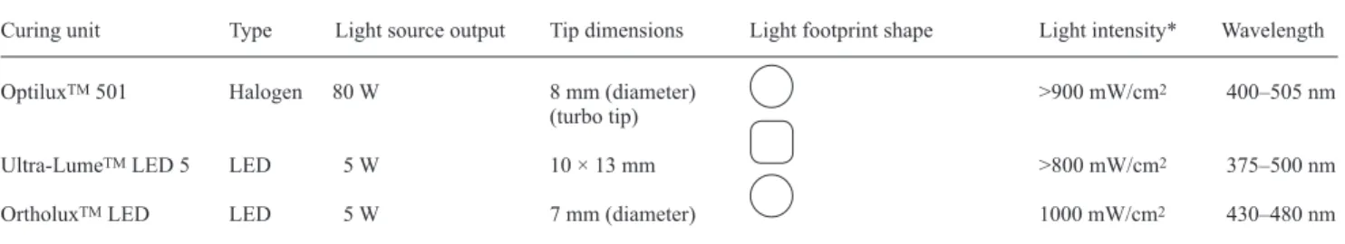
West Collins Orange, California, USA) was used for 40 seconds to bond the teeth of this group. Some of the technical characteristics of these curing units are presented in Table 1.
The adhesive was applied to the bracket base and the bracket was fi rmly pressed onto the fl attest area in the middle of the buccal surface. Any excess adhesive was carefully removed with a probe. The exposure time was equally divided between the mesial and distal part of the bracket only for the groups bonded using Optilux 501 and Ortholux LED. Ultra-Lume LED 5 has a large light-guiding tip and it was directed at the centre of the bracket. For all groups, light exposure took place with the light-guiding tip Table 1 Technical characteristics of the light curing units investigated in this study.
Curing unit Type Light source output Tip dimensions Light footprint shape Light intensity* Wavelength OptiluxTM 501 Halogen 80 W 8 mm (diameter) >900 mW/cm2 400–505 nm
(turbo tip)
Ultra-LumeTM LED 5 LED 5 W 10 × 13 mm >800 mW/cm2 375–500 nm
OrtholuxTM LED LED 5 W 7 mm (diameter) 1000 mW/cm2 430–480 nm
*According to the manufacturer. LED, light-emitting diode.
at an angle of 90 degrees to the tooth surface, and as close as possible to the bracket without touching it.
Testing standardization procedure
Each tooth with the bracket already bonded was mounted on a rectangular acrylic block (Technovit 4071, Heraeus Kulzer, Wehrheim, Germany) that fi tted exactly into the corresponding part on top of the load cell of the Instron testing machine (Instron Corp., Canton, Massachusetts, USA). It was crucial to ensure that the buccal tooth surface was roughly parallel to and projecting slightly above the acrylic surface (Pettemerides et al., 2001). A special standardization procedure was followed in order to ensure that the bracket base would be parallel to the force direction during SBS testing. A custom-made metallic rectangular blade was fi xed with a drop of glue (Cementit White, Merz & Benteli AG, Niederwangen, Switzerland) into the vertical slot of the bracket before pouring the acrylic. This blade was then laid over the mould and was positioned in such a way that the bracket was situated in the middle of the acrylic block with its base parallel to the borders of the block (Staudt et al., 2005).
The embedded teeth were stored for 24 hours at 37°C (Ishikawa et al., 2001; Oesterle and Shellhart, 2001) in water (Fox et al., 1994). The acrylic block was secured in the lower jaw of the testing machine. The shear force was applied by a custom-made jig, which was parallel to the bracket base with its edge parallel to the occlusal border of the bracket base. The samples were stressed in an occlusogingival direction with a crosshead speed of 0.5 mm/ minute. The force values recorded at the point of bond failure were measured in Newtons (N) and were subsequently converted to MegaPascals (MPa or N/mm2). The debonded
enamel surfaces were examined under a light stereomicroscope
at ×10 magnifi cation (Olympus Optical, Hamburg,
Germany) to determine the mode of failure. The adhesive remnant index (ARI) was recorded according to the four-point scale introduced by Årtun and Bergland (1984): 0, no adhesive left on the tooth; 1, less than half the adhesive left on the tooth; 2, more than half the adhesive left on the tooth;
3, all the adhesive left on the tooth with a distinct impression of the bracket mesh.
Statistical analysis
All data are represented as means ± standard deviations. One-way analysis of variance (ANOVA) was applied in order to detect any differences between groups. The least signifi cant difference test was employed to perform post hoc comparisons between groups, and Duncan’s multiple range test was used to classify groups into homogeneous subsets. A Weibull analysis was also carried out in order to plot survival probability curves for each group. The ARI scores were compared using the non-parametric Kruskal–Wallis test. All statistical analyses were performed using the SPSS statistical package (SPSS 11.5, SPSS, Chicago, Illinois, USA). A result was considered statistically signifi cant at P < 0.05.
Results
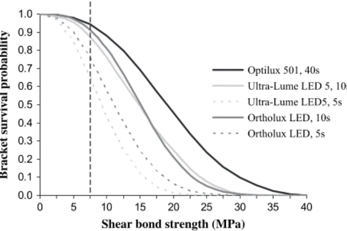
The SBS values recorded were signifi cantly different between groups (P < 0.001; Table 2). Optilux 501 with an exposure time of 40 seconds recorded the highest mean bond strength (19.2 ± 6.8 MPa), while Ultra-Lume LED 5 and Ortholux LED recorded the lowest (9.5 ± 4.3 and 11.3 ± 4.9 MPa, respectively) when used for 5 seconds only. The post hoc comparison revealed the existence of two signifi cantly homogeneous subgroups. The use of both intensive LED devices with an exposure time of 5 seconds led to signifi cantly inferior SBS values. On the other hand, an exposure time of 10 seconds led to bond strength values comparable with those obtained using the halogen lamp for 40 seconds, although the latter exhibited somewhat higher resistance to failure. Applying the Weibull distribution to the data it was possible to plot the bracket survival probability curves for each experimental group (Figure 1). A shear stress of 7.5 MPa, for example, is estimated to produce a bond failure percentage of 5 per cent for Optilux 501, 8 and 12 per cent for Ortholux LED and Ultra-Lume LED 5, respectively, when used for 10 seconds, but more than 25 per cent for both intensive LED units with an exposure time of 5 seconds only.
Table 2 Mean values and comparison of shear bond strength values between the experimental groups.
Group Mean ± SD 95% confi dence interval for mean Homogeneous subsets*
Optilux 501, 40 seconds 19.2 ± 6.8d,e 15.4–23.0 A
Ultra-Lume LED 5, 10 seconds 16.3 ± 7.1d,e 11.8–20.5 A, B
Ortholux LED, 10 seconds 15.9 ± 6.6d,e 11.9–19.2 A, B
Ortholux LED, 5 seconds 11.3 ± 4.9a,b,c 8.6–14.1 C
Ultra-Lume LED 5, 5 seconds 9.5 ± 4.3a,b,c 7.1–11.8 C
SD, standard deviation.
*Signifi cant difference between groups (P < 0.001); A, B, C group classifi cation into homogeneous subsets.
a, b, c, d, eSignifi cant difference (P < 0.05) from the group Optilux 501, Ultra-Lume LED 5 10 seconds, Ortholux LED 10 seconds, Ortholux LED 5
A microscopic evaluation of the bond failure site showed that the great majority of bond failures occurred at the bracket base–adhesive interface (Table 3). No signifi cant differences were found between the experimental groups. The majority of failures were of a cohesive nature (ARI scores 1 and 2).
Discussion
The results of the present study suggest that the new intensive LED curing units may reduce the time necessary to bond orthodontic brackets. An exposure time of 10 seconds achieved SBS values that, in vitro, were comparable with those obtained using a high-power halogen lamp for 40 seconds. When the same LED devices were used for only 5 seconds, the resulting bond strength values were signifi cantly lower.
Ultra-Lume LED 5 uses five LEDs (the main diode with a peak wavelength at 450 nm and four additional diodes with a peak wavelength at 400 nm), which are set into a refl ector that focuses the light into a high-intensity rectangular
footprint of approximately 10 × 13 mm. Due to the size of the light-guiding tip it is impossible to direct the light only on the mesial or distal half of the tooth. Ortholux LED uses a single intensive blue LED, which produces a bandwidth between 430 and 480 nm with a light intensity of approximately
1000 mW/cm2. Its manufacturer recommends an exposure
time of 10 seconds (equally divided mesially and distally) when used to bond metallic brackets and only 5 seconds when used to bond ceramic brackets. Optilux 501 is a high-power halogen light curing lamp that yields a light intensity of more than 900 mW/cm2 (in boost mode with the turbo tip) (Dunn
and Taloumis, 2002; Oberholzer et al., 2003; Kleverlaan and De Gee, 2004). It was used as the positive control, with an exposure time of 40 seconds, in order to compare the new intensive LED units with one of the most powerful conventional halogen-based commercially available devices.
The effective range of the light emission spectrum that can initiate polymerization is narrow. The most common initiator used in visible light-cured adhesives is camphor quinone, which is sensitive to the blue part of the visible light spectrum (360–520 nm), with a peak activity centred around 465 nm (Nomoto, 1997). Halogen lamps produce light when electric current flows through a thin tungsten filament that acts as a resistor. The filament is heated, emitting energy in the form of radiation whose wavelength depends on the temperature reached. High temperatures must be reached in order to achieve visible light emission (Rueggeberg et al., 1996). Preferential production of blue light is impossible and the halogen curing units used in dentistry have special systems to filter out the unwanted portions of the spectrum. As a result, the largest part of the irradiative power is wasted. In contrast, LEDs produce visible light by quantum mechanic effects. A special combination of two different semi-conductors is used to emit a characteristic light with a specifi c narrow spectral distribution. In other words, LED technology is a more effi cient way to convert an electric current into light. LED curing units have been shown to achieve an equal or superior depth of cure in comparison with halogen lamps with approximately the same light intensity when used to polymerize composites (Mills et al., 1999; Jandt et al., 2000).
Reynolds (1975) suggested a minimum ‘clinically acceptable’ bracket bond strength of 6–8 MPa, but the lack of uniformity between bond strength studies (Fox et al., 1994) makes any comparison of strength values between in
vitro studies practically impossible. The results of the present
research confi rm the fi ndings of earlier in vitro investigations on the previous generation of LED curing devices (Dunn and Taloumis, 2002; Bishara et al., 2003). Although these lamps yielded considerably lower light intensity than halogen-based lamps, which served as controls, they achieved equivalent bracket bond strength values when used for the same exposure times. It should be noted, however, that these values cannot be directly transferred to the clinical Table 3 Frequency distribution of the adhesive remnant index
(ARI).
0 1 2 3
Optilux 501, 40 seconds 0 2 11 2 Ultra-Lume LED 5, 10 seconds 0 3 8 4 Ortholux LED, 10 seconds 0 1 11 3 Ortholux LED, 5 seconds 0 0 12 3 Ultra-Lume LED 5, 5 seconds 0 3 9 3 ARI score: 0, no adhesive remaining on tooth; 1, less than half the adhesive remaining on tooth; 2, more than half the adhesive remaining on tooth; 3, all adhesive remaining on tooth.
The Kruskall–Wallis test revealed no statistical difference between groups (P = 0.86).
Figure 1 Survival probability curves plotted for each experimental group using the Weibull distribution. The dotted line corresponds to a mechanical shear stress of 7.5 MPa. The points of intersection on the bracket survival probability curves correspond to the estimated percentage of bond failures.
0.0 0.1 0.2 0.3 0.4 0.5 0.6 0.7 0.8 0.9 1.0 0 5 10 15 20 25 30 35 40
Shear bond strength (MPa)
Bracket survival probability
Optilux 501, 40s Ultra-Lume LED 5, 10s Ultra-Lume LED5, 5s Ortholux LED, 10s Ortholux LED, 5s
situation where the complex ageing of resin materials in the oral environment and unpredictable stress system generated during mastication play a signifi cant role (Eliades and Brantley, 2000). In vivo studies are an indispensable second step to confi rm any conclusions drawn in the laboratory. Conclusions
The results of the present in vitro study show that the recently introduced intensive LED curing devices may reduce the exposure time required to effi ciently bond orthodontic attachments to only 10 seconds. Compared with halogen lamps, the new LED curing units require 10 times less power to operate, which makes them suitable for portable use in cordless devices. They also have a lifetime of 10 000 hours with relatively little degradation, and are resistant to shock and vibration. The fact that they are also relatively inexpensive makes them an extremely promising alternative for bracket bonding in orthodontics.
Address for correspondence Anestis Mavropoulos Department of Orthodontics University of Geneva Barthélemy-Menn 19 CH-1205 Geneva Switzerland E-mail: anestis.mavropoulos@medecine.unige.ch Acknowledgement
The authors would like to thank Dr Maria Cattani for her indispensable help in performing the shear bond strength measurements.
References
Ǻrtun J, Bergland S 1984 Clinical trials with crystal growth conditioning as an alternative to acid-etch enamel pretreatment. American Journal Orthodontics 85: 333–340
Bassiouny M A, Grant A A 1978 A visible light-cured composite restorative. Clinical open assessment. British Dental Journal 145: 327–330 Bishara S E, Ajlouni R, Oonsombat C 2003 Evaluation of a new curing
light on the shear bond strength of orthodontic brackets. Angle Orthodontist 73: 431–435
Dunn W J, Taloumis L J 2002 Polymerization of orthodontic resin cement with light-emitting diode curing units. American Journal of Orthodontics and Dentofacial Orthopedics 122: 236–241
Eliades T, Brantley W A 2000 The inappropriateness of conventional orthodontic bond strength assessment protocols. European Journal of Orthodontics 22: 13–25
Fox N A, McCabe J F, Buckley J G 1994 A critique of bond strength testing in orthodontics. British Journal of Orthodontics 21: 33–43
Haitz R H, Craford M G, Weissman R H 1995 Light emitting diodes. In: Bass M (ed.) Handbook of optics, 2nd edn. McGraw Hill, New York, Chapter 12, pp. 1–39
Ishikawa H, Komori A, Kojima I, Ando F 2001 Orthodontic bracket bonding with a plasma-arc light and resin reinforced glass ionomer cement. American Journal of Orthodontics and Dentofacial Orthopedics 120: 58–63 Jandt K D, Mills R W, Blackwell G B, Ashworth S H 2000 Depth of cure
and compressive strength of dental composites cured with blue light emitting diodes (LEDs). Dental Materials 16: 41–47
Kleverlaan C J, De Gee A J 2004 Curing effi ciency and heat generation of various resin composites cured with high-intensity halogen lights. European Journal of Oral Sciences 112: 84–88
Klocke A, Korbmacher H M, Huck L G, Ghosh J, Kahl-Nieke B 2003 Plasma arc curing of ceramic brackets: an evaluation of shear bond strength and debonding characteristics. American Journal of Orthodontics and Dentofacial Orthopedics 124: 309–315
Lalani N, Foley T F, Voth R, Banting D, Mamandras A 2000 Polymerization with the argon laser: curing time and shear bond strength. Angle Orthodontist 70: 28–33
Mills R W, Jandt K D, Ashworth S H 1999 Dental composite depth of cure with halogen and blue light emitting diode technology. British Dental Journal 186: 388–391
Mitton B A, Wilson N H 2001 The use and maintenance of visible light activating units in general practice. British Dental Journal 191: 82–86 Miyazaki M, Hattori T, Ichiishi Y, Kondo M, Onose H, Moore B K 1998
Evaluation of curing units used in private dental offi ces. Operative Dentistry 23: 50–54
Nakamichi I, Iwaku M, Fusayama T 1983 Bovine teeth as possible substitutes in the adhesion test. Journal of Dental Research 62: 1076–1081 Nomoto R 1997 Effect of light wavelength on polymerization of
light-cured resins. Dental Materials Journal 16: 60–73
Oberholzer T G, Pameijer C H, Grobler S R, Rossouw R J 2003 The effect of different power densities and method of exposure on the marginal adaptation of four light-cured dental restorative materials. Biomaterials 24: 3593–3598
Oesterle L J, Shellhart W C 2001 Bracket bond strength with transillumination of a light-activated orthodontic adhesive. Angle Orthodontist 71: 307–311
Oesterle L J, Shellhart W C, Belanger G K 1998 The use of bovine enamel in bonding studies. American Journal of Orthodontics and Dentofacial Orthopedics 114: 514–519
Pettemerides A P, Ireland A J, Sherriff M 2001 An ex vivo investigation into the use of a plasma arc lamp when using a visible light-cured composite and a resin-modifi ed glass poly(alkenoate) cement in orthodontic bonding. Journal of Orthodontics 28: 237–244
Read M J 1984 The bonding of orthodontic attachments using a visible light cured adhesive. British Journal of Orthodontics 11: 16–20 Reynolds I R 1975 A review of direct orthodontic bonding. British Journal
of Orthodontics 2: 171–178
Rueggeberg F A, Twiggs S W, Caughman W F, Khajotia S 1996 Lifetime intensity profi les of 11 light-curing units. Journal of Dental Research 75: 380 (abstract)
Stahl F, Ashworth S H, Jandt K D, Mills R W 2000 Light-emitting diode (LED) polymerisation of dental composites: fl exural properties and polymerisation potential. Biomaterials 21: 1379–1385
Staudt C B, Mavropoulos A, Bouillaguet S, Kiliaridis S, Krejci I 2005 Time reduction in light-curing: shear bond strength with high-power halogen light. American Journal of Orthodontics and Dentofacial Orthopedics (in press)
Tavas M A, Watts D C 1984 A visible light-activated direct bonding material: an in vitro comparative study. British Journal of Orthodontics 11: 33–37
Wang W N, Meng C L 1992 A study of bond strength between light- and self-cured orthodontic resins. American Journal of Orthodontics and Dentofacial Orthopedics 101: 350–354
Weinberger S J, Foley T F, McConnell R J, Wright G Z 1997 Bond strengths of two ceramic brackets using argon laser, light, and chemically cured resin systems. Angle Orthodontist 67: 173–178