HuD binds to three AU-rich sequences in the 3′-UTR of neuroserpin mRNA and promotes the accumulation of neuroserpin mRNA and protein
10
0
0
Texte intégral
(2) Nucleic Acids Research, 2002, Vol. 30, No. 10 2203. intermediates (24–26). In addition to the poly(A) tail, several cis-acting elements play a role in mRNA stability. Best characterized among them is the AU-rich element located in the 3′-untranslated region (3′-UTR) of several labile mRNAs encoding cytokines, oncoproteins and proteins involved in nervous system development (27–32). Other stability determinants have been identified within the coding regions or the 5′-UTRs of some genes, but the proteins binding to them are mostly uncharacterized (33–36). A number of trans-acting factors that interact with AU-rich sequences have been identified. Two of these, AUF-1/hnRNPD and HuR, modulate the stability of mRNAs containing AU-rich sequences in vivo. HuR, a member of the embryonic lethal abnormal vision (Elav)-like family of proteins (28,32,37,38), stabilizes mRNAs to which it binds by uncharacterized mechanisms (39). Four Elav-like genes have been identified in mammals: HuR (HuA in rodents), Hel-N1 (HuB in rodents), HuC and HuD. While HuR is ubiquitously expressed, the other three genes are only expressed in post-mitotic neurons and in neuroendocrine tumors (40,41). It has been demonstrated that HuD binds to the 3′-UTR of mRNAs encoding proteins such as tau and GAP-43 (29,31). Here we report that HuD is co-expressed with neuroserpin mRNA, and to a lesser extent with neurotrypsin RNA in the rat brain. We demonstrate that HuD protein binds with high affinity to three AU-rich sequences in the 3′-UTR of neuroserpin mRNA, but not to neurotrypsin mRNA. We further demonstrate that stable overexpression of HuD leads to the accumulation of neuroserpin mRNA and protein in rat pheochromocytoma PC12 cells. In addition, we show that exogenous HuD stabilizes neuroserpin 3′-UTR mRNA in co-transfected cells. Our results indicate that HuD regulates neuroserpin expression by controlling the stability of its mRNA through the binding to specific AU-rich sequences in the 3′-UTR. MATERIALS AND METHODS Rats Wistar rats were used. The maintenance and handling of the animals was as recommended by the European Union (European Communities Council Directive of November 24, 1986, 86/609/ EEC). All efforts were made to minimize animal suffering, to reduce the number of animals used and to use alternatives to in vivo techniques. In situ hybridization and immunohistochemistry Under deep pentobarbital anesthesia, rats were perfused through the heart with cold 4% para-formaldehyde in 0.1 M sodium phosphate (pH 7.4). The brains were quickly removed, post-fixed in 4% para-formaldehyde in 0.1 M sodium phosphate (pH 7.4) and cryoprotected in 4% para-formaldehyde + 30% sucrose (w/v) in phosphate-buffered saline (PBS) at 4°C. Subsequently, 25 µm thick coronal sections were cut using a cryostat. In situ hybridization on floating sections was performed as described (42). X-ray films (Hyperfilm β-MAX films; Amersham) were exposed for 15–21 days, developed with Kodak D19 and fixed. Anatomical abbreviations follow Paxinos and Watson (43). To analyze the co-localization of HuD protein and neuroserpin mRNA, a combination of in situ hybridization and immunohistochemistry was performed on. the same tissue section using a double-labeling technique (44). Briefly, after hybridization and washes, the free-floating sections were incubated sequentially with the mouse monoclonal 16A11 anti HuD/HuC antibody (1:200) (45) overnight at 4°C and then with a preadsorbed biotinylated secondary rat anti-mouse antibody (1:200; Vector Laboratories, Burlingame, CA) for 1 h at room temperature, followed by immunocomplex detection using the ABC reagent (Elite kit; Vector Laboratories). Peroxidase was then visualized with diaminobenzidine (0.05%) and H2O2. Sections were mounted on coated slides and air-dried. X-ray films were exposed for 3 weeks. For resolution at the cellular level, the sections were dipped in Hyper-coat LM-1 photographic emulsion (Amersham), and films were exposed for 3 weeks in the cold, developed with D19, fixed, dehydrated and coverslipped. Optical observations were made in a Zeiss Axiophot microscope (Carl Zeiss, Oberkochen, Germany). Preparation of labeled RNA transcripts DNA templates for neurotrypsin and neuroserpin transcripts were synthesized by the polymerase chain reaction using the following oligonucleotides: for neurotrypsin 3′-UTR corresponding to nucleotides 2615–3344 the oligonucleotides were T72615 (5′-GTAATACGACTCACTATAGGGCTATACCAAAGTCTCAGC-3′) and 3344a (5′-GCACGCTGTAGGTAGAAAG-3′). For neuroserpin 3′-UTR corresponding to nucleotides 1343–2908 the oligonucleotides were T71343 (5′GTAATACGACTCACTATAGGGCGAGTACAAAGAAAGCAGG-3′) and 2908a (5′-TATTCTTCCTTACAGGC-3′). For transcript A corresponding to neuroserpin 3′-UTR nucleotides 1343–1674 the oligonucleotides were T71343 and 1674a (5′-ATCATTTTACTACAATTCC-3′). For subfragment B corresponding to neuroserpin 3′-UTR nucleotides 1623–2037 the oligonucleotides were T71623 (5′-GTAATACGACTCACTATAGGGCTGTCTGAGATTTGAAACC-3′) and 2037a (5′GGCCTCTTGATGTCATCC-3′). For subfragment C corresponding to neuroserpin 3′-UTR nucleotides 1977–2443 the oligonucleotides were T71977 (5′-GTAATACGACTCACTATAGGGCCCACATGACTCTACTAGC-3′) and 2443a (5′-GTCTGTGAAAATGTGAGG-3′). For subfragment D corresponding to neuroserpin 3′-UTR nucleotides 2395–2908 the oligonucleotides were T72395 (5′-GTAATACGACTCACTATAGGGCCCTTGGGTTGCAATGTCG-3′) and 2908a. All neurotrypsin and neuroserpin templates were gel-purified. RNA transcripts were synthesized using T7 RNA polymerase (Promega) and purified as described (28). Purification of glutathione-S-transferase (GST)–HuD protein An overnight culture of Escherichia coli BL 21 transformed with pGST–HuD plasmid (28) was diluted 1:50 in LB medium. At an OD 600 of 0.4, the culture was induced with isopropyl β-D-thiogalactopyranoside (0.1 mM) at 30°C. Four hours later, the cells were spun down and resuspended in 10 ml of buffer A (50 mM Tris pH 8.0, 50 mM NaCl, 1 mM EDTA). The cells were lysed by the addition of lysozyme (0.2 mg/ml) and Triton X-100 (1%). The lysate was centrifuged at 12 000 g for 30 min, and the resulting supernatant was collected and passed through 19- and 23-gauge needles several times. It was then incubated with GST–Sepharose beads for 1 h at 4°C, centrifuged at 2000 r.p.m. in an S-4180 rotor (720 g) at 4°C, and washed five.
(3) 2204 Nucleic Acids Research, 2002, Vol. 30, No. 10. times in PBS. The purified GST protein was eluted with 10 mM glutathione in 50 mM Tris–HCl pH 8.0, dialyzed overnight in PBS + 10% glycerol and then pooled and stored at – 70°C. GST protein concentration was determined by comparison with a bovine serum albumin (BSA) curve in an acrylamide gel stained with Coomassie brilliant blue. Agarose RNA gel-shift assay Reaction mixtures (20 µl) contained 50 mM Tris pH 7.0, 0.25 mg/ml tRNA, 0.25 mg/ml BSA, 20 fmol of labeled RNA and protein as indicated. Mixtures were incubated at 37°C for 10 min. Following incubation, 4 µl of dye mixture (50% glycerol, 0.1% bromophenol blue, 0.1% xylene cyanol) was added, and 25% of the reaction mixture was immediately loaded on a 1% agarose gel in TAE buffer (40 mM Tris acetate, 1 mM EDTA). Gels were electrophoresed at 40 V for 3 h and then dried on DE81 paper (Whatman) with a backing of gel drying paper (Hudson City Paper, West Caldwell, NJ). XAR5 films (Eastman Kodak Co.) were exposed for 4–6 h at –70°C. RNase T1 selection assay Reaction mixtures (20 µl) contained 50 mM Tris pH 7.0, 0.25 mg/ml tRNA, 0.25 mg/ml BSA, 10–20 fmol of labeled RNA (100 000–600 000 c.p.m.) and purified GST–HuD (75 ng) or 10 µg of PC12 crude protein extract as indicated. After 10 min incubation at 37°C, 5 U of RNase T1 (Calbiochem, La Jolla, CA) were added to each reaction and incubated at 37°C for a further 10 min. The mixtures were diluted 1:6 with buffer FBB (20 mM Tris–HCl pH 7.0, 0.05 mg/ml tRNA) and filtered through nitrocellulose (BA 85 Schleicher & Schuell). After washing the nitrocellulose twice in FBB, bound HuD–RNA complex was extracted with phenol/chloroform and concentrated by ethanol precipitation. The resulting RNA was dissolved in formamide stop buffer (Gibco-BRL) and denatured at 65°C for 2 min. Samples were analyzed by 10% polyacrylamide/50% urea gel electrophoresis. The gel was fixed with 1:1:8 acetic acid:methanol:water and dried. XAR5 films were exposed overnight at –70°C. Nitrocellulose filter binding assay Reaction mixtures (20 µl) contained 50 mM Tris pH 7.0, 0.25 mg/ml tRNA, 0.25 mg/ml BSA, 20 fmol of labeled RNA and purified GST–HuD as indicated. After 10 min of incubation at 37°C, the mixtures were diluted 1:6 with buffer FBB and filtered using nitrocellulose. After washing the filter twice in FBB, bound radioactivity was determined by Cerenkov counting.. Cell lines, plasmids and transfections PC12 cells were grown in Dulbecco’s modified Eagle’s medium supplemented with 10% horse serum, 5% FCS and 1 mM glutamine (all from Gibco-BRL). To generate cells stably expressing HuD, PC12 cells were transfected using Lipofectamine (Life Technologies) with the pCEFL–AU5HuD expression vector (46). This is a derivative of the pCDNA3 (Invitrogen) vector in which the CMV promoter has been replaced by that of the human elongation factor 1α. Stable cell lines expressing HuD were obtained following selection with the aminoglycoside antibiotic G418 (800 µg/ml). For neuroserpin 3′-UTR-RNA expression, a fragment corresponding to nucleotides 788–2944 of its cDNA was subcloned into the BamHI and XhoI sites of pCDNA3.1 (Invitrogen) and used to transfect PC12 cells. RNA extraction and northern analysis Total RNA from PC12 cells was prepared by standard methods (47). RNAs were fractionated in formaldehyde agarose gels and blotted onto nylon membranes as described (47). As controls for the amount of RNA, we used the 18S rRNA stained with methylene blue. Radioactive probes were prepared by a random priming procedure (48) using the Readyto-go kit (Amersham-Pharmacia). Western blotting To perform immunoblot analysis of AU5HuD expression, PC12 cell protein extracts were prepared following the Dignam C method (47). Protein extracts were electrophoresed in 12% polyacrylamide gels and transferred to nylon (Immobilon P, Millipore) membranes. The filters were washed, blocked with Blotto (5% skimmed milk in PBS, 0.1% Tween-20), and incubated overnight at 4°C with an anti-AU5 antibody (1:1000 dilution). Blots were washed three times for 10 min in PBS + 0.1% Tween-20 and incubated with HRP-conjugated antimouse for 1 h at room temperature. Blots were developed by a peroxidase reaction using the ECL detection system (Amersham). As control, we measured β-actin using an appropriate antibody (sc-1615, Santa Cruz Biotechnology). For neuroserpin, protein extracts were electrophoresed in 10% polyacrylamide gels and transferred to nylon (Immobilon P, Millipore) membranes. The filters were blocked in 3% BSA in TBS + 0.5% Tween-20 and incubated overnight at 4°C with the G47 anti-neuroserpin antibody (1 µg/ml). Blots were washed three times for 10 min in TBS + 0.5% Tween-20 and incubated with HRP-conjugated anti-goat antibody for 1 h at room temperature. Blots were developed by a peroxidase reaction using the ECL detection system (Amersham).. Acrylamide RNA gel-shift and supershift assays Reaction mixtures (10 µl) containing 1 µg of tRNA, 5 fmol of RNA and 7 µg of protein were incubated in reaction buffer (15 mM HEPES pH 7.9, 10 mM KCl, 10% glycerol, 0.2 mM dithiothreitol, 5 mM MgCl2) for 30 min at 25°C and digested with RNase T1 7.5 U/reaction (Calbiochem) for 15 min at 37°C. Complexes were resolved by electrophoresis through native gel (7% acrylamide in 0.25× Tris-borate–EDTA buffer) without loading buffer (160 V, 2 h at 4°C). For supershift assays, 4 µg of antibody were incubated with lysates for 1 h on ice before the addition of radiolabeled RNA; all subsequent steps were as described above.. RESULTS HuD co-localizes with neuroserpin mRNA in the rat brain and binds to its 3′-UTR in vitro Analysis of the previously reported neuroserpin and neurotrypsin cDNA sequences (4,6,49,50) revealed the presence in their 3′-UTR of AU-rich stretches homologous to binding sites for HuD protein in other transcripts (29). Together with the common neuron-specific expression of neuroserpin, neurotrypsin and some Hu genes (HuB, HuC and HuD), this prompted us to investigate whether the expression patterns of.
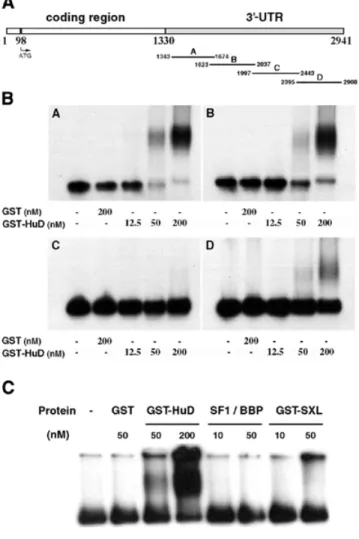
(4) Nucleic Acids Research, 2002, Vol. 30, No. 10 2205. Figure 1. HuD co-localizes with neuroserpin mRNA in the rat brain and binds to its 3′-UTR in vitro. (A) In situ hybridization analysis showing the co-expression of HuD and neuroserpin RNAs in cerebral cortices layer II-V and VIb and in the Pir and RSCx cortices (compare a and c) of post-natal day 5 rats. Neurotrypsin expression (b) is also partially coincident. (B) HuD/HuC proteins and neuroserpin mRNA co-localize at the cellular level in the RSCx of post-natal day 15 rats. Combination of in situ hybridization against neuroserpin with immunohistochemistry using an antiserum against Hu proteins (16A11). Black arrows indicate cells with strong expression of neuroserpin mRNA (dots) and Hu proteins (brownish signal). (C) RNase T1 analysis of HuD–neuroserpin mRNA binding in vitro. The indicated concentrations of GST–HuD or GST proteins were incubated with 32P-labeled 3′-UTR (10 fmol, 200 000–400 000 c.p.m.) of neurotrypsin (left) or neuroserpin (right) mRNAs. Total T1 digests of neurotrypsin (T1 NT) and neuroserpin (T1 NS) RNAs are shown. Black arrows indicate the three HuD-bound RNA fragments corresponding to neuroserpin mRNA. The structures of neurotrypsin and neuroserpin mRNAs are shown at the top.. these three genes were coincident. Expression of HuD, neuroserpin and neurotrypsin in the brain of 5-day-old rats was analyzed by in situ hybridization. HuD and neuroserpin showed a highly coincident regional pattern of expression, with highest levels in cerebral cortex layers II-V and VIb, and the retrosplenial (RSCx) and piriform (Pir) cortices (Fig. 1A, a and c). Neurotrypsin RNA was detected in a subset of these areas, but was virtually absent from others such as cerebral cortex layers II-IV and VIa (Fig. 1A, compare b with a and c). To study in further detail whether neuroserpin and HuD colocalize at the cellular level, we used a combination of in situ hybridization (against neuroserpin mRNA) and immunohistochemistry using an antiserum (16A11) that specifically detects HuC and HuD proteins in the rat brain (41). Expression of neuroserpin RNA was fully coincident with HuC and HuD proteins in the cerebral cortex of 15-day-old rats including cortex layer V and RSCx where HuD is the only member of the Elav family that is expressed (51) (Fig. 1B, see black arrows). Given the role of HuD as a regulator of mRNA stability, we performed T1 RNase digestion assays to elucidate whether. HuD protein could bind neuroserpin mRNA. For comparison, binding to neurotrypsin mRNA containing also AU-rich sequences in its 3′-UTR (GenBank accession no. NM003619) was also investigated. At least three binding sites for GST–HuD were found in the 3′-UTR of neuroserpin mRNA (Fig. 1C, right, black arrows). In contrast, no binding was detected in neurotrypsin mRNA (Fig. 1C, left). In line with the fact that all members of the Elav-like protein family (the neuron-specific HuB, HuC and HuD, and the ubiquitous HuR) share identical binding properties (27,52), the same three bands for neuroserpin mRNA were obtained using GST–HuR protein (data not shown). HuD specifically binds three zones in the 3′-UTR of neuroserpin mRNA Next, we determined the localization of the HuD-binding segments within the 3′-UTR of mouse neuroserpin mRNA. To this end, purified GST–HuD fusion protein was incubated with four partially overlapping in vitro-labeled transcripts (neuroserpin A–D) covering the entire 3′-UTR of neuroserpin mRNA.
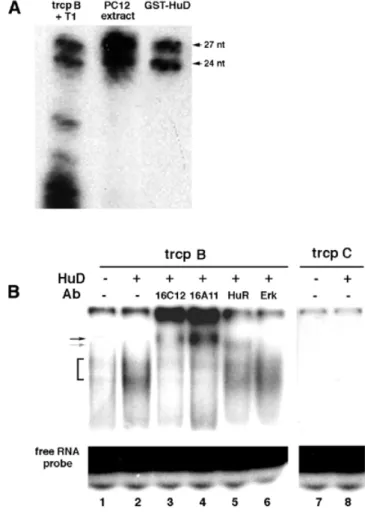
(5) 2206 Nucleic Acids Research, 2002, Vol. 30, No. 10. Figure 3. HuD binds neuroserpin 3′-UTR transcripts with high affinity. RNA– protein complex formation was analyzed by nitrocellulose filter binding assays. A 20 fmol aliquot of each transcript (400 000–600 000 c.p.m.) was incubated with the indicated concentrations of HuD protein for 10 min at 37°C. Plot of the percentage of RNA bound versus HuD concentration. Squares, transcript A; circles, transcript B; triangles, transcript D.. Figure 2. HuD protein binds to three sites in the neuroserpin mRNA 3′-UTR. (A) Scheme of neuroserpin cDNA and the four (A–D) transcripts obtained from the 3 ′-UTR. (B) Agarose gel-shift assays showing binding of HuD to neuroserpin A, B, and D transcripts, but not to neuroserpin C transcript. 32P-labeled transcripts (20 fmol, 400 000–600 000 c.p.m.) were incubated with the indicated concentration of GST or GST–HuD protein. After 10 min incubation mixtures were resolved on 1% agarose gels. (C) Comparative analysis of the binding of HuD and of Sex-lethal (SXL) and SF1/BBP proteins to neuroserpin transcript B (20 fmol, 400 000 c.p.m.). Conditions were as in (B).. (Fig. 2A). GST–HuD–RNA complex formation was analyzed by agarose gel shift assays. High affinity binding of HuD was found for the neuroserpin A, B and D transcripts, but not for neuroserpin C (Fig. 2B). To examine the specificity of this binding we performed gel-shift assays using labeled neuroserpin transcript B and two other RNA-binding proteins, Sex-lethal and SF1/BBP, which show affinity for U-rich or UACUAAC sequences, respectively (53,54). Indicating a specific interaction of HuD, neither Sex-lethal nor SS1/BBP bound to neuroserpin transcript B (Fig. 2C). To measure the interaction between HuD and the transcripts A, B and D, we used a quantitative RNA binding assay (55). Low concentrations (20 fmol) of each labeled transcript were incubated with increasing amounts of HuD protein. The mixtures were then filtered through nitrocellulose to estimate the radioactivity retained in protein complexes. Binding of HuD to neuroserpin A and B transcripts was detected at 1 nM and maximal (70% of input transcript) at 200–500 nM, with a. Kd of 8–10 nM. Lower, but still significant, binding with a midpoint at ∼20 nM was observed for the neuroserpin D transcript (Fig. 3). To precisely identify the sequences bound by HuD in each neuroserpin transcript we performed T1 RNase selection assays. HuD–neuroserpin RNA complexes were allowed to form and then subjected to digestion with T1 RNase. RNA fragments bound to HuD were isolated by adsorption of complexes to nitrocellulose followed by elution with phenol– chloroform. A single 20 nt fragment of neuroserpin A, two fragments of 27 and 24 nt of neuroserpin B, and one 19 nt fragment of neuroserpin D were detected, while as expected from the previous results, no fragments were obtained from the neuroserpin C transcript (Fig. 4A). Likewise, no binding was found using GST protein as control (Fig. 4A). Given the sequence specificity of T1 RNase (cleaves after a G) and the size of the fragments, their sequences were readily located in the 3′-UTR of neuroserpin RNA (Fig. 4B, underlined). The 20 nt fragment in neuroserpin A is the most upstream in the neuroserpin 3′-UTR region and it contains a CUUUnC sequence. The two fragments in neuroserpin B are in tandem, separated by an internal G in a 51 nt sequence, while the 19 nt fragment in neuroserpin D is located further downstream, close to the 3′-end of neuroserpin mRNA (Fig. 4B). All retained fragments are AU-rich sequences that harbor AUUUA or UUAUUUAUU motifs, previously characterized as determinants of mRNA stability (26,32). HuR also bound to the same regions in neuroserpin 3′-UTR mRNA (data not shown). HuD binds to neuroserpin B transcript in PC12 cell extracts To examine whether purified HuD also binds neuroserpin mRNA in cells, we incubated whole cell extracts of PC12 cells with labeled HuD-binding fragments of the 3′-UTR of neuroserpin mRNA. In T1 assays, identical 27 and 24 nt fragments of neuroserpin B transcript (chosen because of its highest affinity) were retained using either PC12 cell extracts or purified GST–HuD protein (Fig. 5A). In contrast, no fragments were retained when using neuroserpin C transcript (data not shown)..
(6) Nucleic Acids Research, 2002, Vol. 30, No. 10 2207. Figure 4. Localization of HuD binding sites in neuroserpin 3′-UTR. (A) HuD binds to three sites in the 3′-UTR of neuroserpin mRNA. RNase T1 analysis of HuD–neuroserpin RNA complexes. GST–HuD or GST proteins (75 ng) were incubated with 32P-labeled neuroserpin transcripts (20 fmol, 400 000– 600 000 c.p.m.) at 37°C for 10 min. RNase T1 (0.5 U) was then added to reaction mixtures before they were filtered through nitrocellulose. Protein-bound RNA fragments were extracted and resolved on 12% denaturing polyacrylamide gels. RNase T1-protected fragments are indicated. T1 digests of each transcript are shown (T1 A, T1 B, T1 C, T1 D). (B) Sequence of the neuroserpin mRNA 3′-UTR showing the precise location of the HuD-binding sites (underlined).. This indicated the presence in PC12 cells of at least one protein able to bind neuroserpin mRNA in the same region as HuD. To further investigate this issue, we transiently transfected PC12 cells with an HuD-expression vector. Extracts from normal and HuD-overexpressing PC12 cells were incubated with the labeled neuroserpin B transcript and subjected to acrylamide gel-shift assays. Two retained bands were found, which were stronger in the case of extracts from HuD-overexpressing cells (Fig. 5B, first two lanes). In contrast, no shifted bands were found using the neuroserpin C transcript (Fig. 5B, right). This result suggested that HuD was present in the retarded complexes, though we cannot rule out the possibility that other Hu proteins could also be included. To confirm the presence of. Figure 5. HuD binds neuroserpin mRNA 3′-UTR in PC12 cells. (A) T1 RNase analysis showing binding of PC12 protein extracts (10 µg) to neuroserpin transcript B (trcp B). GST–HuD protein (75 ng) was used as control. The two protected bands are indicated. (B) Supershift assay showing the presence of HuD and HuR proteins in the retarded bands obtained with PC12 cell extracts. Retarded complexes (bracket) were detected by incubating PC12 protein extracts with neuroserpin transcript B (trcp B, left) but not with transcript C (trcp C, right) (compare lanes 1 and 2 with 7 and 8). Extracts from HuDoverexpressing PC12 cells (lane 2) gave stronger bands than those from normal PC12 cells (lane 1). Incubation with a specific anti-HuD antibody (16C12, lane 3) or an anti-pan-Hu antibody (16A11, lane 4) caused a supershift (black arrow), while incubation with a specific anti-HuR antibody (HuR, lane 5) produced a different supershifted band (gray arrow). No supershifted bands were obtained using an unrelated anti-Erk antibody (Erk, lane 6).. HuD in the retarded bands we performed supershift assays. Incubation of neuroserpin B transcript with an HuD-specific antibody (16C12) or with 16A11 antibody that recognizes HuC and HuD proteins (45) caused a clear supershift (Fig. 5B, lanes 3 and 4). As expected from the previous results, incubation with an anti-HuR antibody resulted also in a (less) shifted band indicating that HuR binds neuroserpin RNA (lane 5). In contrast, an anti-Erk antibody used as control had no effect (lane 6). HuD increases neuroserpin mRNA stability in PC12 cells Elav-like proteins have been proposed to modulate the half-life of their target mRNAs (30,56–58). Given the ability of HuD protein to bind neuroserpin mRNA and their co-expression in vivo, we studied the effect of HuD on neuroserpin mRNA.
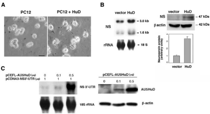
(7) 2208 Nucleic Acids Research, 2002, Vol. 30, No. 10. Figure 6. HuD increases neuroserpin expression in PC12 cells. (A) Stably transfected HuD induces formation of neurite-like processes in PC12 cells. Phase-contrast micrographs of untranfected (left) and HuD-transfected (right) cells. Bar, 250 µ m. (B) Ectopic HuD expression increased neuroserpin mRNA and protein levels in PC12 cells. Four independent experiments were performed. Northern blot analysis (left) shows an increase (∼2-fold) in neuroserpin mRNAs (3.0 and 1.6 kb) in HuD-transfected versus vector-transfected PC12 cells. Western blot analysis shows that the level of neuroserpin protein also increased in HuD-transfected cells (right, top). Quantification of neuroserpin protein expression after normalization to β-actin is shown (right, bottom). (C) Ectopic HuD expression increases the level of co-transfected neuroserpin 3′-UTR mRNA in PC12 cells. Left, northern blot analysis of neuroserpin 3′-UTR mRNA (NS 3′-UTR) levels in cells transfected with increasing amounts of a plasmid encoding a tagged HuD protein (pCEFL–AU5HuD). Right, western blot analysis of the expression of tagged AU5HuD protein in transfected PC12 cells. Expression of β-actin protein was studied as control.. levels in PC12 cells. To this end we used an expression vector for a AU5-tagged HuD protein (pCEFL–AU5HuD). In line with previous reports (31), we observed that stable transfection of HuD in PC12 cells induced the formation of neurites (Fig. 6A). Since ectopic neuroserpin expression in pituitary AtT-20 cells also induced processes (59), we analyzed the expression of neuroserpin mRNA in HuD-transfected PC12 cells. HuD overexpression induced a 2-fold increase in the cellular content of neuroserpin mRNAs (3.0 and 1.6 kb; Fig. 6B, left) and over a 3-fold increase in neuroserpin protein (Fig. 6B, right). To confirm that this effect was a consequence of an increase in neuroserpin RNA stability, the 3′-UTR of neuroserpin cDNA was subcloned in the pCDNA3.1 vector. This construct was used to co-transfect PC12 cells together with increasing amounts of an expression vector for the tagged HuD. We found that the cellular level of neuroserpin 3′-UTR RNA was extremely low in vector-transfected cells, and strongly increased in cells expressing AU5HuD (Fig. 6C, left). Expression of exogenous tagged HuD was assessed by western blotting (Fig. 6C, right). Taken together, our results indicate that HuD enhances the cellular content of neuroserpin by increasing its stability as the result of the binding to specific AU-rich sequences located in the 3′-UTR. DISCUSSION In this study, we report that HuD protein binds with high affinity to the 3′-UTR region of neuroserpin RNA both in vitro and in PC12 cells. Moreover, we show that HuD stabilizes and thereby increases neuroserpin mRNA and protein levels in. these cells. The HuD-mediated increase in neuroserpin mRNA and protein is accompanied by the induction of neurites, which has been observed in AtT-20 cells transfected with neuroserpin (59). Furthermore, we show that HuD protein and neuroserpin RNA are co-expressed in some areas of the rat central nervous system in which HuD, but no other member of the Hu family, is expressed (51). Both HuD and neuroserpin are highly expressed in the hippocampus, cerebral cortex and olfactory bulb, whereas their levels in other areas such as the striatum and cerebellar Purkinje cells are much lower (5,51). In addition, expression of HuD and neuroserpin follow a common pattern during brain development in rodents: both peak at birth (P0, P1) and then decrease slowly to reach adult levels (5,51). These findings reinforce HuD as a candidate to regulate neuroserpin mRNA stability in vivo. We have demonstrated that HuD binds to three AU-rich sequences present in the 3′-UTR of neuroserpin mRNA. T1 RNase assays led to their characterization as being of 20, 51 and 19 nt in length. Since these assays were performed by incubating HuD and neuroserpin mRNA before T1 digestion to prevent the destruction of putative RNA structures important for protein–RNA interaction, the possibility that filter-bound oligonucleotide fragments may correspond to two or more smaller fragments cannot be excluded. Although mutation and accumulation of neuroserpin protein are associated with senile dementia (14) and with epilepsy (15) and it has been demonstrated that neuroserpin has a neuroprotective function after focal ischemic stroke (12,13), little is known about the regulation of neuroserpin gene. In mouse primary hippocampal neurons, neuroserpin gene transcription is induced.
(8) Nucleic Acids Research, 2002, Vol. 30, No. 10 2209. through depolarization by elevated extracellular KCl (60). Binding sites for several transcription factors including Sp1, AP-2 and C/EBP have been identified in the proximal region of the mouse neuroserpin gene promoter (60). In the same study, zif/268 behaves as a silencer of neuroserpin transcription. Our results demonstrate that neuroserpin expression is also regulated post-transcriptionally through the control of the stability of its mRNA by HuD. Since all the neuronal Hu proteins have identical binding properties, in agreement with their coincident spatiotemporal pattern of expression the levels of neuroserpin mRNA in specific central nervous system regions during development could be determined by either a single member of the Hu/Elav-like family of RNA-binding proteins or by combinations of them. The Hu/Elav-like proteins are pleiotropic regulators of gene expression in mammalian cells. Hu proteins stabilize multiple mRNAs. Among them, HuD regulates the expression of several oncogenes, cytokines, genes involved in central nervous system differentiation, and cell cycle regulators such as p21WAF, c-fos, GAP-43, tau and c-myc (31,55,61). Hel-N1 controls the Id helix–loop–helix transcriptional repressor in neural precursors (62). In non-neural cells, HuR regulates the stability of RNAs for tumor necrosis factor alpha (63), the serpin PAI-2 (21), the tumor suppressor neurofibromin (NF1) (64), and vascular endothelial growth factor (30). Hu/Elav-like proteins can stabilize or destabilize specific mRNAs perhaps by influencing the access of degrading enzymes or of other proteins to RNA substrates (32,39,65). Neuroserpin exerts its biological functions in nervous system development or maintenance by the regulation of its cognate serine proteases, especially tPA (10,11). This indicates that neuroserpin levels modulate cell migration, axon outgrowth, and synaptic plasticity in which tPA is critical. Our results define a novel mechanism of regulation of neuroserpin expression acting through the control of the stability of its RNA via the binding of the HuD protein. The post-transcriptional mechanism reported here merits further study for its implication in neuroserpin expression following brain injury and in neuropathological states such as ischemia- or trauma-induced hyperexcitability, or in patients suffering abnormalities associated with pathological neuroserpin accumulation and polymerization. Remarkably, both neuroserpin and HuD genes are up-regulated in schizophrenia (66). Post-transcriptional mechanisms of regulation are considered to be more rapid than those based on the control of the rate of gene transcription. Therefore, the HuD-mediated control of neuroserpin levels may contribute to the response of neuroserpin activity to the sudden changes following acute pathological events. Together with the previous studies showing that HuD regulates the expression of GAP43 and tau, our results indicate that HuD, and perhaps other Hu proteins, may play a crucial role in a number of regulatory key processes in the developing and adult central nervous system. ACKNOWLEDGEMENTS We thank M. F. Marusich for providing us with the Mab16A11 antibody, Dr M. Gorospe for her helpful comments and M. González-Monge and T. Martínez for their excellent technical assistance. A.C. and C.N.-Y. were supported, respectively, by predoctoral fellowships from Ministerio de Educación y Cultura. and Fondo de Investigaciones Sanitarias (FIS) of Ministerio de Sanidad y Consumo of Spain. This work was supported by grants (SAF98-0060 and SAF2001-2291) from Plan Nacional de Investigación y Desarrollo of Spain. REFERENCES 1. Schapira,M. and Patson,P.A. (1991) Serine protease inhibitors (Serpins). Trends Cardiovasc. Med., 1, 146–151. 2. Potempa,J., Korzus,E. and Travis,J. (1994) The serpin superfamily of proteinase inhibitors: structure, function and regulation. J. Biol. Chem., 269, 15957–15960. 3. Mansuy,I.M., van der Putten,H., Schmid,P., Meins,M., Botteri,F.M. and Monard,D. (1993) Variable and multiple expression of Protease Nexin-1 during mouse organogenesis and nervous system development. Development, 119, 1119–1134. 4. Osterwalder,T., Contartese,J., Stoeckli,E.T., Kuhn,T.B. and Sonderegger,P. (1996) Neuroserpin, an axonally secreted serine protease inhibitor. EMBO J., 15, 2944–2953. 5. Krueger,S.R., Ghisu,G.P., Cinelli,P., Gschwend,T.P., Osterwalder,T., Wolfer,D.P. and Sonderegger,P. (1997) Expression of neuroserpin, an inhibitor of tissue plasminogen activator, in the developing and adult nervous system of the mouse. J. Neurosci., 17, 8984–8996. 6. Schrimpf,S.P., Bleiker,A.J., Brecevic,L., Kozlov,S.V., Berger,P., Osterwalder,T., Krueger,S.R., Schinzel,A. and Sonderegger,P. (1997) Human neuroserpin (PI12): cDNA cloning and chromosomal localization to 3q26. Genomics, 40, 55–62. 7. Briand,C., Kozlov,S.V., Sonderegger,P. and Grutter,M.G. (2001) Crystal structure of neuroserpin: a neuronal serpin involved in a conformational disease. FEBS Lett., 505, 18–22. 8. Stoeckli,E.T., Lemkin,P.F., Kuhn,T.B., Ruegg,M.A., Heller,M. and Sonderegger,P. (1989) Identification of proteins secreted from axons of embryonic dorsal-root-ganglia neurons. Eur. J. Biochem., 180, 249–258. 9. Thewke,D.P. and Seeds,N.W. (1996) Expression of hepatocyte growth factor/scatter factor, its receptor, c-met and tissue-type plasminogen activator during development of the murine olfactory system. J. Neurosci., 16, 6933–6944. 10. Hastings,G.A., Coleman,T.A., Haudenschild,C.C., Stefansson,S., Smith,E.P., Barthlow,R., Cherry,S., Sandkvist,M. and Lawrence,D.A. (1997) Neuroserpin, a brain-associated inhibitor of tissue plasminogen activator is localized primarily in neurons. Implications for the regulation of motor learning and neuronal survival. J. Biol. Chem., 272, 33062–33067. 11. Osterwalder,T., Cinelli,P., Baici,A., Pennella,A., Krueger,S.R., Schrimpf,S.P., Meins,M. and Sonderegger,P. (1998) The axonally secreted serine proteinase inhibitor, neuroserpin, inhibits plasminogen activators and plasmin but not thrombin. J. Biol. Chem., 273, 2312–2321. 12. Yepes,M., Sandkvist,M., Wong,M.K., Coleman,T.A., Smith,E., Cohan,S.L. and Lawrence,D.A. (2000) Neuroserpin reduces cerebral infarct volume and protects neurons from ischemia-induced apoptosis. Blood, 96, 569–576. 13. Cinelli,P., Madani,R., Tsuzui,N., Vallet,P., Rülicke,T., Arras,M., Osterwalder,T. and Sonderegger,P. (2001) Neuroserpin, a neuroprotective factor in focal ischemic stroke. Mol. Cell. Neurosci., 18, 443–457. 14. Davis,R.L., Shrimpton,A.E., Holohan,P.D., Bradshaw,C., Feiglin,D., Collins,G.H., Sonderegger,P., Kinter,J., Becker,L.M., Lacbawan,F., Krasnewich,D., Muenke,M., Lawrence,D.A., Yerby,M.S., Shaw,C.M., Gooptu,B., Elliott,P.R., Finch,J.T., Carrell,R.W. and Lomas,D.A. (1999) Familial dementia caused by polymerization of mutant neuroserpin. Nature, 401, 376–379. 15. Takao,M., Benson,M.D., Murrell,J.R., Yazaki,M., Piccardo,P., Unverzagt,F.W., Davis,R.L., Holohan,P.D., Lawrence,D.A., Richardson,R., Farlow,M.R. and Ghetti,B. (2000) Neuroserpin mutation S52R causes neuroserpin accumulation in neurons and is associated with progressive myoclonus epilepsy. J. Neuropathol. Exp. Neurol., 59, 1070–1086. 16. Schleuning,W.D., Medcalf,R.L., Hession,C., Rothenbuhler,R., Shaw,A. and Kruithof,E.K. (1987) Plasminogen activator inhibitor 2: regulation of gene transcription during phorbol ester-mediated differentiation of U-937 human histiocytic lymphoma cells. Mol. Cell. Biol., 7, 4564–4567. 17. Medcalf,R.L. (1992) Cell- and gene-specific interactions between signal transduction pathways revealed by okadaic acid. Studies on the plasminogen activating system. J. Biol. Chem., 267, 12220–12226..
(9) 2210 Nucleic Acids Research, 2002, Vol. 30, No. 10. 18. Sachs,A.B. (1993) Messenger RNA degradation in eukaryotes. Cell, 74, 413–421. 19. Ross,J. (1995) mRNA stability in mammalian cells. Microbiol. Rev., 59, 423–450. 20. Maurer,F. and Medcalf,R.L. (1996) Plasminogen activator inhibitor type 2 gene induction by tumor necrosis factor and phorbol ester involves transcriptional and post-transcriptional events. Identification of a functional nonameric AU-rich motif in the 3′-untranslated region. J. Biol. Chem., 271, 26074–26080. 21. Maurer,F., Tierney,M. and Medcalf,R.L. (1999) An AU-rich sequence in the 3′-UTR of plasminogen activator inhibitor type 2 (PAI-2) mRNA promotes PAI-2 mRNA decay and provides a binding site for nuclear HuR. Nucleic Acids Res., 27, 1664–1673. 22. Coljee,V.W., Rotenberg,M.O., Tresini,M., Francis,M.K., Cristofalo,V.J. and Sell,C. (2000) Regulation of EPC-1/PEDF in normal human fibroblasts is posttranscriptional. J. Cell. Biochem., 79, 442–452. 23. Grabowski,P.J. (1998) Splicing regulation in neurons: tinkering with cell-specific control. Cell, 92, 709–712. 24. Chen,C.Y. and Shyu,A.B. (1995) AU-rich elements: characterization and importance in mRNA degradation. Trends Biochem. Sci., 20, 465–470. 25. Fan,X.C., Myer,V.E. and Steitz,J.A. (1997) AU-rich elements target small nuclear RNAs as well as mRNAs for rapid degradation. Genes Dev., 11, 2557–2568. 26. Xu,N., Chen,C.Y. and Shyu,A.B. (1997) Modulation of the fate of cytoplasmic mRNA by AU-rich elements: key sequence features controlling mRNA deadenylation and decay. Mol. Cell. Biol., 17, 4611–4621. 27. Chung,S., Jiang,L., Cheng,S. and Furneaux,H. (1996) Purification and properties of HuD, a neuronal RNA-binding protein. J. Biol. Chem., 271, 11518–11524. 28. Ma,W.J., Cheng,S., Campbell,C., Wright,A. and Furneaux,H. (1996) Cloning and characterization of HuR, a ubiquitously expressed Elav-like protein. J. Biol. Chem., 271, 8144–8151. 29. Chung,S., Eckrich,M., Perrone-Bizzozero,N., Kohn,D.T. and Furneaux,H. (1997) The Elav-like proteins bind to a conserved regulatory element in the 3′-untranslated region of GAP-43 mRNA. J. Biol. Chem., 272, 6593–6598. 30. Levy,N.S., Chung,S., Furneaux,H. and Levy,A.P. (1998) Hypoxic stabilization of vascular endothelial growth factor mRNA by the RNAbinding protein HuR. J. Biol. Chem., 273, 6417–6423. 31. Aranda-Abreu,G.E., Behar,L., Chung,S., Furneaux,H. and Ginzburg,I. (1999) Embryonic lethal abnormal vision-like RNA-binding proteins regulate neurite outgrowth and tau expression in PC12 cells. J. Neurosci., 19, 6907–6917. 32. Wilusz,C.J., Wormington,M. and Peltz,S.W. (2001) The cap-to-tail guide to mRNA turnover. Nature Rev. Mol. Cell. Biol., 2, 237–246. 33. Shyu,A.B., Greenberg,M.E. and Belasco,J.G. (1989) The c-fos transcript is targeted for rapid decay by two distinct mRNA degradation pathways. Genes Dev., 3, 60–72. 34. Wellington,C.L., Greenberg,M.E. and Belasco,J.G. (1993) The destabilizing elements in the coding region of c-fos mRNA are recognized as RNA. Mol. Cell. Biol., 13, 5034–5042. 35. Yeilding,N.M. and Lee,W.M. (1997) Coding elements in exons 2 and 3 target c-myc mRNA downregulation during myogenic differentiation. Mol. Cell. Biol., 17, 2698–2707. 36. Stripecke,R., Oliveira,C.C., McCarthy,J.E. and Hentze,M.W. (1994) Proteins binding to 5′ untranslated region sites: a general mechanism for translational regulation of mRNAs in human and yeast cells. Mol. Cell. Biol., 14, 5898–5909. 37. Vakalopoulou,E., Schaack,J. and Shenk,T. (1991) A 32-kilodalton protein binds to AU-rich domains in the 3′ untranslated regions of rapidly degraded mRNAs. Mol. Cell. Biol., 11, 3355–3364. 38. Zhang,W., Wagner,B.J., Ehrenman,K., Schaefer,A.W., DeMaria,C.T., Crater,D., DeHaven,K., Long,L. and Brewer,G. (1993) Purification, characterization and cDNA cloning of an AU-rich element RNA-binding protein, AUF1. Mol. Cell. Biol., 13, 7652–7665. 39. Brennan,C.M. and Steitz,J.A. (2001) HuR and mRNA stability. Cell. Mol. Life Sci., 58, 266–277. 40. Szabo,A., Dalmau,J., Manley,G., Rosenfeld,M., Wong,E., Henson,J., Posner,J.B. and Furneaux,H.M. (1991) HuD, a paraneoplastic encephalomyelitis antigen, contains RNA-binding domains and is homologous to Elav and Sex-lethal. Cell, 67, 325–333.. 41. Wakamatsu,Y. and Weston,J.A. (1997) Sequential expression and role of Hu RNA-binding proteins during neurogenesis. Development, 124, 3449–3460. 42. Cuadrado,A., Bernal,J. and Muñoz,A. (1999) Identification of the mammalian homolog of the splicing regulator Suppressor-of-whiteapricot as a thyroid hormone regulated gene. Mol. Brain Res., 71, 332–340. 43. Paxinos,G. and Watson,C. (1998) The Rat Brain in Stereotaxic Coordinates, 4th Edn. Academic Press, London. 44. Guadaño-Ferraz,A., Escámez,M.J., Rausell,E. and Bernal,J. (1999) Expression of type 2 iodothyronine deiodinase in hypothyroid rat brain indicates an important role of thyroid hormone in the development of specific primary sensory systems. J. Neurosci., 19, 3430–3439. 45. Marusich,M.F., Furneaux,H.M., Henion,P.D. and Weston,J.A. (1994) Hu neuronal proteins are expressed in proliferating neurogenic cells. J. Neurobiol., 25, 143–155. 46. Teramoto,H., Crespo,P., Coso,O.A., Igishi,T., Xu,N. and Gutkind,J.S. (1996) The small GTP-binding protein Rho activates c-Jun N-terminal kinases/stress-activated protein kinases in human kidney 293T cells. Evidence for a Pak-independent signaling pathway. J. Biol. Chem., 271, 25731–25734. 47. Sambrook,J., Fritsch,E.F. and Maniatis,T.E. (1989) Molecular Cloning: A Laboratory Manual, 2nd Edn. Cold Spring Harbor Laboratory Press, Cold Spring Harbor, NY. 48. Feinberg,A.P. and Vogelstein,B. (1983) A technique for radiolabeling DNA restriction endonuclease fragments to high specific activity. Anal. Biochem., 132, 6–13. 49. Gschwend,T.P., Krueger,S.R., Kozlov,S.V., Wolfer,D.P. and Sonderegger,P. (1997) Neurotrypsin, a novel multidomain serine protease expressed in the nervous system. Mol. Cell. Neurosci., 9, 207–219. 50. Proba,K., Gschwend,T.P. and Sonderegger,P. (1998) Cloning and sequencing of the cDNA encoding human neurotrypsin. Biochim. Biophys. Acta, 1396, 143–147. 51. Okano,H.J. and Darnell,R.B. (1997) A hierarchy of Hu RNA binding proteins in developing and adult neurons. J. Neurosci., 17, 3024–3037. 52. Gao,F.B., Carson,C.C., Levine,T. and Keene,J.D. (1994) Selection of a subset of mRNAs from combinatorial 3′ untranslated region libraries using neuronal RNA-binding protein Hel-N1. Proc. Natl Acad. Sci.USA, 91, 11207–11211. 53. Singh R., Valcárcel,J. and Green,M.R. (1995) Distinct binding specificities and functions of higher eukaryotic polypyrimidine tract-binding proteins. Science, 268, 1173–1176. 54. Berglund,J.A., Chua,K., Abovich,N., Reed,R. and Rosbach,M. (1997). The splicing factor BBP interacts specifically with the pre-mRNA branchpoint sequence UACUAAC. Cell, 89, 781–787. 55. Carey,J., Cameron,V., de Haseth,P.L. and Uhlenbeck,O.C. (1983) Sequence-specific interaction of R17 coat protein with its ribonucleic acid binding site. Biochemistry, 22, 2601–2610. 56. Myer,V.E., Fan,X.C. and Steitz,J.A. (1997) Identification of HuR as a protein implicated in AUUUA-mediated mRNA decay. EMBO J., 16, 2130–2139. 57. Joseph,B., Orlian,M. and Furneaux,H. (1998) p21(waf1) mRNA contains a conserved element in its 3′-untranslated region that is bound by the Elav-like mRNA-stabilizing proteins. J. Biol. Chem., 273, 20511–20516. 58. Wang,W., Caldwell,M.C., Lin,S., Furneaux,H. and Gorospe,M. (2000) HuR regulates cyclin A and cyclin B1 mRNA stability during cell proliferation. EMBO J., 19, 2340–2350. 59. Hill,R.M., Parmar,P.K., Coates,L.C., Mezey,E., Pearson,J.F. and Birch,N.P. (2000) Neuroserpin is expressed in the pituitary and adrenal glands and induces the extension of neurite-like processes in AtT-20 cells. Biochem. J., 345, 595–601. 60. King,P.H., Levine,T.D., Fremeau,R.T.,Jr and Keene,J.D. (1994) Mammalian homologs of Drosophila ELAV localized to a neuronal subset can bind in vitro to the 3′ UTR of mRNA encoding the Id transcriptional repressor. J. Neurosci., 14, 1943–1952. 61. Berger,P., Kozlov,S.V., Cinelli,P., Kruger,S.R., Vogt,L. and Sonderegger,P. (1999) Neuronal depolarization enhances the transcription of the neuronal serine protease inhibitor neuroserpin. Mol. Cell. Neurosci., 14, 455–467. 62. Chagnovich,D. and Cohn,S.L. (1996) Binding of a 40-kDa protein to the N-myc 3′-untranslated region correlates with enhanced N-myc expression in human neuroblastoma. J. Biol. Chem., 271, 33580–33586. 63. Dean,J.L., Wait,R., Mahtani,K.R., Sully,G., Clark,A.R. and Saklatvala,J. (2001) The 3′ untranslated region of tumor necrosis factor alpha mRNA is.
(10) Nucleic Acids Research, 2002, Vol. 30, No. 10 2211. a target of the mRNA-stabilizing factor HuR. Mol. Cell. Biol., 21, 721–730. 64. Haeussler,J., Haeusler,J., Striebel,A.M., Assum,G., Vogel,W., Furneaux,H. and Krone,W. (2000) Tumor antigen HuR binds specifically to one of five protein-binding segments in the 3′-untranslated region of the neurofibromin messenger RNA. Biochem. Biophys. Res. Commun., 267, 726–732.. 65. Ford,L.P., Watson,J., Keene,J.D. and Wilusz,J. (1999) ELAV proteins stabilize deadenylated intermediates in a novel in vitro mRNA deadenylation/degradation system. Genes Dev., 13, 188–201. 66. Hakak,Y., Walker,J.R., Li,C., Wong,W.H., Davis,K.L., Buxbaum,J.D., Haroutunian,V. and Fienberg,A.A. (2001) Genome-wide expression analysis reveals dysregulation of myelination-related genes in chronic schizophrenia. Proc. Natl Acad. Sci. USA, 98, 4746–4751..
(11)
Figure

Documents relatifs