Cite this article as: Etz CD, Weigang E, Hartert M, Lonn L, Mestres CA, Di Bartolomeo Ret al. Contemporary spinal cord protection during thoracic and thoracoabdominal aortic surgery and endovascular aortic repair: a position paper of the vascular domain of the European Association for Cardio-Thoracic Surgery. Eur J Cardiothorac Surg 2015;47:943–57.
Contemporary spinal cord protection during thoracic and
thoracoabdominal aortic surgery and endovascular aortic repair:
a position paper of the vascular domain of the European Association
for Cardio-Thoracic Surgery
†
Christian D. Etz
a,‡, Ernst Weigang
b,‡, Marc Hartert
c, Lars Lonn
d, Carlos A. Mestres
e,f, Roberto Di Bartolomeo
g,
Jean E. Bachet
h, Thierry P. Carrel
i, Martin Grabenwöger
j, Marc A.A.M. Schepens
kand Martin Czerny
l,m,*
a Department of Cardiac Surgery, Herzzentrum Leipzig-University Hospital, Germany and Mount Sinai School of Medicine, New York, NY, USA b Department of Vascular and Endovascular Surgery, Evangelisches Krankenhaus St. Hubertus, Berlin, Germany
c Department of Cardiothoracic and Vascular Surgery, University of Mainz, Mainz, Germany
d Department of Vascular Surgery and Cardiovascular Radiology, Faculty of Health Science, Rigshospitalet Copenhagen, Copenhagen, Denmark e Department of Cardiovascular Surgery, Hospital Clinic Barcelona, Spain
f Heart and Vascular Institute, Cleveland Clinic Abu Dhabi, Abu Dhabi, United Arab Emirates
g Department of Cardiovascular Surgery, Policlinico Sant’Orsola-Malpighi, Università di Bologna, Bologna, Italy h Nogent sur Marne, France
i Department of Cardiovascular Surgery, Inselspital, University Hospital Bern, Bern, Switzerland j Department of Cardiovascular Surgery, Hospital Hietzing, Vienna, Austria
k Department of Cardiothoracic Surgery, AZ Sant-Jan, Brugge, Belgium
l Department of Cardiovascular Surgery, University Hospital Zurich, Zurich, Switzerland
mDepartment of Cardiovascular Surgery, University Heart Center Freiburg— Bad Krozingen, Freiburg, Germany
* Corresponding author. Department of Cardiovascular Surgery, University Heart Center Freiburg— Bad Krozingen, Hugstetterstrasse 55, 79106 Freiburg, Germany. Tel: +49-761-27028180; fax: +49-761-27025500; e-mail: martin.czerny@uniklinik-freiburg.de (M. Czerny).
Received 1 September 2014; received in revised form 14 January 2015; accepted 29 January 2015
Abstract
Ischaemic spinal cord injury (SCI) remains the Achilles heel of open and endovascular descending thoracic and thoracoabdominal repair. Neurological outcomes have improved coincidentially with the introduction of neuroprotective measures. However, SCI ( paraplegia and paraparesis) remains the most devastating complication. The aim of this position paper is to provide physicians with broad information regarding spinal cord blood supply, to share strategies for shortening intraprocedural spinal cord ischaemia and to increase spinal cord tol-erance to transitory ischaemia through detection of ischaemia and augmentation of spinal cord blood perfusion. This study is meant to support physicians caring for patients in need of any kind of thoracic or thoracoabdominal aortic repair in decision-making algorithms in order to understand, prevent or reverse ischaemic SCI. Information has been extracted from focused publications available in the PubMed database, which are cohort studies, experimental research reports, case reports, reviews, short series and meta-analyses. Individual chap-ters of this position paper were assigned and after delivery harmonized by Christian D. Etz, Ernst Weigang and Martin Czerny. Consequently, further writing assignments were distributed within the group and delivered in August 2014. Thefinal version was submitted to the EJCTS for review in September 2014.
Keywords:Spinal cord injury• Surgery • TEVAR • Thoracic aorta • Thoracoabdominal aorta
BACKGROUND
Over half a century after the first successful surgery for aneur-ysms of the descending thoracic aorta (DTA) and thoracoabdom-inal aorta (TAAA) by Etheredge (in 1955) and De Bakey (in 1956),
ischaemic spinal cord injury (SCI) remains the most devastating complication after repair by any modality. In 1993, Svensson described the risk of SCI after open surgery according to the ‘Crawford classification’—15% of Type I, 31% of Type II, 7% of Type III and 4% of Type IV aneurysm patients suffered post-operative SCI. In the past two decades, the neurological outcome of open DTA/TAAA repair has improved coincidentally with the introduction of several neuroprotective adjuncts and by cerebrospinal fluid (CSF) drainage [1–9]. In spite of numerous
†Presented at the 28th Annual Meeting of the European Association for
Cardio-Thoracic Surgery, Milan, Italy, 11–15 October 2014.
‡Thefirst two authors contributed equally to this work.
© The Author 2015. Published by Oxford University Press on behalf of the European Association for Cardio-Thoracic Surgery. All rights reserved.
REP
O
RT
European Journal of Cardio-Thoracic Surgery 47 (2015) 943–957
POSITION STATEMENT
strategies designed to reduce the risk of SCI, its occurrence is relevant [1,3,4,10–15] (Tables1and2).
Thoracic endovascular aortic repair (TEVAR) offers a less-invasive approach diminishing the magnitude of repair-associated injury by avoiding thoracotomy and aortic cross-clamping, minim-izing perioperative end-organ ischaemia and the insult to the re-spiratory system, but is still associated with a significant risk of SCI.
In 2007, Coselli presented the largest series of open TAAAs so far of 2286 patients reflecting a significant improvement in spinal cord protection with 3.3% for Type I, 6.3% for Type II, 2.6% for Type III and 1.4% for Type IV TAAAs—the current benchmark for endovascular repair. SCI is an individual disaster with a profound impact on early mortality, longevity and healthcare cost, and eventually a significant socioeconomic issue [16,17]. Conradet al. [18] stratified SCI after open and endovascular DTA/TAAA repair by deficit severity and determined its impact on early and late sur-vival: the 30-day mortality was significantly higher in patients with SCI (and varied with the severity of the SCI); 5-year mortality more than doubled with SCI and reached 100% among patients withflaccid paralysis.
Thefinal goal to eliminate SCI has not yet been reached. Since ischaemic SCI is multifactorial in origin, contemporary spinal cord protection requires an integrated strategy and a multimodality ap-proach. The succesful treatment of the—often old and frail— patient with extensive aortic pathology remains a challenge for all members of the multidisciplinary aortic team.
Common risk factors for SCI after DTA/TAAA repair are the follow-ing: (i) Aneurysm extent—related to the number of segmental arter-ies (SAs) compromised, e.g. occluded, sacrificed or reimplanted during repair, (ii) location—aneurysms affecting the lumbar region
(with less robust collateralization when compared with the thor-acic region where intercostal arteries (ICAs) provide instant back-upflow originating, e.g. from both internal mammary arteries), (iii) extended aortic cross-clamp times during open repair— particularly when adding prolonged (normothermic, or only mild-to-moderate hypothermic) distal circulatory arrest to segmental inflow compromise [19–21], while deep hypothermic circulatory arrest (DHCA) and distal aortic perfusion might be protective [19,22–27] and (iv) perioperative hypotension [e.g. after weaning from CBP [28], or during the early postoperative period (e. g. during temporary atrialfibrillation) [29,30]]—presenting a widely underesti-mated risk factor responsible for a significant number of cases of postoperative ischaemic injury resulting in delayed-onset SCI: up to 83% of all patients developing SCI after open repair and 87% of cases after endovascular repair, respectively [19,29,31–35].
After TEVAR and open repair, a‘post-implantation syndrome’ may aggravate the risk for delayed SCI [36]. Recent advances in the understanding of the anatomy and physiology of the collateral network supplying the spinal cord have led to a new experimental strategy of the‘staged repair’ [37,38]. This strategy has been vali-dated in a retrospective analysis, might be applicable to open and endovascular repair and might be particularly suitable for hybrid repairs. Nonetheless, reoperative DTA/TAAA repair might be sig-nificantly safer with hypothermic circulatory arrest (HCA) for two reasons: it protects the cord from intraoperative ischaemic insults by decreasing metabolism; secondly, it reduces the incidence of postoperative haemodynamic instability resulting from intraopera-tive visceral ischaemia [23,25,29].
Based on these considerations, contemporary concepts for perioperative spinal cord protection involve the following: (i) the maintenance of higher than normal arterial blood pressure values, (ii) the drainage of CSF and (iii) the reattachment of critical SAs in open repair (particularly if staging is not an option) [5,13,29,39–43]. The prevention of spinal cord ischaemia requires the knowledge-able use of these adjuncts and a thorough understanding of the anatomy and physiology of spinal cord blood supply, the appro-priate monitoring modalities and the characteristics of haemo-dynamic support, surgical and interventional techniques, and their interaction.
INTRODUCTION
Aortic disease, including DTA and TAAA, is the 12th leading cause of overall death in the USA: between 43 000 and 47 000 patients die annually in the USA from diseases of the aorta and its branches [44]. While the exact numbers for Europe are not readily available, an estimated 110 000–125 000 Europeans die annually from aortic disease. The natural history of DTA/TAAA is devastating and the 5-year survival rates range 13–50% [45–48]. The majority of patients with DTA/TAAA are in their 60s, 70s and 80s and have difficulties in tolerating the physiological insult of open surgical repair. In Europe, probably fewer than 5000 (<2000 in the USA) DTA/TAAA per year are treated, with a hospital mortality rate of up to 20% and a 1-year survival rate of only 60–70%; as a consequence, probably 4 of 5 patients with DTA/TAAA decline or are not offered surgery because it is anticipated that they will have prohibitive operative mortality and morbidity.
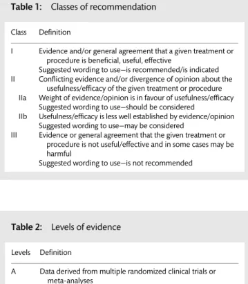
Open repair and TEVAR both severely compromise the blood supply to the spinal cord: by extensive SA sacrifice during surgery, the simultaneous SA occlusion with the deployment of covered stent-grafts or the interruption of collateral perfusion during aortic Table 1: Classes of recommendation
Class Definition
I Evidence and/or general agreement that a given treatment or procedure is beneficial, useful, effective
Suggested wording to use—is recommended/is indicated II Conflicting evidence and/or divergence of opinion about the
usefulness/efficacy of the given treatment or procedure IIa Weight of evidence/opinion is in favour of usefulness/efficacy
Suggested wording to use—should be considered
IIb Usefulness/efficacy is less well established by evidence/opinion Suggested wording to use—may be considered
III Evidence or general agreement that the given treatment or procedure is not useful/effective and in some cases may be harmful
Suggested wording to use—is not recommended
Table 2: Levels of evidence Levels Definition
A Data derived from multiple randomized clinical trials or meta-analyses
B Data derived from a single randomized clinical trial or large non-randomized studies
C Consensus of opinion of experts and/or small studies, retrospective studies, registries
C.D. Etzet al. / European Journal of Cardio-Thoracic Surgery 944
cross-clamping (or with large-bore sheaths in place during pro-longed endovascular procedures) leaving the hypogastric arteries not being perfused and the collateral network thus being deprived of its major distal inflow source. Accordingly, the major risk factors for ischaemic SCI during TAA/A repair are the following: (i) the extent of aortic graft replacement or endovascular coverage (i.e. the complexity of the repair), (ii) the presence of acute aortic dissection —particularly, with extensive, acute SA malperfusion due to the for-mation of a false lumen and (iii) the degree of urgency (i.e. limited time for proper planning, no option for staging or perioperative haemodynamic instability). The risk may vary in the range 4–7% after TEVAR for DTA, 2–28% after elective descending aortic surgery and up to 40% after emergency repair of extensive TAAAs (Table3).
The impact of prior distal aortic operations—i.e. abdominal aortic aneurysm repair—on SCI risk after reoperative surgery for more proximal aortic disease affecting the descending or the TAAA has been the focus of controversial expert debates. Retrospective series suggest a decreased risk of acute SCI, also supported by experimental evidence and the concept of a‘staged repair’—while others discuss an increase in the risk of SCI [37,38,
55–57]. The key to understanding the pathophysiology behind reoperative repair might be to assess how the previous repair affected the collateral inflow—i.e. if the hypogastric arteries have been sacrificed or the left subclavian artery has been overstented, the risk during subsequent repair might be increased.
The pathophysiology of SCI in DTA/TAAA surgery is essentially an ischaemia–infarction model caused by a variety of mechanisms (Table4). It has been assumed that injury arises primarily as a con-sequence of two mechanisms: (i) an intraoperative insult after temporary interruption of spinal cord blood supply during surgery of duration sufficient to irreversibly damage cell bodies and nerve tracts in the spinal cord and (ii) the second insult was thought to occur postoperatively: permanent reduction in blood supply sec-ondary to sacrifice of critical blood vessels—the thoracic (intercos-tal) and lumbar SAs—to a level incompatible with cord viability.
While thefirst pathomechanism primarily affects open surgical repair, thesecond mechanism also limits endovascular repair due to the sudden simultaneous occlusion of SA inflow when a stent-graft is deployed. In addition, perfusion to the hypogastric arteries supplying distal collateral inflow may be compromised by large-bore sheaths during the procedure. Starting with aortic cross-clamping or circulatory arrest during open aortic repair and followed by the sacrifice or exclusion of SAs, arterial blood supply to the spinal cord is acutely reduced, possibly triggering oedema and subsequently an increased production of CSF (Fig.1). Elevated CSF pressure generates a gradient hindering arterial blood from entering the spinal canal, and arterial blood supply to the spinal cord is progressively reduced leading to a vicious circle of progres-sive ischaemic SCI.
Significant progress has been made in understanding the para-spinous and intraspinal arterial collateral network supporting the spinal cord during deprivation of major sources of direct arterial blood supply [37,58]. Contemporary strategies to prevent acute SCI in DTA/TAAA surgery or TEVAR primarily aim at minimizing the duration of ischaemia during procedures by means of improv-ing perfusion pressure andflow as well as tissue oxygen delivery, and enabling early detection of spinal cord ischaemia to permit immediate intervention (Table 5). Intraoperatively, numerous adjuncts have been implemented to reduce SCI. Currently, left heart bypass (LHB), CSF drainage, reimplantation of the most im-portant SAs, hypothermia and maintenance of an adequate mean arterial pressure (MAP) are thought to be effective measures.
Electrophysiological assessment is helpful in detection of ischae-mia in the monitored neural tracts, the use somatosensory evoked potentials (SSEPs) or motor evoked potentials (MEPs) and has been studied extensively in thoracoabdominal aortic surgery and TEVAR. Additionally, these methods might be able to detect situa-tions of marginal blood flow resulting in neuronal dysfunction (i) at a time when the neurons are still salvageable and the insult is potentially reversible and (ii) allowing for guidance of therapeuti-cal interventions to relieve acute ischaemia.
SPINAL CORD BLOOD SUPPLY
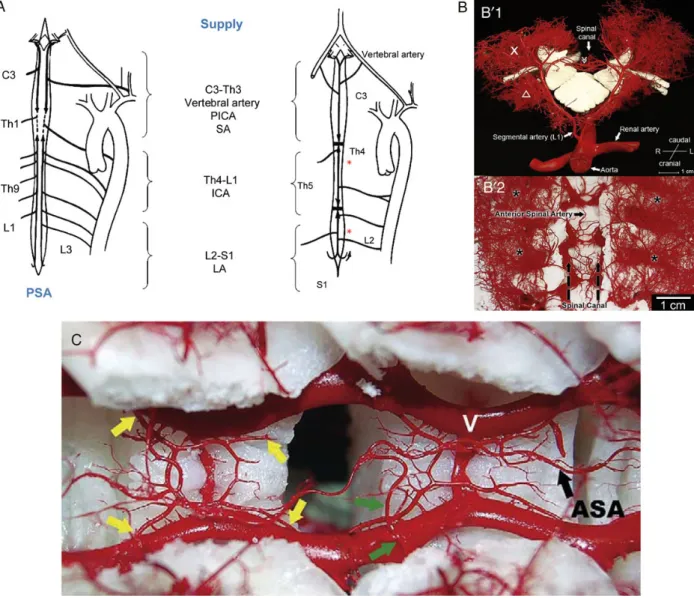
Arterial blood supply to the spinal cord is provided by the anterior spinal artery (ASA) arising cranially from both vertebral arteries to supply its anterior portion. A pair of posterior spinal arteries (PSAs) also arising from the vertebral arteries supplies the poster-ior spinal cord. Caudally, the ASA receives arterial collateral blood from the internal iliac arteries and the sacral arteries, and from the inferior mesenteric artery. Additional supply is provided by paired intercostal and lumbar SAs that originate from the DTA and ab-dominal aorta (Fig.2A).
Two different paradigms are used to explain the elusive nature of spinal cord circulation, one based on anatomical (direct, seg-mental supply) and the other on less anatomical and rather dynamic demand-depending (collateral) blood supply [42]. A thorough understanding of the anatomy of the blood supply of the spinal cord appears essential for developing strategies to prevent SCI. Direct visualization of these vessels is arduous and most surgeons therefore continue to rely on a few classic anatom-ical studies. The most influential of these has been the treatise by Albert W. Adamkiewicz (1850–1921), whose meticulously detailed drawings suggest that the most important input to the ASA is a single dominant branch of an SA in the lower thoracic or upper lumbar region, which is now often referred to as the Artery of Adamkiewicz, who in 1881 published his thesis entitled, Die Blutgefaesse des menschlichen Rueckenmarks’ at the University of Cracow [59]. His concept became the accepted doctrine forover a century—and the rationale to justify reimplantation of intercostals and lumbar arteries in TAAA surgery—even after Guy Lazorthes in 1971 postulated a new concept he had developed since the 1960s, based on three main arteries, each arising from several regional seg-mental arteries, supplying the cervical, thoracic and lumbosacral region of the spinal cord [60–62].
The clinical relevance of these concepts is controversial: the opponents of Adamkiewicz argue that SA reimplantation during TAA/A repair is the best possible strategy for preserving spinal cord blood supply [63–68]. Despite various painstaking and in-ventive attempts to avoid ischaemic SCI with this approach, there continues to be a definite, seemingly irreducible incidence of SCI after treatment of extensive TAAA [65,68–70]. Furthermore, reat-taching intercostal or lumbar SAs—a daunting undertaking during open surgical repair—is not possible with current endovascular techniques. (Table3summarizes the incidence of SCI after both open surgical and endovascular repair.)
Imaging techniques to identify SAs are considered critical to spinal cord function are controversial. In the 1990s, selective inter-costal angiography was introduced to preoperatively identify the Artery of Adamkiewicz [63,65]. Then, radiological imaging tech-nology evolved and Nojiriet al. [71] proposed preoperative detec-tion of the Artery of Adamkiewicz using intra-arterial computed tomographic angiography. Recently, it became possible to identify
REP
O
Table 3: Contemporary incidence of ischaemic SCI with permanent dysfunctionaccording to aneurysm extent—reported by international centres of excellence in endovascular (TEVAR) and open surgical aortic repair
Year N Incidence of ischaemic SCI with permanent dysfunction
according to aneurysm extent
DTA (%) Thoracoabdominal/Crawford (%) Technical/perioperative management
Type I Type II Type III Type IV Segmental arteries CSF drainage Neuromonitoring
Endovascular (TEVAR)
Greenberget al. [32] 2008 352 1 10 19 5 3 Occluded Yesa None
Gravereauxet al. [49] 2001 53 5.7 b b b b Occluded Yes, with extensive coverage None
Conradet al. [18] 2008 105 7 b b b b Occluded Yes None
Bavariaet al. [50] 2007 140 3 b b b b Occluded Not consistently None
Feezoret al. [51] 2008 326 10 b b b b Occluded None in most patients None
Stoneet al. [52] 2006 74 10.4c b b b b Occluded d None
Open surgery
Greenberget al. [32] 2008 372 1 14 22 10 2 Reimplanted or bypassed Yesa None
Conradet al. [18]e 2008 471 7 24 20 13 2 Reimplanted T9-L1, if patent Yes None
Fehrenbacheret al. [22]f 2010 343 1 4.3 5.4 3.1 0 Reimplantated T8—coeliac axis Not routinely MEP
Coselliet al. [11]g 2007 2286 b 3.3 6.3 2.6 1.4 Reimplanted in 61% In 27% None
Bavariaet al. [50] 2007 94 14h b b b b d Not consistently None
Zoliet al. [53] 2010 609 2.3 2.5 11.5 3.9 2.2 Total sacrifice In 59% MEP/SSEP
Sundtet al. [30] 2011 99 3i 0 0 0 0 Reimplanted, T9-L1 None None
Schepenset al. [54] 2009 571 b Overall paraplegia 5.3%, paraparesis 3% Reimplanted, T8-L1 Yes MEP/SSEP
Safiet al. [39] 2005 1106 d d 10.7% d d No reimplant in 61% Yes None
Stoneet al. [52] 2006 83 7.2c b b b b d d Not reported
Perfusion, temperature and the anaesthesiological perioperative management for open repair varied significantly among reference centres, e.g. Sundtet al.: DHCA at 18°C; Schepens et al.: moderate hypothermia at 32°C, DHCA only if proximal clamping is impossible; Zoliet al.: full cardiopulmonary bypass, partial cardiopulmonary bypass, left heart bypass and DHCA.
SCI: spinal cord injury; TEVAR: thoracic endovascular aortic repair; DTA: descending thoracic aorta; CSF: cerebrospinal fluid; MEP: motor evoked potential; SSEP: somatosensory evoked potential; DHCA: deep hypothermic circulatory arrest.
a‘At the discretion of the treating physician’. b
Excluded.
cDTA patients treated for degenerative pathology excluding ruptures. dNot reported.
eIntraoperative epidural cooling (EC) to 25–27°C until reperfusion of the lower extremities. f
Singularly operated in DHCA, SA reimplant, no CSF drain (!).
gSelective perfusion (balloon catheters) to the coeliac and superior mesenteric arteries, renals intermittently with 4°C crystalloid, left heart bypass in 40%, 60%‘clamp-and-sew’ 32–34°C.
hThe Gore TAG non-randomized multicentre trial: significantly higher incidence of symptomatic aneurysms (38 vs 21%,P = 0.007) in the surgical control group, historically and retrospectively acquired; surgeons
performing the open procedures were from various surgical backgrounds; there was a variable volume of thoracic aortic surgery performed in each contributing centre, and a variable use of spinal cord protection techniques. For example, in the open repair group 75% of the paraplegic patients died in hospital.
iDelayed paraplegia secondary to persistent hypotension; the comparison is not representative as patient characteristics are not uniform, e.g. urgent/emergent procedures—see original data for details.
C.D. Etz et al. / Eur opean Journal of Cardio-Thor acic Surgery 946
what was thought to be the Artery of Adamkiewicz using magnetic resonance angiography [72–75].
Within the spinal canal, there is an axial network of small arteries that connect with each other as well as with major arteries that supply the spinal cord [37]. Blood supply to the spinal cord is even more complex and pathologically modified in patients with aortic diseases. In almost 25% of these patients, most SAs are occluded and spinal cord integrity is maintained by an extensive collateral network in which lumbar arteries and the pelvic circulation are re-sponsible for main blood supply. Reimplantation of SAs increases aortic cross-clamp time and possibly aggravates intraoperative spinal cord hypoperfusion due to blood loss via back bleeding. Probably, a substantial percentage of reimplanted SAs occlude early. Alternative surgical techniques for reimplantation include latero-lateral aortic patch reimplantation or the use of small-calibre (≤5 mm) bypass conduits such as vein grafts or vascular prostheses.
The alternative paradigm suggests—in addition to the radicular arteries—that the spinal cord also has a complex collateral circula-tion. It is hypothesized that there is also an axial network of small
arteries in the spinal canal, perivertebral tissues and paraspinal muscles that receives input from the subclavian, internal thoracic, lumbar and hypogastric arteries (Figs2B and C and3). These small arteries are connected with each other and with the ASA and PSA providing bloodflow to the spinal cord. This network can increase bloodflow from one source when another is impaired. Conversely, a steal effect can occur—spinal cord blood flow can be reduced if an alternative lower resistance pathway becomes patent elsewhere in the circulation. The concept of collateral circulation is most prob-ably the reason why the maintenance of high arterial blood pres-sure and cardiac index may reduce SCI in TAAA surgery.
Reimplantation of significant patent SAs has been associated with decreased rates of SCI [43]. However, if a particular branch is small or occluded, a great deal of time may be spent without benefit—possibly even causing harm to the spinal cord increasing the risk of intraoperative SCI. Intercostal reimplantation (IRP) may jeopardize spinal cord blood flow by back bleeding. Even after successful revascularization of a dominant SA, symptomatic SCI may be observed. On the contrary, IRP may jeopardize spinal cord Table 4: Mechanisms of spinal cord ischaemia in open repair and during thoracic endovascular aortic repair (TEVAR)
Insult Effect
Open repair
(Prolonged) aortic cross-clamping Acute loss of direct (SAs) and indirect (collateral network) cord perfusion Decrease in mean arterial pressure (e.g. due to anaesthesia and
extracorporeal circulation)
Insufficient spinal cord perfusion pressure (resulting in acute, generalized malperfusion of the cord)
Increase in CSF pressure Counteracts spinal cord perfusion pressure triggering a‘spinal compartment syndrome’ Loss of critical SAs Acute loss of direct spinal cord perfusion
Insufficient distal perfusion pressure (on pump/no pulsatility) Inadequate distal inflow to the collateral network Arterial steal phenomenon via patent SAs after opening the
aneurysm sac
Reduced SCPP! oedema of the spinal cord Reperfusion injury after cross-clamping Spinal cord oedema (beginning a‘vicious cycle’)
Postoperative thrombosis of the spinal cord-supplying vessels May be responsible for delayed paraplegia (e.g. after TEVAR) TEVAR
TEVAR covering of left subclavian artery, intercostal and lumbar SAs, hypogastric arteries and sacral arteries
Reduction in proximal, medial and distal direct and collateral arterial blood flow to the spinal cord
Previous distal aortic surgery If the hypogastric axis was compromised/sacrificed, distal collateral inflow is compromised
Severe peripheral vascular disease Reduction in (predominantly distal) collateral inflow also due to the impairment of flow provided to the collateral network by the hypogastric arteries
CSF: cerebrospinal fluid; SA: segmental arteries.
Figure 1:Spinal cord bloodflow and perfusion pressure during thoracic aortic occlusion. The changes (arrows) represent the response to aortic cross-clamping per se. : increase; : decrease; ICP: intracranial pressure.
REP
O
bloodflow by back bleeding. By using ligation or clipping of SAs ( preferably prior to opening the aneurysm sac), the bloodflow is directed to the spinal cord by collateral vessels and the need for revascularization becomes futile. Therefore, it might be more im-portant to consider the superior and inferior supply of the spinal cord via the subclavian arteries and the internal iliac network (e.g. the hypogastric arteries). At least unilateral internal iliac artery perfusion should be strictly maintained. Careful consideration is also warranted in the treatment of common and internal iliac aneurysms particularly in endovascular repair. A branched stent-graft should be used if possible—otherwise primary embolization, e.g. with Amplatzer plugs seems to cause (at least) less buttock is-chaemia. However, patients with poor pelvic circulation are critic-ally dependent on the above-mentioned specific ICAs. In these cases, reimplantation of critical SAs (Th8-L4) may be considered.
STRATEGIES TO SHORTEN THE INTRAOPERATIVE
DURATION OF SPINAL CORD ISCHAEMIA
The duration of aortic cross-clamping has a close relationship to the risk of SCI. For this reason, one major objective of surgery is to keep the overall ischaemic time short.
Open TAAA repair starts by clamping the proximal and distal aorta to isolate partially (sequential or staged repair) or totally the diseased segment if the patient is operated on with LHB. Unless distal aortic perfusion via extracorporeal circulation support is initiated, there is no or only minimal bloodflow below the cross-clamp. Spinal cord perfusion is sustained only via the vertebral, cervical and subclavian arteries. Thus, spinal cord perfusion pres-sure (SCPP = radicular artery end prespres-sures minus the greater of venous or CSF pressures) may be compromised [76,77]. Important arteries arising from the aortic aneurysm sac are no longer per-fused. In case of back bleeding, the steal phenomenon will add-itionally reduce both collateral network pressure and thereby perfusion pressure of the ASA. Therefore, it is important to avoid back bleeding instantly by oversewing (or preferably prior to aneur-ysm sac opening by clip occlusion); blocking of the corresponding
SAs with small catheters after opening the aneurysm is another option (with caution in patients with connective tissue disease) if an island repair is planned. A novel approach termed‘minimally invasive segmental artery coil embolization’ (MISACE) is an elegant alternative recently introduced as an option allowing for endovas-cular staging to precondition spinal cord blood supply, avoiding ‘steal’ and type II endoleaks and shortening cross-clamp time. Several methods have been used to provide reperfusion on the one hand and avoid back bleeding of these arteries on the other hand. One approach is to preserve a fragment of the back wall of the aneurysm where large SAs arise and use it during aortic recon-struction with the idea to restore critical perfusion. Another ap-proach is to attach critical SAs via a second prosthesis into the aortic prosthesis. Furthermore, to prevent significant steal via back bleeding, non-critical arteries are occluded with surgical clips from outside or oversewn from inside. A common mistake is to aim for reimplantation of the vigorous‘back bleeders’—but these are the arteries that are sufficiently collateralized—rather than those SAs that do not bleed back. As the time required for reim-plantion of all SAs potentially increases the risk of SCI, intraopera-tive neurophysiological monitoring (IOM) may direct the surgeon during sequential cross-clamping and provide information about which vessels are essential for sufficient spinal cord perfusion. However, the success of this strategy is controversial and the rates of SCI with this strategy have not proved to be superior.
Because of the key role of pelvic circulation mainly provided by the hypogastric arteries, many patients are critically dependent on distal (retrograde) perfusion from LHB. Partial LHB provides a con-trolled perfusion of the distal aorta by directing blood from the left atrium to distal segments. Flow is controlled by a centrifugal pump or a complete circuit with a membrane oxygenator accord-ing to the department policy. Duraccord-ing partial LHB with a proximal aortic cross-clamp, the distal aortic cross-clamp can be moved from proximal to distal as repair of the descending aorta pro-gresses to minimize end-organ ischaemia, a technique that has been termed‘sequential repair’ by Coselli et al. A large amount of retrospective data suggests that the use of LHB in extensive TAAA reduces the risk of ischaemic complications. The absence of an oxygenator in the LHB system necessitates less heparinization, which is associated with considerable reduction in bleeding. However, it effectively improves oxygenation during one-lung ventilation, especially as these patients frequently are smokers with varying extent of chronic obstructive pulmonary disease (COPD) [78]. In addition, LHB allows for selective perfusion of mesenteric branch vessels through separate balloon-blocking catheters. Summarizing, LHB facilitates both afterload reduction as well as cooling and rewarming, avoids vasodilators, increases distal aortic pressure for patients dependent on caudal vessels, reduces an increased CSF pressure and decreases the risk of visceral ischae-mia and spinal cord ischaeischae-mia by permitting selective organ and segmental artery perfusion. However, LHB does not appear to be the ultimate methodology for all cases: this especially applies to Crawford Type I and II aneurysms, where the rate of SCI is still high.
STRATEGIES TO INCREASE SPINAL CORD
TOLERANCE TO TRANSIENT ISCHAEMIA
The highly metabolic grey substance of the spinal cord is more sensitive to ischaemia than the white substance. Under nor-mothermic conditions, the central nervous system poorly tolerates Table 5: Strategies to prevent and treat spinal cord ischaemia
Minimizing spinal cord ischaemic time
Multisegmental, sequential reconstruction of the aorta Stepwise or staged or sequential clamping of the aneurysm
(if anatomy permits to do so) Increasing tolerance to ischaemia
Deliberate utilization of mild systemic hypothermia
Optional deep hypothermic circulatory arrest and/or selective spinal cord hypothermia by epidural cooling
Pharmacological neuroprotection/ischaemic preconditioning (‘staged repair’)
Augmenting spinal cord perfusion
Deliberate proximal and distal hypertension Cerebrospinal fluid (CSF) drainage Reimplantation of segmental arteries
Preservation of subclavian artery and hypogastric artery flow/left heart bypass/distal aortic perfusion
Early detection of spinal cord ischaemia IOM (MEP and SSEP)
Fast track concept and serial postoperative neurological examination
IOM: intraoperative neurophysiological monitoring; MEPs: motor evoked potentials; SSEPs: somatosensory evoked potentials.
C.D. Etzet al. / European Journal of Cardio-Thoracic Surgery 948
ischaemia, manifesting neuronal dysfunction and injury within 5 min after the cessation of bloodflow. When incomplete ischae-mia is produced, SCI generally does not occur with an aortic cross-clamping time of less than 15 min. As the cross-clamp time is prolonged, the risk of SCI gradually increases. It is important to note that the risk of SCI is closely related to the body core tem-perature during lower body circulatory arrest, initiated by placing the cross-clamp: Kamiyaet al. [79] found a 6-fold increase in the incidence of SCI in their subgroup analysis of patients undergoing prolonged distal circulatory arrest at only moderate hypothermia. Experimentally, the safe period of distal arrest has been shown to be widely overestimated and irreversible SCI at 28°C occurs earlier than expected [80]. The only intervention in humans that has consistently proved to be effective in protecting the central nervous system from ischaemia during the absence of bloodflow is hypothermia [81–83].
Additionally, delayed postoperative rewarming might have a positive effect on ischaemia tolerance of the spinal cord and therefore is part of the postoperative protocol at some institutions. However, there is not yet enough clinical evidence or prospective randomized studies to proof this concept.
The protective effect of hypothermia is thought to be primarily a consequence of the decreased metabolic demands associated with reduced spinal cord oxygen consumption. However, hypo-thermia may also protect the cell by stabilizing membranes and attenuating the inflammatory and excitotoxic responses to ischae-mia during reperfusion. Further protection of the spinal cord tissue has been attempted with regional spinal cord hypothermia (epidural cooling) [70]. However, besides contamination issues, re-sponsive hyperperfusion and consecutive development of oedema are feared by some after cooling is ended.
Figure 2:(A) Blood supply to the spinal cord. Schematic drawing of the spinal cord with indications of areas supplied by the posterior and the anterior spinal arteries. Radicular arteries are variable in location. The inflow to spinal arteries is divided into three main supply zones ASA: anterior spinal artery; PSA: posterior spinal artery; PICA: posterior inferior cerebellar artery; SA: segmental arteries; ICA: intercostal arteries; LA: lumbar arteries. (B) Anatomy of the collateral network from experimental casts, sagittal (B01) and dorsal (B02) views. Macroscopic appearance of the pair of dorsal segmental vessels at L1. The dorsal process is removed. In B01, the X designates the paraspinous muscular vasculature providing extensive longitudinal arterioarteriolar connections in B01 and B02; the triangle indicates iliopsoas muscle; the double arrow indicates anterior spinal artery [reprinted from Etzet al. [37] Copyright (2011) with permission from Elsevier]. (C) Relationship of the ASA and the repetitive epi-dural arcades in a Yorkshire pig model. V indicates the epiepi-dural venous plexus. Anterior to the extensive venous plexus, four arteriolar branches (yellow arrows) con-tribute to one circular epidural arcade. This pattern is repeated at the level of each vertebral segment. These vascular structures connect segments side to side as well as longitudinally. Green arrows depict the anterior radiculomedullary artery, which connects directly with the anterior spinal artery [reprinted from Etzet al. [37] Copyright (2011) with permission from Elsevier].
REP
O
In addition to lowering oxygen consumption, the risk of intra-operative spinal cord ischaemia may be avoided or minimized by improving oxygen delivery via an increase of SCPP. Demand side interventions prolong ischaemic tolerance by decreasing the need of oxygen (barbiturates and hypothermia), while reducing the levels of neurotoxins released during ischaemia and/or their dele-terious effects (naloxone and hypothermia). The spinal cord may be directly protected against neuronal injury at the cellular level by reducing hyperaemic and inflammatory responses (hypother-mia, steroids and free radical scavengers). Supply side interven-tions increase spinal cord blood supply and tissue oxygen delivery by maximizing collateral bloodflow to the spinal cord, reducing spinal fluid pressure, increasing arterial blood pressure and the cardiac index during and after the repair, preventing steal and guaranteeing sufficient oxygenation during aortic cross-clamping. The observation that similar reductions in SCI can be achieved by combining different therapies basically reflects the complexity of spinal cord blood supply and neuronal injury.
STRATEGIES TO AUGMENT SPINAL CORD
PERFUSION
As previously elaborated on, spinal cord perfusion during aortic surgery depends on (i) the ASAflow from radicular vessels arising above the proximal cross-clamp and supplied by proximal aortic pressure, (ii) from vessels arising from the aorta below the distal
cross-clamp, depending on distal aortic perfusion via LHB or extracorporeal circulation with an oxygenator and (iii) from the central venous and CSF pressure. During proximal aortic cross-clamping, the MAP inceases considerably and needs pharmaco-logical correction to control left ventricular afterload. The elevated cerebral blood pressure during proximal aortic cross-clamping may result in an overproduction of CSF and an elevation of CSF pressure. Elevated CSF pressure further reduces the SCPP. If CSF pressure exceeds both ASA and PSA pressure, the spinal cord blood flow ceases and oxygen supply is interrupted—the spinal cord is suffering ischaemia. Full or partial recovery from delayed postoperative SCI after open or endovascular repair has been reported and emphasizes the effectiveness of acute interventions to improve spinal cord perfusion, if applied instantly. Postoperative events such as hypotension due, for instance, to haemorrhage or increased CSF pressure may also increase the risk of SCI after open and endovascular repair. Therefore, maintaining adequate spinal cord perfusion by increasing arterial pressure and augmenting cardiac output, together with preventing hypotension, lowering CSF pressure and reducing central venous pressure (CVP), is important for the prevention of spinal cord ischaemia.
CSF production rises during ischaemia, causing an increased CSF pressure soon after cross-clamping. To minimize spinal cord ischae-mia, CSF drainage is used to maintain a low CSF pressure while im-proving net perfusion pressure. The physiological basis for lumbar CSF drainage is given by the SCPP being a direct function of the MAP minus lumbar CSF pressure (or alternatively central venous pressure). Therefore, an increased CSF pressure decreases the Figure 3:A schematic diagram of the blood supply to the spinal cord demonstrates the relationships, relative sizes and the interconnections among the segmental arteries (SAs), the anterior radiculomedullary arteries (ARMAs), the epidural arcades and the anterior spinal artery (ASA). The longitudinal anastomoses along the dorsal processes of the spine as well as dorsal communications (interstitial connections) between the right and the left branches of segmental arteries are also shown [reprinted from Etzet al. [37] Copyright (2011) with permission from Elsevier].
C.D. Etzet al. / European Journal of Cardio-Thoracic Surgery 950
SCPP. Draining CSF has the potential to increase the SCPP by decreasing the CSF pressure. Experiences during open surgical DTA/TAAA repair have shown that this intervention has a positive effect on neurological outcome. In general, introduction of a CSF catheter is performed preoperatively, but can also be performed postoperatively when neurological symptoms develop. It is highly advisable to insert a CSF drain in all patients undergoing TAA/A surgery or thoracoabdominal EVAR and measure CSF pressure for at least 48 h postoperatively. If CSF pressure was allowed to rise postoperatively in combination with a period of blood pressure instability, late onset SCI due to spinal cord oedema may occur. CSF should be drained into a sealed reservoir to achieve a CSF pressure of 10 mmHg; some institutions alternatively aim for the preoperative ‘opening pressure’ immediately after CSF catheter placement as an individual baseline pressure of the patient. CSF drainage appears to be a safe method even in patients subjected to full anticoagulation for extracorporeal circulation. Complica-tions associated with this technique occur in up to 1% of patients and include intracranial hypotension, subdural haematoma, intracranial haemorrhage, remote cerebellar haemorrhage, spinal headache, persistent CSF leak, intraspinal haematoma, catheter fracture, meningitis and direct SCI. Some institutions insert the CSF catheter on the evening prior to surgery to avoid or anticipate bleeding complications. The most serious complications appear to be associated with intracranial hypotension from rapid or too much CSF drainage. Precautions, such as continuous measurement of CSF pressure, controlled intermittent CSF drainage and assess-ment of coagulation function, decrease the risks associated with CSF drainage.
Augmentation of the MAP (in combination with CSF drainage if not already present) is another technique for the treatment of spinal cord ischaemia. In general, vasopressor agents such as nor-epinephrine are administered to maintain an MAP of 80–100 mmHg to ensure an SCPP of at least 70 mmHg. A more recent clinical study suggested that failure to maintain a patient’s individ-ual preoperative arterial baseline pressure during the early post-operative period after TAA/A repair is strongly associated with delayed postoperative SCI [29]. The MAP can be further increased in 5 mmHg steps in case of persisting SCI. When arterial pressure increases, it is also important to assure a satisfying cardiac output and to guarantee an optimal oxygen delivery (control of haemoglo-bin). Inconsistent arterial pressure control may also partly explain the controversy surrounding the effectiveness of CSF drainage as an exclusive means to decrease CSF pressure. Hypotension from bleeding or other causes is often associated with the onset of SCI after TAAA repair. Nevertheless, clinical observations suggest that SCI may as well contribute to hypotension due to generalized vasoplegia. In some patients, spinal cord ischaemia-associated hypotension is caused by neurogenic shock with autonomic dys-function. In this situation, hypotension may not be the cause but represent an early sign of SCI and the beginning of a vicious cycle. An immediate treatment of hypotension associated with spinal cord ischaemia is necessary to prevent permanent SCI. Finally, ar-terial pressure should be monitored carefully when antihyperten-sive therapy is resumed after successful open or endovascular TAA/A repair to avoid unintentional hypotension. Nitroprusside derivatives should be strictly avoided due to possible arterio-venous shunting. The benefits of postoperative arterial pressure increase must be weighed against the risk of bleeding and the risks associated with temporary arterial pressure elevation. Anaes-thetic staff needs to be well trained in the management of TAAA surgery and the postoperative patient to prevent large variations
in blood pressure during and early after the procedure. Equally, an intensive care unit that is familiar with all aspects of postoperative care after TAA/A repair is very important to provide maximal haemodynamic stability. Many patients with late onset SCI have a documented period of instability prior to symptoms.
Spinal cord perfusion can be surgically augmented by reattach-ment of SAs into the vascular graft if the surgeon respects the ana-tomical paradigm that direct segmental blood flow is the most important intervention to reduce the risk of SCI. Large SAs with little or no back bleeding may be particularly important for spinal cord perfusion. Alternatively, occlusion or oversewing of strong back bleeding SAs has been advocated to improve spinal cord perfusion by preventing an arterial steal effect and shortening intraoperative ischaemic time [3]. As most reports combine IRP with other strategies, it is hard to determine how much reattach-ment of SAs contributes on its own to improved results, even though it is frequently presented as the factor primarily respon-sible for reducing SCI.
On the other side, a significant reduction in the risk of SCI without IRP was obtained by increasing ischaemic tolerance and maximizing collateral circulation to the spinal cord. Significantly, this technique maintains high proximal arterial blood pressure during aortic occlusion. Thesefindings show that spinal cord in-farction can almost always be prevented without any IRP if ischae-mic protection and collateral circulation to the spinal cord are sufficient [3]. Although this approach considerably reduced imme-diate SCI, delayed SCI still occurred in a few patients days to weeks after surgery [29]. The occurrence of delayed SCI shows the limita-tions of perioperative ischaemic protection and of the mainten-ance of collateral circulation strategies to prevent infarction [29]. Recent advances in magnetic resonance angiography have per-mitted a more precise imaging of the ASA and the expected most important SAs in patients with TAAA. It provides a method to ana-tomically identify SAs for potential reimplantation. Non-selective IRP may also be protective by additionally increasing perfusion pressure in the collateral circulation and feeding the greater ra-dicular artery. This suggests that any SA can supply blood to the spinal cord and may evolve into collateral circulation to the ASA. In patients with reduced collateral circulation, reimplanting any SA in the critical zone of T8 to L1 may permanently increase per-fusion pressure in the collateral network. This may be the import-ant factor to avoid spinal cord infarction, regardless of whether specific identified intercostals are reimplanted.
Techniques that rely on extensive IRP based on changes in evoked potentials (EPs) may be successful not because SAs identi-fied by ischaemic changes were reimplanted, but because reim-planting so many SAs increased the perfusion pressure in the collateral circulation. By reimplanting SAs as an aortic button using a side clamp after the distal anastomosis is completed, it is possible to achieve high SA patency without significantly increas-ing aortic occlusion times. These findings suggest that factors related to spinal cord ischaemia as well as collateral circulation account for most of the SCI risk in TAAA surgery and that IRP, al-though not necessary to prevent SCI in most patients, is critically important in a few. Since we do not yet know how to identify the very patients who will sustain SCI without IRP, it is important to reimplant SAs only without substantially increasing intraoperative spinal cord ischaemic time and surgical morbidity, even in those who would not sustain SCI without IRP in order to maximize the benefit for the ones at risk.
In TEVAR, it is not possible to preserve bloodflow in SAs. If the left subclavian artery requires coverage by the stent-graft to
REP
O
enable complete exclusion of the aneurysm or to allow for a better proximal landing zone, subclavian arterialflow should be preserved by prior transposition of the subclavian artery onto the left common carotid artery. Another approach to preserve left subclavian arteryflow in TEVAR is to perform a left carotid to sub-clavian bypass graft with ligation or coil embolization of the prox-imal left subclavian artery stump. Maintaining bloodflow in the left subclavian artery is important for spinal cord perfusion as its branches supply the ASA. Meanwhile, there is substantial evidence available supporting routine preservation of the left subclavian artery [84,85].
STRATEGIES TO DETECT SPINAL CORD
ISCHAEMIA
Early detection of spinal cord ischaemia is important as it permits early intervention before ischaemia evolves to infarction. SSEPs and MEPs are established methods of spinal cord monitoring during TAAA surgery and TEVAR. The clinical objectives for SSEP/ MEP monitoring are to ensure adequate spinal cord perfusion throughout the procedure, to identify critical vessels for reimplan-tation and to establish an MAP adequate for spinal cord perfusion. Decreased EP amplitudes have proved to correlate with spinal cord ischaemia, but the sensitivity and specificity of these techni-ques for detection of spinal cord ischaemia remain to be deter-mined. Intraoperative changes or loss of EP signals are not always caused by spinal cord ischaemia. A functioning peripheral nerve is required to generate both SSEP and MEP signals. Therefore, per-ipheral nerve ischaemia from any cause will affect the associated SSEP or MEP amplitudes. Vascular malperfusion of a lower ex-tremity can cause a loss of peripheral EP in the absence of spinal cord ischaemia if bloodflow to the limb is significantly impaired. Lower extremity malperfusion may be caused by aortic dissection itself, atheroembolism or most commonly by arterial cannulation of the femoral artery for extracorporeal circulation. Similar to mal-perfusion, aortic cross-clamping without distal aortic perfusion results over time in fading EP signals from the lower extremities. Acute intraoperative stroke may also produce EP changes. They can be distinguished from changes caused by spinal cord ischae-mia by comparing signals recorded at different sites along the neural conduction pathway. Stroke is associated with selective loss of cortical signals and typically affects the EP from both upper and lower extremities.
SSEP recordings measured via the sensory cortex can be affected by ischaemia of the peripheral nerves, the spinal cord, the brainstem, the sensory cortex and additionally by technical and anaesthesiological factors (Fig.4). An advantage of SSEP mon-itoring is that it is relatively safe to perform and easy to interpret by comparing the amplitude and latency of SSEPs recorded from the upper and lower extremities. Thefidelity of SSEPs is improved with neuromuscular blockade under general anaesthesia. Although high concentrations of inhaled anaesthetics, thiopental or propo-fol can attenuate cortical SSEP signals, a balanced general and inhaled anaesthetic provides consistent conditions for intraopera-tive SSEP monitoring. Anatomically, the SSEP travels cephalad via the peripheral nerve and enters the dorsal roots of the spinal cord corresponding to the stimulated nerve. It traverses the dorsal horn and ascends the spinal cord via the dorsal spinal cord that med-iates proprioception and vibration. A potential limitation of SSEP monitoring is that spinal cord ischaemia confined to the anterior
spinal cord may cause a selective motor deficit with intact sensa-tion. In this situation, SSEP monitoring may fail to detect spinal cord ischaemia. This anatomical picture is likely an oversimpli fica-tion, because SSEPs from the lower extremity are thought to include a contribution from the spinocerebellar pathways that are located deeper in the spinal cord. Since the latter contribution is vascularized by the ASA, it is possible that the SSEP may respond to selective anterior ischaemia by the effect on this component of the pathway. Alternatively, anterior ischaemia may steal blood from the posterior perfusion, leading to SSEP changes. As SSEPs are primarily a white substance pathway in the spinal cord and largely devoid of synaptic connections, they may react less sensi-tively than MEP pathways that include synapses. However, SSEPs recorded in the spinal cord are known to be sensitive to hypoten-sion and have been used to gauge deliberate hypotenhypoten-sion during TAAA surgery. The sensitivity of SSEPs to distal perfusion has resulted in substantial false-positive changes. As aortic cross-clamping compromises perfusion of the anterior spinal cord and results primarily in motor deficits, it is not surprising that SSEP monitoring during TAAA surgery with distal aortic perfusion has not reduced the incidence of neurological deficits.
Monitoring of motor pathways, particularly in case the function of alpha motor neurons is included, is a sensitive measure of an-terior spinal cord function. To ensure that only motor pathways are stimulated, electrical or magnetic stimulation of the cerebral cortex is used to produce descending volleys of activity in the cor-ticospinal tracts. Following the pathway of motor function, MEPs elicited through transcortical electrical stimulation appear to be a more specific monitor (Fig.4). Transmission may be evaluated by recording from the distal spinal cord using epidural recordings (evoked spinal cord volley, epi-MEP), from a peripheral nerve (neurogram) or from muscles (compound muscle action poten-tials,). Unfortunately, epi-MEPs are less sensitive to the degree of spinal cord ischaemia because they do not involve the anterior horn cell and their axons are less sensitive to ischaemia than grey matter.
MEP monitoring has been used to identify SAs critical for re-attachment following the acute loss of lower extremity MEP signals during TAAA repair. MEP recording during surgery may guide the physician in determining the optimum postoperative blood pressure. In patients with a significant risk of spinal cord is-chaemia, sequential cross-clamping of the aorta may identify the critical segments of the aorta that provide important blood supply to the spinal cord. MEPs may therefore be used to guide the need and level of intercostal and lumbar SA reattachement. Although this method of monitoring spinal cord function may be useful in studying the effectiveness of adjuncts to lower the risk of SCI, it sometimes provides false-positive results; particularly since the neurological function of the spinal cord may be affected by anaesthetic agents that potentially depress the syn-aptic function of the cerebral cortex and spinal grey substance. In particular, the amplitude of the MEP is sensitive to neuromus-cular blocking agents and many general anaesthetic agents. General anaesthetic regimens utilizing intravenous infusions of remifentanil, ketamine, propofol or etomidate without muscular blockade or carefully controlled incomplete neuro-muscular blockade are often required to maintain satisfactory MEP signals during operation.
Recently, near-infrared spectroscopy has been successfully introduced into a clinical pilot study to non-invasively detect spinal cord ischaemia during open thoracic/thoracoabdominal repair. Sensitivity and response time are promising, but further C.D. Etzet al. / European Journal of Cardio-Thoracic Surgery
research has to validate this method experimentally and clinically to define its role in relation to IOM to detect spinal cord ischaemia [86]. Finally, another eventual limitation used as an argument by some is that the method of IOM is complex and renders the pro-cedure even more cumbersome due to the equipment required in the operation theatre and the contribution of a neurophysiolo-gist during the entire procedure [87]. Not all institutions are able to provide such an environment.
TEVAR—SPECIAL CONSIDERATIONS
TEVAR has made us rethink the pathophysiology of spinal cord is-chaemia. Coverage of the thoracic aorta without revascularization of SAs feeding the spinal cord was expected to produce higher rates of spinal cord ischaemia than actually observed. TEVAR may be performed with CSF drainage. Other adjuncts believed to be necessary in avoiding spinal ischaemia such as revascularization of important intercostal branches cannot be employed and, still, the rates of SCI are low thereby supporting the collateral network concept (and the strategy of SA sacrifice in open TAA/A repair). TEVAR has less influence on the patient’s perfusion physiology, ensures cardiovascular stability and offers shorter or no organ is-chaemic periods as aortic cross-clamping is not necessary during TEVAR, thereby avoiding distal hypotension (except for the duration
of large-bore sheaths placed in the iliac arteries during the proced-ure) and negative effects on spinal cord perfusion associated with open TAAA surgery. Distal aortic perfusion remains uninterrupted, guaranteeing a continuous bloodflow to the spinal cord and ex-cluding a steal effect via SAs after opening the aneurysm. Delayed paraplegia may occur due to (micro) embolism caused by athero-sclerotic debris or blood clotsflushed into the spinal cord vascula-ture after being mobilized from the aneurysm sac during partial and/or temporary perfusion which may occur in type II endoleaks.
Reperfusion injury after open surgical aortic replacement can occur when cytotoxic metabolites formed during cross-clamping reach the reimplanted SAs. Considering avoidance of SCI after TEVAR, it must be remembered that the extent of repair is of im-portance to determine the risk of SCI. The reduced risk of SCI in TEVAR compared with open TAAA surgery is multifactorial. If a series contain patients with a shorter length of covered aorta, they will inevitably show lower rates of spinal cord ischaemia. Likewise, when a dissected aorta is stented, retrograde perfusion of the false lumen via communications in the membrane maintains SA blood supply. This is clearly not a phenomenon that occurs after surgical repair and complete exclusion of the lesion.
However, the issue of SCI still remains with TEVAR because of (i) the inability to revascularize covered SAs, (ii) a period of hypoten-sion for TEVAR deployment, (iii) the persistence of the risk of em-bolization from aortic atheromatous lesions and (iv) the possibility Figure 4:Left upper panel: physiological somatosensory evoked potentials (SSEPs). Right upper panel: pathophysiological SSEPs. Left lower panel: physiological motor evoked potentials (MEPs). Right lower panel: pathophysiological MEPs.
REP
O
of compromise of distal perfusion due to large-bore sheaths used for stent-graft introduction during the procedure. Consequently, SCI remains the most devastating complication also after TEVAR. Independent proven risk factors for the development of delayed-onset SCI are (i) perioperative MAP of less than 70 mmHg, (ii) CSF drainage complications, (iii) previous abdominal aortic aneurysm repair (if the hypogastric arteries have been compromised), (iv) sig-nificant preoperative renal insufficiency, (v) left subclavian artery coverage without revascularization and (vi) the use of three or more stent-grafts (reflecting the lengths of the covered segments as well as the lengths of the procedural time). However, others have shown that the impact of simultaneous closure of two independent arterial spinal-cord supplying vascular territories (in particular in combin-ation with intraoperative hypotension) is the most important risk factor for symptomatic SCI irrespective of the covered length or pre-vious aortic repair, underscoring the importance of the collateral network concept [88]. A thorough consideration of the risk profile in patients requiring TEVAR remains essential. Careful haemodynamic monitoring is vital and prophylactic measures for spinal cord protec-tion should be considered in patients whose thoracic aortas require extensive coverage and those with other independent risk factors.
When TEVAR is performed in patients with chronic atheroscler-otic aneurysm in contrast to the ones with acute aortic dissection, collaterals may have developed with time and are able to com-pensate for acute SA occlusion. Many studies have underlined the importance of these individual collateral arterial networks supply-ing the spinal cord in patients undergosupply-ing TEVAR. Neurophysio-logical monitoring is viewed as an effective method to detect spinal cord ischaemia during these procedures [89]. In patients with deteriorating SSEPs or MEPs, a decrease in the SCPP and/or the CVP as well as an increase in the MAP is obligatory to ensure sufficient collateral spinal cord perfusion.
Recently, an approach to enhance collateralization has been reported as minimally invasive selective segmental artery coil embolization before TEVAR or open repair (MISACE) [90]. The method seems to be effective but extensive clinical work has to be done before a recommendation can be made.
SUMMARY
In essence, the surgical community is divided by their respective hypotheses as to the cause of SCI after TAA/A repair. Those who are convinced that SCI is the consequence of chronic hypoperfu-sion after sacrifice of SAs critical to spinal cord blood supply do reimplant SAs, trading prolonged intraoperative spinal cord is-chaemia for the achievement of arguably superior postoperative perfusion [10,69,91–96]. Others are convinced that the blood supply to the spinal cord depends on a highly variable collateral system capable of perpetuating sufficient spinal cord perfusion even after radical sacrifice of (almost all) SAs under stable haemo-dynamic conditions; this encourages them to omit reimplantation, shortening intraoperative spinal cord ischaemia by cutting down aortic cross-clamp time [3,97].
Reimplantation remains the most widespread strategy for pre-serving spinal cord function. In 2000, Jacobset al. [98] reported a significant reduction in neurological complications—to 2.3%—with the monitoring of MEPs in a series of 170 patients with TAAA, using a reimplantation approach with LHB and CSF drainage. van Dongenet al. [91] reported a 4.2% rate of postoperative paraplegia in a series of 118 patients, using hypothermia, LHB and a reim-plantation strategy guided by MEP and SSEP monitoring. In 2002,
Donget al. reported a 5.4% SCI rate in a series of 56 TAA/A opera-tions utilizing MEP and SSEP monitoring with a reimplantation ap-proach. The majority of studies that sought to prevent ischaemia by reimplantating SAs with particular focus on the area between T7 and L2 anticipated that SCI is the consequence of hypoperfu-sion after sacrifice [10,69]. Several studies have attempted to dem-onstrate the arguable superiority of this approach. In contrast, in 1994 and 1996, SCI rates as low as 3% in DTA/TAAA repair without SA reimplantation were described both in a series of 110 by Acher et al. [97], and in 95 consecutive patients by Grieppet al. [99].
In 2004, Ohtsuboet al. proposed the selective perfusion of the Artery of Adamkiewicz to prevent intraoperative spinal cord is-chaemia [100]. Furukawaet al. in their most recent contribution proposed a sophisticated, integrated intraoperative approach: se-lective intraoperative perfusion of the identified artery to prevent ischaemia during aortic cross-clamping, temporary clamping of SAs during aortic cross-clamping to prevent steal once the aneur-ysm sac is opened (along with neuroprotective adjuncts like CSF drainage and high MAP) and reconstruction of those SAs deemed relevant for the supply of the Artery of Adamkiewicz to restore native spinal cord perfusion [100]. Although only a very small series, 44% of the reconstructed SAs were occluded in the post-operative follow-up, and the only case of SCI occurred in the group with SA reconstruction [101]. Acher, a former proponent of SA sacrifice who had switched to a very sophisticated reimplanta-tion strategy in 2005 using preoperative magnetic resonance angiographic localization to identify the Artery of Adamkiewicz, has stated recently (in December 2010) that‘it remains unclear whether intercostal reimplantation reduces paraplegia risk, as we had initially proposed’ [43,97].
SCI remains a multifactorial problem with several aetiologies, contributing factors and underlying aortic pathologies and may vary considerably among different patient cohorts. No single spinal cord protecting method is currently able to provide absolute safety. Included is SCI as a consequence of the underlying pathology, ischaemic injuries from loss of distal aortic perfusion, ischaemic in-juries from loss of critical intercostal and lumbar SAs during the procedure, and other perioperative factors such as hypotension resulting in delayed SCI. Advanced contemporary surgical and an-aesthetic methods include reduction of aortic cross-clamp times, retrograde perfusion via partial LHB, hypothermia, reattachment of SAs, CSF drainage, MAP augmentation and IOM, and have improved the safety of thoracic and TAAA repair and TEVAR.
The objective is to rapidly identify the ischaemic condition and restore spinal cord perfusion with an attempt to minimize the duration of spinal cord ischaemia. However, even the com-bination of these various techniques does not entirely abolish the problem. The practice of coverage of the thoracic aorta by TEVAR and the exclusion of potentially relevant SAs with relative-ly low rates of SCI suggest that an exclusiverelative-ly anatomical basis concerning spinal cord ischaemia is not a realistic scenario, and that the actual individual functionality of the patient’s collateral network, anaesthetic stability and duration of ischaemia seem to play a major role. Despite all these advances and an improved understanding of spinal cord perfusion, spinal cord ischaemia and infarction causing postoperative SCI remains an important and debilitating complication of all thoracic and thoracoabdom-inal aortic procedures, be it open or endovascular. Associated morbidity and mortality justify the routine clinical application of techniques to prevent and treat SCI. It would be more than welcome to gain evidence by randomized controlled trials to eventually develop widely acceptable algorithms to prevent this C.D. Etzet al. / European Journal of Cardio-Thoracic Surgery