Promoter methylation of the DNA repair gene MGMT in astrocytomas is frequently associated with G:C → A:T mutations of the TP53 tumor suppressor gene
5
0
0
Texte intégral
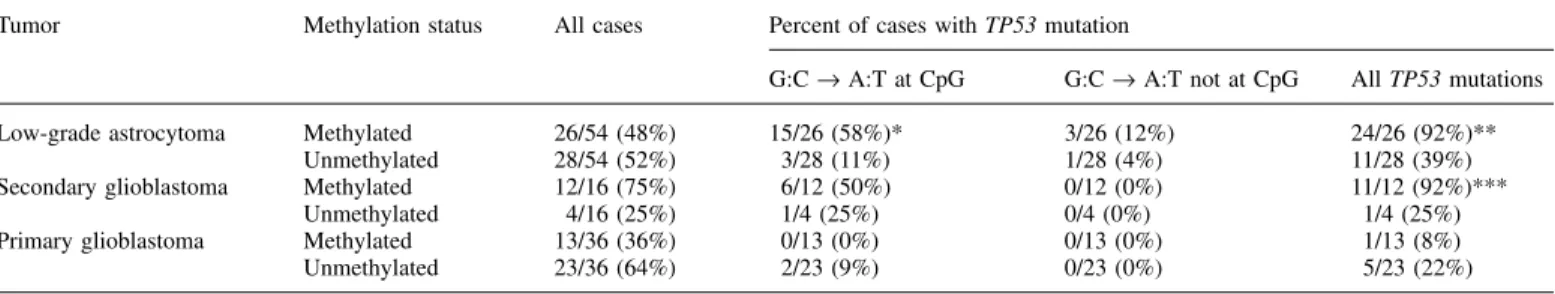
(2) M.Nakamura et al.. Table I. Correlation between MGMT promoter methylation and TP53 mutations in astrocytic brain tumours Tumor. Low-grade astrocytoma Secondary glioblastoma Primary glioblastoma. Methylation status. Methylated Unmethylated Methylated Unmethylated Methylated Unmethylated. All cases. 26/54 28/54 12/16 4/16 13/36 23/36. (48%) (52%) (75%) (25%) (36%) (64%). Percent of cases with TP53 mutation G:C → A:T at CpG. G:C → A:T not at CpG. All TP53 mutations. 15/26 (58%)* 3/28 (11%) 6/12 (50%) 1/4 (25%) 0/13 (0%) 2/23 (9%). 3/26 (12%) 1/28 (4%) 0/12 (0%) 0/4 (0%) 0/13 (0%) 0/23 (0%). 24/26 (92%)** 11/28 (39%) 11/12 (92%)*** 1/4 (25%) 1/13 (8%) 5/23 (22%). Significantly different between methylated and unmethylated cases (*P ⫽ 0.0004, **P ⬍ 0.0001, ***P ⬍ 0.03). using the CpGenome™ DNA Modification Kit (Intergen, Oxford, UK) as described previously (24). Control methylated DNA (Intergen) and unmethylated DNA (normal blood) were treated with bisulfite by the same method. Primer sequences for the methylated and unmethylated reactions have been reported by Esteller et al. (20,25). Sense primers were located at the 3⬘ end of exon 1 and antisense primers were at the 5⬘ end of intron 1, the region where the presence of an enhancer element has been reported (17). PCR was carried out in a 10 µl volume containing PCR buffer (20 mM Tris pH 8.4, 50 mM KCl), 2 mM MgCl2, dNTPs (250 mM each), primers (0.5 mM each), 0.5 U PLATINUM® Taq DNA polymerase (Gibco-BRL, Cergy Pontoise, France) and ~40 ng bisulfite-modified DNA. Amplification was carried out in a Robocycler (Stratagene) with initial denaturing at 95°C for 5 min followed by 35 cycles of denaturing at 95°C for 50 s, annealing for 50 s at 59°C and extension for 50 s at 72°C, and then a final extension for 2 min at 72°C. Amplified products were electrophoresed on a 3% agarose gel, and were visualized by ethidium bromide. For each PCR reaction, we included methylated and unmethylated DNA, and normal blood DNA without bisulfite modification, as positive and negative controls. TP53 mutations TP53 mutations in all glioblastomas and most low-grade diffuse astrocytomas except for nine cases were reported previously (5,8,9). For these nine cases, pre-screening for mutations by PCR–SSCP analysis was carried out in exons 5–8 of the TP53 gene. Sequencing primers used were as follows: 5⬘-TCT GTC TCC TTC CTC TTC CTA C-3⬘ (sense) and 5⬘-AAC CAG CCC TGT CGT CTC TCC A-3⬘ (antisense) for exon 5; 5⬘-CTG GGG CTG GAG AGA CGA CA-3⬘ (sense) and 5⬘-GCC ACT GAC AAC CAC CCT TA-3⬘ (antisense) for exon 6; 5⬘-TGC CAC AGG TCT CCC CAA GG-3⬘ (sense) and 5⬘-GGG TCA GAG GCA AGC AGA GG-3⬘ (antisense) for exon 7; 5⬘-TCC TTA CTG CCT CTT GCT TC-3⬘ (sense) and 5⬘-TCT CCT CCA CCG CTT CTT GT-3⬘ (antisense) for exon 8. Samples which showed mobility shifts in SSCP analysis were further analyzed by DNA sequencing. After PCR amplification with the same set of primers, PCR products were sequenced on a Genetic Analyzer (ABI PRISM™ 310, Perkin-Elmer Biosystems) using ABI PRISM BigDye Terminator Cycle Sequencing Ready Reaction Kits (Perkin-Elmer Applied Biosystems).. Results Frequency of MGMT methylation MGMT promoter methylation was detected in 26 of 54 (48%) low-grade astrocytomas and in 12 of 16 (75%) secondary glioblastomas that had progressed from low-grade astrocytomas (Table I, Figure 1). In five out of the 12 cases of secondary glioblastomas with MGMT methylation, the preceding lowgrade astrocytoma in the same patient also showed methylation, whereas in the remaining seven cases, promoter methylation was only observed after progression to glioblastoma. The frequency of MGMT methylation was significantly lower in primary (de novo) glioblastomas (13 of 36, 36%, P ⫽ 0.0155, Table I) than in secondary glioblastomas. We assessed 11 samples of normal brain tissue (five of which were adjacent to low-grade astrocytomas with MGMT methylation), but none showed MGMT methylation. Correlation between MGMT methylation and TP53 mutations The majority of low-grade astrocytomas with MGMT methylation (24/26, 92%) contained a TP53 mutation, whereas only 1716. Fig. 1. Methylation-specific PCR of MGMT promoter in low-grade astrocytomas (II) and a glioblastoma (IV). In low-grade astrocytoma from patient 33, only unmethylated base is present, while in a tumour from patient 38, both methylated and unmethylated bases are observed. In patient 59 with two biopsies for low-grade astrocytoma and glioblastoma, methylated bases are detected already in low-grade astrocytoma. S, molecular size marker; U, PCR product amplified by unmethylated-specific primers. M, PCR product amplified by methylated-specific primers. PC, positive control for unmethylated and methylated DNA; NC, negative control (DNA from normal blood sample).. 11 out of 28 (39%) low-grade astrocytomas without MGMT methylation contained a TP53 mutation (Table I, P ⬍ 0.0001). G:C → A:T transition mutations at CpG sites were significantly more frequent in low-grade astrocytomas with MGMT methylation (15/26, 58%) than in those without MGMT methylation (3/28, 11%: P ⬍ 0.0004, Table I). In secondary glioblastomas, TP53 mutations were significantly more frequent in cases with MGMT methylation (92%) than in those without (25%; P ⫽ 0.027). In primary glioblastomas, the difference was not significant, probably because this glioblastoma subtype rarely contains TP53 mutations. Correlation between MGMT methylation and age of patients There was no significant difference in age of patients with lowgrade astrocytomas between those with and without MGMT methylation. The patients with glioblastomas with MGMT methylation were significantly younger (47 ⫾ 13 years) than those without MGMT methylation (55 ⫾ 13 years, P ⫽ 0.0267). Discussion The present study shows that MGMT promoter methylation is frequent (75%) in secondary glioblastomas. Low-grade astrocytomas, the less malignant precursor lesions, also showed MGMT methylation in 48% of cases. Methylation-specific PCR only indicates whether or not specific cytosine residues in the MGMT promoter are methylated, but does not directly reveal loss of gene expression. However, methylation identified with the primers used in this study has been shown to be associated with loss of MGMT protein expression in a variety of human tumours including gliomas, in contrast to retention of protein expression in the majority of tumours lacking.
(3) MGMT methylation in astrocytomas. methylation (20). Thus, our results strongly suggest that loss of expression of the MGMT gene by promoter methylation is frequent in the pathway leading to secondary glioblastomas. We have also shown that MGMT methylation is often associated with a TP53 mutation in secondary glioblastomas and low-grade astrocytomas. Except for two cases, all lowgrade astrocytomas with MGMT methylation contained a TP53 mutation, the majority of which were G:C → A:T transition mutations (18/26, 69%) and of these, most were at CpG sites (15/18, 83%). In contrast, only 11 out of 28 (39%) low-grade astrocytomas lacking MGMT methylation contained a TP53 mutation; in these cases, G:C → A:T mutations were also less frequent (14%). For comparison, the frequency of G:C → A:T TP53 mutations in 749 human brain tumours listed in the IARC TP53 database is 52%, of which 66% are located at CpG sites (www.iarc.fr/p53/index.html); this frequency is lower than that of our cases with MGMT methylation. A similar correlation has been reported for colon carcinomas (25), for which a strong association was found between MGMT methylation and the presence of G → A mutations in the K-ras gene: 71% (36/51) of the tumours with a G → A mutation had MGMT methylation, whereas only 32% (12/37) of those with other K-ras mutations (not involving G → A transitions) and 35% (55/156) of the tumours without K-ras mutations had MGMT methylation (25). These and our results suggest that epigenetic silencing of MGMT by promoter methylation may lead to preferential occurrence of G:C → A:T transitions in transformation-associated genes in human neoplasms. It is of interest that spontaneous G:C → A:T transitions are detected more frequently in the adenine phosphoribosyl transferase gene of Chinese hamster ovary cells lacking MGMT activity (28%) than in those expressing MGMT (5%) (26). The aetiology of human brain tumours is still largely unknown. With the exception of the very rare causation by therapeutic irradiation (22), epidemiological studies have failed to unequivocally identify environmental carcinogens that operate in the evolution of gliomas. Several endogenous pathways may lead to G:C → A:T transitions at CpG sites, which are the most frequently observed TP53 mutations in human brain tumours (27). The best-characterized underlying mechanism is the deamination to thymine of 5-methylcytosine that is clustered at CpG sites. This is considered to occur spontaneously or to be factor-mediated, e.g. through the action of oxygen radicals or by nitric oxide produced by nitric oxide synthase in conditions of chronic inflammation (28). The possibility exists that the loss of MGMT-mediated repair due to methylation causes accumulation of G:C → A:T mutations resulting from O6-alkylG → T mispairing. However, analyses of tumour samples do not allow unequivocal distinction between the two major underlying mechanisms, i.e. C → T transitions in the transcribed strand of the gene resulting from either cytosine mutation (following deamination) or from mutation of the paired guanine on the complementary strand due to O6-alkylguanine mispairing with T. It has been shown that O6-methylation of the guanine moiety at CpG islands is not efficiently repaired by MGMT if normal 5-methylcytosine is present in the TP53 sequence (27). This raises the possibility that TP53 mutations at CpG sites are not a result of deamination of 5-methylcytosine alone. They may, in addition, result from endogenous or exogenous factors that produce DNA adducts at the O6 position of guanine. A great variety of adducts at this position have been shown to be. substrates for repair by MGMT (12). Such adducts typically result from exposure to N-nitrosamides and related alkylating agents that cause brain tumours in rats (29) but there is no evidence that these carcinogens are involved in the aetiology of human brain tumours. If MGMT methylation is directly associated with preferential occurrence of TP53 mutations, one would expect that normal tissue surrounding tumours might also show MGMT methylation. Lack of MGMT activity (Mer– status) has been found in ~60% of histologically normal brain samples adjacent to a primary brain tumour (15,16,30). The incidence of Mer– status in non-neoplastic cerebral tissue from brain tumour patients was age-dependent, increasing from 21% in children (⬍19 years) to 75% in adults over 50 (30). In contrast, the Mer– status was found in only 12% of normal brain specimens from patients operated for conditions other than primary brain tumours, and was not age-dependent (30). In the present study, we assessed 11 normal brain tissue samples (five of which were from an area adjacent to a low-grade astrocytoma with MGMT methylation), but no single case showed MGMT methylation. One possibility would be that MGMT methylation occurs after the development of low-grade astrocytomas but before acquisition of a TP53 mutation. TP53 mutations constitute an early event in the pathway leading to secondary glioblastomas; ⬎65% of low-grade astrocytomas contain TP53 mutations (5,9,31) and this fraction does not increase during malignant progression. In a clonal assay, it was shown that in low-grade astrocytomas, the fraction of mutated cells is low (8–21%) and increases during progression, reaching values of ⬎95% in glioblastomas (32), suggesting that TP53 mutation is first present in a small fraction of tumour cells which, because of their growth advantage, then gradually expand during progression (32,33). However, it cannot be excluded that the lower fraction of mutated cells in low-grade astrocytomas is a result of contamination with non-neoplastic reactive astrocytes. As the present results are based on DNA from biopsies rather than cultured cells, identification of MGMT methylation and TP53 mutation does not necessarily imply that both events occurred in the same tumour cell. In fact, methylation was more frequent in secondary glioblastomas than in low-grade astrocytomas from which they were derived. The activity of MGMT can be modified in several ways. O6-benzylguanine efficiently inactivates the MGMT protein, thereby increasing the chemotherapeutic effectiveness of methylating and chloroethylating agents in vitro and in human tumour xenograft models (34). Other factors that may affect levels and activity of MGMT include polymorphisms in the MGMT gene, exposure to formaldehyde, presence of metal ions and the extent of DNA depurination (12,35). In an analysis of 152 adult gliomas, MGMT activity was inversely correlated with the age of patients (16). In the present study, the mean age of patients with low-grade astrocytoma with MGMT methylation was similar to that of patients with tumours lacking methylation. The glioblastoma patients with MGMT methylation were younger than those without MGMT methylation, but this is likely to reflect the younger age of patients with secondary glioblastoma compared to primary glioblastoma (5). O6-Alkylguanine adducts are produced by cytostatic monomethylating agents (procarbazine, temozolomide, dacarbazine), bifunctional chloroethylnitrosoureas that are used for the 1717.
(4) M.Nakamura et al.. treatment of gliomas including 1,3-bis-(2-chloroethyl)-1nitrosourea (BCNU) and 1-(2-chloroethyl)-3-cyclohexyl1-nitrosourea (CCNU), and related cancer therapeutic drugs (36). Brain tumours expressing low levels of MGMT have been found to be more sensitive to chemotherapy, and the therapeutic efficacy was enhanced by depletion of MGMT by O6-benzylguanine in vitro and in patients (12,34,37). Patients with malignant gliomas expressing high levels of MGMT had a shorter time to treatment failure and death following radiation plus adjuvant chemotherapy with BCNU (38). Furthermore, the frequency of the Mer– phenotype among tumours recurring after surgery, radiation and alkylating agent-based chemotherapy was 7-fold lower than in tumours treated with surgery alone and 6-fold lower than in tumours recurring after surgery and radiation (15). Recently, Esteller et al. (39) assessed MGMT promoter methylation in anaplastic astrocytomas and glioblastomas that were subsequently treated with BCNU, and showed that MGMT promoter methylation was significantly associated with longer overall survival and time till progression. The present study shows that MGMT methylation is significantly more frequent in secondary glioblastomas that progressed from low-grade astrocytomas than in primary glioblastomas (75 versus 36%). It is still a matter of controversy whether the prognosis of patients with secondary glioblastoma is better than (40) or similar to (41) that of patients with primary (de novo) glioblastomas. It thus remains to be shown whether susceptibility to chemotherapy differs between primary and secondary glioblastomas and whether this correlates with MGMT methylation. Acknowledgements This work was supported by a grant from the Foundation for Promotion of Cancer Research, Japan.. References 1. Kleihues,P., Burger,P.C., Collins,V.P., Newcomb,E.W., Ohgaki,H. and Cavenee,W.K. (2000) Glioblastoma. In Kleihues,P. and Cavenee,W.K. (eds) Pathology and Genetics of Tumours of the Nervous System. IARC Press, Lyon, pp 29–39. 2. Kleihues,P. and Ohgaki,H. (2000) Phenotype vs genotype in the evolution of astrocytic brain tumours. Toxicol. Pathol., 28, 164–170. 3. von Deimling,A., Louis,D.N., von Ammon,K., Petersen,I., Hoell,T., Chung,R.Y., Martuza,R.L., Schoenfeld,D.A., Yasargil,M.G., Wiestler,O.D. and Seizinger,B.R. (1992) Association of epidermal growth factor receptor gene amplification with loss of chromosome 10 in human glioblastoma multiforme. J. Neurosurg., 77, 295–301. 4. Lang,F.F., Miller,D.C., Koslow,M. and Newcomb,E.W. (1994) Pathways leading to glioblastoma multiforme: a molecular analysis of genetic alterations in 65 astrocytic tumours. J. Neurosurg., 81, 427–436. 5. Watanabe,K., Tachibana,O., Sato,K., Yonekawa,Y., Kleihues,P. and Ohgaki,H. (1996) Overexpression of the EGF receptor and p53 mutations are mutually exclusive in the evolution of primary and secondary glioblastomas. Brain Pathol., 6, 217–224. 6. Biernat,W., Tohma,Y., Yonekawa,Y., Kleihues,P. and Ohgaki,H. (1997) Alterations of cell cycle regulatory genes in primary (de novo) and secondary glioblastomas. Acta Neuropathol., 94, 303–309. 7. Tohma,Y., Gratas,C., Biernat,W., Peraud,A., Fukuda,M., Yonekawa,Y., Kleihues,P. and Ohgaki,H. (1998) PTEN (MMAC1) mutations are frequent in primary glioblastomas (de novo) but not in secondary glioblastomas. J. Neuropathol. Exp. Neurol., 57, 684–689. 8. Fujisawa,H., Reis,R.M., Nakamura,M., Colella,S., Yonekawa,Y., Kleihues,P. and Ohgaki,H. (2000) Loss of heterozygosity on chromosome 10 is more extensive in primary (de novo) than in secondary glioblastomas. Lab. Invest., 80, 65–72. 9. Watanabe,K., Sato,K., Biernat,W., Tachibana,O., von Ammon,K., Ogata,N., Yonekawa,Y., Kleihues,P. and Ohgaki,H. (1997) Incidence and timing of p53 mutations during astrocytoma progression in patients with multiple biopsies. Clin. Cancer Res., 3, 523–530.. 1718. 10. Nakamura,M., Yang,F., Fujisawa,H., Yonekawa,Y., Kleihues,P. and Ohgaki,H. (2000) Loss of heterozygosity on chromosome 19 in secondary glioblastomas. J. Neuropathol. Exp. Neurol., 59, 539–543. 11. Nakamura,M., Yonekawa,Y., Kleihues,P. and Ohgaki,H. (2001) Promoter hypermethylation of the RB1 gene in glioblastomas. Lab. Invest., 81, 77–82. 12. Pegg,A.E. (2000) Repair of O6-alkylguanine by alkyltransferases. Mutat. Res., 462, 83–100. 13. Margison,G.P. and Kleihues,P. (1975) Chemical carcinogenesis in the nervous system. Preferential accumulation of O6-methylguanine in rat brain deoxyribonucleic acid during repetitive administration of N-methylN-nitrosourea. Biochem. J., 148, 521–525. 14. Goth,R. and Rajewsky,M.F. (1974) Persistence of O6-ethylguanine in ratbrain DNA: correlation with nervous system-specific carcinogenesis by ethylnitrosourea. Proc. Natl Acad. Sci. USA, 71, 639–643. 15. Silber,J.R., Blank,A., Bobola,M.S., Ghatan,S., Kolstoe,D.D. and Berger,M.S. (1999) O6-methylguanine-DNA methyltransferase-deficient phenotype in human gliomas: frequency and time to tumour progression after alkylating agent-based chemotherapy. Clin. Cancer Res., 5, 807–814. 16. Silber,J.R., Bobola,M.S., Ghatan,S., Blank,A., Kolstoe,D.D. and Berger,M.S. (1998) O6-methylguanine-DNA methyltransferase activity in adult gliomas: relation to patient and tumour characteristics. Cancer Res., 58, 1068–1073. 17. Danam,R.P., Qian,X.C., Howell,S.R. and Brent,T.P. (1999) Methylation of selected CpGs in the human O6-methylguanine-DNA methyltransferase promoter region as a marker of gene silencing. Mol. Carcinogen., 24, 85–89. 18. Watts,G.S., Pieper,R.O., Costello,J.F., Peng,Y.M., Dalton,W.S. and Futscher,B.W. (1997) Methylation of discrete regions of the O6methylguanine DNA methyltransferase (MGMT) CpG island is associated with heterochromatinization of the MGMT transcription start site and silencing of the gene. Mol. Cell. Biol., 17, 5612–5619. 19. Qian,X.C. and Brent,T.P. (1997) Methylation hot spots in the 5⬘ flanking region denote silencing of the O6-methylguanine-DNA methyltransferase gene. Cancer Res., 57, 3672–3677. 20. Esteller,M., Hamilton,S.R., Burger,P.C., Baylin,S.B. and Herman,J.G. (1999) Inactivation of the DNA repair gene O6-methylguanine-DNA methyltransferase by promoter hypermethylation is a common event in primary human neoplasia. Cancer Res., 59, 793–797. 21. Kleihues,P. and Cavenee,W.K. (2000) Pathology and Genetics of Tumours of the Nervous System. International Agency for Research on Cancer, Lyon. 22. Bru¨ stle,O., Ohgaki,H., Schmitt,H.P., Walter,G.F., Ostertag,H. and Kleihues,P. (1992) Primitive neuroectodermal tumours after prophylactic central nervous system irradiation in children. Association with an activated K-ras gene. Cancer, 69, 2385–2392. 23. Herman,J.G., Graff,J.R., Myo¨ ha¨ nen,S., Nelkin,B.D. and Baylin,S.B. (1996) Methylation-specific PCR: a novel PCR assay for methylation status of CpG islands. Proc. Natl Acad. Sci. USA, 93, 9821–9826. 24. Nakamura,M., Watanabe,T., Klangby,U., Asker,C.E., Wiman,K.G., Yonekawa,Y., Kleihues,P. and Ohgaki,H. (2001) P14Arf deletion and methylation in genetic pathways to glioblastomas. Brain Pathol., 11, 159–168. 25. Esteller,M., Toyota,M., Sanchez-Cespedes,M., Capella,G., Peinado,M.A., Watkins,D.N., Issa,J.P., Sidransky,D., Baylin,S.B. and Herman,J.G. (2000) Inactivation of the DNA repair gene O6-methylguanine-DNA methyltransferase by promoter hypermethylation is associated with G to A mutations in K-ras in colorectal tumourigenesis. Cancer Res., 60, 2368–2371. 26. Aquilina,G., Biondo,R., Dogliotti,E., Meuth,M. and Bignami,M. (1992) Expression of the endogenous O6-methylguanine-DNA-methyltransferase protects Chinese hamster ovary cells from spontaneous G:C to A:T transitions. Cancer Res., 52, 6471–6475. 27. Bentivegna,S.S. and Bresnick,E. (1994) Inhibition of human O6methylguanine-DNA methyltransferase by 5-methylcytosine. Cancer Res., 54, 327–329. 28. Ohshima,H. and Bartsch,H. (1994) Chronic infections and inflammatory processes as cancer risk factors: possible role of nitric oxide in carcinogenesis. Mutat. Res., 305, 253–264. 29. Kleihues,P. and Rajewsky,M.F. (1984) Chemical neuro-oncogenesis: role of structural DNA modifications, DNA repair and neural target cell population. Prog. Exp. Tumor Res., 27, 1–16. 30. Silber,J.R., Blank,A., Bobola,M.S., Mueller,B.A., Kolstoe,D.D., Ojemann,G.A. and Berger,M.S. (1996) Lack of the DNA repair protein O6-methylguanine-DNA methyltransferase in histologically normal brain adjacent to primary human brain tumours. Proc. Natl Acad. Sci. USA, 93, 6941–6946. 31. Reifenberger,J., Ring,G.U., Gies,U., Cobbers,L., Oberstrass,J., An,H.X., Niederacher,D., Wechsler,W. and Reifenberger,G. (1996) Analysis of p53 mutation and epidermal growth factor receptor amplification in recurrent.
(5) MGMT methylation in astrocytomas gliomas with malignant progression. J. Neuropathol. Exp. Neurol., 55, 822–831. 32. Sidransky,D., Mikkelsen,T., Schwechheimer,K., Rosenblum,M.L., Cavenee,W.K. and Vogelstein,B. (1992) Clonal expansion of p53 mutant cells is associated with brain tumour progression. Nature, 355, 846–847. 33. Ishii,N., Tada,M., Hamou,M.F., Janzer,R.C., Meagher-Villemure,K., Wiestler,O.D., Tribolet,N. and Van Meir,E.G. (1999) Cells with TP53 mutations in low grade astrocytic tumours evolve clonally to malignancy and are an unfavorable prognostic factor. Oncogene, 18, 5870–5878. 34. Dolan,M.E. and Pegg,A.E. (1997) O6-benzylguanine and its role in chemotherapy. Clin.Cancer Res., 3, 837–847. 35. Chan,C.L., Wu,Z., Ciardelli,T., Eastman,A. and Bresnick,E. (1993) Kinetic and DNA-binding properties of recombinant human O6-methylguanineDNA methyltransferase. Arch. Biochem. Biophys., 300, 193–200. 36. Meer,L., Schold,S.C. and Kleihues,P. (1989) Inhibition of the hepatic O6alkylguanine-DNA alkyltransferase in vivo by pretreatment with antineoplastic agents. Biochem. Pharmacol., 38, 929–934.. 37. Friedman,H.S., Kokkinakis,D.M., Pluda,J. et al. (1998) Phase I trial of O6-benzylguanine for patients undergoing surgery for malignant glioma. J. Clin. Oncol., 16, 3570–3575. 38. Belanich,M., Pastor,M., Randall,T. et al. (1996) Retrospective study of the correlation between the DNA repair protein alkyltransferase and survival of brain tumour patients treated with carmustine. Cancer Res., 56, 783–788. 39. Esteller,M., Garcia-Foncillas,J., Andion,E., Goodman,S.N., Hidalgo,O.F., Vanaclocha,V., Baylin,S.B. and Herman,J.G. (2000) Inactivation of the DNA-repair gene MGMT and the clinical response of gliomas to alkylating agents. N. Engl. J. Med., 343, 1350–1354. 40. Winger,M.J., Macdonald,D.R. and Cairncross,J.G. (1989) Supratentorial anaplastic gliomas in adults. The prognostic importance of extent of resection and prior low-grade glioma. J. Neurosurg., 71, 487–493. 41. Dropcho,E.J. and Soong,S.J. (1996) The prognostic impact of prior low grade histology in patients with anaplastic gliomas: a case-control study. Neurology, 47, 684–690. Received March 6, 2001; revised June 22, 2001; accepted June 26, 2001. 1719.
(6)
Figure
Documents relatifs
This study was conducted to evaluate the promoter methylation status of p16 INK4a and E-cadherin genes in 22 specimens of cervical carcinomas, four cervical cancer cell lines
Les noms de famille pour ainsi dire, sont caractéristiques d'une ethnie, d'une caste dont l'action peut être suivie à la trace par la seule présence de tels patronymes ou
L'idée d'une liberté d'indifférence n'en est pas moins très significative, si elle indique, non pas que nous pouvons consta ter, mais que nous voudrions constater
The mechanism by which Hsp70 can use ATP hydroly- sis to pull and unfold polypeptide segments in stably mis- folded and aggregated proteins, or pull and unfold alter- native
In recent studies carried out under anoxic conditions iodide sorption onto pyrite was not detectable at high total dissolved iodine concentrations (10 -4 –10 -2 M) [ 13 , 14
The optimal scheme was determined during an extensive study of the atomic energy levels and auto-ionising states of gold, carried out by means of in-source resonance
Ainsi, 46 % des entrants en contrat de professionnalisation de niveau au moins égal au baccalauréat n’ont connu aucune période de chômage, contre 36 % de ceux ayant un niveau
THE ROLE OF METHYLATION IN INVERTEBRATE GENOME EVOLUTION A general association between methylation of transcription units and protein conservation in invertebrates emerges from
