Annals of Oncology 19 (Supplement 7): vii28–vii30, 2008 doi:10.1093/annonc/mdn480
symposium article
Lung cancer: new opportunities—changing algorithm
in staging
W. Weder
University Hospital of Zurich, Thoracic Surgery, Zurich, Switzerland
introduction
The stage of the disease of patients with non-small-cell lung cancer (NSCLC) should be evaluated according to the extent of the primary tumour (T-stage) as well as the spread to regional or distant lymph nodes (N-stage) or organs (M-stage). The TNM staging system is still the best predictor for prognosis and has the most influence on the selection of a tailored therapy. In general, surgery is the treatment of choice for patients in whom the NSCLC is confined to the lung and hilar lymph nodes. When lymph node metastases are present in the ipsilateral mediastinum, a multimodality approach is indicated, including chemo- or chemo/radiotherapy followed by surgery for cases which would result in a complete resection. When the contralateral lymph nodes in the mediastinum are involved or distant metastases present, chemotherapy,
radiotherapy or both are the treatment of choice and surgery is limited to few indications only.
non-invasive staging
Non-invasive staging is based primarily on chest computed tomography (CT) and, if available, on positron emission tomography (PET). The combination of both is best applied with an integrated PET–CT approach. Despite the fact that these imaging methods provide important information, we have to take into account that the rate of positive as well as false-negative results from these imaging methods is substantial and, therefore, tissue confirmation by invasive staging methods are mandatory for most situations, except if systemic disease is already proven. Many studies have evaluated the value of CT and PET for staging NSCLC over recent years. Since the N-stage is the most discriminating descriptor for prognosis with the greatest implication for decision making on treatment, the main focus in staging NSCLC is on mediastinal lymph node staging.
In localized lung cancer confined to the lungs with or without infiltration of the chest wall, accurate staging of the mediastinal lymph node involvement is the most important aspect for defining initial management. CT is very accurate in detecting enlarged lymph nodes. Lymph node size itself, however, is not very predictive in staging, since normal size lymph nodes may contain metastases in up to 20% of cases and enlarged lymph nodes of >1.0–1.5 cm may be benign in up to 30%–40% [1]. For the clinical decision making that is relevant to the patient this information is insufficient.
The use of PET with 2-[fluorine-18]fluoro-2-deoxy-D-glucose (FDG) has proved to be clearly superior compared with CT for mediastinal lymph node staging. Most reliable is the fact that the negative predictive value of lymph nodes assessed by PET is comparably high, as is that of
mediastinoscopy. Therefore, mediastinal lymph nodes which are negative on a PET scan need no further assessment and this information can be accepted for most patients with stage I–II NSCLC. Exceptions are centrally located tumours or tumours with a poor uptake of glucose on PET scan, such as bronchial alveolar cell carcinomas.
Assessment of the lymph nodes by histology or cytology is indicated for PET-positive lymph nodes in the mediastinum as well as for enlarged lymph nodes of >1.5 cm on CT and the above-mentioned situations in which PET has a high rate of a false negativity. This recommendation is based on the fact that the positive predictive value of the PET scan is only 79% [2] and might be higher where inflammatory mediastinal lymph node disease is endemic. Integrated PET–CT scans allow a more precise anatomical correlation of the radionuclide uptake and the anatomical structure within the mediastinum, usually the lymph nodes, compared with a visual correlation of the information by the reader. This improves the quality of invasive staging.
invasive non-surgical staging
Invasive non-surgical techniques for biopsy of mediastinal lymph nodes or pathologic tissue include transbronchial needle aspiration (TBNA), either ‘blind’ or with the guidance of endoscopic ultrasonography (EBUS–TBNA), and ultrasound-guided fine-needle aspiration through the route of the oesophagus, the so-called EUS–FNA. TBNA has been applied for many years by a ‘blind’ technique, where the bronchoscopist punctures lymph nodes adjacent to the tracheobronchial tree. This might be successful when lymph nodes are enlarged and located in areas that are clearly defined bronchoscopically by landmarks from inside the tracheobronchial tree, such as the subcarinal region (lymph node station 7) or at the
tracheobronchial angle (lymph node station 4). The sensitivity is reported to be 76% and the false-negative rate is 29% in clinical N2 disease (Table 1). The high false-negative rate for a blind puncture is not surprising. Nevertheless, this technique is valuable as a primary diagnostic test in the above-described
symposium
article
ªThe Author 2008. Published by Oxford University Press on behalf of the European Society for Medical Oncology. All rights reserved. For permissions, please email: [email protected]
circumstances or if EBUS-TBNA is not available. It can be complemented by mediastinoscopy if the suspected proof of cancer involvement of the lymph nodes fails.
EBUS–TBNA improves the accuracy compared with blind TBNA and may allow successful puncture of lymph nodes that are mildly enlarged and not completely adjacent to the tracheobronchial tree. Histology can be provided for all lower paratracheal and subcarinal lymph nodes, and even those in the higher mediastinum. The limitations are lymph nodes in the posterior and inferior part of the mediastinum. For these situations EUS–FNA has to be used, which allows the puncture of lymph nodes in the inferior mediastinum (lymph node stations 8 and 9) as well as in the aorta–pulmonary window (lymph node station 5) and in the posterior part of subcarinal and paratracheal lymph nodes (lymph node stations 7 and 4). A large meta-analysis reports a sensitivity of 88% with a specificity of 91%, which results in a positive predictive value of 98% and negative predictive value of 77% [2] (Table 1). The combination of EBUS–FNA and EUS–FNA allows a near complete minimal invasive mediastinal staging. Most of the studies, however, have been performed in patients with a high suspicion of lymph node involvement and lymph nodes are enlarged and/or PET positive. Nevertheless, it is generally accepted that these techniques are suitable to provide histological confirmation of suspicious mediastinal lymph nodes. It has to be recognized that due to their low negative predictive value these techniques are insufficient to exclude mediastinal lymph node disease and surgical staging is indicated for those situations.
invasive surgical staging
Lymph nodes in the mediastinum can be surgically reached either by cervical mediastinoscopy, video-assisted thoracoscopy (VATS) or an open technique through a mini-thoracotomy. Cervical mediastinoscopy is by far the most commonly used technique and usually performed under general anaesthesia. Through a small cervical incision above the sternum the mediastinoscope is inserted and lymph node stations adjacent to the trachea or the right and left main bronchus on both sides can be assessed. This includes the highest mediastinal lymph
node station 1, the left and right superior paratracheal lymph node station (2L and 2R), the left and right inferior
paratracheal station (4L and 4R), and the subcarinal lymph node station (7). Lymph nodes can be biopsied or completely resected. Some groups have further developed the technique and perform a complete lymph node resection by video-assisted mediastinoscopy (VAMLA). Even more extensive is the technique of transcervical extended mediastinal lymph adenectomy (TEMLA). These two techniques were developed in order to completely remove all mediastinal lymph nodes in order to minimize the false-negative results by finding even micrometastases in lymph nodes. So far this technique is applied by a few groups only and its clinical meaning has to be assessed by further studies.
Mediastinoscopy is used by trained thoracic surgeons routinely and morbidity of the procedure is very low in experienced hands. The European Society of Thoracic Surgeons (ESTS) created guidelines for preoperative as well as
intraoperative mediastinal staging. It is recommended that the left and right superior as well as inferior paratracheal nodes (2 and 4) and the subcarinal lymph nodes (7) are biopsied to achieve a proper analysis of the extent of the disease in the mediastinum. As in most invasive procedures, the quality of mediastinoscopy depends on the experience of the surgeon and varies substantially. The advantage of mediastinoscopy compared with EUS–FNA is the more complete mediastinal mapping. Not only lymph nodes which are indicated as pathologic by imaging methods are assessed, but also lymph nodes of normal size can be biopsied or even resected, as can those on the contralateral side. The limitation of
mediastinoscopy is the fact that some stations are not accessible for anatomic reasons, such as the station in the aorta–pulmonary window (5) and the lower oesophageal lymph nodes (8) or lymph nodes in the pulmonary ligament (9). Additionally, lymph nodes in the upper posterior mediastinum are not accessible. Nevertheless, mediastinoscopy remains the gold standard for reliable mediastinal staging.
guidelines from the ESTS for baseline
mediastinal lymph node staging
The basic imaging modality in lung cancer is the CT scan of the chest. If PET is not available, invasive staging is advised in every patient since the accuracy of CT scan only is not sufficient for mediastinal lymph node staging. An exception to this rule can be made for patients with a peripheral stage-I NSCLC and normal size lymph nodes. EBUS and EUS–TBNA are first choice and sufficient when a metastasis can be proven. Negative findings have to be confirmed by mediastinoscopy.
When PET is applied and mediastinal nodes are negative, despite a tumour being PET-positive, no further tissue confirmation is needed. The following subgroups are
exceptions to this rule: central tumours, low FDG uptake in the primary tumour and enlarged lymph nodes >1.5 cm on CT. In these situations invasive staging remains indicated. All mediastinal lymph nodes that are positive on PET–CT scan should be confirmed either cytologically or histologically. This can be performed either by TBNA, EBUS–FNA or EUS–FNA.
Table 1. Value of techniques for staging metastatic mediastinal nodesa Technique Sensitivity Specificity NPV PPV
% % % % CT 57 82 83 56 PET 84 89 93 79 Blind TBNA 76 96 71 100 EUS–FNA 88 91 77 98 Mediastinoscopy 81 100 91 100
aProportion of patients with metastatic mediastinal nodes in the study
cohorts.
NPV, negative predictive value; PPV, positive predictive value; CT, computed tomography; PET, positron emission tomography; TBNA, transbronchial needle aspiration; EUS–FNA, endoscopic oesophageal ultrasound-guided fine needle aspiration.
Annals of Oncology
symposium article
As discussed above, the negative predictive value is low and therefore a negative result must be confirmed by a cervical mediastinoscopy.
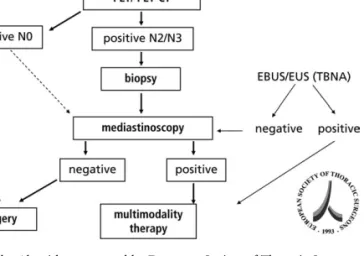
The algorithm proposed by the ESTS (Figure 1) has to be adopted in each institution according to the availability of the various methods, especially PET–CT, skilled mediastinoscopists or skilled invasive bronchoscopists with sufficient experience in these techniques.
restaging after induction therapy
Patients which have been diagnosed initially as stage III disease generally undergo induction chemo- or chemo/radiotherapy. Restaging of the mediastinal lymph nodes prior to surgery is
necessary in order to avoid incomplete resection. It has been shown that patients with complete resection after downstaging would benefit from a surgical procedure. Imaging modalities alone, such as CT scan, PET or PET–CT, are not accurate enough for restaging, except for those with a complete response after chemo- or chemo/radiotherapy. Tissue information either by endoscopic techniques or by mediastinoscopy or thoracoscopy is needed.
Re-mediastinoscopy is more difficult to perform due to extensive scaring of the mediastinal structures and is associated with a higher risk for morbidity. It is recommended that patients who are foreseen as cases for induction therapy preferably should be staged initially by an endoscopic technique (TBNA or EBUS/EUS–TBNA) in order to reserve mediastinoscopy for restaging. There are not yet sufficient data on endoscopic techniques in restaging and, currently, each centre has to rely on its available expertise.
disclosures
No significant relationships.
references
1. de Langen AJ, Raijmakers P, Riphagen I et al. The size of mediastinal lymph nodes and its relation with metastatic involvement: a meta-analysis. Eur J Cardio Thorac Surg 2006; 29: 26–29.
2. Toloza EM, Harpole L, McCrory DC. Noninvasive staging of non-small cell lung cancer: a review of the current evidence. Chest 2003; 123: 137–146. 3. de Leyn P, Lardinois D, van Schil PE et al. ESTS guidelines for preoperative lymph
node staging for non-small cell lung cancer. Eur J Cardio Thorac Surg 2007; 32: 1–8.
Figure 1. Algorithm proposed by European Society of Thoracic Surgeons for staging mediastinal lymph nodes (station 3).