Characterization of the N-linked oligosaccharides of megalin (gp330) from rat kidney
10
0
0
Texte intégral
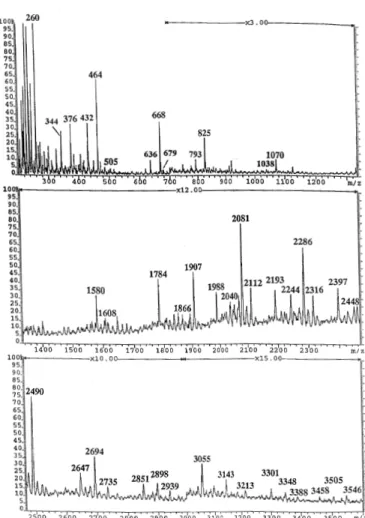
(2) W.Morelle et al.. Fig. 1. Summary of overall experimental strategy employed to characterize megalin N-glycans.. Structural analysis strategy To facilitate the release of N-glycans without resorting to detergent denaturation, megalin was first digested with cyanogen bromide. Glycans were then released from the resulting peptides/glycopeptides by digestion with peptide Nglycosidase F (PNGase F). PNGase-F released oligosaccharides were separated from peptides and glycopeptides using a Sep-Pak, and their methylated derivatives were characterized by fast atom bombardment mass spectrometry (FAB-MS) before and after sequential exoglycosidase digestions and by linkage analysis. Because of the availability of only limited amounts of material, the oligosaccharides were analyzed as mixtures. Structural assignments were based on molecular weight and fragment ion information (the latter derived from unassisted fragmentation in the normal FAB-MS experiment), susceptibility to exoglycosidase digestions, and linkage data. The overall structural strategy is summarized in Figure 1. Monosaccharide composition of N-glycans The monosaccharide composition of the mixture of PNGase F released oligosaccharides from megalin was determined by gas chromatographic/mass spectrometric (GC/MS) analysis of the trimethylsilylated derivatives of the methyl glycosides and methyl esters. Mannose, fucose, galactose, N-acetylgalactosamine, N-acetylglucosamine, and N-acetylneuraminic acid were all detected (data not shown). FAB-MS of N-glycans released from megalin by PNGase F N-Glycans were released from megalin glycopeptides by digestion with PNGase F. After purification by Sep-Pak, they were permethylated and analyzed by FAB-MS. The permethylation derivatization of oligosaccharides as well as increasing the sensitivity of detection of molecular ions, also causes predictable fragmentation, which give characteristic “maps” of fragment ions produced by A-type cleavage at each amino sugar residue (Dell et al., 1994). The composition of the molecular ions as deduced from their precise m/z values, when considered in conjunction with methylation analysis data and the range of nonreducing A-type fragment ions produced, allow important structural conclusions to be drawn on picomolar amounts of components. 296. Fig. 2. FAB-mass spectrum of permethylated N-glycans from megalin. NGlycans were released from megalin CNBr glycopeptides by digestion with PNGase F.. Data from FAB-MS analyses of permethylated PNGase Freleased glycans are shown in Figure 2 and summarized in Table I. A heterogeneous mixture of oligosaccharides was observed, affording about 15 major molecular ions and more than 30 minor ones. Based on the FAB-MS data and currently accepted models of eukaryotic N-glycan biosynthesis, the major N-glycans may be conveniently grouped into the following two classes: (1) high mannose structures having from five to nine mannoses and no core fucosylation and (2) complex type structures based on a trimannosyl core and extended with the terminal sequences tabulated in Table I. Notable features of these data are: (1) complex structures are more abundant than high mannose structures; (2) the majority of the molecular ions attributable to complex structures have compositions consistent with bi- or triantennary structures; (3) the A-type fragment ions indicate that the major antennae have HexNAc, HexHexNAc, Hex2HexNAc, NeuAcHexHexNAc, or NeuAcHexHexNAc2, at their non-reducing ends; (4) no fragment ions of composition Hex2HexNAc2+ and Hex3HexNAc3+ indicative of poly-Nacetyllactosamine are observed; (5) there is no evidence for Lewisx although there is a minor signal corresponding to its lacdiNAc counterpart (composition FucHexNAc2+); in addition there is a minor but significant signal for lacdiNAc itself (HexNAc2+ at m/z 505). These data suggest that very few.
(3) Characterization of N-linked oligosaccharides of megalin (gp330). Table I. Assignments of molecular and fragment ions observed in FAB spectrum of permethylated N-glycans of megalin Signal (m/z). Assignment. 260. HexNAc+. 376. NeuAc+. Loss of methanol gives 344. 464. HexHexNAc+. Loss of methanol gives 432. 505. HexNAc2+. (minor). 668. Hex2HexNAc+. 679. FucHexNAc2. +. 825. NeuAcHexHexNAc+. Loss of methanol gives 793. 1070. NeuAcHexHexNAc2+. Loss of methanol gives 1038. 1580. Hex5HexNAc2 + Na+. 1608. Hex3HexNAc4+. 1784. Hex6HexNAc2 + Na+. 1866. Hex4HexNAc4 + Na+. 1907. Hex3HexNAc5 + Na+ (major). 1988. Hex7HexNAc2 + Na+. 2040. FucHex4HexNAc4 + Na+. 2081. FucHex3HexNAc5 + Na+ (major). 2112. Hex4HexNAc5 + Na+. 2193. Hex8HexNAc2 + Na+. 2244. FucHex5HexNAc4 + Na+. 2286. FucHex4HexNAc5 + Na+ (major). 2316. Hex5HexNAc5 + Na+. 2397. Hex9HexNAc2 + Na+. 2448. FucHex6HexNAc4 + Na+. 2490. FucHex5HexNAc5 + Na+ (major). 2647. NeuAcFucHex4HexNAc5 + Na+. 2694. FucHex6HexNAc5 + Na+ (major). 2735. FucHex5HexNAc6 + Na+. 2851. NeuAcFucHex5HexNAc5 + Na+ (minor). 2898. FucHex7HexNAc5 + Na+. 2939. FucHex6HexNAc6 + Na+. 3055. NeuAcFucHex6HexNAc5 + Na+. 3143. FucHex7HexNAc6 + Na+. 3213. NeuAc2FucHex5HexNAc5 + Na+ (minor). 3301. NeuAcFucHex6HexNAc6 + Na+ (minor). 3348. FucHex8HexNAc6 + Na+ (minor). 3388. FucHex7HexNAc7 + Na+ (minor). 3458. NeuAc2FucHex5HexNAc6 + Na+ (minor). 3505. NeuAcFucHex7HexNAc6 + Na+ (minor). 3546. NeuAcFucHex6HexNAc7 + Na+ (minor). antennae carry fucose. Since the most abundant molecular ions have compositions containing one fucose it appears that most complex structures are core fucosylated. Linkage analysis of total glycan population Linkage analysis on the PNGase F released glycans and their desialylated counterparts gave the data shown in Table II. These results are fully consistent with high mannose and. Loss of methanol gives 636 (minor). complex type structures being the major constituents of the Nglycan population. Key features of these data are as follows. (1) The abundant 2-linked Man indicates that the majority of the complex glycans are biantennary but low levels of 2,4-linked Man and 2,6-linked Man suggest that minor tri- and/or tetraantennary structures are present; (2) Gal, Man and GlcNAc are the major nonreducing sugars; (3) after desialylation, 6-linked Gal and 297.
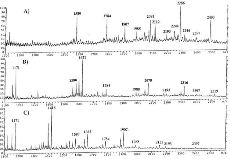
(4) W.Morelle et al.. Table II. GC-MS analysis of partially methylated alditol acetates obtained from the PNGase F released N-glycans of megalin Elution time (min). Characteristic fragment ions. Assignment. Relative abundance. 16.93. 115, 118,131, 162, 175. Terminal fucose. 0.30. 18.53. 102, 118, 129, 145, 161, 162, 205. Terminal mannose. 0.30. 18.82a. 102, 118, 129, 145, 161, 162, 205. Terminal galactose. 0.45. 19.77. 129, 130, 161, 190. 2-Linked mannose. 1.00. 19.87d. 118, 233. 4-Linked galactose. 20.07c. 118, 129, 161, 234. 3-Linked galactose. 0.23. 20.62b. 99, 102, 118, 129, 162, 189, 233. 6-Linked galactose. 0.11. 20.76b. 118, 129. 3,4-Linked galactose. 0.02. 21.01. 130, 190, 233. 2,4-Linked mannose. 0.02. 21.45. 129, 130, 189, 190. 2,6-Linked mannose. 0.03. 21.62. 118, 129, 189, 234. 3,6-Linked mannose. 0.1. 22.08. 118, 333. 3,4,6-Linked mannose. 0.24. 22.62. 117, 159, 203, 205. Terminal GlcNAc. 0.47. 23.12. 117, 159, 203, 205. Terminal GalNAc. 0.03. 23.53. 117, 159, 233. 4-Linked GlcNAc. 0.44. 24.94. 117, 159, 261. 4,6-Linked GlcNAc. 0.07. aSignals. more intense after treatment of N-glycans with V.cholerae sialidase. not observed after treatment of N-glycans with V.cholerae sialidase. cSignals not observed after treatment of N-glycans with coffee bean a-galactosidase. dSignals observed after treatment of N-glycans with V.cholerae sialidase. bSignals. 3,4-linked Gal disappear and there is a concomitant increase in terminal Gal and 4-linked Gal, indicating that sialic acid residues were attached to the 6-position of Gal and to the 3-position of the 3,4-linked Gal prior to desialylation; (4) the minor 3,4-linked Gal is therefore likely to be derived from the minor terminal epitope NeuAcHexHexNAc2 suggested by the FAB data (m/z 1070); (5) 3-linked Gal is present, the majority of which is retained after desialylation and is therefore likely to be derived from the Hex2HexNAc moieties suggested by the FAB data (m/z 668 in Figure 2); (6) a very minor amount of terminal GalNAc is present; (7) some 4,6-linked GlcNAc is present but most of the GlcNAc is terminal, or 4-linked; (8) 4,6-linked GlcNAc supports the presence of core α6-fucosylation; (9) the major 3,4,6-linked Man indicates that most of the complex structures are bisected. Endo-β-galactosidase digestion In order to establish if the N-glycans released by PNGase-F contain polylactosaminyl chains, N-glycans were subjected to digestion with endo-β-galactosidase from Bacteroides fragilis. The products were permethylated, and examined by FAB-MS after Sep-Pak purification (data not shown). The treatment with this enzyme did not result in any significant change, indicating the absence of polylactosaminyl chains. Sequential exo-glycosidase digestions To define the anomeric configurations as well as to confirm tentative sequences, N-glycans released by PNGase F were subjected to digestion with α-sialidase, α-galactosidase, βhexosaminidase, β-galactosidase, and α-fucosidase (see Figure 1 for information on the sequences of enzymes used in different experiments). Aliquots were taken after each diges298. tion, permethylated, and examined by FAB-MS after reverse phase Sep-Pak C18 purification. After neuraminidase treatment, the FAB-MS data indicated that, as expected, all sialylated molecular ions previously described (Figure 2 and Table I) were reduced in molecular weight consistent with the loss of one or two sialic acid residues. Thus, the NeuAc residues are in normal α linkages. The terminal sequence Hex2HexNAc revealed by the FABMS data (Figure 2 and Table I) was expected to contain the Galα1–3Gal group which is widely expressed in glycoconjugates originating from nonprimate mammals (Galili et al., 1987). To confirm its presence the desialylated glycans were treated with coffee bean α-galactosidase. After α-galactosidase treatment, the A-type ion at m/z 668 (Hex2HexNAc) disappeared while that at m/z 464 (HexHexNAc) was unaffected. The molecular ions at m/z 2316, 2490, and 2694 were significantly reduced in intensity concomitant with a significant increase in the abundance of the molecular ions at m/z 2112 and 2286 (Figure 3A), consistent with the removal of a terminal α-Gal. Comparison of linkage data before and after αgalactosidase treatment indicated that removal of terminal αGal residues is accompanied by loss of the 3-linked Gal (Table II). These data establish that α-Gal is attached to the 3-position of Gal. Thus, the FAB data and the linkage analysis suggest that the complex-type glycans Hex5HexNAc5 (m/z 2316), FucHex5HexNAc5 (m/z 2490), and FucHex6HexNAc5 (m/z 2694) carry the nonreducing terminal sequence Galα1–3Gal14GlcNAc. However, it is interesting to note that the molecular ion at m/z 2490 is still quite prominent after α-galactosidase treatment. Therefore, it is likely that a second isomeric structure which correspond to a normal biantennary, bisected, core fucosylated glycan is present (see Figure 6)..
(5) Characterization of N-linked oligosaccharides of megalin (gp330). Fig. 3. FAB-mass spectra of permethylated N-glycans from megalin after α-galactosidase digestion (A), β-N-acetylhexosaminidase digestion (B) and βgalactosidase digestion (C). Megalin N-glycans were released from megalin CNBr glycopeptides by digestion with PNGase F. The released glycans were sequentially digested with α-sialidase, α-galactosidase, β-N-acetylhexosaminidase, and β-galactosidase. Aliquots were taken after each digestion, permethylated and purified by Sep-Pak.. After β-N-acetylhexosaminidase treatment, the HexNAc+ ion at m/z 260 was greatly reduced in intensity while the HexHexNAc+ at m/z 464, as expected, was unaffected. The molecular ions at m/z 1907 (Hex3HexNAc5), and 2081 (FucHex3HexNAc5) were abolished concomitant with the appearance of a new signal at m/z 1171 (Hex3HexNAc2). In addition, another new molecular ion was observed at m/z 1621 (Hex4HexNAc3), consistent with the loss of two β-HexNAcs from the original molecular ion at m/z 2112 and the loss of two β-HexNAcs and one fucose from the original molecular ion at m/z 2286 (Figure 3B). In addition, the molecular ion at m/z 2490 (FucHex5HexNAc5) was abolished concomitant with the appearance of two ions at m/z 2070 (Hex5HexNAc4), and 2316 (Hex5HexNAc5) which are approximately in equal abundance. Thus, possibly due to steric hindrance, the bisecting Nacetylglucosamine is partially resistant to the β-N-acetylhexosaminidase (Yamashita et al., 1983). Since the β-N-acetylhexosaminidase is contaminated by an α-fucosidase (<2%), the removal of terminal HexNAc residues is accompanied by the removal of terminal α-Fuc residues. Comparison of linkage data before and after β-N-acetyl-hexosaminidase treatment indicated that loss of terminal β-GlcNAc residues, β-GalNAc. residues, and α-Fuc residues is accompanied by a decrease in 3,4,6-linked Man, a concomitant increase in 3,6-linked Man, loss of 4,6-linked GlcNAc, and a concomitant increase in 4linked GlcNAc, loss of 4-linked Gal and a concomitant increase of terminal Gal. After β-galactosidase treatment, a prominent A-type ion was observed at m/z 260 (HexNAc+) and the A-type ion at m/z 464 (HexHexNAc+) disappeared. Four major molecular ions were observed which correspond to Hex3HexNAc2 (m/z 1171), Hex3HexNAc3 (m/z 1416), Hex3HexNAc4 (m/z 1662), and Hex3HexNAc5 (m/z 1907), indicating that the components were efficiently degalactosylated by β-galactosidase from bovine testes (Figure 3C). Methylation analysis showed that the loss of terminal β-Gal residues is accompanied by a decrease in 4-linked GlcNAc and a concomitant increase in terminal GlcNAc. The signals at m/z 1580, 1784, 1988, 2193, and 2397 were unaffected by the above exoglycosidase digestions, a result that is consistent with the assignment of high mannose structures to these ions. Linkage data corroborated the assignment since terminal Man and 2-linked-Man were the major residues. 299.
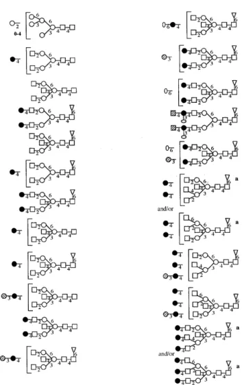
(6) W.Morelle et al.. Fig. 4. FAB-mass spectrum of permethylated N-glycans from megalin after β-galactosidase and β-N-acetylhesosaminidase digestion. Megalin N-glycans were released from megalin CNBr glycopeptides by digestion with PNGase F. The released glycans were sequentially digested with α-sialidase, α-galactosidase and with β-galactosidase and β-N-acetylhexosaminidase. Aliquots were taken after each digestion, permethylated and purified by Sep-Pak.. In a separate experiment, PNGase-F released oligosaccharides after α-sialidase and α-galactosidase treatment were subjected to digestion with β-galactosidase and β-N-acetylhexosaminidase in order to check that the complex type glycans could be efficiently trimmed to Hex3HexNAc2 (Figure 4). The products were examined by FAB-MS after permethylation and Sep-Pak purification. The FAB-MS spectrum was characterized by a major molecular ion at m/z 1171 consistent with composition Hex3HexNAc2 and an abundant A-type fragment ion at m/z 872 (Hex3HexNAc+). Other than Hex3HexNAc2, the major structures present in PNGase F pool after these treatments were Hex5HexNAc2, Hex6HexNAc2, Hex7HexNAc2, Hex8HexNAc2, and Hex9HexNAc2. These FAB data indicated that, as expected, the complex-type glycans were fully trimmed to Hex3HexNAc2 (m/z 1171), while putative highmannose structures were unaffected. PNGase-F released oligosaccharides were also treated sequentially with α-sialidase, α-galactosidase, β-galactosidase, and α-fucosidase in order to determine the fucosyl linkages. The reactions were monitored by FAB-MS after permethylation (Figure 5). Comparison of linkage data before and after α-fucosidase indicates that loss of fucosyl residues is 300. accompanied by loss of the 4,6-linked GlcNAc and a concomitant increase in 4-linked GlcNAc. These data establish that fucose is attached to the 6-position of 4,6-linked GlcNAc and confirmed the presence of core α6-fucosylation. Assignment of oligosaccharide structures The proposed structures for the major oligosaccharides are shown in Figure 6. The glycans fall into two classes, namely, high mannose and complex. The FAB-MS data and the linkage analysis data suggest that the major oligosaccharides correspond to biantennary, bisected structures with (α1,6)-core fucosylation. The major nonreducing epitopes in the complextype glycans are: GlcNAc, Galβ1–4GlcNAc (LacNAc), NeuAcα2–6Galβ1–4GlcNAc (sialylated LacNAc), Galα1– 3Galβ1–4GlcNAc and GalNAcβ1–4[NeuAcα2–3]Galβ1– 4GlcNAc (Sda). The high sensitivity achieved in the FAB-MS analyses of the total glycan population allowed the detection of very minor components giving molecular ions at masses above m/z 3500. These correspond to tri- and tetraantennary structures and/or bi- and tri-antennary structures with the presence of a bisecting GlcNAc residue. The very low abundance of these components has to date precluded precise structural analysis..
(7) Characterization of N-linked oligosaccharides of megalin (gp330). Fig. 5. FAB-mass spectra of permethylated N-glycans from megalin after β-galactosidase digestion (A) and α-fucosidase digestion (B). Megalin N-glycans were released from megalin CNBr glycopeptides by digestion with PNGase F. The released glycans were sequentially digested with α-sialidase, α-galactosidase, βgalactosidase, and α-fucosidase. Aliquots were taken after each digestion, permethylated and purified by Sep-Pak.. Discussion Characterizing the glycosylation in a glycoprotein of the size of megalin is very challenging because the glycans make only a small contribution to the mass of the total glycoprotein. Thus, highly sensitive structural strategies are essential for success. In this regard the mixture analysis capability of FAB-MS is paramount because purification steps with their inevitable sample losses can be kept to a minimum. Despite the high degree of heterogeneity exhibited by the N-glycans of megalin we have successfully acquired sufficient data to unambiguously assign the major structures (Figure 6) and to draw conclusions about the antennae compositions of the remaining minor components. The major N-glycans in megalin belong to the high mannose and biantennary complex type families. Complex structures are more abundant than high mannose structures. Most of biantennary complex glycans have bisected structures and are core fucosylated (Figure 6). Minor tri- and tetraantennary complex structures are also present. The major nonreducing epitopes in the complex-type glycans are: GlcNAc, Galβ1– 4GlcNAc (LacNAc), NeuAcα2–6Galβ1–4GlcNAc (sialylated LacNAc), Galα1–3Galβ1–4GlcNAc, and GalNAcβ1–4[Neu-. Acα2–3]Galβ1–4GlcNAc (Sda). These results are in good agreement with lectin blotting data of purified megalin (Ziak et al., 1999a). The structural element Galα1–3Galβ1–4GlcNAc has been found on several glycoproteins, such as bovine thyroglobulin (Spiro and Bhoyroo, 1984), bovine lactotransferrin (Coddeville et al., 1992). Analysis of the acceptor specificity of the α(1–3)D-galactosyltransferase purified from calf serum has demonstrated that the α(1–3)-D-galactosyltransferase competes with the α(2–6) and α(2–3) sialyltransferases for the N-acetyllactosamine units in a mutually exclusive way (Blanken and Van den Eijnden, 1985). In our study, the Galα1–3Galβ1–4GlcNAc unit was found to be located on one antenna of three N-glycans. Similar results were obtained for the glycoprotein of Friend murine leukemia virus (Geyer et al., 1984), whereas in the glycopeptides derived from a Lewis lung carcinoma cell subline, the α1–3Gal unit was found linked to both antennae of a diantennary glycan (Debray et al., 1991). However, it should be noted that, in our study, the structural element Galα1–3Galβ1– 4GlcNAc could be present on two antennae of minor components giving molecular ions at m/z 3143 (FucHex7HexNAc6) and at m/z 3388 (FucHex7HexNAc7). 301.
(8) W.Morelle et al.. Fig. 6. Structures of megalin N-glycans. Superscript a indicates that these glycans could correspond to triantennary structures with a bisecting GlcNAc residue as shown or tetraantennary structures lacking the bisecting GlcNAc.. A feature of megalin is the presence of the structural element GalNAcβ1–4[NeuAcα2–3]Galβ1–4GlcNAc (Sda) in very minor amounts in the N-glycans. α-linked GalNAc occurs as a constituent of the blood group A determinant, GalNAcα1– 3[Fucα1–2]Galβ1, which is located at the nonreducing termini of oligosaccharides linked either N- or O-glycosidically to peptides or to glycolipids (Watkins, 1980). By contrast, oligosaccharides containing β-linked GalNAc residues are quite unusual. For example, the sequence GalNAcβ1–4GlcNAc (LacdiNAc), with the exception of the pituitary glycohormones (Baenziger and Green, 1988) has been rarely observed in glycoproteins in higher animals (see Dell and Khoo, 1993; Van den Eijnden et al., 1995; Dell et al., 1995, and references cited therein). β-GalNAc residues have been found to be present at the nonreducing termini of O-linked oligosaccharides of a cloned murine cytotoxic T lymphocyte line (Conzelmann and Kornfeld, 1984) and are a constituent of the very rare blood group determinant called Cad (Blanchard et al., 1983). Glycophorin from erythrocytes of Cad-positive individuals contains 302. O-linked oligosaccharides with the structure GalNAcβ1– 4[NeuAcα2–3]Galβ1–3[NeuAcα2–6]GalNAc-Ser/Th. These β-GalNAc residues are also part of the determinant for the blood group Sda. Sda activity has previously been found in erythrocytes, Tamm-Horsfall glycoprotein (Hard et al., 1992) and urinary mucin (Cartron et al., 1982). The minor GalNAccontaining N-glycans characterized in the present study should have Sda activity as well. It is interesting to note that, in our study, no terminal GalNAcβ1–4Gal or NeuAcα2–6[GalNAcβ1–4]Gal element was found. This is in agreement with biosynthetic studies with β-N-acetylgalactosaminyltransferases from guinea-pig kidney (Serafini-Cessi and Dall’Olio, 1983), human kidney (Piller et al., 1986), human blood plasma (Takeya et al., 1987), and human urine (Serafini-Cessi et al., 1988), which have demonstrated that the enzymes require α2–3 linked sialic acid in the acceptor. Another characteristic feature of the complex type glycans of megalin is that many of these glycans are incompletely galactosylated. This phenomenon might be caused by the steric effect of a bisecting N-acetylglucosamine residue (Yamashita et al., 1983, 1988). The contribution of N-linked oligosaccharides to the properties of megalin can only be speculated on at this time. In general, the variety of functions attributed to N-linked oligosaccharides is very broad (Varki, 1993). Assuming that megalin has a general role of binding multiple ligands, speculation can be limited to those types of functions of N-linked oligosaccharides that are consistent with this role. N-Linked oligosaccharides are known to be involved in cell–cell interactions, cell–matrix interactions, hormone–receptor binding, antibody–antigen binding, and virus and bacterial pathogenesis (Varki, 1993). All of our structural data were carefully scrutinized for evidence of KDN, and none was detected. Of particular relevance are the results of our GC-MS sugar composition experiments where data from standards demonstrated that levels of KDN as low as 1% of the total sialic acid content would be detectable. Thus, our data are in accord with recent immunological studies from which it was concluded that the oligo/poly (α2,8-KDN) on megalin of various tissue sources is most likely to be carried on the O-glycans (Ziak et al., 1999a,b). Experiments are now under way to characterize KDN glycoforms of megalin. Materials and methods Isolation of megalin Renal megalin was purified as previously described (Ziak et al., 1999a). Briefly, microsomes from rat renal cortex were isolated by standard differential centrifugation and were resuspended to a final concentration of 3–4 mg/ml protein in 50 mM Tris–HCl (pH 7.5) containing protease inhibitors. CHAPS was added to a final concentration of 10 mM. After 60 min on ice, the extract was centrifuged at 100,000 × g for 60 min. The soluble extract was dialyzed for 18 h against 50 mM Tris–HCl (pH 7.5) containing 1 mM CHAPS and loaded at a flow rate of 90 ml/h onto a DEAE-Sephacel column (XK 16/20) equilibrated with the same buffer. The flow-through fraction containing the megalin was loaded on a lentil lectin-Sepharose column and lectin-bound glycoproteins were eluted using.
(9) Characterization of N-linked oligosaccharides of megalin (gp330). 50 mM Tris–HCl (pH 7.5) containing 10 mM CHAPS, 500 mM α-methylmannoside, and 10 mM EDTA. Finally, the glycoprotein sample was concentrated and applied onto a HR 16/50 gel filtration column packed with Sephacryl S-400. Fractions immunoreactive for oligo/poly α2,8 KDN were pooled and lyophilized. Preparation of CNBr fragments of megalin The lyophilized sample (∼200 µg) was dissolved in 100 µl of a solution of CNBr in 70% (v/v) formic acid and incubated in the dark for 5 h. The reaction was terminated by drying in vacuo after the addition of 500 µl of water.. internal diameter, J&W Scientific). The partially methylated alditol acetates were dissolved in hexanes prior to on-column injection at 65°C. The GC oven was held at 65°C for 1 min before being increased to 290°C at a rate of 8°C/min. For monosaccharide composition analysis, glycans were methanolyzed with 0.5 methanolic-HCl at 80°C for 16 h, re-Nacetylated with 500 µl of methanol, 10 µl of pyridine, and 50 µl of acetic anhydride, and then treated with the Tri-Sil TMSderivatizing reagent (Pierce) for 15 min at room temperature. GC-MS analysis of the TMS derivatives was performed on the same system using a temperature gradient of 140°C to 200°C at 5°C/min, increased to 300°C at 10°C/min.. PNGase F digestion PNGase F (EC 3.5.1.52, Roche Molecular Biochemicals) digestion was carried out on CNBr fragments of megalin in ammonium bicarbonate buffer (50 mM, pH 8.4) for 16 h at 37°C using 0.6 U of enzyme. The reaction was terminated by lyophilization and the products were purified on a Sep-Pak C18 (Waters Corporation) as described previously (Dell et al., 1994).. Acknowledgments. Sequential exoglycosidase digestions. Abbreviations. These were carried out on released glycans using the following enzymes and conditions: neuraminidase (from Vibrio cholerae, EC 3.2.1.18, Roche Molecular Biochemicals): 50 mU in 100 µl of 50 mM ammonium formate buffer, pH 5.5, for 48 h; α-galactosidase (from green coffee beans, EC 3.2.1.22, Roche Molecular Biochemicals): 0.5 U in 100 µl of 50 mM ammonium formate buffer, pH 6.0, for 24 h and then for an additional 24 h with a second aliquot of enzyme; N-acetyl-β-D-hexosaminidase (from beef kidney, EC 3.2.1.30, Roche Molecular Biochemicals): 0.2 U in 100 µl of 50 mM ammonium formate buffer, pH 4.6, initially for 24 h and then for an additional 24 h with a second aliquot of enzyme; β-galactosidase (from bovine testes, EC 3.2.1.23, Roche Molecular Biochemicals): 10 mU in 100 µl of 50 mM ammonium formate buffer, pH 4.6, for 48 h; α-L-fucosidase (from bovine kidney, EC 3.2.1.51, Roche Molecular Biochemicals): 0.2 U in 100 µl of 100 mM ammonium acetate buffer, pH 4.5, for 48 h. All enzyme digestions were incubated at 37°C and terminated by boiling for 3 min before lyophilization. For sequential enzyme digestions, an appropriate aliquot was taken after each digestion and permethylated for FAB-MS analysis after purification on a Sep-Pak C18 (Waters Corporation).. FAB, fast atom bombardment; Hex, hexose; HexNAc, Nacetylhexosamine; KDN, 2,8-deaminoneuraminic acid; lacdiNAc, GalNAcβ1–4GlcNAcβ1-; lacNAc, Galβ1– 4GlcNAcβ1-; Man, mannose; GalNAc, N-acetylgalactosamine; GlcNAc, N-acetylglucosamine; MS, mass spectrometry; PNGase F, peptide N-glycosidase F; u, mass unit.. Chemical derivatization and FAB-MS analysis Permethylation using the sodium hydroxide procedure was performed as described previously (Dell et al., 1994). FABMS spectra were acquired using a ZAB-2SE2FPD mass spectrometer fitted with a cesium ion gun operated at 30 kV. Data acquisition and processing were performed using the VG Analytical Opus software. Solvents and matrices were as described previously (Dell et al., 1994). Monosaccharide composition and linkage analysis Partially methylated alditol acetates were prepared form permethylated samples for GC-MS linkage analysis as described (Albersheim et al., 1967). GC-MS analysis was carried out on a Fisons Instruments MD800 machine fitted with a DB-5 fused silica capillary column (30 m × 0.32 mm,. This work was supported by the Biotechnology and Biological Sciences Research Council, the Wellcome Trust (Grants 030825 and 046294), and the Mizutami Foundation for Glycoscience, Tokyo. W.M. is a recipient of a FEBS Fellowship.. References Albersheim,P., Nevins,D.J., English,P.D. and Karr,A. (1967) A method for the analysis of sugars in plant cell wall polysaccharides by gas-liquid chromatography. Carbohydr. Res., 5, 340–345. Bachinsky,D., Zheng,G., Niles,J., McLaughlin,M., Abbate,M. andres,G., Brown,D. and McCluskey,R. (1993) Detection of two forms of Gp330. Their role in Heymann nephritis. Am. J. Pathol., 143, 598–611. Baenziger,J.U. and Green,E.D. (1988) Pituitary glycoprotein hormone oligosaccharides—structure, synthesis and function of the asparagine-linked oligosaccharides on lutropin, follitropin and thyrotropin. Biochim. Biophys. Acta, 947, 287–306. Blanchard,D., Cartron,J.-P., Fournet,B., Montreuil,J., Van Halbeek,H. and Vliegenthart,J.F.G. (1983) Primary structure of the oligosaccharide determinant of blood-group CAD specificity. J. Biol. Chem., 258, 7691–7695. Blanken,W.M. and Van den Eijnden,D.H. (1985) Biosynthesis of terminal Galα1–3Galβ1–4GlcNAc-R oligosaccharide sequences on glycoconjugates. Purification and acceptor specificity of a UDP-Gal:N-acetyllactosaminide α1–3 galactosyltransferase from calf thymus. J. Biol. Chem., 260, 12927–12934. Cartron,J.-P., Kornprobst,M., Lemonier,M., Lambin,P., Piller,F. and Salmon,C. (1982) Isolation from human urines of a mucin with bloodgroup Sda activity. Biochem. Biophys. Res. Commun., 106, 331–337. Chatelet,F., Brianti,E., Ronco,P., Roland,J. and Verroust,P. (1986) Ultrastructural-localization by monoclonal-antibodies of brush border antigens expressed by glomeruli. 2. Extrarenal distribution. Am. J. Pathol., 122, 512–519. Christensen,E., Nielsen,S., Moestrup,S., Borre,C., Maunsbach,A., Deheer,E., Ronco,P., Hammond,T. and Verroust,P. (1995) Segmental distribution of the endocytosis receptor gp330 in renal proximal tubules. Eur. J. Cell Biol., 66, 349–364. Coddeville,B., Strecker,G., Wieruszeski,J.-M., Vliegenthart,J.F.G., Van Halbeek,H., Peter-Katalinic,J., Egge,H. and Spik,G. (1992) Heterogeneity of bovine lactotransferrin glycans. Characterization of α-D-Galp (1–3)-β-DGal- and α-NeuAc- (2–6)-β-D-GalpNAc- (1–4)-β-D-GlcNAc-substituted N-linked glycans. Carbohydr. Res., 236, 145–164.. 303.
(10) W.Morelle et al.. Conzelmann,A. and Kornfeld,S. (1984) β-linked N-acetylgalactosamine residues present at the nonreducing termini of O-linked oligosaccharides of a cloned murine cytotoxic T lymphocyte line are absent in a Vicia villosa lectin resistant mutant cell line. J. Biol. Chem., 259, 12528–12535. Debray,H., Dus,D., Wieruszeski,J.-M., Strecker,G. and Montreuil,J. (1991) Structures of the α (1–3)-galactose-containing asparagine-linked glycans of a Lewis lung-carcinoma cell subline resistant to aleuria-aurantia agglutinin—elucidation by H-1-NMR spectroscopy. Glycoconjugate J., 8, 29– 37. Dell,A. and Khoo,K.-H. (1993) Covalent structure determination of glycopolymers. Curr. Opin. Struc. Biol., 3, 687–693. Dell,A., Reason,A.J., Khoo,K.-H., Panico,M., Mc Dowell,R.A. and Morris,H.R. (1994) Mass spectrometry of carbohydrate-containing bipolymers. Methods Enzymol., 230, 108–132. Dell,A., Morris,H.R., Easton,R.L., Panico,M., Patankar,M., Oehninger,S., Koistinen,R., Koistinen,H., Seppala,M. and Clark,G.F. (1995) Structural analysis of the oligosaccharides derived from glycodelin, a human glycoprotein with potent immunosuppressive and contraceptive activities. J. Biol. Chem., 270, 24116–24126. Galili,U., Clark,M.R., Shohet,S.B., Buehler,J. and Macher,B.A. (1987) Evolutionary relationship between the natural anti-Gal antibody and the Galα1– 3Gal epitope in primates. Proc. Natl. Acad. Sci. USA, 84, 1369–1373. Geyer,R., Geyer,H., Stirm,S., Hunsmann,G., Schneider,J., Dabrowski,U. and Dabrowski,J. (1984) Major oligosaccharides in the glycoprotein of friend murine leukemia-virus. Structure elucidation in the glycoprotein by onedimensional and two-dimensional proton nuclear magnetic-resonance and methylation analysis. Biochemistry, 23, 5628–5637. Guhl,B., Ziak,M. and Roth,J. (1998) Unconventional antigen retrieval for carbohydrate and protein antigens. Histochem. Cell Biol., 110, 603–611. Hammond,T.G., Majewski,R.R., Kaysen,J.H., Goda,F.O., Navar,G.L., Pontillon,F. and Verroust,P.J. (1997) Gentamicin inhibits rat renal cortical homotypic endosomal fusion: role of megalin. Am. J. Physiol.-Renal Physiol., 41, F117–F123. Hard,K., Van Zadelhoff,G., Moonen,P., Kamerling,J.P. and Vliegenthart,J.F.G. (1992) The Asn-linked carbohydrates chains of human Tamm-Horsfall glycoprotein of one male. Novel sulfated and novel Nacetylgalactosamine-containing N-linked carbohydrates chains. Eur. J. Biochem., 209, 895–915. Kanalas,J.J. and Makker,S.P. (1991) Identification of the rat heymann nephritis autoantigen (gp 330) as a receptor-site for plasminogen. J. Biol. Chem., 266, 10825–10829. Kerjaschki,D. and Farquhar,M.G. (1982) The pathogenic antigen of Heymann nephritis is a membrane glycoprotein of the renal proximal tubule brushborder. Proc. Natl. Acad. Sci. USA, 79, 5557–5561. Kerjaschki,D. and Farquhar,M.G. (1983) Immunocytochemical localization of the Heymannn nephritis antigen (gp 330) in glomerular epithelial-cells of normal lewis rats. J. Exp. Med., 157, 667–686. Kerjaschki,D., Miettinen,A. and Farquhar,M.G. (1987) Initial events in the formation of immune deposits in passive Heymann nephritis—gp330-antigp330 immune-complexes form in epithelial coated pits and rapidly become attached to the glomerular-basement-membrane. J. Exp. Med., 166, 109–128. Kounnas,M.Z., Chappell,D.A., Strickland,D.K. and Argraves,W.S. (1993) Glycoprotein-330, a member of the low-density-lipoprotein receptor family, binds lipoprotein-lipase in vitro. J. Biol. Chem., 268, 14176–14181. Lundgren,S., Hjalm,G., Hellman,P., Ek,B., Juhlin,C., Rastad,J., Klareskog,L., Akerstom,G. and Rask,L. (1994) A protein involved in calcium sensing of the human parathyroid and placental cytotrophoblast cells belongs to the LDL-receptor protein superfamily. Exp. Cell Res., 212, 344–350. Moestrup,S.K., Birn,H., Fischer,P.B., Petersen,C.M., Verroust,P.J., Sim,R.B., Christensen,E.I. and Nexo,E. (1996) Megalin-mediated endocytosis of transcobalamin-vitamin-B12 complexes suggests a role of the receptor in vitamin-B12 homeostasis. Proc. Natl. Acad. Sci. USA, 93, 8612–8617. Moestrup,S.K., Cui,S., Vorum,H., Bregengård,C., Bjorn,S.E., Norris,K., Gliemann,J. and Christensen,E.I. (1995) Evidence that epithelial glycoprotein 330/megalin mediates uptake of polybasic drugs. J. Clin. Invest., 96, 1404– 1413.. 304. Nykjaer,A., Dragun,D., Walther,D., Vorum,H., Jacobsen,C., Herz,J., Melsen,F., Christensen,E.I. and Willnow,T.E. (1999) An endocytic pathway essential for renal uptake and activation of the steroid 25- (OH) vitamin D-3. Cell, 96, 507–515. Orlando,R.A., Rader,K., Authier,F., Yamazaki,H., Posner,B.I., Bergeron,J.J.M. and Farquhar,M.G. (1998) Megalin is an endocytic receptor for insulin. J. Am. Soc. Nephrol., 9, 1759–1766. Piller,F., Blanchard,D., Huet,M. and Cartron,J.-P. (1986) Identification of a αNeuAc (2–3)β-D-galactopyranosyl N-acetyl-β-D-galactosaminyltransferase in human kidney. Carbohydr. Res. 149, 171–184. Saito,A., Pietromonaco,S., Loo,A.K. and Farquhar,M.G. (1994) Complete cloning and sequencing of rat gp330/“megalin”, a distinctive member of the low density lipoprotein receptor gene family. Proc. Natl. Acad. Sci. USA, 91, 9725–9729. Serafini-Cessi,F. and Dall’Olio,F. (1983) Guinea-pig kidney β-N-acetylgalactosaminyltransferase towards Tamm-Horsfall glycoprotein. Requirement of sialic acid in the acceptor for transferase activity. Biochem. J., 215, 483– 489. Serafini-Cessi,F., Malagolini,N. and Dall’Olio,F. (1988) Characterization and partial purification of β-N-acetylgalactosaminyltransferase from urine of Sd (a+) individuals. Arch. Biochem. Biophys., 266, 573–582. Spiro,R.G. and Bhoyroo,V.D. (1984) Occurence of α-D-galactosyl residues in the thyroglobulins from several species. Localization in the saccharide chains of the complex carbohydrate units. J. Biol. Chem., 259, 9858–9866. Takeya,A., Hosomi,O. and Kogure,T. (1987) Identification and characterization of UDP-GalNAc-NeuAc-α-2–3-Gal-β1-4Glc (NAc)-β-1–4- (GalNAc to Gal)N-acetylgalactosaminyltransferase in human blood plasma. J. Biochem. (Tokyo), 101, 251–259. Van den Eijnden,D.H., Neeleman,A.P., Van der Kanpp,W.P.W., Bakker,H., Agterberg,M. and Van Die,I. (1995) Novel glycosylation routes for glycoprotein. The LacdiNAc pathway. Biochem. Soc. Trans., 23, 175–179. Varki,A. (1993) Biological roles of oligosaccharides: all the theories are correct. Glycobiology, 3, 97–130. Watkins,W.M. (1980) In Harris,H. and Hirschhorn,K. (eds.), Advances in Human Genetics, Vol. 10. Plenum Press, New York, pp. 1–116, 379–385. Willnow,T.E., Goldstein,J.L., Orth,K., Brown,M.S. and Herz,J. (1992) Lowdensity-lipoprotein receptor-related protein and gp330 bind similar ligands, including plasminogen activator-inhibitor complexes and lactoferrin, an inhibitor of chylomicron remnant clearance. J. Biol. Chem., 267, 26172–26180. Yamashita,K., Hitoi,A., Matsuda,Y., Tsuji,A., Katunuma,N. and Kobata,A. (1983) Structural studies of the carbohydrate moieties of rat kidney γglutamyltranspeptidase. An extremely heterogeneous pattern enriched with nonreducing terminal N-acetylglucosamine residues. J. Biol. Chem., 258, 1098–1107. Yamashita,K., Tachibana,Y., Matsuda,Y., Katunuma,N., Kochibe,N. and Kobata,A. (1988) Comparative studies of the sugar chains of aminopeptidase N and dipeptidylpeptidase IV purified from rat kidney brush-border membrane. Biochemistry, 27, 5565–5573. Zheng,G., Bachinsky,D.R., Stamenkovic,I., Strickland,D.K., Brown,D. andres,G. and McCluskey,R.T. (1994) Organ distribution in rats of 2 members of the low-density-lipoprotein receptor gene family, gp 330 and lrp/α2mr and the receptor-associated protein (RAP). J. Histochem. Cytochem., 42, 531–542. Zheng,G., Marino,M., Zhao,J. and McCluskey,R.T. (1998) Megalin (gp330): a putative endocytic receptor for thyroglobulin (Tg). Endocrinology, 139, 1462–1465. Ziak,M., Kerjaschki,D., Farquhar,M.G. and Roth,J. (1996) The single poly α 2,8 deaminoneuraminic acid bearing glycoprotein in rat kidney corresponds to megalin. Mol. Biol. Cell, 7, 3466. Ziak,M., Kerjaschki,D., Farquhar,M.G. and Roth,J. (1999a) Identification of megalin as the sole rat kidney sialoglycoprotein containing poly α 2,8 deaminoneuraminic acid. J. Am. Soc. Nephrol., 10, 203–209. Ziak,M., Meier,M. and Roth,J. (1999b) Megalin in normal tissues and carcinoma cells carries oligo/poly α2,8 deaminoneuraminic acid as a unique posttranslational modification. Glycoconj. J., 16, 185–188..
(11)
Figure


+4


Documents relatifs
Two examples of these recent advances are the Santa Fe approach or Complexity approach and the AgentBased Computational Economics approach, that is, the computational study of
As predicted by the model, we show that in most genomes (i) direct repeats are more numerous than inverted repeats, (ii) CDR are in large excess, (iii) there is a negative
The results of this analysis indicate that the extended SAPPhIRE model is a viable representation of the essential elements in a concurrent design study and that it can facilitate
The transition-state calculation brings the 1 t, 2 e theoretical transition energy into c( exact )> agreement with the measured 2.3 eV absorption energy, although
These results of stochiometry and space distribution of the Mn atoms, are in good support of the α network being a reasonable idealization of the Mn chemical order in i-AlPdMn
Among the various types of models available, we have deliberately con- sidered the model which introduces an additional internal variable governed by a differential equation in
It is an immediate consequence of the analytical continuation property 6.7 and Wolpert’s theorem 5.7 that complex Fenchel-Nielsen coordinates are Darboux co- ordinates for the
