COMMENTARY
Antigens of Lung Cancer: Results of the Third
International Workshop on Lung Tumor and
Differentiation Antigens
Rolf A. Stahel, Walter R. Gilks, Thomas Schenker*
This commentary describes the results of the "cluster" analysis of the Third International Workshop on Lung Tumor and Differentiation Antigens, which was held under the patronage of the International Association for the Study of Lung Cancer in Zurich, September 8-11, 1993. Antibodies and an-tigens were grouped into clusters on the basis of the phenotypic reaction of antibodies with cell lines and tissues, with the main focus being the characterization of lung cancer antigens and an-tibodies. This workshop extended the findings of two previous International Workshops on Small-Cell Lung Cancer Antigens that were held in London in 1987 (/) and 1990 (2). Seven clusters were described at these two earlier workshops (an eighth cluster, cluster-3, had been described at the first work-shop, but was rejected at the second workshop). Eight new clusters were described at this workshop.
The Third International Workshop on Lung Tumor and Dif-ferentiation Antigens had three goals. The first goal was to iden-tify clusters of antibodies based on similar reactivity against cell lines and tissues in order to define antigens associated with lung tumors and lung differentiation. In contrast to the two previous workshops, which were largely restricted to antibodies reacting with small-cell lung cancer, it was decided to broaden the scope of the workshop to include antibodies reacting with all types of lung tumors or normal lung tissues. This decision was made after determining (a) that many of the antibodies examined during the previous workshops reacted with non-small-cell lung cancer, (b) that there was a growing interest in studies on bronchial differentiation and dysplasia, and (c) that many more clusters of antibodies are likely to be found with reactivities that would be classified as being restricted to small-cell lung cancer. The second goal of the workshop was to exchange information about the molecular nature and function of the members of pre-viously defined clusters. The third goal was to exchange infor-mation about preclinical and clinical studies in lung cancer with antibodies or conjugates.
Since the first workshop, our knowledge of particular anti-gens recognized by clustered antibodies has greatly increased. This increase is primarily due to the molecular characterization of some of the antigens—in particular those of cluster-1
tified as the neural cell adhesion molecule (2)], cluster-2 [iden-tified as epithelial glycoprotein-2 or the product of the GA733-2 gene (2,5)], and cluster-w4 [shown to be identical to the Leukocyte Workshop antigen CD24, with the exception of one amino acid (4)]. As the complementary DNA (cDNA) probes encoding these antigens were available, it was decided to add the screening of a panel of cells transfected with the genes en-coding the respective antigens to the cluster analysis conducted by the workshop participants.
The details of the workshop, including the results of the cen-tral data analysis, the statistical analysis, a list of the participat-ing laboratories, and individual papers describparticipat-ing the antigens or preclinical and clinical studies with the antibodies, are being published in a supplement to the International Journal of Cancer (5). Here, we summarize the major findings of this workshop, including the cluster analysis and the present state of knowledge regarding the antigens identified and assigned to clusters.
Twenty-eight laboratories submitted antibodies to the work-shop. Of 60 new antibodies or antibodies that had not been as-signed to clusters at the previous workshops, 32 could be placed in clusters at the current workshop. The decisions regarding the assignment to particular clusters or the definition of new clusters were made entirely on the basis of phenotypic observations on cell lines and tissues. As in the previous workshops, provisional clusters were designated with the prefix "w" if no more than two antibodies from the same laboratory formed the cluster and the nature of their antigen had not been fully defined. The an-tibodies included in the present workshop were assigned to 15 clusters, with 8 of these clusters (cluster-lC and clusters 9-15) being newly defined. Cluster-1 and cluster-8 were each assigned six antibodies at this workshop; however, of all the clusters, cluster-1 continues to have the largest total number of
an-*Affiliations of authors: R. A. Stahel, T. Schenker, Division of Medical On-cology, Department of Medicine, University Hospital, Zurich, Switzerland.
W. R. Gilks, Medical Research Council Biostatistics Unit, Institute of Public Health, Cambridge, England.
Correspondence to: R.A. Stahel, Laboratory of Oncology, Haldeliweg 4, 8044 Zurich, Switzerland.
See "Note" section following "References."
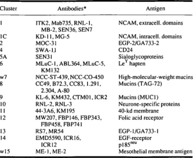
tibodies. Cluster-6 and cluster-8, which have been provisional clusters defined primarily on the basis of oligosaccharide bind-ing at the second workshop, have now for the first time been defined by phenotypic analysis. The antibodies defining these clusters and the present knowledge about the respective antigens are summarized in Table 1.
Cluster-1
Four antibodies from four laboratories were grouped with two antibodies observed to react with cluster-1 antigens in the pre-vious workshop. With the exception of antibody Mab735, which was described as reacting with an epitope of 10 or more units of a-(2,8)-/V-linked polysialic acid side chains of NCAM, all anti-bodies were found to react with NCAM transfectants of mouse L-cells. The tissue distribution confirmed strong binding to small-cell lung cancer, neuroblastoma, nerve, central nervous system, Leydig cells of the testis, and thyroid follicles; little or no binding to epithelial cells was observed. All antibodies lost reactivity in routinely fixed tissues. As expected, binding was seen on subpopulations of lymphoid cells, with the exception of antibody SEN7, which has been reported to have little or no binding on peripheral blood lymphocytes (6). Collaborative work by participants in the workshop resulted in a topographic mapping of antigenic epitopes on NCAM (7).
Cluster-lC
Two antibodies from the same laboratory that had been raised directly against human NCAM clustered very closely and were designated to cluster-1C. Both bind to intracellular NCAM epitopes and thus have a different pattern of reactivity than cluster-1 antibodies. Antibody KD11 was shown to recognize the most C-terminal part of transmembrane isoforms; antibody MG5 specifically recognizes an undecapeptide in the N-terminal region of exon 18 (8).
Table 1. Cluster analysis of the Third International Workshop on Lung Tumor and Differentiation Antigens
Cluster 1 1C 2 4 5A 6 w7 g 9 10 11 12 13 14 w!5 Antibodies* ITK2,Mab735,RNL-l, MB-2, SEN36, SEN7 KD-ll.MG-5 MOC-31 SWA-1.1 SEN31
MLuC-1, ABL364, MLuC-5, KM 132 NCC-ST-439, NCC-CO-450 CC49, B72.3, CC83, 1.291, 2.304, A-80 KL-6, KM432, CTM01, ICR2 RNL-2, RNL-3 44-3A6, KM 195 MW207, FBP146, FBP343, FBP458, FBP741 RS7, MR54 EMD5590,ICR16, ICR12 ME-l.ME-2 Antigen
NCAM, extracell. domains
NCAM, intracell. domains EGP-2/GA733-2 CD24 Sialoglycoproteins LeY hapten High-molecular-weight mucins Mucins (TAG-72) Mucins (MUC1) Neurone-specific proteins 40-kd membrane Folic acid receptor
EGP-1/GA733-1 EGF-receptor pl85ncu
Mesothelial membrane antigen
•Eleven of the antibodies are controls (ITK2, RNL-1, SEN36, MOC-31, SWA-11, SEN31, NCC-ST-439, NCC-CO-450, A-80, RNL-2, and 44-3A6).
peripheral blood mononuclear cells and lymphoid cell lines. Protein sequence analysis revealed a heavily glycosylated and unusually short protein of 80 amino acids, virtually identical to the recently cloned leukocyte activation antigen CD24 except for a single valine-alanine substitution resulting from a single base polymorphism. Biochemical analysis has confirmed the presence of a phosphoinositol tail by which the antigen is anchored in the cell membrane. The linear peptide sequence leucine-alanine-proline on the protein core of the antigen lo-cated close to the phosphoinositol anchor has been identified as one of the epitopes (9).
Cluster-2
No new antibodies grouped with the cluster-2 control by phenotypic cluster analysis. Since the last workshop, progress has been made in the molecular characterization of this antigen by several laboratories (3,8). The antigen named epithelial glycoprotein-2 or the product of the GA733-2 gene was cloned both from a lung adenocarcinoma cell cDNA expression library using the monoclonal antibody KS1/4 as probe and from a colon carcinoma cell cDNA expression library using the antibodies GA733 and HEA 125 as probes. At the workshop, the current knowledge about the epitopes recognized by cluster-2 anti-bodies as well as evidence for a putative 30-kd ligand with specificity for cluster-2 antigen was presented.
Cluster-4
No new antibodies reacting with cluster-4 antigen were iden-tified. The antigen has been cloned from a small-cell lung can-cer expression library (5). Cluster-4 message was found to be highly overexpressed in small-cell lung cancer compared with
Cluster-5 and Cluster 5A
No new antibodies reacting with cluster-5 and cluster 5A an-tigens were identified at this workshop. The anan-tigens recognized by cluster-5 and cluster-5A antibodies represent a group of N-linked sialoglycoproteins strongly expressed in about 50% of small-cell lung cancer tissues, with little expression on epithelial cells and no expression on white blood cells. Cluster-5 and cluster-5A antigens are coexpressed in small-cell lung cancer cell lines and tissues, but their molecular relationship has long remained undefined. Evidence for the simultaneous expression of the cluster-5 and cluster-5A epitopes on a single membrane molecule which is shed in low quantities into the serum of patients with small-cell lung cancer has been obtained by im-munological means. This was accomplished by first generating a mouse monoclonal anti-anti-idiotype antibody against a rat monoclonal anti-idiotype antibody in the cluster-5 system and then by conducting heterologous cluster-5 and cluster-5A anti-body sandwich assays on the sera of patients with small-cell lung cancer (70).
Cluster-6
Four antibodies from three laboratories were found to group together at this workshop. Antibody MLuC-1 recognition of the blood group LeY hapten as an antigen has been described (77).
In the previous workshop, two antibodies that were not available to the current workshop defined cluster-w6 by both weak pheno-typic clustering and by oligosaccharide binding. The availability of additional antibodies that grouped closely together allowed the discontinuation of the provisional "w" designation. The tis-sue distribution for cluster-6 antibodies reported in this workshop includes binding to small-cell lung cancer and non-small-cell lung cancer, broad epithelial reactivity, and lack of binding to neural tissues and white blood cells.
Cluster-w7
Two control antibodies from the same laboratory that react with cluster-w7 antigen were used by the panel of this workshop. One of the antibodies clustered with the negative controls, suggesting loss of immunoreactivity. No new antibody clustering with the remaining cluster-w7 control was identified.
Cluster-8
Five antibodies from two laboratories clustered together with the cluster-w8 control antibody A-80. In the previous workshop, cluster-w8 antibodies grouped only weakly by phenotypic analysis but exhibited similarities in oligosaccharide-binding studies; biochemical analyses indicated that they bound the blood group A trisaccharide. At this workshop, the phenotypic clustering was much closer. The tissue distribution for cluster-8 antibodies reported in this workshop included binding to non-small-cell lung cancer, broad epithelial reactivity, and lack of reactivity with neural tissues or with white blood cells. A previous study (72) has charac-terized the antibodies B72.3, CC49, and CC83, which recognize a high-molecular-weight mucin designated TAG72.
Cluster-9
Four antibodies from four laboratories grouped together and were newly designated to cluster-9. One of these antibodies, CTM01, is reported to recognize the mucin MUC-1. Another antibody that recognizes an epitope, termed BrE-3, on the pep-tide core of MUC-1 was phenotypically only weakly associated with cluster-9. A similarly weak association with cluster-9 was also observed for a second antibody, RS5-4H6, which had been characterized as reacting with undefined high-molecular-weight mucins. It is likely that this finding might be due to different carbohydrate epitopes that can be present on the same mucin core protein. The tissue distribution of cluster-9 antibodies demonstrated binding to small-cell lung cancer and non-small-cell lung cancer, broad epithelial reactivity, white blood non-small-cell reactivity, and lack of binding to neural cells.
CIuster-10
Two antibodies from one laboratory closely grouped together and were newly designated to cluster-10. The antibodies
recog-nize a family of intracellular neuron-specific proteins ranging from 135 to 25 kd in molecular mass. These proteins have been shown to derive from one gene, NSP (13). Phenotypic analysis at this workshop confirmed the intracellular expression of this antigen predominantly in neuroendocrine tissues, including small-cell lung cancer. The reaction was lost in routinely fixed tissues. Evidence for the subcellular localization of these proteins in the endoplasmatic reticulum was presented.
Cluster-11
Two IgG antibodies from two laboratories phenotypically grouped together and were newly designated to cluster-11. The antigen of one of these antibodies, 44-3A6, has been biochemi-cally described as a 40-kd membrane-associated protein (14). At the workshop, it was reported that the gene coding for this nega-tively charged protein has recently been cloned and termed Nag gene (14). No sequence homologies have been identified so far. The phenotypic analysis obtained at the workshop demonstrated lack of immunofluorescence activity on viable cells. Examina-tion of tissues revealed antibody binding to non-small-cell lung cancer, limited epithelial reactivity, and lack of neural or white blood cell reactivity.
Cluster-12
Five antibodies from two laboratories grouped together and were newly designated to cluster-12. The antibodies of the FBP series have been obtained by immunization with human folate binding protein, a membrane glycoprotein with specific affinity for folic acid and methotrexate (75). Phenotypic analysis in the current workshop demonstrated binding to non-small-cell lung cancer (in particular, adenocarcinoma), limited epithelial reac-tivity, and some binding to monocytes. No antibody binding was seen in routinely fixed tissues. Additional antibodies, generated against ovarian carcinoma, were recently demonstrated to recog-nize human folate-binding proteins by expression cloning. Since antifolate agents, including methotrexate, enter the cell through the folate-binding protein, the expression of these antigens in tumor cells might be important to maintain the sensitivity of tumor cells to these drugs.
Cluster-13
Two antibodies from two laboratories of the same institution closely grouped together and were designated to cluster-13. The antigen recognized by antibody RS7 has been affinity purified and partially sequenced. Sequence analysis indicated identity to the product of the GA733-1 gene (76). Together with the gene GA733-2, which encodes epithelial glycoprotein-2 or cluster-2 antigen, GA733-1 forms a family of genes coding for type I transmembrane proteins of unknown function (77). No other antibodies against the GA733-1 gene product (epithelial glycoprotein-1) had been identified previously. Phenotypic analysis at this workshop demonstrated antigen expression on non-small-cell lung cancer, limited epithelial expression, and lack of expression in neural tissues or white blood cells. No binding was seen on routinely fixed tissues.
Cluster-14
Three antibodies from two laboratories grouped together and were designated to cluster-14. Two of these antibodies (EMD5590 and ICR16) (18) were shown to bind to the external domain of the epidermal growth factor receptor, and one has been shown to be specific for the external domain of pl85neu
(ICR12). Despite recognizing different molecules, the three anti-bodies had a similar binding pattern in the cell lines and tissues examined during this workshop. Binding was seen in non-small-cell lung cancer, mesothelioma, and a limited number of epithelia but not on normal neural tissues or white blood cells. None of the antibodies bound on routinely fixed tissues.
Cluster-wl5
Two antibodies raised in one laboratory against a meso-thelioma cell line closely grouped together and were designated to cluster-wl5. Nothing is known about their respective an-tigens, except that they are expressed on the external cell membrane surface. Phenotypic analysis in the workshop demonstrated binding to non-small-cell lung cancer (in par-ticular, squamous cell carcinoma) and mesothelioma, with only limited epithelial reactivity. Also, weak binding to neural cells and tissues has been observed. The antibodies lost activity in routinely fixed tissues. The antibody ME1 strongly stains all malignant mesothelioma tissues and a proportion of poorly dif-ferentiated lung adenocarcinomas (19).
Judged on the ability to define clusters of antibodies and an-tigens, the Third International Workshop has been a success, doubling the number of recognized clusters from seven to 15. The classification system developed at these workshops comple-ments the classification system developed by others for leuko-cyte antigens because the majority of antibodies directed against solid tumor antigens were selected by their lack of reactivity with white blood cells. The strength of the statistical analysis based on phenotypic reactivity of the antibodies was docu-mented (20) and was found to ideally complement the clustering of antibodies determined by the analysis of cells transfected with the cDNA of cloned antigens.
The clustering of antibodies has both stimulated and facili-tated investigations on their potential clinical use in diagnosis and therapy. The use of a common nomenclature allows a better comparison of investigations by different laboratories and potentially avoids duplicate work. Most importantly, the work-shop has brought together investigators working on similar molecules and has fostered fruitful collaborations between dif-ferent groups. This effort, hopefully, should increase the pace of advances, not only by improving our understanding of the roles that individual antigens play in lung biology and carcinogenesis, but also through provision of information that might soon be utilized in lung cancer detection and treatment.
With the addition of antibodies to non-small-cell lung cancer, the current workshop has increased in scope compared with the previous workshops. Results communicated at the workshop, how-ever, clearly suggest that most of the antigens clustered have also been identified in other solid tumors, exemplified by cluster-2 and cluster-8 in colon carcinoma, cluster-12 in ovarian carcinoma, and cluster-12 in breast cancer. It has thus become evident that in order
to facilitate the identification of additional antibodies and anti-gens to group in new and existing clusters, a useful and common nomenclature must be further developed, not only for the benefit of lung cancer specialists, but also for the benefit of those study-ing other solid tumors and lung diseases. Specifically, a future workshop might include research groups working with antigens in other solid tumors. Laboratories interested in contributing to the development of such a workshop should contact us.
References
(/) Souhami RL, Beverley PCL, Bobrow LG: Antigens of small-cell lung can-cer. First International Workshop. Lancet 2:325-326,1987
(2) Souhami RL, Beverley PCL, Bobrow LG, et al: Antigens of lung cancer: results of the Second International Workshop on Lung Cancer Antigens. J Natl Cancer Inst 83:609-612,1991
(3) Szala S, Froehlich M, Scollon M, et al: Molecular cloning of cDNA for the carcinoma-associated antigen GA733-2. Proc Natl Acad Sci U S A 87:3542-3546,1990
(4) Jackson D, Waibel R, Weber E, et al: CD24, a signal-transducting molecule expressed on human B cells, is a major surface antigen on small cell lung carcinoma. Cancer Res 52:5264-5270, 1992
(5) Stahel RA (ed): Third International IASLC Workshop on Lung Tumor and Differentiation Antigens. Int J Cancer Suppl 8. In press
(<5) Waibel R, Mannhart M, O'Hara CJ, et al: Monoclonal antibody SEN7 recog-nizes a new epitope on the neural cell adhesion molecule present on small cell lung cancer but not on lymphocytes. Cancer Res 53:2840-2845, 1993 (7) Gerardy-Schan R, Eckhardt M, Ledermann J, et al: Topography of NCAM
epitopes recognized by cluster-1 antibodies. A consensus view. Int J Can-cer Suppl 8. In press
(8) De Leji L, Helfrich W, Stein R, et al: SCLC cluster-2 antibodies detect the pancarcinoma/epithelial glycoprotein-2. Int J Cancer Suppl 8. In press (9) Weber E, Lehmann HP, Beck-Sickinger AG, et al: Antibodies to the
pro-tein core of the small cell lung cancer workshop cluster-w4 and to the leukocyte workshop antigen CD24 recognize the same short protein se-quence leucine-alanine-proline. Clin Exp Immunol 93:279-285, 1993 (10) Lehmann HP, Weber E, Schenker T, et al: Immunological evidence for
co-expression of cluster-5 and cluster-5A small cell lung cancer antigen on a single molecule. Int J Cancer Suppl 8. In press
(//) Leoni F, Colnaghi MI, Canevari S, et al: Glycolipids carrying LeY are
preferentially expressed on small-cell lung cancer cells as detected by the monoclonal antibody MLuC 1. Int J Cancer 51:225-231, 1992
(12) Muraro R, Kuroki M, Wunderlich D, et al: Generation and characterization of B72.3 second generation monoclonal antibodies reactive with tumor-as-sociated glycoprotein 72 antigen. Cancer Res 48:4588-4596, 1988 (13) Roebroek AJ, van de Velde HJ, Van Bokhoven A, et al: Cloning and
ex-pression of alternative transcripts of a novel neuroendocrine-specific gene and identification of its 135-kDa translational product. J Biol Chem 268:13439-13447,1993
(14) Radosevich JA, Ma YX, Lee I, et al: Monoclonal antibody 44-3A6 as a probe for a novel antigen found on human lung carcinomas with glandular differentiation. Cancer Res 45:5808-5812, 1985
(15) Franklin WA, Weintraub M, Edwards D, et al: New anti-cancer antibody cluster-12 reacts with human folate receptors present on adenocarcinoma. Int J Cancer Suppl 8. In press
(16) Stein R, Basu A, Goldenberg DM, et al: Characterization of cluster-13: the epithelial/carcinoma antigen recognized by monoclonal antibody RS7. Int J Cancer Suppl 8. In press
(17) Linnenbach AJ, Seng B, Robbins S, et al: Retroposition in a family of car-cinoma-associated antigen genes. Mol Cell Biol 13:1507-1515, 1993 (18) Modjtahedi H, Eccles SA, Box G, et al: Immunotherapy of human tumour
xenografts overexpressing the EGF receptor with rat antibodies that block growth factor-receptor interaction. BrJ Cancer 67:254-261,1993 (79) O'Hara CJ, Corson JM, Pinkus GS, et al: ME1. A monoclonal antibody
that distinguishes epithelial-type malignant mesothelioma from pulmonary adenocarcinoma and extrapulmonary malignancies. Am J Pathol 136:421-428,1990
(20) Gilks WR, Stahel RA, Walker N, et al: Statistics of the data analysis of the Third International Workshop on Lung Tumor and Differentiation An-tigens. Int J Cancer Suppl 8. In press
Note
Manuscript received January 1, 1994; accepted February 4, 1994.