Nephrol Dial Transplant (2011) 26: 1033–1041 doi: 10.1093/ndt/gfq488
Advance Access publication 13 August 2010
Podoplanin-positive cells are a hallmark of encapsulating
peritoneal sclerosis
Niko Braun
1,2, Dominik M. Alscher
1,2, Peter Fritz
2,3, Ilka Edenhofer
4,5, Martin Kimmel
1,
Ariana Gaspert
6, Fabian Reimold
1, Beata Bode-Lesniewska
6, Urs Ziegler
7, Dagmar Biegger
8,
Rudolf P. Wüthrich
4and Stephan Segerer
4,51
Department of Internal Medicine, Division of General Internal Medicine and Nephrology, Robert-Bosch-Hospital, Stuttgart, Germany,2Institute of Digital Medicine, Stuttgart, Germany,3Department of Diagnostic Medicine, Division of Pathology, Robert-Bosch-Hospital, Stuttgart, Germany,4Division of Nephrology, University Hospital Zurich, Zurich, Switzerland,5Institute of Anatomy, University of Zurich, Zurich, Switzerland,6Department of Surgical Pathology, University Hospital Zurich, Zurich, Switzerland,7Center of Microscopy and Image Analysis, University of Zurich, Zurich, Switzerland and8Margarete Fischer-Bosch Institute of Clinical Pharmacology, Stuttgart, Germany
Correspondence and offprint requests to: Stephan Segerer; E-mail: [email protected]
Abstract
Background. Encapsulating peritoneal sclerosis (EPS) and simple peritoneal sclerosis are important complica-tions of long-term peritoneal dialysis (PD). Podoplanin is expressed by mesothelial cells and lymphatic vessels, which are involved in inflammatory reactions in the peri-toneal cavity.
Methods. We studied 69 peritoneal biopsies from patients on PD (n = 16), patients with EPS (n = 18) and control bi-opsies taken at the time of hernia repair (n = 15) or append-ectomy (n = 20). Immunohistochemistry was performed to localize podoplanin. Additionally, markers of endothelial cells, mesothelial cells, myofibroblasts (smooth muscle actin), proliferating cells, and double labelling for smooth muscle actin/podoplanin were used on selected biopsies. Results. Podoplanin was present on the endothelium of lymphatic vessels in the submesothelial fibrous tissue and on mesothelial cells. In patients on PD and in biopsies with appendicitis, the mesothelial cells demonstrated a cuboidal appearance and circumferential podoplanin stain-ing, with gaps between the cells. The number of lymphatic vessels was variable, but prominent at sites of fibrosis. In patients with EPS, a diffuse infiltration of podoplanin-positive cells with a fibroblastic appearance was present in 15 out of 18 biopsies. This pattern was focally present in 3 out of 16 on PD and none in the 35 controls. The podoplanin-positive cells did not express the endothelial marker or the mesothelial marker (calretinin).
Conclusions. EPS is characterized by a population of po-doplanin and smooth muscle actin double-positive cells. Podoplanin might be a suitable morphological marker sup-porting the diagnosis and might be involved in the patho-genesis of EPS.
Keywords: encapsulating peritoneal sclerosis; EPS; peritoneal dialysis; podoplanin
Introduction
Encapsulating peritoneal sclerosis (EPS) is an uncommon but potentially life-threatening complication of peritoneal dialysis (PD) [1]. The key factors for the diagnosis of EPS are clinical symptoms of bowel obstruction, a typical radiological picture and extensive thickening of the peri-toneal membrane (resulting in cocooning of the bowel) [2]. Morphological features of EPS are mesothelial de-nudation, peritoneal fibroblast swelling, interstitial fibro-sis, angiogenesis with increased numbers of capillaries and mononuclear cell infiltration [3,4]. The peritoneal membrane is affected by fibrin deposits, which may lead to adherences and permanent scarring [2].
Simple sclerosis on the other hand is characterized by increased thickening of the peritoneal membrane, loss of peritoneal function with high transporter status, but no clinical symptoms of bowel obstruction [5,6]. EPS can be differentiated from simple sclerosis by the deposition of fibrin and the increased thickness of the degenerative compact zone. Angiogenesis, vasculopathy, new mem-brane formation and fibrosis did not distinguish between simple sclerosis and EPS [7]. The histological picture is therefore relatively unspecific, particularly in early stages, and morphological markers are currently not available.
Podoplanin (also known as aggrus, hT1α or M2A re-cognized by the antibody D2-40) is a member of a family of type-1 transmembrane sialomucin-like glycoproteins [8]. The protein has an extracellular domain (with
abun-© The Author 2010. Published by Oxford University Press on behalf of ERA-EDTA. All rights reserved. For Permissions, please e-mail: [email protected]
dant Ser and Thr residues as potential O-glycosylation sites), a single transmembrane portion and a short cyto-plasmic tail [9]. As a glycoprotein, it can bind chemokines and modulate inflammatory reactions. Podoplanin was ori-ginally described to be lost in rat puromycin nephritis and was named according to the loss of foot processes in this model (flat feet in latin, pes planus) [10]. Later it was found to be a good marker for lymphatic endothelial cells, but it is also expressed by peritoneal mesothelial cells [11]. As podoplanin can bind chemokines, it may modulate the in-flammatory milieu (on mesothelial cells and on lymphatic vessels) and therefore might be involved in the injury process of both simple sclerosis and EPS [12]. Podoplanin can be detected by the antibody D2-40, which is suitable for routine staining of formalin-fixed and paraffin-embedded tissues. This antibody enables the localization of mesothe-lial cells, and cells of mesothemesothe-lial origin in combination with markers of endothelial origin.
The goal of this study was to localize podoplanin in peritoneal biopsies from patients on PD with and without clinical signs of EPS.
Materials and methods
Study population
Sixty-nine tissue samples with different peritoneal pathological states were either selected from the files of the Department of Pathology or ran-domly from a database of 220 patients. All patients had given informed consent for their tissue to be used for research purposes. Patients with appendicitis were selected randomly. Biopsies were submerged in buf-fered formalin and embedded in paraffin following routine protocols. The biopsies were incorporated into paraffin blocks so that sections were cut in a right angle to the surface. Histological features were scars in the submucosal layer and small to moderate lymphoplasmacellular infiltra-tions in the tunica propria and/or subserosal layer. For purposes of com-parison with study patients, 15 specimens were selected randomly from patients who had undergone hernia repairs (direct or indirect). Peritoneal biopsies from patients on PD were also included (n = 16). The tissue was taken from the visceral peritoneum, from areas located at least 10 cm away from the peritoneal catheter entry side. Indications for surgery were herniotomy, leakage or catheter removal because of switching to haemo-dialysis. In addition, we collected tissues from all patients with EPS from our Department of Nephrology or sent for additional investigations to the Department of Pathology (n = 18).
For diagnosis, we used criteria stated by Nakamoto [13]: stage 1 (pre-EPS period) characterized by loss of ultrafiltration, development of high transport state, hypoproteinaemia, bloody dialysate, ascites and calcifica-tion of peritoneum; stage 2 (inflammacalcifica-tion period), increases in C-reactive peptide level, white blood cell count, fever, weight loss, appetite loss and diarrhoea; stage 3 (encapsulating or progressive period), disappearance of signs of inflammation and appearance of signs of ileus (nausea, vomiting, abdominal pain, constipation, abdominal mass and ascites); and stage 4 (ileus or complete period), anorexia, complete ileus and abdominal mass. Our patients with EPS were in stages 3 and 4. Peritoneal biopsies were formalin-fixed and paraffin-embedded following routine protocols. All patients in the EPS group underwent preoperative CT scans; intraopera-tive findings were documented.
Immunohistochemistry
Immunohistochemistry was performed as previously described [14]. De-waxed and rehydrated tissue sections were incubated in 3% hydrogen per-oxide (to block endogenous peroxidases). The antigen retrieval was performed in an autoclave oven, using the antigen retrieval solution (Vector, Burlingame, CA). The primary antibodies were applied for 1 h or overnight. Incubation with biotinylated secondary reagents (Vector) for 30 min was followed by the ABC reagent (Vector). 3′3′Diaminobenzidine (DAB, Sigma, Taufkirchen, Germany) with metal enhancement (resulting in a black colour product) was used as a detection system.
A monoclonal mouse antihuman podoplanin antibody (D2-40, Signet Laboratories, Dedham, MA) was used on all biopsies [15,16]. As controls we used human tonsils, including the replacement of the antibody by di-luent of isotype-matched control antibodies. These controls did not dem-onstrate positive staining (not illustrated). All sections were evaluated by an observer blinded to the specimen’s diagnosis. The following monoclo-nal mouse antibodies were used on selected biopsies on consecutive sec-tions: anti-CD31 (JC70A, DakoCytomation, Glostrup, Denmark) for endothelial cells, anti-calretinin (Dak Calret 1, DakoCytomation, Glostrup, Denmark) and anti-smooth muscle actin (1A4, DakoCytomation, Glostrup, Denmark) for myofibroblasts and anti-KI-67 (2D3, InnoGenex, San Ramon, CA) for proliferating cells.
The appearance of podoplanin-positive infiltrating cells was scored as follows by an observer blinded to the diagnosis of the biopsies: 0: positive podoplanin staining on lymphatics and mesothelial cells, but not on single cells with fibroblastic appearance; 1: focal accumulation of podoplanin-positive cells with fibroblastic appearance; 2: diffuse accumulation of podoplanin-positive cells with fibroblastic appearance.
Immunofluorescence
Double immunofluorescence for D2-40 and smooth muscle actin was per-formed similarly as previously described [15]. Another antigen retrieval with microwave treatment was performed between the two staining proce-dures. Both primary antibodies were replaced by isotype control antibodies.
Statistics
To calculate specificity, sensitivity and predictive values, a contingency table was created (InStat® software, Version 3.05, Intuitive Software for Science, San Diego, CA). The row/column association was tested by the Fisher’s exact test. The means of scores were compared by the non-parametric Kruskal–Wallis test. A P < 0.05 was considered to be statistically significant.
Results
Podoplanin in normal peritoneal biopsies and in inflammatory peritoneal lesions
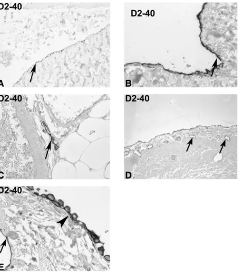
The clinical features of the study population were sum-marized in Table 1. In order to describe the distribution of D2-40-positive cells in the abdominal cavity of patients not involved in PD, we studied peritoneal biopsies taken during hernia repair (n = 15, Figure 1A–C). The mesothe-lial cell layer was positive for D2-40 (as previously de-scribed) and helped to define the peritoneal surface of the peritoneal biopsies (Figure 1A, B; [17]). Even in these normal control biopsies, the mesothelial cell layer was commonly lost, most likely due to the tissue handling. When the mesothelium was conserved, it demonstrated a flat appearance (Figure 1B). Podoplanin was predominantly localized on the apical side of the mesothelial cells and at times intercellularly (lateral sides, Figure 1B). A low num-ber of lymphatic vessels were present in the submesothelial fibrotic tissue and the adjacent fat tissue (Figure 1C). These lymphatic vessels were commonly associated with larger arteries and veins (Figure 1C). Overall, the number of D2-40-positive vessels in the submesothelial tissue and the adjacent fat tissue was low.
Biopsies taken at the time of appendectomy were used as an example of an inflammatory lesion in the abdominal cavity (n = 20). In the majority of biopsies, the mesothe-lium demonstrated a plump, cuboidal appearance with gaps between the mesothelial cells (Figure 1D, E). D2-40 was present circumferentially on these activated mesothelial
Table 1. Clinical data of study patients
Variable Normal peritoneum Appendicitis PD EPS
n 15 20 16 18
Age (years; mean ± SD) 58.4 ± 19.9 40.4 ± 35.6 49.5 ± 15.1 45.7 ± 13.5
PD—duration in months 24 ± 25.7 76 ± 37.2 (** vs PD)
Peritonitis 5 in 384 months 1:77 34 in 1,017 months 1:30 (* vs PD) PDF Neutral 2/16 6/18 Acidic 4/16 2/18 Both or N.D. 10/16 11/18 Icodextrin 1/6 10 N.D. 8/9 10 N.D. (* vs PD) Diabetes 0/15 0/20 5/16 3/18 Smoker 4/12 2 N.D. 7/19 1 N.D. 2/15 1 N.D. 7/14 4 N.D. Hypertension 4/20 2/20 7/16 11/18 Hb (g/dL ± SD [13–18]) 13.8 ± 1.7 13.7 ± 1.1 10.8 ± 1.9 10.0 ± 4.1 Leukocytes (G/L ± SD [4.0–11.3]) 8.0 ± 3.0 10.4 ± 2.9 7.2 ± 2.0 9.1 ± 4.04 Phosphate (mmol/L [0.68–1.68]) N.D. N.D. 1.78 ± 0.67 1.50 ± 0.54 Calcium (mmol/L [1.90–2.70]) 2.32 ± 0.12 2.35 ± 0.09 2.37 ± 0.22 2.24 ± 0.47 PTH (pmol/L [1.1–7.3]) N.D. N.D. 19.1 ± 17.3 27.2 ± 25.38 Urea-N (mg/dL [10–25]) N.D. N.D. 68.38 ± 40.68 40.09 ± 17.04 (* vs PD) Creatinine (mg/dL [0.5–1.4]) 1.4 ± 1.7 1.0 ± 0.3 8.3 ± 4.0 7.0 ± 1.90
PD, peritoneal dialysis; EPS, encapsulating peritoneal sclerosis; PDF, peritoneal dialysis fluid; Hb, haemoglobin; N.D., not determined; PTH, parathy-roid hormone, *: P < 0.05, **: P < 0.01.
Fig. 1. Expression of podoplanin in controls. Immunohistochemistry was performed on tissue sections from peritoneal biopsies taken at the time of hernia repair (A–C) or during appendectomy (D, E) with the monoclonal antibody D2-40 against podoplanin (orig. ×100 in D; ×250 in A, C; ×400 in B, E). The normal mesothelium cells were positive for podoplanin on the apical and intercellular region of the relatively flat mesothelial cells. Lymphatic vessels were present close to larger arteries in the submesothelial interstitial tissue (arrow). In biopsies from patients with appendicitis, the mesothelial cells were cuboid, with gaps between the cells (arrowhead, lymphatic vessels are labelled by arrows in D, E). This activated mesothelium commonly demonstrated circumferential podoplanin staining.
cells (Figure 1E). Focal accumulation of lymphatic vessels was present at sites of inflammatory cell accumulations. Expression of podoplanin in peritoneal biopsies in patients on PD
Sixteen biopsies from patients on PD were included in the study (Figure 2A, B). The transporter status was available in seven patients prior to biopsies, of whom 1 was low, 3 were low average, 1 high average and 1 high. In two biop-sies, the mesothelium was found to be preserved. In both cases, the mesothelium had a cuboidal appearance, similar
to what was described for the patients with appendicitis (Figure 2B). The number of D2-40-positive vessels was quite variable ranging from a single positive vessel per biopsy to a very prominent accumulation of D2-40-positive vessels in the thickened peritoneal membrane (Figure 2A). Three biopsies demonstrated a diffuse infiltration of podo-planin-positive cells.
Expression of podoplanin in EPS
Eighteen biopsies from patients on PD with clinical signs of EPS (radiological picture, symptoms of intestinal
ob-Fig. 2. Podoplanin in patients on peritoneal dialysis. Immunohistochemistry was performed on tissue sections from peritoneal biopsies taken from patients on PD (A–B) or on PD with signs of EPS (C–E) with the monoclonal antibody D2-40 against podoplanin (orig. ×100 in A, C, D; ×250 in B; × 400 in E). A prominent number of lymphatic vessels were present in the submesothelial interstitial tissue (arrows in A). A single layer of mesothelial cells was present on the surface of the biopsies, but with a cuboidal appearance (arrow in B) and with gaps between the cells (arrowhead in B). The morphological picture of EPS demonstrated the absence of a mesothelial cell layer on the surface (arrow in E). A high number of podoplanin-positive cells with the morphological appearance of fibroblasts are embedded in the extracellular matrix of EPS (C–E).
struction) were included (Figure 2C–E, Figure 3). The find-ings in the CT scans were calcifications (4/18), cocooning (11/18), peritoneal thickening (11/18), ascites after cessa-tion of PD (7/18) and bowel dilatacessa-tion (7/18). All patients underwent surgery and showed the typical features of EPS (ascites and fluid loculation, peritoneal thickening, adhe-sions, bowel dilatation and cocooning with thickening of the visceral peritoneum). The transporter status prior to stopping PD was in five patients, high average, and in two patients, high.
In the biopsies, the mesothelial cells on the surface were absent in all biopsies (Figure 2E, arrow). The podo-planin staining demonstrated a striking pattern with a high number of positive cells embedded in the sclerotic matrix (Figure 2E, Figure 3A). These podoplanin-positive cells had the morphological appearance of fibroblasts (Figure 2E). To further define the involved cell types, consecutive sections of four biopsies were further studied for the expression of CD31 (as an endothelial marker, Figure 3C), calretinin (a marker of mesothelial cells, Figure 3D) and smooth muscle actin (for myofibroblasts, Figure 3B, F). CD31 was positive on the endothelium of arteries and veins as expected, but the
podoplanin-positive‘infiltrating’ cells were CD31 negative (Figure 3C). Scattered lymphatic vessels were CD31 posi-tive (as this marker does not discriminate between blood and lymphatic endothelial cells). Therefore, the podoplanin-positive cell population is unlikely of endothelial origin. Calretinin was only found to be expressed by a small num-ber of cells close to the surface, but the vast majority of podoplanin-positive cells were negative (Figure 3D). The diffuse pattern of these cells mirrored the staining pattern for smooth muscle actin (Figure 3A, B, E, F). KI-67 (a cell proliferation marker) was expressed by scattered cells with focal accumulations of these cells (not illustrated).
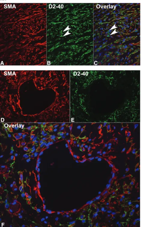
The pattern of smooth muscle actin-positive myofibro-blasts mirrored the distribution of podoplanin-positive cells; therefore, we performed immunofluorescence to confirm the double-positive cells (Figure 4). Smooth muscle actin-positive myofibroblasts embedded in the fi-brotic tissue (Figure 4A, D) were also positive for podopla-nin (Figure 4B, E). An overlay of the two reaction patterns is illustrated in Figure 4 (C, F). The smooth muscle actin-positive cells of vessel walls were podoplanin negative (Figure 4D, E). On the other hand, the podoplanin-positive
Fig. 3. Characteristics of the podoplanin-positive cells in EPS. Immunohistochemistry was performed on tissue sections from peritoneal biopsies taken from patients with EPS with a monoclonal antibody against podoplanin (A, E), smooth muscle actin (B, F), CD31 (C) or against calretinin (D, orig. × 100 in A–D; ×250 in E, F). A high number of D2-40 and smooth muscle actin-positive cells were present in a similar pattern of distribution (A, B and E, F). The vast majority of the D2-40-positive cells were CD31 and calretinin negative (C, D). Some CD31-positive vessels (arrowheads in C) and some calretinin-positive cells (arrow in D) were present.
lymphatic vessels were smooth muscle actin negative (Figure 4A, B).
In Figure 5A, the results are illustrated according to the time on PD and the number of peritonitis episodes. There is an overlap of the time on PD between patients with EPS and
the patients on PD of ~50 months. The PD patients with the pattern were not the ones with long time on PD (Figure 5A). The fibrotic zone was significantly thicker in the patients with EPS [as compared to the patients on PD (P < 0.05) and the control biopsies (P < 0.001)]. The biopsies from patients
Fig. 4. Double immunofluorescence for podoplanin and smooth muscle actin in EPS. Double immunofluorescence was performed on biopsies with EPS for smooth muscle actin (A, D) and podoplanin (with the monoclonal antibody D2-40, (B, E), orig. × 400). The overlay with the nuclear counterstain (DAPI) is illustrated in (C) and (F). Please note the podoplanin-positive lymphatic vessels (arrows in B), which are smooth muscle actin negative. The‘heart’ illustrated in F is a larger vein with a single layer of smooth muscle cells (D), which are podoplanin negative (E).
Fig. 5. Description of the time on PD, fibrosis and podoplanin scores. (A) The study population is illustrated according to the time on PD, the number of peritonitis episodes and the podoplanin scores. Please note that there is an overlap of ~50 months between the patients with EPS and without EPS, but the patients on PD with the pattern fall within the first 40 months of dialysis (w/o, without). (B) The thickness of the fibrosis zone was measured in a blinded fashion (*P < 0.05, **P < 0.01, ***P < 0.001). (C) The mean podoplanin scores (± SEM) are illustrated (**P < 0.01, ***P < 0.001).
670.7 180.1 43.8 0 100 200 300 400 500 600 700 800
controls PD (no EPS) PD (with EPS)
Thickness (micrometer)
***
*
**
0.1 0.6 1.6 0.0 0.2 0.4 0.6 0.8 1.0 1.2 1.4 1.6 1.8 2.0controls PD (no EPS) PD (with EPS)
Mean Podoplanin Score
***
**
C
B
on PD also demonstrated thickening of the submesothelial fibrotic zone (P < 0.01 as compared to control biopsies).
The podoplanin scores were significantly higher in EPS, as compared to patients on PD without EPS and controls (Figure 5C). An area of podoplanin-positive cells embed-ded in a fibrotic matrix was present in 15 out of 18 biopsies in patients with EPS, in 3 out of 16 patients on PD without signs of EPS, but none in the 35 controls. The three patients on PD who demonstrated a diffuse presence of podoplanin-positive cells were treated for 4, 5 and 22 months.
In this retrospectively collected biopsies, the staining pat-tern was significantly associated with EPS (P < 0.0001). It resulted in a sensitivity of 0.83 (95% confidence interval 0.59–0.96) and a specificity of 0.94 (95% confidence inter-val 0.84–0.99). The positive predictive value was 0.83 (95% confidence interval 0.5857–0.9642) and the nega-tive predicnega-tive value 0.94 (95% conf idence interval 0.8377–0.9877). The likelihood ratio was 14.2.
Discussion
The main finding of this study was the discovery of a po-doplanin and smooth muscle actin double-positive cell type, which forms the majority of cells in the biopsies from patients with EPS. The diagnosis of EPS must be based on a typical clinical, radiologic picture. Unfortunately, the histological picture is not specific, and markers differenti-ating between simple sclerosis and EPS are not available at the moment.
In a direct comparison of PD-associated simple sclerosis and EPS, the morphological differences were found to be a significantly thicker sclerosis zone in EPS (750 versus 45μm), fibrin deposition, fibroblast swelling, the presence of inflammation, vascular alterations, up-regulation of vas-cular endothelial growth factor and the presence of tissue calcification [18–21]. The thickness of sclerosis demon-strated a bimodal distribution without intermediate stages [18]. In EPS, the fibrin deposition is accompanied by a de-creased number of mast cells and a decrease in mast cell tryptase [4]. Mast cell tryptase has a strong fibrinogenoly-tic activity [22]. With our study, we add a new marker to the morphological evaluation of biopsies from patients with EPS. The majority of cells in the sclerosis zone ex-pressed podoplanin in combination with the typical myofi-broblast marker (smooth muscle actin). Morphologically, these cells represent fibroblasts. The diffuse appearance of podoplanin-positive cells separated the majority of cases with EPS from simple sclerosis. In contrast to other mar-kers previously used (e.g. smooth muscle actin), which was found to be commonly present, podoplanin-positive cells with a fibroblastic appearance were only found in a minority of patients on PD without signs of EPS (3 out of 16) [20]. None of the biopsies taken during hernia repair, appendicitis and in uraemic patients before PD was in-itiated (n = 9, not illustrated) demonstrated the podoplanin pattern. Therefore, this pattern had a good specificity and negative predictive value. We are currently planning a larger multicenter study to include a higher number of patients. Particularly early cases of EPS need to be compared with late cases of simple sclerosis (matched for the time on PD).
From the clinical point of view, simple sclerosis and EPS are two different entities, the latter with an aggressive course with life-threatening bowel obstruction [21]. It is still unclear whether there is a subclinical transition phase (early EPS; [21]). In our study, three patients had no clin-ical signs of EPS, but the morphologclin-ical appearance and the podoplanin-positive pattern at least focally. One patient presented with ascites and an early form of EPS seems possible (also we have no data to confirm this). In one pa-tient, the biopsy was taken close to the catheter where fibro-sis might be present. It is currently unclear whether the podoplanin-positive pattern is a reflection of an early dis-ease course or a risk factor for the development of EPS. On the other hand, three biopsies in the EPS group did not demonstrate the typical pattern of podoplanin-positive cells. Follow-up was available in only one podoplanin-negative patient where all other diagnostic criteria con-firmed EPS. Whether this is due to a sampling error or a technical problem with the staining technique is currently not clear. Further studies are clearly needed including EPS samples from different sources to evaluate the clinical use-fulness of D2-40 in routine material.
The source of the double-positive cells remains un-defined. In detailed electron-microscopic studies, the heal-ing process in the rat abdominal cavity was evaluated [23]. Following physical injury, the new mesothelium developed from subperitoneal connective tissue cells. These cells demonstrated subcellular similarities with primitive mes-enchymal cells and with fibroblasts in the subperitoneal stroma. The combination of smooth muscle actin and podoplanin expression might indicate a cell type with an intermediate phenotype between myofibroblasts and differ-entiated mesothelial cells. In the studies by Raftery, the mes-enchymal cells finally differentiated into mesothelial cells [23]. When the podoplanin-positive cells would be a cell population attempting to rebuild the mesothelial integrity, the absence of a basement membrane might prevent the dif-ferentiation, keeping the cells in an immature fibroblastic and profibrotic cell type in EPS. The denudation of the peri-toneal cavity could be the driving force for the process.
The transition of mesothelial cells into a fibroblastic phenotype (called epithelial mesenchymal transition) has recently found a lot of attention [24–27]. A part of the fi-broblasts in the sclerotic zone might be derived from mesothelial cells. This might explain why these cells still express podoplanin as a mesothelial cell marker. As the transition of mesothelial cells into fibroblasts has been de-monstrated to be an early event involved in simple scle-rosis [25], this hypothesis would not be consistent with our data, as the majority of patients on PD but without signs of EPS did not demonstrate double-positive cells. As illustrated in Figure 5A, the PD patients without EPS but with the pattern all have a time on PD lower than 50 months. Therefore, the podoplanin pattern was not just associated with time on PD.
Conclusion
In summary, we describe a new cell marker combination in-volved in patients with EPS. Podoplanin might turn out to be
useful in routine morphological evaluation as well as a driv-ing force of this devastatdriv-ing condition. We formulated sev-eral new hypotheses which can now be tested in vitro and in vivo.
Acknowledgements. S.S. is supported by grants from the University of Zurich, Baxter and the Swiss National Science Foundation (32003B_ 129710). N.B. is supported by the Robert-Bosch Foundation.
Conflict of interest statement. The study was supported by a Grant from Baxter. Otherwise, the authors do not declare conflicts of interest.
References
1. Alscher DM, Reimold F. New facts about encapsulating peritoneal sclerosis as a sequel of long-term peritoneal dialysis - what can we do? Minerva Urol Nefrol 2007; 59: 269–279
2. Augustine T, Brown PW, Davies SD et al. Encapsulating peritoneal sclerosis: clinical significance and implications. Nephron Clin Pract 2009; 111: c149–c154, discussion c154
3. Honda K, Nitta K, Horita S et al. Histologic criteria for diagnosing encapsulating peritoneal sclerosis in continuous ambulatory periton-eal dialysis patients. Adv Perit Dial 2003; 19: 169–175
4. Alscher DM, Braun N, Biegger D et al. Peritoneal mast cells in peri-toneal dialysis patients, particularly in encapsulating periperi-toneal scler-osis patients. Am J Kidney Dis 2007; 49: 452–461
5. Williams JD, Craig KJ, Topley N et al. Morphologic changes in the peritoneal membrane of patients with renal disease. J Am Soc Nephrol 2002; 13: 470–479
6. Williams JD, Craig KJ, von Ruhland C et al. The natural course of peritoneal membrane biology during peritoneal dialysis. Kidney Int Suppl 2003; S43–S49
7. Sherif AM, Yoshida H, Maruyama Y et al. Comparison between the pathology of encapsulating sclerosis and simple sclerosis of the peri-toneal membrane in chronic periperi-toneal dialysis. Ther Apher Dial 2008; 12: 33–41
8. Raica M, Cimpean AM, Ribatti D. The role of podoplanin in tumor progression and metastasis. Anticancer Res 2008; 28: 2997–3006 9. Ogasawara S, Kaneko MK, Price JE et al. Characterization of
anti-podoplanin monoclonal antibodies: critical epitopes for neutralizing the interaction between podoplanin and CLEC-2. Hybridoma (Larchmt) 2008; 27: 259–267
10. Breiteneder-Geleff S, Matsui K, Soleiman A et al. Podoplanin, novel 43-kd membrane protein of glomerular epithelial cells, is down-regulated in puromycin nephrosis. Am J Pathol 1997; 151: 1141–1152 11. Kalof AN, Cooper K. D2-40 immunohistochemistry–so far! Adv Anat
Pathol 2009; 16: 62–64
12. Kerjaschki D, Regele HM, Moosberger I et al. Lymphatic neoangio-genesis in human kidney transplants is associated with immuno-logically active lymphocytic infiltrates. J Am Soc Nephrol 2004; 15: 603–612
13. Nakamoto H. Encapsulating peritoneal sclerosis–a clinician’s ap-proach to diagnosis and medical treatment. Perit Dial Int 2005; 25: S30–S38
14. Heller F, Lindenmeyer MT, Cohen CD et al. The contribution of B cells to renal interstitial inflammation. Am J Pathol 2007; 170: 457–468
15. Segerer S, Banas B, Wornle M et al. CXCR3 is involved in tubuloin-terstitial injury in human glomerulonephritis. Am J Pathol 2004; 164: 635–649
16. Segerer S, Bohmig GA, Exner M et al. Role of CXCR3 in cellular but not humoral renal allograft rejection. Transpl Int 2005; 18: 676–680
17. Ordonez NG. D2-40 and podoplanin are highly specific and sensitive immunohistochemical markers of epithelioid malignant mesotheli-oma. Hum Pathol 2005; 36: 372–380
18. Garosi G, Di Paolo N, Sacchi G et al. Sclerosing peritonitis: a noso-logical entity. Perit Dial Int 2005; 25: S110–S112
19. Braun N, Reimold F, Biegger D et al. Fibrogenic growth factors in encapsulating peritoneal sclerosis. Nephron Clin Pract 2009; 113: c88–c95
20. Honda K, Oda H. Pathology of encapsulating peritoneal sclerosis. Perit Dial Int 2005; 25: S19–S29
21. Garosi G, Di Paolo N. Inflammation and gross vascular alterations are characteristic histological features of sclerosing peritonitis. Perit Dial Int 2001; 21: 417–418
22. Schwartz LB, Bradford TR, Littman BH et al. The fibrinogenolytic activity of purified tryptase from human lung mast cells. J Immunol 1985; 135: 2762–2767
23. Raftery AT. Regeneration of parietal and visceral peritoneum: an electron microscopical study. J Anat 1973; 115: 375–392
24. Hirahara I, Ishibashi Y, Kaname S et al. Methylglyoxal induces peritoneal thickening by mesenchymal-like mesothelial cells in rats. Nephrol Dial Transplant 2009; 24: 437–447
25. Del Peso G, Jimenez-Heffernan JA, Bajo MA et al. Epithelial-to-mesenchymal transition of mesothelial cells is an early event during peritoneal dialysis and is associated with high peritoneal transport. Kidney Int Suppl 2008; 108: S26–S33
26. Margetts PJ, Bonniaud P, Liu L et al. Transient overexpression of TGF-{beta}1 induces epithelial mesenchymal transition in the rodent peritoneum. J Am Soc Nephrol 2005; 16: 425–436
27. Vargha R, Endemann M, Kratochwill K et al. Ex vivo reversal of in vivo transdifferentiation in mesothelial cells grown from peri-toneal dialysate effluents. Nephrol Dial Transplant 2006; 21: 2943–2947