HAL Id: hal-02633595
https://hal.inrae.fr/hal-02633595
Submitted on 27 May 2020
HAL is a multi-disciplinary open access
archive for the deposit and dissemination of sci-entific research documents, whether they are pub-lished or not. The documents may come from teaching and research institutions in France or abroad, or from public or private research centers.
L’archive ouverte pluridisciplinaire HAL, est destinée au dépôt et à la diffusion de documents scientifiques de niveau recherche, publiés ou non, émanant des établissements d’enseignement et de recherche français ou étrangers, des laboratoires publics ou privés.
Distributed under a Creative Commons Attribution - NonCommercial - NoDerivatives| 4.0 International License
Matthieu Pasco, Rihab Loudhaief, Armel Gallet
To cite this version:
Matthieu Pasco, Rihab Loudhaief, Armel Gallet. The cellular homeostasis of the gut: what the Drosophila model points out. Histology and Histopathology, Universidad de Murcia, 2015, 30 (3), pp.277-292. �hal-02633595�
Summary. The digestive tract is subjected to many aggressions throughout animal life. Since disruptions of gut physiology impact on animal fitness and survival, maintenance of gut integrity and functionality is essential for the individual. Over the last 40 years, research on rodents has aimed at understanding how cellular homeostasis of the digestive tract is maintained when challenged with disruptions. Following the discovery of stem cells in the digestive tract of Drosophila, a flurry of studies made an important contribution to our understanding of how the proliferation and the differentiation of these cells are controlled and participate in the renewal of the digestive tract. Insights into these mechanisms in Drosophila have revealed many similarities with mammalian intestinal stem cells. For instance, the highly conserved EGFR, JAK/STAT, Wingless/Wnt, Hedgehog, Integrins, BMP/TGFβ, Hippo and Insulin pathways all participate in adult intestinal cellular homeostasis. Here, we provide a literature review of recent advances in the field highlighting the adult Drosophila midgut as a convenient model for dissecting mechanisms involved in the maintenance of the cellular homeostasis of the digestive tract in conventionally reared conditions. In addition, we shed light on recently published data putting Drosophila forward as a genetic tool to decipher the mechanisms underlying intestinal diseases and intestinal tumour
progression.
Key words: Intestinal homeostasis, Stem cell maintenance, Symmetric and asymmetric division, Intestinal diseases, Cancers
Introduction
Gut physiology appears essential for the development, fitness and health of animals. One of the main functions of the digestive tract (DT) is food digestion and subsequent uptake of nutrients essential for life. The intestine is an organ endowed with an innate immune response and acts as a first barrier against ingested pathogens. It is therefore increasingly recognized that intestine function is essential for animal and human longevity. Chronic intoxications (due to bacteria, virus, toxins, chemicals…) inducing injuries of the gut promote inflammation, chronic diseases or even, in the worst cases, cancers (Radtke and Clevers, 2005; Gersemann et al., 2011; Ren and Fang, 2011; Rizzo et al., 2011; Sun and Irvine, 2011). Moreover, genetically predisposed organisms are more susceptible to develop bowel diseases or cancers upon chronic infection of the digestive tract (Garrett et al., 2010; Apidianakis and Rahme, 2011; Christofi and Apidianakis, 2013). Aging is also characterized by an overall decline of the intestinal immune function and tissue homeostasis maintenance that in turn can affect lifespan due to the occurrences of diseases (failure of nutrient absorption, susceptibility to infection and superinfections, cancers,
Review
The cellular homeostasis of the gut:
what the Drosophila model points out
Matthieu Y. Pasco*, Rihab Loudhaief* and Armel GalletSophia Agrobiotech Institute, UMR INRA 1355/CNRS 7254/Nice
-Sophia Antipolis University, BP 167, 06903 -Sophia Antipolis Cedex - France * These authors contribute equally to this work
Offprint request to: Armel Gallet, Institut Sophia Agrobiotech, UMR INRA 1355/CNRS 7254/Université Nice Sophia-Antipolis, 400 route des Chappes, BP 167, 06903 Sophia Antipolis Cedex, France. e-mail: gallet@unice.fr
etc…), especially in humans whose average lifespan lengthens (DeVeale et al., 2004; Rossi et al., 2008; Biteau et al., 2008, 2010; Alper, 2010).
In order to keep its physiological functions at an optimum throughout life, the DT has to maintain its integrity and functionality as long as possible. This is what we refer to as homeostasis. Since the mid 70’s, a flurry of studies carried out in vertebrates has provided significant insight into how gut integrity and physiology are maintained over time. The initial discovery was the identification of somatic stem cells residing in adult tissue capable of renewing all the differentiated cells types of the intestine (Cheng and Leblond, 1974). Although the main discoveries on gut renewal were successfully carried out in vertebrates (Radtke and Clevers, 2005; Barker et al., 2012; Vanuytsel et al., 2013), these do not provide a convenient system for studying gut replenishment in response to many different experimental or environmental challenges, being both expensive and time consuming. The discovery of intestinal stem cells in the Drosophila adult DT a few years ago has put forward flies as a system for studying DT homeostasis (Micchelli and Perrimon, 2006; Ohlstein and Spradling, 2006). Hence, in this review, we will outline all the recent advances made in adult intestinal homeostasis using Drosophila as a system model. To avoid overlap with the numerous reviews describing the molecular events triggered in response to gut damage (Charroux and Royet, 2010; Apidianakis and Rahme, 2011; Biteau et al., 2011; Lucchetta and Ohlstein, 2012; Christofi and Apidianakis, 2013; Hombria and Serras, 2013), herein we will focus on how the Drosophila gut renews its epithelium and controls cellular homeostasis at steady state conditions. We will also discuss how external factors may challenge gut homeostasis. Finally we will put forward some examples showing the usefulness of the Drosophila model to understand mechanisms involved in human intestinal diseases and cancers.
Drosophila melanogaster as a system model
Most of the studies in Drosophila have been made in the central part of the adult DT, the midgut, and more particularly in a region of the posterior midgut called R4 (Buchon et al., 2013) (http://flygut.epfl.ch/) or P1-P2 (Marianes and Spradling, 2013). This region is mainly involved in the absorption of nutrients derived from food previously digested in the anterior midgut and central acidic region (Buchon et al., 2013; Marianes and Spradling, 2013). The R4 region also participates to the reabsorption of electrolytes (Shanbhag and Tripathi, 2009). Therein, the Drosophila posterior midgut physiologically resembles the small intestine of vertebrates.
At the tissue level, the Drosophila posterior midgut displays an apico-basal arrangement of its cell-cell junctions similar to what is observed in the mammalian DT with the septate junction (acting as the vertebrate
tight junction) positioned apical to the adherens junction. By contrast, in the Drosophila anterior midgut the position of the septate and adherens junctions are reversed (Baumann, 2001; St Johnston and Ahringer, 2010; Goulas et al., 2012;). Moreover, while the vertebrate small intestine looks like “roller coaster” with alternating crypts and villi, the drosophila midgut displays a flat architecture.
The cellular composition of the drosophila intestine closely resembles that of vertebrates. There are roughly three types of cells: the progenitor cells, the secreting cells and the absorptive cells. The drosophila midgut is made of the Intestinal Stem cells (ISCs) and the Enteroblasts (EBs) (ISC+EB make up the progenitor cells), the secretory enteroendocrine cells (ee) and the absorptive Enterocytes (ECs) (Micchelli and Perrimon, 2006; Ohlstein and Spradling, 2006). In Drosophila, ISCs are located basally in the epithelium. They are the only cells undergoing mitosis and the only supply of cells for gut replenishment. The EBs, the daughter cells of the ISCs, differentiate into either ECs (90% of the differentiated cells) or ee (10% of the differentiated cells). In vertebrates, ISCs, upon mitosis, give birth to transient-amplifying (TA) cells that are capable of undergoing 3 to 4 more divisions over a period of 2-3 days. Progenitor cells are located in the crypts with ISCs at the bottom and TA cells just above. These TA cells acquire their fate choice (absorptive vs. secretory lineage) soon after birth while migrating upwards of the villus (Takashima et al., 2013).
ECs are the main cell type of the Drosophila midgut. As in vertebrates, they are involved in the absorption of nutrients and water. Their apical surface is covered by microvilli (forming the brush border) and faces the lumen of the intestine (Shanbhag and Tripathi, 2009). In vertebrates most of the digestive enzymes are secreted by other organs (salivary glands, stomach and pancreas), while in Drosophila this function is mainly fulfilled by the ECs (Buchon et al., 2013; Marianes and Spradling, 2013). In addition, Drosophila ECs are involved in the production of antimicrobial peptides, whereas in vertebrates the Paneth cells are specifically dedicated to this function (Santaolalla and Abreu, 2012). Finally, ee are chemosensory cells secreting peptide hormones regulating gut physiology, food intake, metabolisms and probably behaviour in response to luminal contents (Veenstra et al., 2008; Tolhurst et al., 2012). In vertebrates, Goblet cells secrete luminal mucus involved both in the protection of gut lining and the intestinal transit. In Drosophila, the peritrophic membrane (secreted by the ECs) replaces mucus and functions to protect the gut (Kuraishi et al., 2011). Despite these small differences, Drosophila is a convenient model for studying the cellular and molecular mechanisms governing gut homeostasis due to the simplicity of its gut architecture (Shanbhag and Tripathi, 2009), the conservation of the signalling pathways (Vanuytsel et al., 2013; this review) and the multitude of existing tools (Singh et al., 2012).
ISC maintenance
While ISCs give birth to EBs, they also have to maintain their own pool during the entire lifespan: this is what we call ISC maintenance. First and most essential for ISC maintenance is their survival. Intriguingly, many studies have observed that most of the signalling pathways that are involved in the control of ISC division (e.g. Wg/Wnt, EGFR, JAK/STAT, Hippo, Integrins and BMP signalling pathways, see below) are not required for ISC survival, since inhibiting these pathways individually or in combination does not promote ISC death (Lin et al., 2008, 2010, 2013; Karpowicz et al., 2010; Liu et al., 2010; Biteau and Jasper, 2011; Xu et al., 2011; Tian and Jiang, 2014). This suggests that, at steady state, ISC might be immortal. However, it was shown that the transcription factor FOS is required for ISC survival, since, in its absence, ISC are rapidly lost and this can be rescued by the overexpression of the anti-apoptotic protein p35 (Biteau and Jasper, 2011). Furthermore, Buchon et al. (2009) observed that silencing of the transcription factor c-jun in ISC/EB promoted loss of ISC. Interestingly, both FOS and c-Jun act downstream of the JNK signalling pathway which is known to exert a cytoprotective activity, enabling cells to survive stress (Shaulian, 2010). In agreement with this, Bond and Foley (2012) observed that inhibiting the JNK kinase in ISCs reduced their number in the midgut. These results suggest that the JNK signalling pathway may be involved in ISC survival, though further investigations are necessary to definitively prove the implication of the JNK signalling in ISC survival.
Symmetric or asymmetric division, what makes the choice?
One of the main questions concerning ISC homeostasis is to understand how the choice of symmetrical or asymmetrical division is made. The answer is of prime importance, because symmetrical division generates either two short-lived EB daughter cells, which can induce a loss of ISCs, or two ISC daughter cells with unlimited potential to divide, giving rise to stem cell tumours. By contrast, an asymmetric division allows the self-renewal of the ISC as well as replenishment of dying differentiated cells.
Many observations suggest that in Drosophila midgut the main mode of ISC division is asymmetric. Using ISC lineage tracing, four independent labs have recently estimated that 2/3 of ISCs divide asymmetrically while 1/3 of ISCs divide symmetrically with a stochastic compensatory neutral drift where the loss of ISC, owing to EB cell fate choice, is compensated for by the appearance of two new ISC due to the symmetrical division of the neighbouring ISC (O’Brien et al., 2011; de Navascues et al., 2012; Goulas et al., 2012; Tian and Jiang, 2014).
At the level of ISC, the mechanism making the choice between symmetric vs. asymmetric division
begins to be unravelled (Fig. 1). At least in Drosophila, Notch signalling appears to play an important role in this choice. Thus, the transmembrane ligand, Delta, which is only expressed by the ISCs, binds to its receptor, Notch, on neighbouring EBs. Upon asymmetric division, the ISC daughter cell maintains the expression of delta, while the EB daughter cell expresses Notch target genes and down regulates delta expression (Ohlstein and Spradling, 2007; Maeda et al., 2008; Perdigoto et al., 2011). Accordingly, inhibiting either delta expression in ISC or N signalling in EB promotes ISC tumours (e.g. too much ISC/ISC symmetric division) (Micchelli and Perrimon, 2006; Ohlstein and Spradling, 2006, 2007). Allison Bardin and Colleagues have beautifully demonstrated that the activation of Notch signalling in EB is necessary to repress the activity of two transcription factors, Hairless and Daughterless, involved in ISC proliferation. Thus, the inhibition of Hairless or Daughterless in ISCs promotes their disappearance and differentiation into EBs. Moreover, delta expression is lost in hairless mutant ISCs. Conversely, Hairless overexpression increases the number of dividing ISCs (Bardin et al., 2010). By subtly manipulating Notch activity levels, they further demonstrated that a high level of Notch signalling is required in one of the two daughter cells for it to commit to an EB fate (Perdigoto et al., 2011). Therefore, once the ISC has completed its mitosis, one daughter cell keeps the expression of Delta (the future ISC) while the other daughter cell (the future EB) strongly activates the Notch pathway. The activation of Notch limits symmetrical division by repressing factors involved in proliferation, allowing the cell to exit the cell cycle and commit in a process of differentiation (Fig. 1).
It was recently shown that the BMP and Notch signalling pathways act antagonistically (Tian and Jiang, 2014). First, the BMP signalling is asymmetrically activated with a strong activation in ISC and a low activation in EB. Second, suppressing BMP signalling in ISC promotes symmetric EB/EB divisions at the expense of both asymmetric ISC/EB and symmetric ISC/ISC (as observed in absence of Notch signalling in EB, see above) divisions. Third, over-activation of BMP signalling in ISC promotes symmetric ISC/ISC divisions, which can be suppressed by concomitant over-activation of Notch signalling. Conversely, inhibiting Notch signalling suppresses the loss of BMP signalling (e.g. the symmetric EB/EB division) and even promotes the appearance of small clusters of ISC (e.g. the loss of Notch signalling phenotype) (Tian and Jiang, 2014). Therefore the levels of activation of these two pathways within ISC and EB fine-tune the cell fate choice of the progenitor daughter cells (Fig. 1). Interestingly, while Delta is a transmembrane ligand intrinsically produced by the ISC only able to bind to its receptor, Notch, in neighbouring cells (through cell-cell contact), BMP ligands originate from many sources (visceral mesoderm, trachea and EC, see below) and act at a distance. Therefore, these two ligands allow the
integration of information derived from different cells, compartments, and tissues, enabling a tight control of the mode of ISC division according to micro-environmental cues.
Interestingly, the Drosophila PDGF/VEGF-like signalling pathway, Pvf2/Pvr, is also implicated in EB cell fate choice. The ligand Pvf2 and its receptor Pvr are both expressed in ISC, and their absence inhibits the commitment of ISCs toward EBs (Choi et al., 2008; Bond and Foley, 2012). Whether there is a dialog between Notch, BMP and Pvf2/Pvr signalling to control ISC asymmetric division is not yet known.
One interesting question is how the decision to make symmetric or asymmetric division is made by the dividing cells. During Drosophila embryonic neurogenesis, the Par complex is responsible for the asymmetric division of neuroblasts and is itself asymmetrically localized inside the neuroblasts. During mitosis, the Par complex interacts with the mitotic spindle to orient it along the apico-basal axis and then segregates with the neuroblast daughter cells (Prehoda, 2009). Interestingly, the Par complex is also expressed in ISC and EB of the adult midgut. During ISC mitosis, it is apically located and then inherited by the
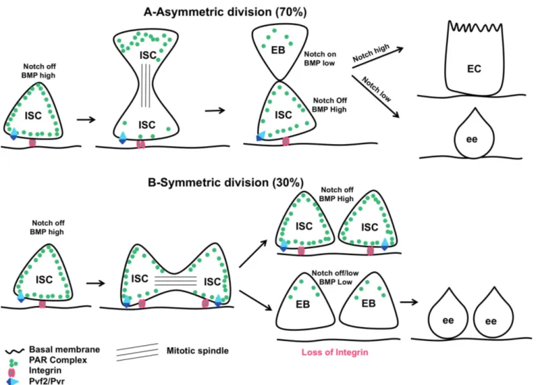
Fig. 1. Symmetric or asymmetric division, what is making the choice? A. Asymmetric division: At steady state, 70% of ISCs divide asymmetrically. Both
BMP and Notch signalling are asymmetrically activated with BMP signalling ON in ISC and Notch signalling ON in EB. While the Par complex is uniformly distributed in ISC, it becomes asymmetrically localized during ISC mitosis where it is more apically distributed. The Par complex is necessary to orient the mitotic spindle perpendicular to the basement membrane. Whereas the Integrins are required to keep the ISC attached to the basement membrane (the stem cell niche), a sine qua non condition to maintain the stemness, the most apical daughter cell will become an EB. The Drosophila PDGF/VEGF-like signalling pathway Pvf2/Pvr is necessary for the EB cell fate choice. Then the EB will further differentiate either into enterocyte (EC) or enteroendocrine cells (ee) depending on the level of Notch signalling activity. B. Symmetric division: At steady state, 30% of ISCs divide symmetrically because of orientation of the mitotic spindle parallel to the basement membrane. Both daughter cells will be either ISCs or EBs. If the daughter cells remain attached to the basement membrane (the stem cell niche) thanks to Integrins, they will stay ISCs. In absence of Integrins, daughter cells move away from the basement membrane and they will become EBs. Because of the low level of Notch signalling activity due to the absence of the ligand Delta (normally expressed at the surface of ISC), the twin EB will differentiate into ee.
differentiating EB (Fig. 1). Silencing components of the Par complex in ISC abrogates the apico-basal orientation of the mitotic spindle that becomes more parallel oriented to the basement membrane. As a consequence, the development of small ISC and ee tumours is observed (Goulas et al., 2012), suggesting that in the absence of Par complex, ISC divides symmetrically giving rise to two identical daughter cells, as is the case upon loss of Notch signalling (see above). Moreover, silencing Integrins (implicated in the attachment of gut lining to the basement membrane (Wolfenson et al., 2013)) in ISC impairs both the asymmetric localization of Par complex components and the orientation of the mitotic spindle (Goulas et al., 2012). Therefore Integrins appear to regulate the mode of ISC division through the asymmetric localization of the Par complex, which in turn will orient the mitotic spindle to promote asymmetric division (Fig. 1). Under homeostatic conditions, one can imagine a model in which Integrins are required to maintain ISC close to the stem cell niche (the niche being defined as the microenvironment providing all the signals, cytokines, and cellular interactions and architecture necessary for the maintenance of stem cell properties (Scadden, 2014)). During mitosis, the Integrins and the Par complex allow the orientation of the mitotic spindle along the apico-basal axis thereby retaining the apico-basal daughter cell in the
vicinity of its niche to maintain its stemness. Instead, the apical daughter cell moves away from the niche and commits to differentiation. When the orientation of the mitotic spindle is aligned to the basement membrane the ISC divides symmetrically and both daughter cells become either ISCs or EBs (Fig. 1). If the daughter cells remain close to the basement membrane, they stay ISC, if they move away from it, they become EBs (Fig.1). In agreement with this Goulas et al. (2012) observed that a small fraction of mitotic structures displays a symmetric distribution of the Par complex that might correspond to the 30% of symmetric division normally observed (O'Brien et al., 2011; de Navascues et al., 2012; Goulas et al., 2012; Tian and Jiang, 2014). Knowing that BMP signalling is required for the stemness of ISC (Tian and Jiang, 2014), it would be interesting to determine whether BMP signalling might regulate factors such as Integrins in ISC allowing, first, to keep the ISC attached to the basement membrane (and therefore close to its niche) and, second, the apico-basal orientation of the mitotic spindle to promote asymmetric division.
It is not yet known whether Integrins play a similar role in vertebrates, but it has been shown that knocking down one of the numerous Integrin subunits in mouse induces ISC proliferation (Jones et al., 2006). It is also interesting to note that although a function of the Par-complex in ISC mitosis has yet to be demonstrated in
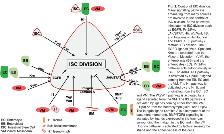
Fig. 2. Control of ISC division.
Many signalling pathways emanating from many sources are involved in the control of ISC division. Some pathways stimulate the ISC division such as EGFR, Pvf2/Pvr,
JAK/STAT, Hh, Wg/Wnt, IIS, and Integrins while Hpo/Yki and BMP/TGFβ pathways restrain ISC division. The EGFR ligands (Vein, Sptz and Krn) are secreted from the Visceral Mesoderm (VM), the enteroblasts (EB) and the enterocytes (EC). Pvf2/Pvr pathway acts autonomously in ISC. The JAK/STAT pathway is activated by Upd/IL-6 ligand coming from the EB, EC and the VM. The Hh pathway is activated by the Hh ligand originating from the EC, ISC and VM. The Wg/Wnt pathway is activated by a ligand secreted from the VM. The IIS pathway is activated by ligands coming either from the VM (Dilp3) or from the haemolymph (Dilp2 and Dilp5). The Integrin ligand Laminin A is a component of the basement membrane. BMP/TGFβ signalling is activated by ligands expressed in the tracheas surrounding the midgut, in the EC and in the VM. Hpo/Yki pathway is activated by factors sensing the shape and the adhesiveness of the cells.
vertebrates, it is asymmetrically located at the apical surface of mouse ISC (Quyn et al., 2010). Recently, it has been shown that mice ISCs preferentially divide symmetrically giving birth to either two ISCs or to two TA cells. There is also a phenomenon of neutral drift to maintain the right number of ISCs (Lopez-Garcia et al., 2010; Snippert et al., 2010). However, regarding the orientation of the mitotic spindle, nothing is yet clear. Indeed Lopez-Garcia et al. (2010) found that the mitotic spindle of ISC in the mouse crypt base can adopt all possible angles with respect to the apico-basal axis of the crypt (with about 20% oriented parallel to apicobasal axis) while Quyn et al. (2010) found that the mitotic spindle of dividing ISCs in the crypt base of the mouse and human small intestine is mainly oriented along the apicobasal axis (55% and 60% respectively). Thus, it has been proposed that even though the spindle is oriented perpendicular to the epithelial sheet, the daughter cells do not end up in divergent locations due to the spatial constraints of the crypt epithelium. As a consequence, each daughter cell remains in contact with their niche and hence would not adopt different fates (Snippert et al., 2010).
Control of ISC division
In healthy animals, the gut is completely renewed in about 8 days whether it is in Drosophila or in vertebrates (Ohlstein and Spradling, 2006; Jiang et al., 2009; Choi et al., 2011). Old ECs are eliminated and replaced by new ones derived from progenitor cells. This potential for self-renewal is due to the capacity of ISC to divide and give rise to daughter cells committed to the differentiation fate on a nearly daily basis. Understanding what is controlling ISC division is of prime importance, since it is now well established that overproliferation of stem cells promotes cancer (Radtke and Clevers, 2005). Studies of Drosophila gut homeostasis have highlighted many conserved signalling pathways involved in the control of ISC division (Fig. 2).
EGFR signalling
One of the main signalling pathways involved in the stimulation of ISC division is the EGFR pathway. In Drosophila, there are three EGFR ligands expressed in the intestine: Vein, Spitz and Keren. At steady state, Vein is expressed in the visceral mesoderm (VM) surrounding the gut epithelium (Biteau and Jasper, 2011; Jiang et al., 2011; Xu et al., 2011), Spitz is expressed in EB (Buchon et al., 2010; Xu et al., 2011) and Keren is expressed in both EB (Xu et al., 2011) and EC (Jiang et al., 2011) though they are present at low levels. All three ligands activate the EGFR pathway in ISCs through the activation of the small GTPase Ras and the kinase ERK that further phosphorylates the transcription factor FOS (Ren et al., 2010; Biteau and Jasper, 2011; Jiang et al., 2011; Xu et al., 2011). A prolonged inhibition of EGFR
signalling in ISCs causes them to be almost completely depleted after 4 weeks. The disappearance of ISCs is not due to cell death, but to their commitment toward the differentiation. Although Vein is probably the most important ligand for stimulating ISC division, the three ligands are somewhat redundant. Thus, after two weeks of inhibition of vein expression in the VM, the number of ISC is maintained even though the number of both ISC mitotic figures and EBs is reduced (Biteau and Jasper, 2011; Jiang et al., 2011; Xu et al., 2011). Interestingly a similar function for the EGFR/ErbB signalling pathway was recently identified in vertebrates (Wong et al., 2012).
Another EGFR related signalling pathway, the Pvf2/Pvr pathway, is also involved in the control of ISC division. It has been shown that inhibiting this pathway in ISCs slows down their division (Choi et al., 2008; Bond and Foley, 2012) and, as Pvf2/Pvr is also involved in ISC commitment toward EB (see above), small clones of ISC, which will never differentiate, accumulate over time (Bond and Foley, 2012). Interestingly, the small Ras GTPase (Bond and Foley, 2012) and the p38b MAPK (Park et al., 2009) were recently shown to act downstream of Pvr in ISC. Therefore cross talk between the EGFR and Pvf2/Pvr pathways at the level of Ras might exist allowing ISC division to be tightly controlled.
JAK/STAT signalling
While in vertebrates there is a wide array of cytokines activating the JAK/STAT pathway (Quintas-Cardama and Verstovsek, 2013), in Drosophila there are just 3 ligands named Unpaired (Upd), Upd2 and Upd3. At steady state, only Upd is expressed and secreted by both the VM surrounding the midgut (Lin et al., 2010) and the EB (Liu et al., 2010; Osman et al., 2012). Consequently the JAK/STAT pathway is activated, though weakly, in both ISC and EB (Buchon et al., 2009; Jiang et al., 2009; Beebe et al., 2010; Liu et al., 2010; Ren et al., 2010). Whereas it is generally accepted that over-activation of the JAK/STAT pathway in ISCs increases their rate of division (Jiang et al., 2009; Beebe et al., 2010; Lin et al., 2010; Liu et al., 2010; Xu et al., 2011), the effect of inhibiting JAK/STAT signalling is less clear. Some studies show that inhibiting the JAK/STAT pathway in ISCs does not alter their division (Buchon et al., 2009; Jiang et al., 2009; Beebe et al., 2010), whereas other groups find that ISCs disappear from the gut epithelium in the absence of JAK/STAT signalling in ISC (Lin et al., 2010; Liu et al., 2010; Xu et al., 2011) or upon inhibition of Upd production in EB (Osman et al., 2012). Interestingly, these later experiments monitored the presence of ISCs over a longer period and found ISCs to disappear after inhibiting JAK/STAT signalling in ISC for 3 weeks or more. These discrepancies suggest that either the inhibition of JAK/STAT signalling is not efficient enough in all the above experiments, explaining why a
longer period is necessary to observe the loss of ISCs, or (and) that other parallel pathways could partially compensate for the lack of JAK/STAT signalling in ISC. Although these two possibilities are not exclusive, there is a line of evidence sustaining the "compensatory" hypothesis. Indeed, the inhibition of EGFR signalling in ISC suppresses the JAK/STAT pathway overexpression-dependant ISC division. Reciprocally, the absence of JAK/STAT signalling in ISC abolishes the EGFR-dependent ISC mitosis (Buchon et al., 2010; Jiang et al., 2011; Xu et al., 2011). Noteworthy, in mammals, the STAT3 pathway seems to play a role in stimulating ISC proliferation (Grivennikov et al., 2009; Pickert et al., 2009).
Wingless (Wg)/Wnt signalling
In Drosophila (see below), as well as in vertebrates (Vanuytsel et al., 2013), the Wg/Wnt pathway is involved in the stimulation of ISC division. Indeed Wg/Wnt is secreted from the VM and activates signalling in ISC. Removing Wg from the VM or the intracellular components of the Wg/Wnt pathway within ISC reduces the rate of ISC division (Lin et al., 2008; Cordero et al., 2012b). Wg/Wnt signalling probably acts through the regulation of its target gene myc (Cordero et al., 2012b), which encodes a transcription factor involved in cell cycle regulation in both vertebrates and invertebrates. In agreement with this, it was recently shown that Myc is involved in ISC division at steady state, since silencing myc in ISC blocks their division (Ren et al., 2013). Interestingly, in ISC, myc is also a transcriptional target of the EFGR, JAK/STAT and Hpo/Yki (see below) pathways (Cordero et al., 2012a; Ren et al., 2013) and might be a convergent target gene of many signalling pathways controlling ISC division. This overlapping regulatory system of myc transcription may circumvent any deficiency in one of the aforementioned pathways. In line with this, the loss of ISCs caused by the inhibition of Wg signalling can be rescued by the overexpression of intracellular components of EGFR pathway in ISC. Conversely, the reduced rate of ISC division owing to the loss of EGFR signalling is rescued by overexpressing intracellular components of the Wg pathway within ISC (Xu et al., 2011). However myc is probably not the sole endpoint of these pathways to control ISC division since overexpression of Wg, Upd or EGFR ligands, but not Myc (Ren et al., 2013), is sufficient to induce ISC proliferation (Lin et al., 2008, 2010; Jiang et al., 2009; Beebe et al., 2010; Buchon et al., 2010; Liu et al., 2010; Jiang et al., 2011; Xu et al., 2011). This suggests that other activated downstream factors or target genes of these pathways participate in the stimulation of ISC division in conjunction with Myc.
Hedgehog signalling
There is one Hedgehog (Hh) ligand in Drosophila
and three in mammals. Hh peptides are novel cholesterol-linked secreted morphogens that are involved in many developmental processes (Gallet, 2011). Very recently Hh signalling was found to control ISC division in the adult Drosophila midgut (Li et al., 2014). Thus, the authors demonstrated that down-regulating Hh signalling in ISCs reduced their rate of division. Noteworthy, the source of Hh ligand comes from different origins, hh being expressed in the VM as well as in EC and ISC. Consequently, hh had to be silenced in all of these sources to observe a decrease in ISC proliferation (Li et al., 2014). Interestingly Hh is also involved in the regulation of ISC proliferation in vertebrates, although, contrary to the Drosophila gut, Hh peptides repress ISC proliferation (Vanuytsel et al., 2013).
Integrins
Very recent data in Drosophila show that the focal adhesion molecule Integrin, (Wolfenson et al., 2013) and more particularly the α1, α3 and βPS Integrin subunits, accumulate at the basal surface of ISC contacting the basement membrane (Goulas et al., 2012; Lin et al., 2013). Furthermore, Lin et al. (2013) observed that ISC mutant for Integrin subunits rapidly disappeared. This loss is probably due to both the slowdown of ISC division and, with time, the differentiation of non-dividing ISCs, since no ISC death was observed. Moreover, the Integrin ligand Laminin A and the downstream signalling components Talin and Integrin-linked kinase are also required for ISC division. A recent study using RNAi-targeted knockdown of the βPS subunits confirmed the role of Integrins in controlling ISC division (Okumura et al., 2014). Surprisingly, individual overexpression of components of the EGFR, JAK/STAT or Wg/Wnt signalling pathways did not rescue the Integrin-dependent loss of ISCs (Lin et al., 2013). It is likely that adhesion of ISCs to the basement membrane maintains them close to the VM that provides many of the cytokines/growth factors required for their division. Impairing ISC adhesion moves ISCs away from their niche, rendering them unable to perceive growth factors emanating from the VM. Strikingly, and in contrast to these results, Goulas and colleagues (2012) observed, using RNAi constructs targeting Integrin subunits or intracellular components of the Integrin signalling pathway, an increase in ISC division. How could one reconcile these opposing data? One could assume that the RNAi used by Goulas and colleagues (2012) impairs asymmetric cell division (see above), but not ISC adhesion to the basement membrane. As a consequence, the two daughter cells would still have enough Integrins to remain in their niche and stay ISC. Conversely, in ISC bearing integrin null mutant alleles, there are no functional Integrins to maintain the two daughter cells close to their niche, and as a result they become EBs (Fig. 1).
division of ISC also require signals and interactions emanating from the underlying mesenchyme (Yeung et al., 2011). It would be interesting to know whether Integrins play a similar role in controlling ISC division in vertebrates.
BMP/TGFβ signalling
So far, we have only described signalling pathways involved in the stimulation of ISC division. However, to avoid any ISC overproliferation, which could cause breakdown of gut epithelium and ultimately give rise to tumours, some negative feedback mechanisms are necessary.
In vertebrates, the activation of BMP/TGFβ signalling plays a role in regulating ISC division. BMP is secreted from the underlying mesenchyme and signals to the gut epithelium to restrict ISC proliferation (Vanuytsel et al., 2013). Interestingly, such mechanisms appear to be conserved in Drosophila (Guo et al., 2013; Li et al., 2013a; Tian and Jiang, 2014). Three recent studies carried out in the Drosophila adult midgut show that the BMP pathway is strongly activated in EC and ISC (Guo et al., 2013; Li et al., 2013a; Tian and Jiang, 2014). Decreasing the level of BMP signalling within ISC promotes ISC overproliferation (Guo et al., 2013; Tian and Jiang, 2014), though this effect seems indirect as it depends on the activation of the EGFR pathway within ISC (Guo et al., 2013; Li et al., 2013a).
Two BMP homologues, Decapentaplegic (Dpp) and Glass bottom boat (Gbb), are expressed in many different cell types in the Drosophila midgut. These ligands are expressed by the tracheal cells surrounding the midgut (Guo et al., 2013; Li et al., 2013a; Tian and Jiang, 2014), the VM (Guo et al., 2013) and EC (Tian and Jiang, 2014). Interestingly, Li et al. (2013) state that ISC proliferation is restricted by Dpp secreted from the tracheas while Guo and colleagues (2013) claim that the VM is the source of the functional Dpp. Finally, Tian and Jiang (2014) find that the Dpp/Gbb heterodimer is secreted by EC to control ISC proliferation. Hence, the functional source of BMP ligands in Drosophila is still controversial, and additional studies are needed to solve this discrepancy. In fact, it is possible that all the above BMP/Dpp-producing tissues could be involved in the restriction of ISC proliferation. In agreement with this, Tian and Jiang (2014) observed that Dpp::GFP expressed in EC is secreted on the basal side of the EC and accumulates between gut lining, the underlying basement membrane, and the VM. They further demonstrate a role of Collagen IV, a component of the basement membrane, in Dpp::GFP retention. Therefore, regardless of whether BMP ligands are secreted from the trachea, the VM or the EC, they accumulate at the basal side of the gut epithelium where ISCs lie. Accordingly, the ISC niche not only includes many factors stimulating ISC division (EGF, Wg/Wnt and Upd ligands), but also ligands (BMP) that negatively control ISC proliferation. Interestingly, it also seems that activation of the
JAK/STAT pathway in the VM directly or indirectly induces dpp expression (Guo et al., 2013). Hence, the activation of the JAK/STAT pathway promotes ISC division, but also stimulates the activation of the BMP signalling, which in turn restricts ISC division. Such a negative feedback mechanism prevents ISC overproliferation, which could have dramatic consequences for the gut physiology (e.g. hyperplasia of the epithelium or in the worst case, ISC tumours).
Hippo (Hpo)/Yorkie (Yki) signalling
Another potential negative regulator of the ISC division is the well-conserved Hpo/Yki pathway (Badouel and McNeill, 2011). Activation of the pathway triggers a cascade of phosphorylation ultimately resulting in the phosphorylation of the transactivator Yki, which as a consequence is retained in the cytoplasm. Upon inhibition of the Hippo kinase, Yki is no longer phosphorylated and translocated into the nucleus to activate its target genes. Among them, some are involved in cell proliferation (Yu and Guan, 2013). In vertebrates as in invertebrates, the nuclear translocation of Yki (named Yap in vertebrates) has been implicated in the stimulation of ISC proliferation upon gut injury (Karpowicz et al., 2010; Ren et al., 2010; Shaw et al., 2010; Staley and Irvine, 2010; Vanuytsel et al., 2013). In the Drosophila midgut, down-regulating upstream components of the Hpo pathway or overexpressing yki in ISCs greatly increases the rate of ISC division owing to the activation of Yki target genes involved in cell proliferation (e.g. myc, cycE, and Bantam) or cell survival (e.g. diap1). However, silencing yki in ISC does not impact on ISC division rate (Karpowicz et al., 2010; Ren et al., 2010; Shaw et al., 2010; Poernbacher et al., 2012; Ren et al., 2013; Huang et al., 2014). Thus, in steady state conditions, the Hpo pathway is ON and retains Yki in the cytoplasm. Interestingly, it was recently shown that the chromatin-remodeling protein Brahma binds directly to Yki and acts downstream of the Hpo pathway to control ISC division. Silencing brahma in ISCs slows down their division and rescues the hyperproliferation induced by the inhibition of the Hpo pathway (Jin et al., 2013). Therefore at steady state, the main role of the Hpo pathway is to limit the activation of Yki target genes involved in cell proliferation (e.g. Bantam, myc or cycE), thereby keeping the level of ISC division low. Noteworthy, one of the Yki target genes, Bantam, is a microRNA (Huang et al., 2014). Bantam is expressed in both ISC and ee but, unlike yki, silencing Bantam in ISCs reduced their rate of division (without affecting the differentiation of EB into EC). This suggests that Bantam, like myc, could be the target of multiple signalling pathways involved in the control of ISC division. Therefore, it seems that positive and negative outputs from different signalling pathways are integrated at the level of the transcriptional regulation of genes involved in the control of cell cycle. The
chromatin-remodeling protein Brahma or the microRNA Bantam are good illustrations of such intermingled regulation allowing the adaptation of ISC proliferation to environmental cues.
Interestingly, all the aforementioned signalling pathways are also activated upon bacterial intoxication. Indeed, many recent studies of the drosophila midgut have shed light on the mechanisms involved in the restoration of gut integrity after exposure to various aggressions. In response to stressful challenges, such as infection of the gut by pathogenic bacteria, ISC proliferation is strongly increased. This regenerative response allows restoring large parts of the intestinal epithelium, allowing the individual to overcome the intoxication (for more details see these reviews and references herein: Lucchetta and Ohlstein, 2012; Pitsouli et al., 2009; Kuraishi et al., 2013).
Progenitor cells and differentiation
Given that EB can differentiate into all the cell types making up the gut epithelium, what determines the fate choice of EB cells? In Drosophila, as in vertebrates, one major signalling pathway involved in this process is the Notch signalling pathway (Micchelli and Perrimon, 2006; Ohlstein and Spradling, 2006, 2007). Thus, the transmenbrane ligand Delta is expressed in ISCs and binds to its receptor Notch present at the surface of newborn EBs. This leads to the cleavage and intracellular relocalization of the Notch intracellular domain, activation of its target genes (Perdigoto and Bardin, 2013), and differentiation of EB into EC, which depends on high levels of Notch signalling. At lower levels of Notch acitivity, EB differentiates into ee (Fig. 3) (Micchelli and Perrimon, 2006; Ohlstein and Spradling, 2006, 2007; Bardin et al., 2010; Beebe et al.,
2010). Peculiarly, many studies have observed ee occurring as doublets throughout the midgut (Micchelli and Perrimon, 2006; Ohlstein and Spradling, 2006; Jiang et al., 2009; Perdigoto et al., 2011; our unpublished data). This phenomenon can be explained by symmetrical division of ISC giving rise to two EBs. In the absence of ISC expressing Delta, Notch is no longer strongly activated in the EB. Consequently the two EBs will differentiate into two ee staying in close proximity (Fig. 1). Interestingly, it has been proposed that a prolonged ISC-EB cell-cell contact is necessary to allow Notch signalling to reach the threshold necessary for the differentiation of EB into EC (Maeda et al., 2008). In agreement with this, the adherens junction molecules, DE-Cadherin and Armadillo/βCatenin, strongly mark the plasma membranes involved in the cell-cell contact between the ISC and the EB (Micchelli and Perrimon, 2006; Ohlstein and Spradling, 2006, 2007; Maeda et al., 2008; Choi et al., 2011). Moreover, Maeda and collegues (2008) observed that reducing ISC/EB cell-cell adhesion promotes either EB misdifferentiation or differentiation of EB into ee. Notably, Choi and colleagues (2011) did not observe such phenotypes though ECs appeared to be smaller than usual. Therefore, more work is required to decipher the relationship between cell adhesion and Notch signalling in the control of EB differentiation. Noteworthy, in the mammalian intestine, it is well-established that Notch signalling plays an important role in cell fate determination (Vanuytsel et al., 2013).
JAK/STAT signalling is also involved in the cell fate choice made by EB. In the absence of JAK/STAT signalling, undifferentiated EBs accumulate (Buchon et al., 2009; Jiang et al., 2009; Beebe et al., 2010; Lin et al., 2010; Liu et al., 2010; Ren et al., 2010; Osman et al., 2012). In addition, the contributions of Notch and JAK/STAT signalling to the terminal differentiation of
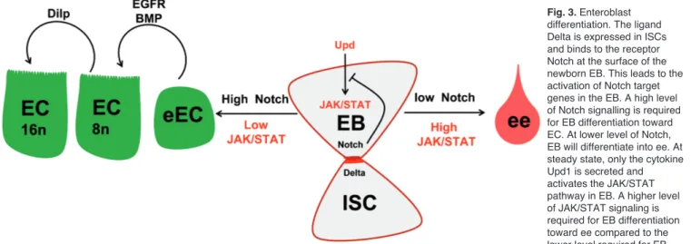
Fig. 3. Enteroblast
differentiation. The ligand Delta is expressed in ISCs and binds to the receptor Notch at the surface of the newborn EB. This leads to the activation of Notch target genes in the EB. A high level of Notch signalling is required for EB differentiation toward EC. At lower level of Notch, EB will differentiate into ee. At steady state, only the cytokine Upd1 is secreted and activates the JAK/STAT pathway in EB. A higher level of JAK/STAT signaling is required for EB differentiation toward ee compared to the lower level required for EB differentiation toward EC. A high level of Notch signalling in EB diminishes the level of JAK/STAT activation. Conversely, a lower level of Notch signalling in EB enables a stronger activation of JAK/STAT pathway. Both EGFR and BMP/TGFβ signalling are involved in EC maturation. Finally the Insulin-like growth factor Dilp3 is required for EC growth (by increasing their polyploidy) in presence of abundant food.
EB are interdependent, since each pathway regulates the expression of the ligand for the other pathway. Indeed, while JAK/STAT positively regulates the expression of delta in ISC (Jiang et al., 2009; Beebe et al., 2010), Notch signalling negatively regulates the expression of upd in EB (Liu et al., 2010). Consequently, a balance between the levels of activation of either pathway determines EB cell fate choice. Thus, a high level of Notch signalling in EB associated with a low level of JAK/STAT activation promotes differentiation of EB into EC. Conversely, low levels of Notch signalling in EB enable strong activation of the JAK/STAT pathway and promote EB differentiation into ee (Fig. 3) (Lin et al., 2010; Liu et al., 2010). A similar function for JAK/STAT signalling in vertebrates has yet to be identified.
Studies carried out in Drosophila also highlight the involvement of many other pathways in the terminal differentiation of EB into EC. The BMP and EGFR signalling pathways both contribute to EC maturation, as reduced BMP signalling slows down EC maturation (Li et al., 2013b; Tian and Jiang, 2014) and reduced EGFR signalling impacts EC shape (Buchon et al., 2010). Furthermore, BMP signalling also plays an anti-apoptotic role in EC (Li et al., 2013b). How Notch, JAK/STAT, BMP and EGFR signalling are coordinated to control EB differentiation and maturation remains unexplored, but Drosophila will be an ideal tool to carry out such studies.
Extrinsic factors regulating midgut homeostasis In this part, we detail a few novel mechanisms that may regulate ISC and cellular homeostasis of the gut. ISC proliferation, EB differentiation and EC turn-over are not only controlled by factors intrinsic to gut structure, but also by external factors including the circadian clock and nutrient availability.
Circadian clock
The circadian clock is an ancient molecular pathway that synchronizes organisms with daily environmental cues such as light intensity and temperature oscillations. Circadian rhythms are thought to influence the cell cycle, and there is some evidence that the clock plays a role in regeneration and proliferation. The Drosophila circadian pacemaker comprises the transcription factor partners Clock (CLK) and Cycle (CYC), which are negatively regulated by PER and Timeless (TIM). One transcriptional target of CLK/CYC is PER itself, which represses its own production and causes the cyc transcriptional rhythms that underlie circadian rhythms (Hardin, 2011). Interestingly, Karpowics and colleagues (2013) observed that upon gut damage, ISC mitosis increases at dawn, suggesting the involvement of the circadian clock in gut damage response. They further observed an accumulation of per mRNA in the early evening, while PER normally accumulates in the nucleus
of ICS and EC in the late night/early morning. Then they also found that while, at steady state, the loss of the circadian clock does not alter cellular homeostasis of the gut, its absence (using mutants for per and cyc) affects regeneration of the gut lining in response to damage (Karpowicz et al., 2013) with the disappearance of the peak of mitoses and reduced rate of ISC proliferation and fly survival. It remains to be determined how the circadian clock is coupled with other signalling pathways involved in gut regeneration. In addition, it is not yet known whether the role of the circadian clock in gut regeneration is conserved in vertebrates. It is also possible that environmental inputs such as temperature could modulate ISC division. Drosophila likely provides a good model for deciphering the links between gut homeostasis and environmental conditions.
Nutrient availability and Insulin signalling pathway (IIS)
In Drosophila there are eight insulin-like peptides, designated Dilp1—8. These Dilps have unique properties, and varying tissue and temporal expression patterns, and established roles in adult physiology including regulation of gut homeostasis (Kannan and Fridell, 2013).
Interestingly in Drosophila, it was shown that nutrient availability modulates ISC division. Indeed, flies starved of protein have less ISC/EB than flies reared on a protein-rich diet. Nonetheless, refeeding quickly (in less than 4 days) increases the size of the gut and the number of ISC/EB back to levels comparable to those of control animals, indicating that ISC/EB number is tightly controlled by nutrition (McLeod et al., 2010; O'Brien et al., 2011). Accordingly, flies fed a rich diet have higher rates of ISC division than flies fed a poor diet (Choi et al., 2011; O'Brien et al., 2011). Moreover, the 2/3 asymmetric-1/3 symmetric ratio of ISC division is reversed in response to a diet rich in proteins allowing the gut to grow rapidly. In this condition, EB rapidly differentiate into ECs in order to adapt nutrient absorption to food availability. Upon food withdrawal, the ratio of asymmetric to symmetric division returns to normal and a wave of apoptosis takes place, thereby shrinking the gut (O'Brien et al., 2011). Hence the ratio of asymmetric to symmetric divisions of ISC is not fixed, as initially thought, but can be modulated according to nutritional cues. The IIS pathway is therefore a good candidate for coupling food intake with ISC proliferation and gut growth. Accordingly, silencing dInR, encoding for the receptor of Dilps, or components of the IIS pathway in ISC/EB reduces the rate of ISC division (Amcheslavsky et al., 2009; Biteau et al., 2010; Choi et al., 2011; O'Brien et al., 2011).
Interestingly, compelling data highlight the fact that both a local and a systemic production of Dilps intervene to control ISC division, each source of Dilps likely responding to specific inputs. Systemic Dilps are secreted into the haemolymph by brain-specific neurons and ablating these neurons reduces ISC division
(Amcheslavsky et al., 2009; Biteau et al., 2010). Among the systemic Dilps secreted, dILP2 is probably involved in the systemic regulation of ISC division since its overexpression in brain neurons increases the rate of ISC division (Amcheslavsky et al., 2009). It has also been shown that feeding flies on a rich diet moderately increases the levels of dilp2 and dilp5 mRNA in the brain neurons, though their production is delayed relative to the change in diet (O'Brien et al., 2011). However, it was previously shown that the level of the transcription of dilps mRNA by brain neurons is not an efficient readout for monitoring systemic Dilps secretion (Geminard et al., 2009). Indeed, Dilps are stored in intercellular vesicles and are rapidly secreted (in less than 15min.) into the haemolymph in response to metabolic signals (Geminard et al., 2009).
Remarkably, Dilp3 was found to be quickly, strongly, and locally produced by the VM in response to a rich diet and this could be reversed upon withdrawal of food (O'Brien et al., 2011). Silencing dilp3 in the VM decreases the rate of ISC division irrespectively of plentiful nutritional availability and conversely, the overexpression of dilp3 in the VM stimulates ISC proliferation in fasted flies (O'Brien et al., 2011). It would now be interesting to investigate the relative importance of each source of Dilps (e.g. systemic vs. paracrine) in the regulation of ISC proliferation, whether they respond to identical cues and whether they can have redundant activities.
Nutrient availability could also be a major signal driving terminal differentiation, growth and death of EC. Indeed a protein-rich diet was shown to promote EC growth, owing to increased polyploidy (with a majority of 8n and 16n EC instead of 4n and 8n under normal conditions), and rapid EC turnover (1 week), while on poor diet, EC turnover slowed down during the first week and then ceased (Choi et al., 2011). Then the authors showed that activation of the IIS pathway in EC was responsible for their growth and rapid turnover in presence of rich food (Choi et al., 2011). Hence, the IIS pathway could function as a sensor coupling EB cell fate choice and EC growth with nutrient availability in the gut lumen. In line with this, the rapid local production of Dilp3 by the VM following food intake can, in addition to promoting ISC proliferation (see below), support EC growth and turnover (O'Brien et al., 2011).
Therefore, in the adult Drosophila midgut, ISCs interpret a nutrient cue to “break homeostasis” and drive growth of the gut when food is abundant. The niche (e.g. the VM) production of Insulin-like growth factor allows an immediate response while the systemic production of Insulin-like growth factor is likely involved in a long lasting adaptation of growth to nutrient availability. We can imagine a two step scenario in which the first step involves local production of Dilp3 in response to food abundance to rapidly increase organ size and nutrient absorption by the gut. In a second step, elevated levels of circulating Dilp2 and 5, which are secreted from brain neurons, in response to increased availability of nutrients
in the haemolymph, promotes growth of the whole individual and the storage of nutrients. The data described above using Drosophila could provide a foundation for future studies in vertebrates.
Drosophila as a tool to decipher intestinal diseases Various studies have demonstrated that failure to maintain cellular homeostasis and integrity of the gut contributes to the development of bowel diseases and/or cancers. The appearance of human gastrointestinal cancers is mainly caused by mutations occurring in components of the signalling pathways described above. It is likely that the high frequency of gastrointestinal malignancies reflects the importance of these signalling pathways (e.g. EGFR/Ras, Wg/Wnt, Notch, JAK/STAT, Hippo/Yki (YAP) and Hh) in controlling ISC division. Here, we will focus on some examples emphasizing the usefulness of Drosophila as a genetic tool to decipher the mechanisms underlying intestinal diseases.
For example, the tightly regulated process of ISC self-renewal controlled by Wg/Wnt signalling in stem and progenitor cells is subverted in cancer cells allowing malignant proliferation to take place (Reya and Clevers, 2005). Almost invariably, intestinal tumours carry activating mutations in the Wg/Wnt pathway, which mainly target tumour suppressor genes such as APC. Allelic loss and somatic mutations of APC tumour suppressor genes represent the most frequent molecular events in colorectal cancer (Nathke, 2004). Indeed, APC loss promotes the appearance of adenomas that when coupled with secondary oncogenic mutations in Ras (acting downstream of EGFR signalling) or in Smad (acting downstream of BMP/TGFβ signalling) can develop into aggressive adenocarcinomas giving rise to metastases (Reya and Clevers, 2005; Berg and Soreide, 2012). Interestingly, recent data on Drosophila shed light on the underlying mechanisms linking loss of APC with the appearance of tumours. Thus, in the Drosophila midgut, loss of APC leads to ISC over-proliferation and intestinal hyperplasia, a process which also involves non-autonomous cross-talk with the JAK/STAT and EGFR pathways (Lee et al., 2009; Cordero et al., 2012a). Drosophila genetics also permitted demonstrating that in addition to the loss of APC-mediated ISC over-proliferation, oncogenic mutations in Ras or Raf (acting downstream of EGFR) are required to block EB differentiation and down-regulate DE-Cadherin, two steps promoting intestinal outgrowth and tumour progression (Wang et al., 2013).
Over-activation of the Notch pathway is also implicated in various cancers (Geissler and Zach, 2012; Guilmeau, 2012). Particularly, in many cases of colorectal cancer owing to the loss of APC, the Notch ligand, Jagged1, is transcriptionally up-regulated arguing for a cooperation between Notch and Wg/Wnt signalling in promoting cancer (Bertrand et al., 2012; Guilmeau, 2012). Over-activation of Hh signalling was also found in a variety of human malignancies. A wide range of
digestive tract tumours, including most of those originating in the esophagus, stomach, biliary tract, and pancreas, display increased Hh pathway activity, often in conjunction with deregulation of other signalling pathways such as Notch and Wg/Wnt (Bertrand et al., 2012; Geissler and Zach, 2012). Conversely, while the constitutive activities of the Wg/Wnt, EGFR, Notch and Hh pathways are responsible for tumour appearance, development and metastasis, the loss of BMP and Hippo pathways activity also contribute to the development of intestinal cancers (Reya and Clevers, 2005; Avruch et al., 2012; Bertrand et al., 2012; Yu and Guan, 2013). The discovery that all of these pathways have conserved functions in flies renders Drosophila as a useful tool for deciphering crosstalks taking place to promote tumour growth.
Cancer has long been considered as a genetic disease. However, accumulating evidence supports the involvement of infectious agents in the development of cancers. This is especially true for those organs that are continuously exposed to microorganisms, such as the large intestine. Bacteria may initiate oncogenesis due to, first, their production of cell-damaging toxins and second, the induction of pro-inflammatory cytokines by the host in response to the damage, both events facilitating tumorigenesis in animals susceptible to developing cancers (Collins et al., 2011; Sun and Irvine, 2011; Tjalsma et al., 2012; Christofi and Apidianakis, 2013). Once again, Drosophila melanogaster has emerged as a powerful tool to unravel the underlying mechanisms. Adult flies bearing a latent oncogenic allele of the mammalian ortholog K-Ras (Ras1ACT) and fed
with the human pathogenic bacteria P. aeruginosa develop huge intestinal dysplasia (Apidianakis et al., 2009). Indeed Ras1ACT can synergize with the
inflammatory signals (JAK/STAT and JNK pathways) triggered by the bacteria to induce stem cell-originating tumours. This ultimately leads to intestinal dysplasia, a pre-malignant condition characterized by profound ISC or progenitor proliferation, impaired differentiation, epithelial multilayering and alterations in apicobasal polarity (Apidianakis et al., 2009). More recently, Bangi et al. (2012) demonstrated that P. aeruginosa intestinal infection cooperates with the oncogenic allele Ras1ACT
to activate the pro-inflammatory JNK pathway inducing expression of its transcriptional target, metalloprotease 1 (MMP1). An excess of MMP1 provokes the degradation of the basement membrane and facilitates the delamination of proliferating RasACTmutant ISCs (Bangi
et al., 2012).
Helicobacter pylori (H. pylori), another Human pathogenic bacterium colonizing gastric mucosae, was found to be the primary cause of upper gastrointestinal disorders, such as acute and chronic gastritis, peptic ulcer disease, and gastric cancer, which is the second most common cause of cancer-related deaths worldwide (Makola et al., 2007; Atherton and Blaser, 2009). Strains of H. pylori that can inject their CagA effector protein into host cells are known to be more virulent, but the
potential contributions of host genetics to pathogenesis are not well-understood (Atherton and Blaser, 2009). Using Drosophila melanogaster, Wandler and Guillemin (2012) showed that CagA acts through the activation of JNK signalling to induce apoptosis and disrupt tissue integrity. Moreover, in the presence of an oncogenic allele of Ras, CagA expression promotes growth and invasion of tumours in a JNK-dependent manner (Wandler and Guillemin, 2012). Therefore, the host genetic background can influence the outcome of H. pylori (or P. aeruginosa) pathogenesis and hence disease progression.
In conclusion, the conservation of molecular, cellular and tissue structures, as well as the high conservation of oncogenes and tumour suppressors between flies and mammals, justify Drosophila as a model for studying mammalian cancers and infections. Conclusion
Since Drosophila has emerged as an easily manageable model to study mechanisms involved in gut renewing and replenishment, much progress has been made. The insights gained from the studies discussed in this review show the particular importance of controlling self-renewal and regenerative capacity of the intestinal epithelium in order to maintain cellular homeostasis. This homeostasis is tightly controlled by a panoply of signalling pathways ensuring appropriate renewal of the epithelium and protecting the organism from potential aggressors present in daily ingested food. A coordinate network of more than ten signalling pathways exhibiting more or less redundancy is implicated in ISC maintenance and division, and EB differentiation. Consequently, in human, deregulations in these pathways lead to the appearance of intestinal diseases such as cancers. The discovery that all of these pathways have conserved functions in fly renders Drosophila a good model for deciphering crosstalks taking place to promote tumour growth and to understand the relationship between the presence of oncogenic alleles and bacterial intoxication.
Very recently, thanks to a Drosophila study, enteroendocrine cells (ee) have emerged as a new piece of the puzzle in regulating cellular homeostasis of the gut. Indeed, it appears that the neuropeptide Bursicon secreted by ee can restrain ISC proliferation by limiting the production of the EGFR ligand by the VM (Scopelliti et al., 2014). Knowing that the apical pole of the ee faces the lumen and that their basal pole is in direct contact with the VM, hormones secreted by ee are probably key regulators of the gut homeostasis, adapting ISC division and EB differentiation in function of the luminal content.
Moreover, the regenerative capacity of the intestinal epithelium can be influenced by extrinsic factors such as the circadian clock and the quality and abundance of nutrients. Accordingly, all future studies should take into account extrinsic regulations of gut homeostasis. Thus,
numerous discrepancies between independent labs reviewed here could be explained by differences in rearing/feeding conditions. In addition, all the studies in mouse were performed with conventionally fed mice. Knowing that food quality can potentially modulate the mode of ISC division, it would be interesting to study how nutrients may influence ISC division in mice. Finally, several studies validate the adult Drosophila midgut as a useful model for studying the behaviour of stem cells under homeostatic conditions and during pathogenic infection of the DT. The conservation of molecular, cellular and tissue structures between flies and mammals suggests that flies can be used to shed light on several aspects of biology relevant to human disease, including epithelial regeneration in the context of cancer and intestinal bowl diseases.
Acknowledgements. We are grateful to J. Colombani and C. Géminard for their advice and corrections on the manuscript. A special thank to D. Andersen. MYP was supported by the ANR-13-CESA-0003-01 grant. RL was supported by the Ministère de l’Enseignement Supérieur et de la Recherche. AG was supported by the CNRS.
References
Alper S. (2010). Model systems to the rescue: The relationship between aging and innate immunity. Commun. Integr. Biol. 3, 409-414. Amcheslavsky A., Jiang J. and Ip Y.T. (2009). Tissue damage-induced
intestinal stem cell division in Drosophila. Cell Stem Cell 4, 49-61. Apidianakis Y. and Rahme L.G. (2011). Drosophila melanogaster as a
model for human intestinal infection and pathology. Dis. Model. Mech. 4, 21-30.
Apidianakis Y., Pitsouli C., Perrimon N. and Rahme L. (2009). Synergy between bacterial infection and genetic predisposition in intestinal dysplasia. Proc. Natl. Acad. Sci. USA 106, 20883-20888.00 Atherton J.C. and Blaser M.J. (2009). Coadaptation of Helicobacter
pylori and humans: ancient history, modern implications. J. Clin. Invest. 119, 2475-2487.
Avruch J., Zhou D. and Bardeesy N. (2012). YAP oncogene overexpression supercharges colon cancer proliferation. Cell Cycle 11, 1090-1096.
Badouel C. and McNeill H. (2011). SnapShot: The hippo signaling pathway. Cell 145, 484-484.e1.
Bangi E., Pitsouli C., Rahme L.G., Cagan R. and Apidianakis Y. (2012). Immune response to bacteria induces dissemination of Ras-activated Drosophila hindgut cells. EMBO Rep. 13, 569-576. Bardin A.J., Perdigoto C.N., Southall T.D., Brand A.H. and Schweisguth
F. (2010). Transcriptional control of stem cell maintenance in the Drosophila intestine. Development 137, 705-714.
Barker N., van Oudenaarden A. and Clevers H. (2012). Identifying the stem cell of the intestinal crypt: strategies and pitfalls. Cell Stem Cell 11, 452-460.
Baumann O. (2001). Posterior midgut epithelial cells differ in their organization of the membrane skeleton from other drosophila epithelia. Exp. Cell. Res. 270, 176-187.
Beebe K., Lee W.C. and Micchelli C.A. (2010). JAK/STAT signaling coordinates stem cell proliferation and multilineage differentiation in the Drosophila intestinal stem cell lineage. Dev. Biol. 338, 28-37.
Berg M. and Soreide K. (2012). EGFR and downstream genetic alterations in KRAS/BRAF and PI3K/AKT pathways in colorectal cancer: implications for targeted therapy. Discov. Med. 14, 207-214. Bertrand F.E., Angus C.W., Partis W.J. and Sigounas G. (2012).
Developmental pathways in colon cancer: crosstalk between WNT, BMP, Hedgehog and Notch. Cell 11, 4344-51.
Biteau B. and Jasper H. (2011). EGF signaling regulates the proliferation of intestinal stem cells in Drosophila. Development 138, 1045-1055.
Biteau B., Hochmuth C.E. and Jasper H. (2008). JNK activity in somatic stem cells causes loss of tissue homeostasis in the aging Drosophila gut. Cell Stem Cell 3, 442-455.
Biteau B., Karpac J., Supoyo S., Degennaro M., Lehmann R. and Jasper H. (2010). Lifespan extension by preserving proliferative homeostasis in Drosophila. PLoS Genet. 6, e1001159.
Biteau B., Hochmuth C. E. and Jasper H. (2011). Maintaining tissue homeostasis: dynamic control of somatic stem cell activity. Cell 9, 402-11.
Bond D. and Foley E. (2012). Autocrine platelet-derived growth factor-vascular endothelial growth factor receptor-related (Pvr) pathway activity controls intestinal stem cell proliferation in the adult Drosophila midgut. J. Biol. Chem. 287, 27359-27370.
Buchon N., Broderick N.A., Chakrabarti S. and Lemaitre B. (2009). Invasive and indigenous microbiota impact intestinal stem cell activity through multiple pathways in Drosophila. Genes Dev. 23, 2333-2344.
Buchon N., Broderick N.A., Kuraishi T. and Lemaitre B. (2010). Drosophila EGFR pathway coordinates stem cell proliferation and gut remodeling following infection. BMC Biol. 8, 152.
Buchon N., Osman D., David F.P., Fang H.Y., Boquete J.P., Deplancke B. and Lemaitre B. (2013). Morphological and molecular characterization of adult midgut compartmentalization in Drosophila. Cell Rep. 3, 1725-1738.
Charroux B. and Royet J. (2010). Drosophila immune response: From systemic antimicrobial peptide production in fat body cells to local defense in the intestinal tract. Fly 4, 40-47.
Cheng H. and Leblond C.P. (1974). Origin, differentiation and renewal of the four main epithelial cell types in the mouse small intestine. V. Unitarian Theory of the origin of the four epithelial cell types. Am. J. Anat. 141, 537-561.
Choi N.H., Kim J.G., Yang D.J., Kim Y.S. and Yoo M.A. (2008). Age-related changes in Drosophila midgut are associated with PVF2, a PDGF/VEGF-like growth factor. Aging Cell 7, 318-324.
Choi N.H., Lucchetta E. and Ohlstein B. (2011). Nonautonomous regulation of Drosophila midgut stem cell proliferation by the insulin-signaling pathway. Proc. Natl. Acad. Sci. USA 108, 18702-18707. Christofi T. and Apidianakis Y. (2013). Ras-oncogenic Drosophila
hindgut but not midgut cells use an inflammation-like program to disseminate to distant sites. Gut 4, 54-9.22429.
Collins D., Hogan A.M. and Winter D.C. (2011). Microbial and viral pathogens in colorectal cancer. Lancet Oncol. 12, 504-512. Cordero J.B., Stefanatos R.K., Myant K., Vidal M. and Sansom, O.J.
(2012a). Non-autonomous crosstalk between the Jak/Stat and Egfr pathways mediates Apc1-driven intestinal stem cell hyperplasia in the Drosophila adult midgut. Development 139, 4524-45235. Cordero J.B., Stefanatos R.K., Scopelliti A., Vidal M. and Sansom O.J.
(2012b). Inducible progenitor-derived Wingless regulates adult midgut regeneration in Drosophila. EMBO J. 31, 3901-3917. de Navascues J., Perdigoto C.N., Bian Y., Schneider M.H., Bardin A.J.,