Accepted
Article
DR. VIOLAINE SMAÏL-FAUGERON (Orcid ID : 0000-0002-3197-6650) Article type : Original Article
Evaluation of intraosseous computerized injection system
(QuickSleeper™) versus conventional infiltration anaesthesia in
paediatric oral healthcare: a multicenter, single-blind, combined
split-mouth and parallel-arm randomized controlled trial
Violaine Smaïl-Faugeron*1,2, Michèle Muller-Bolla2,3, Jean-Louis Sixou4, Frédéric Courson1,2
1
Assistance Publique-Hôpitaux de Paris, Hôpital Bretonneau, Service d'Odontologie, Paris, France
2
Université Paris Descartes - Sorbonne Paris Cité, Faculté de Chirurgie Dentaire, Unité de Recherches Biomatériaux Innovants et Interface EA4462, Montrouge, France
3 Department of Pediatric Dentistry, UFR d’Odontologie de Nice Sophia Antipolis, Université Côte
d’Azur, CHUN, Nice, France
4
Université Européenne de Bretagne, Université de Rennes 1 and Centre Hospitalier Universitaire de Rennes, Rennes, France
*Corresponding author: Unité de Recherches Biomatériaux Innovants et Interface EA4462, 1 rue Maurice Arnoux, 92120 Montrouge, France. Tel: + 33 1 58 07 67 82; Fax: + 33 1 58 07 68 99; e-mail: violaine.smail-faugeron@parisdescartes.fr
Accepted
Article
Acknowledgments
We thank all patients and their parents for participating in the trial. We thank Laura Smales (BioMedEditing, Toronto, Canada) for editing the article.
Conflict of Interest
This study was not externally funded. There is no conflict of interest to declare.
Author contributions
V.S.F., F.C and M.M.B conceived the ideas; V.S.F. collected the data; V.S.F and J.L.S. analyzed the data; and V.S.F., F.C., M.M.B. and J.L.S. all contributed to the writing.
Summary
Background Conventional infiltration anaesthesia (CIA) is the most frequently used in paediatric oral
healthcare. However, other techniques are available, such as intraosseous anaesthesia (IOA), that can beneficiate from newly developed technologies. Aim To compare the pain caused by CIA and IOA delivered by the computerized system (QuickSleeper™) in children. Design We used an innovative design consisting in simultaneously conducting a multicenter split-mouth and parallel-arm randomized controlled trial (RCT) to allow for increased power. The primary outcome was pain reported by the patient on a visual analogue scale (0-10 cm) concerning the insertion of the needle and injection. Results A total of 30 children were included in the split-mouth RCT and 128 in the parallel-arm RCT. We combined treatment effect estimates by using an inverse-variance weighting
Accepted
Article
metaanalysis approach. Pain scores were significantly decreased with IOA vs CIA (mean difference -0.69 cm, 95% confidence intervals -1.13 to -0.25 cm). For each patient enrolled in the split-mouth RCT, about 5 were enrolled in the parallel-arm RCT, which allowed for not losing any eligible patients. Conclusion Pain during the insertion of the needle and injection was less with IOA versus CIA in children. The design of this study allowed for increasing statistical power and using all generated evidence. (ClinicalTrials.gov NCT02084433)
Keywords
: Anaesthesia, intraosseous, Quicksleeper™, pain, first permanent molar, paediatricdentistry
Introduction
Local anaesthesia is frequently indicated in paediatric oral healthcare [1]. Two types of anaesthesia exist: conventional intra-mucosal infiltration anaesthesia (CIA), frequently used by practitioners, and intraosseous anaesthesia (IOA), which delivers the local anesthetic within the spongy bone adjacent to the tooth to anesthetize. Needle- and injection-related fear are common sources of dental anxiety in children in case of CIA. Indeed, the needle is not only visible, but also injecting too quickly in the mucosa can lead to pain or discomfort. With IOA, the fear of the needle would be decreased because the pen grip is very close to the needle. Also, pain during injection would be decreased because IOA can be delivered by a computerized system (QuickSleeper™ system), which delivers local anaesthesia at a constant rate and pressure. Indeed, some authors showed that 83,9% of 50 children felt no pain or only mild discomfort with QuickSleeper™ system (scores 0 to 2 on the visual analogue scale [VAS]) [2], and that 69,7% of 30 patients preferred IOA compared to CIA [3]. To our best knowledge, IOA studies of pain felt by children have been only observational or
non-Accepted
Article
randomized comparative studies. Moreover, a recent systematic review and Evidence-Based Clinical Practice Guidelines showed insufficient evidence supporting the injection technique as superior to others and concluded that more rigorous and high-quality studies are needed [1, 4].
The randomized controlled trial (RCT) is the gold standard for evaluating the effectiveness of interventions [5]. The design most frequently used is the parallel-arm RCT [6], but the split-mouth RCT is also commonly used in dental clinical trials [7]. Because every patient receives each intervention, the split-mouth design allows for removing a lot of inter-individual variability from the estimates of the treatment effect and increasing the statistical power. Nevertheless, recruitment in a split-mouth RCT requires patients to have symmetrical disorders (e.g., carious lesions on at least two first permanent molars) to allow random allocation of the interventions [8]. Conducting simultaneously a parallel-arm RCT and a split-mouth RCT would allow enrolling all patients and maximizing power.
In this study, we aimed to compare pain during the insertion of the needle and injection with IOA against CIA for treating first permanent molars with deep carious lesions or moderate to high severity (MIH) lesions in children and adolescents. Secondary objectives were to compare the two anaesthesia types in terms of pain latency, the need for additional anaesthesia and pain felt during the treatment. We hypothesized that intraosseous anaesthesia delivered by the QuickSleeper™ may reduce pain more than anaesthesia delivered by the conventional infiltration technique. We used an innovative approach, by simultaneously conducted a parallel-arm RCT and a split-mouth RCT.
Accepted
Article
Methods
All procedures performed in studies involving human participants were in accordance with the ethical standards of the national research committee and with the 1964 Helsinki declaration and its later amendments or comparable ethical standards. The protocol of the trial was approved by the local research ethics committee (no. 13466) and the national authorities regulating confidentiality (no. 14.217). We communicated any important protocol modifications to the ethics committee. The protocol was prospectively registered in ClinicalTrials.gov (NCT02084433) and was published [9]. The study was conducted in paediatric dentistry departments of three French universities (Nice, Paris and Rennes). This manuscript was written following the guidelines of CONSORT (Consolidated Standards of Reporting Trials) (Supplementary Table S1).
Study design
The study design consisted in simultaneously conducting a multicentric split-mouth RCT and a parallel-arm RCT. For the split-mouth RCT, two permanent first molars belonging to the same dental arch were included per patient, and for the parallel-arm RCT, only one permanent first molar was included per patient.
Inclusion criteria
The following inclusion criteria were established: patients between 7-15 years of age; mentally and generally disease-free; cooperative (score 0 to 2 on the Venham distress scale) [10]; with one (for the parallel-arm trial) or at least two (for the split-mouth trial) first permanent molars with deep caries (i.e., dentinal lesion involving more than 50% of the entire dentin thickness evaluated by radiography) or MIH lesion, with preserved pulp vitality as determined by clinical and radiographic
Accepted
Article
observations (treatments could be conservative or endodontic limited to pulpotomy); without use of analgesic drug for 48 hr before randomization. The non-inclusion criteria were periodontal alterations (periodontal pockets or dental mobility) or radiological defects (bone loss, furcation or periapical radiolucency). The exclusion criteria were per-operative clinical and radiographic observations of irreversible radicular pulpal inflammation or pulpal necrosis. All patients and parents received oral and written information about the study, which was carried out according to the Declaration of Helsinki, following approval of the local research ethics committee. Oral agreement was obtained from all individual patients and parents included in the study.
Interventions
Before both types of anaesthesia, we applied the topical anaesthesia Xylocaïne® visqueuse 2% (AstraZeneca, Rueil Malmaison, France) for 1 to 2 min on previously dried mucosa. For CIA, we used a 16-mm-long needle for para-apical maxillary anaesthesia, and a 35-mm-long needle for locoregional mandibular anaesthesia. IOA was carried out using the QuickSleeper™ system (Dental Hi Tec, Cholet, France) following the three-step procedure described by the manufacturer [11]. For IOA, we used a 30-gauge, 9 or 16-mm-long needle. The anesthetic solution used in both techniques was 4% articaine with 1: 200,000 adrenalin. Anaesthesia in all cases was carried out by a single practitioner (one of the four investigators) in each of the three centers.
Outcomes
All outcomes were recorded on the day of the intervention. The primary outcome measure was pain felt during the insertion of the needle and injection of the anesthetic, assessed at the end of the anaesthesia, with a VAS ranging from 0 cm (no pain) to 10 cm (very much pain) [12]. We evaluated three secondary outcomes: latency, the need for additional anaesthesia during the treatment and
Accepted
Article
pain felt during the treatment. The latency was assessed examining the sensitivity of the vestibular sulcus for CIA or lingual sulcus for IOA by using a probe at the end of the injection (an exam was conducted every minute until the sulcus was insensitive to the probe). Patient assessed the pain felt during the treatment with the VAS at the end of the dental treatment session.
Sample size
For the split-mouth RCT, we estimated the sample size for the primary outcome by taking into account the correlation induced by paired nature of the data. In a previous trial, the corresponding standard deviation in the VAS score was estimated at 1.2 [2]. Assuming that the standard deviation is equal in the two randomization groups and that the correlation between the pain scores for the same subject in the first and second treatment is 0.6, the difference in VAS scores would have a standard deviation of 1.10. With a type I error risk of 0.05, we needed 30 patients to guarantee 80% power to detect a minimum true difference of 0.6 points in mean pain experienced during IOA and CIA. For the parallel-arm RCT, under the same assumptions described previously, we estimated the sample size at 128.
Assignment of interventions
For anaesthesia allocation, we used a computer-generated, permuted-block randomization sequence, with two block sizes randomly varied. The random sequences were stratified by center for both RCTs and by dental arch for the parallel-arm RCT. The practitioner obtained each randomization allocation via a centralized, secure web-based interface (RandoWeb). For the split-mouth RCT, one permanent first molar was randomly allocated to one of the techniques (e.g. IOA) and the other permanent first molar belonging to the same dental arch in the same child was allocated to the other technique (e.g. CIA). A 7- to 21-day interval was established between the two procedures. For
Accepted
Article
the parallel-arm RCT, one patient with one permanent molar first was randomly allocated to one of the techniques (IOA or CIA). Patients — evaluators — were unaware of which of the two techniques was used, whereas practitioners were not.
Data collection and management
First, data were collected by investigators by using a paper form. The form was pilot-tested by the coordinating center. Second, data were entered electronically by the coordinating center. Participant files were stored in a secure and accessible place and manner. A Data Monitoring Committee was not needed because of the short duration of patient participation and known minimal risks. Adverse events were collected and reported.
Statistical methods
The unit of analysis was the tooth for the split-mouth RCT, and the patient for the parallel-arm RCT. For pain and latency, we reported the mean differences between groups and the associated 95% confidence intervals (95% CI). For the need of additional anaesthesia, we reported odds ratio between groups and the associated 95% CI. For the split-mouth RCT, the results were analyzed by Student’s t test for paired samples (because of the paired nature of data). We also compared intervention effect estimates between the split-mouth and parallel-arm RCTs by z ratio tests (the difference in effect estimates between the two RCTs divided by its standard error). Because the effect estimates from the two RCTs were consistent, we combined them by using an inverse-variance weighting meta-analysis approach: the combined effect estimate is a weighted average of the two effect estimates, each effect estimate being weighted by the inverse of its variance [8, 13, 14]. We also performed a subgroup analysis by arch (the same statistical methods were applied). For the primary outcome, we did an exploratory post hoc subgroup analysis by type of disorders (deep
Accepted
Article
caries, MIH, or both deep caries and MIH) (the same statistical methods were applied). Finally, we compared the pain felt by boys versus girls and assessed the role of age using a post hoc multivariate analysis. All the analyses followed the intention-to-treat principle [15]. We did not plan any interim analysis and early stopping.
Results
Patient recruitment started in January 2015 and stopped in February 2017. The flow of participants in the trial is in Figure 1. For each patient enrolled in the split-mouth RCT, about 5 were enrolled in the parallel-arm RCT, which allowed for not losing any eligible patients. In total, 158 children aged 7 to 15 years for whom parental consent had been obtained were deemed eligible for inclusion. Of these, 30 (mean age 9.0 years, SD 2.3) participated to the split-mouth RCT and 128 (mean age 10.4 years, SD 2.5) to the parallel-arm RCT and randomly allocated to IOA (n = 63) or CIA (n = 65). On average, one cartridge containing the anesthetic solution was injected per anaesthesia. Finally, no harm was detected. Patient characteristics are in Table 1.
Split mouth RCT
The main results are shown in Table 2.
Primary outcome
The mean VAS scores were 0.73 ± 1.31 cm for the IOA and 1.43 ± 1.45 cm for the CIA groups. The mean (95% CI) for the difference in paired proportions was -0.70 ± 0.36 cm (-1.44 to 0.04), indicating that patients felt less pain on the insertion of the needle and the injection with IOA compared to CIA, but this difference was not statistically significant (p = 0.06).
Accepted
Article
Secondary outcomes
Latency
The mean latency was 1.07 ± 0.25 min for the IOA and 2.83 ± 2.64 min for the CIA groups. The mean (95% CI) for the difference in paired proportions was -1.77 ± 0.51 (-2.77 to -0.77), indicating that latency was statistically decreased with IOA compared to CIA with a statistically significant difference (p = 0.001).
Need for additional anaesthesia during the treatment
Four patients needed additional anaesthesia: two in IOA group (one additional intrapulpal anaesthesia and one additional intraligamentary anaesthesia in mandibular arch), and two patients in CIA group (one additional IOA on maxillary arch and one para-apical anaesthesia in mandibular arch). There was no statistically significant difference between the two groups (odd ratio (OR) = 1.00, 95%CI 0.15 to 6.74, p = 1.00).
Pain felt during the treatment
The mean VAS scores were 1.07 ± 1.76 cm for the IOA and 0.53 ± 0.82 cm for the CIA groups. The mean (95% CI) for the difference in paired proportions was 0.53 ± 0.37 cm (-0.19 to 1.25), indicating that patients felt less pain during treatment with CIA compared to IOA, but this difference was not statistically significant (p = 0.14).
Subgroup analysis
Accepted
Article
Primary outcome
On the maxillary arch, the mean VAS scores were 0.53 ± 1.19 cm for the IOA and 1.33 ± 1.54 cm for the CIA groups. The mean (95% CI) for the difference in paired proportions was -0.81 ± 0.51 cm (-1.89 to 0.29), indicating that patients felt less pain on the insertion of the needle and the injection with IOA compared to CIA, but this difference was not statistically significant (p = 0.14).
On the mandibular arch, the mean VAS scores were 0.93 ± 1.44 cm for the IOA and 1.53 ± 1.41 cm for the CIA groups. The mean (95% CI) for the difference in paired proportions was -0.60 ± 0.53 cm (-1.74 to 0.54), indicating that patients felt less pain on the insertion of the needle and the injection with IOA compared to CIA, but this difference was not statistically significant (p = 0.28).
Secondary outcomes
On the maxillary arch, the mean latency was 1.07 ± 0.26 min for the IOA and 2.33 ± 1.05 min for the CIA groups. The mean (95% CI) for the difference in paired proportions was 1.27± 0.29 (1.84 to -0.69), indicating that latency was statistically decreased with IOA compared to CIA with a statistically significant difference (p = 0.0003). On the mandibular arch, the mean latency was 1.07 ± 0.26 min for the IOA and 3.33 ± 3.58 min for the CIA groups. The mean (95% CI) for the difference in paired proportions was -2.27 ± 1.03 (-4.28 to -0.25), indicating that latency was statistically decreased with IOA compared to CIA with a statistically significant difference (p = 0.03).
On the maxillary arch, the mean VAS scores were 0.93 ± 2.02 cm for the IOA and 0.20 ± 0.56 cm for the CIA groups. The mean (95% CI) for the difference in paired proportions was 0.73 ± 0.56 cm (- 0.36 to 1.83), indicating that patients felt less pain during the treatment with CIA compared to IOA, but this difference was not statistically significant (p = 0.17). On the mandibular arch, the mean VAS
Accepted
Article
scores were 1.20 ± 1.52 cm for the IOA and 0.87 ± 0.92 cm for the CIA groups. The mean (95% CI) for the difference in paired proportions was 0.33 ± 0.54 cm (-0.73 to 1.39), indicating that patients felt less pain during the treatment with CIA compared to IOA, but this difference was not statistically significant (p = 0.51).
Post hoc subgroup analysis for the primary outcome
Results are summarized in Supplementary Table S5.
For the subgroup “deep caries”, the mean VAS scores were 1.43 ± 1.90 cm for the IOA and 1.43 ± 1.51 cm for the CIA groups. The mean (95% CI) for the difference in paired proportions was 0 cm (-2.14 to (-2.14, p=1).
For the subgroup “MIH”, the mean VAS scores were 1 ± 1.41 cm for the IOA and 1.41 ± 1.58 cm for the CIA groups. The mean (95% CI) for the difference in paired proportions was -0.33 cm (-2.09 to 1.42), indicating that patients felt less pain on the insertion of the needle and the injection with IOA compared to CIA, but this difference was not statistically significant (p = 0.67).
For the subgroup “both deep caries and MIH”, the mean VAS scores were 0.21 ± 0.58 cm for the IOA and 1.50 ± 1.45 cm for the CIA groups. The mean (95% CI) for the difference in paired proportions was -1.29 cm (-2.18 to -0.40), indicating that patients felt less pain on the insertion of the needle and the injection with IOA compared to CIA, with a statistically significant difference (p = 0.008).
Accepted
Article
Parallel-arm RCT
The main results are shown in Table 2.
Primary outcome
The mean VAS scores were 1.17 ± 1.40 cm for the IOA and 1.86± 1.81 cm for the CIA groups. The mean (95% CI) for the difference in paired proportions was -0.69 ± 0.29 cm (-1.25 to -0.12), indicating that patients felt statistically less pain on the insertion of the needle and the injection with IOA compared to CIA statistically significant, with a statistically significant difference (p = 0.02).
Secondary outcomes
Latency
The mean latency was 1.63 ± 0.97 min for the IOA and 3.08± 2.11 min for the CIA groups. The mean (95% CI) for the difference in paired proportions was -1.44 ± 0.29 (-2.02 to -0.87), indicating that latency was statistically decreased with IOA compared to CIA with a statistically significant difference (p = 2.87.10-6).
Need for additional anaesthesia during the treatment
Eighteen patients needed additional anaesthesia: seven in IOA group (two additional IOA, two additional para-apical anaesthesia, two additional intraligamentary anaesthesia and one additional intrapulpal anaesthesia), and eleven patients in CIA group (four additional IOA, two additional para-apical anaesthesia, and five additional intraligamentary anaesthesia). There was no statistically significant difference between the two groups (odd ratio (OR) = 0.61, 95%CI 0.22 to 1.70, p = 0.34).
Accepted
Article
Pain felt during the treatment
The mean VAS scores were 0.90 ± 1.51 cm for the IOA and 0.88 ± 1.64 cm for the CIA groups. The mean (95% CI) for the difference in paired proportions was 0.03 ± 0.28 cm (-0.52 to 0.58), indicating that patients felt less pain during treatment with CIA compared to IOA, but this difference was not statistically significant (p = 0.92).
Subgroup analysis
Results are summarized in Supplementary Table S3.
Primary outcome
On the maxillary arch, the mean VAS scores were 1.25 ± 1.40 cm for the IOA and 1.64 ± 1.65 cm for the CIA groups. The mean (95% CI) for the difference in paired proportions was -0.40 ± 0.41 cm (-1.21 to 0.42), indicating that patients felt less pain on the insertion of the needle and the injection with IOA compared to CIA, but this difference was not statistically significant (p = 0.34).
On the mandibular arch, the mean VAS scores were 1.11 ± 1.41 cm for the IOA and 2.06 ± 1.91 cm for the CIA groups. The mean (95% CI) for the difference in paired proportions was -0.94 ± 0.40 cm (-1.75 to -0.14), indicating that patients felt less pain on the insertion of the needle and the injection with IOA compared to CIA, with a statistically significant difference (p = 0.02).
Secondary outcomes
On the maxillary arch, the mean latency was 1.57 ± 0.92 min for the IOA and 2.81 ± 1.80 min for the CIA groups. The mean (95% CI) for the difference in paired proportions was 1.24 ± 0.38 (1.97 to
-Accepted
Article
0.50), indicating that latency was statistically decreased with IOA compared to CIA with a statistically significant difference (p = 0.002). On the mandibular arch, the mean latency was 1.69 ± 1.02 min for the IOA and 3.32 ± 2.36 min for the CIA groups. The mean (95% CI) for the difference in paired proportions was -1.64 ± 0.45 (-2.52 to -0.75), indicating that latency was statistically decreased with IOA compared to CIA with a statistically significant difference (p = 0.0005).
On the maxillary arch, five patients needed additional anaesthesia: one in IOA group and four patients in CIA group. There was no statistically significant difference between the two groups (mean OR = 0.61, 95% CI 0.22 to 1.70, p = 0.20). On the mandibular arch, 13 patients needed additional anaesthesia: six in IOA group and seven patients in CIA group. There was no statistically significant difference between the two groups (mean OR = 0.61, 95% CI 0.22 to 1.70, p = 0.72).
On the maxillary arch, the mean VAS scores were 1.18 ± 1.49 cm for the IOA and 0.87 ± 1.98 cm for the CIA groups. The mean (95% CI) for the difference in paired proportions was 0.31 ± 0.47cm (-0.61 to 1.23), indicating that patients felt less pain during the treatment with CIA compared to IOA, but this difference was not statistically significant (p = 0.51). On the mandibular arch, the mean VAS scores were 0.69 ± 1.51 cm for the IOA and 0.88 ± 1.30 cm for the CIA groups. The mean (95% CI) for the difference in paired proportions was -0.20 ± 0.35 cm (-0.87 to 0.48), indicating that patients felt less pain during the treatment with IOA compared to CIA, but this difference was not statistically significant (p = 0.56).
Accepted
Article
Post hoc subgroup analysis for the primary outcome
Results are summarized in Supplementary Table S5.
For the subgroup “deep caries”, the mean VAS scores were 1.31 ± 1.57 cm for the IOA and 1.79 ± 1.83 cm for the CIA groups. The mean (95% CI) for the difference in paired proportions was -0.47 cm (-1.24 to 0.29), indicating that patients felt less pain on the insertion of the needle and the injection with IOA compared to CIA, but this difference was not statistically significant (p = 0.22).
For the subgroup “MIH”, the mean VAS scores were 0.86 ± 0.90 cm for the IOA and 2.80 ± 1.93 cm for the CIA groups. The mean (95% CI) for the difference in paired proportions was -1.94 cm (-3.62 to -0.26), indicating that patients felt less pain on the insertion of the needle and the injection with IOA compared to CIA, with a statistically significant difference (p = 0.03).
For the subgroup “both deep caries and MIH”, the mean VAS scores were 0.93 ± 1.03 cm for the IOA and 1.47 ± 1.59 cm for the CIA groups. The mean (95% CI) for the difference in paired proportions was -0.54 cm (-1.52 to 0.44), indicating that patients felt less pain on the insertion of the needle and the injection with IOA compared to CIA, with a statistically significant difference (p = 0.04).
Combined treatment effect from split-mouth and parallel-arm RCTs
For all outcomes, findings from the two types of trials were consistent: for pain felt during insertion of the needle and injection, p value was equal to 0.98; for latency, p value was equal to 0.58; for need for additional anaesthesia, p value was equal to 0.66; and for pain felt during treatment, p value was equal to 0.27. Consequently, for all outcomes, findings could be combined by using an inverse-variance weighting meta-analysis approach (see forest plots in Figure 2). The main results are shown in Table 3.
Accepted
Article
Primary outcome
Based on VAS score, patients felt less pain on the insertion of the needle and the injection with IOA compared to CIA, with a statistically significant mean difference of -0.69 (95% CI 1.13 to -0.25, p = 0.002) with a reduction of half of one standard deviation.
Secondary outcomes
For secondary outcomes, latency was statistically decreased with IOA compared to CIA (mean difference -1.52, 95% CI -2.02 to -1.03, p =1.99.10-9). We found no statistically significant differences for need for additional anaesthesia (mean OR 0.68, 95% CI 0.28 to 1.68, p = 0.41) and pain felt during treatment (mean difference 0.22, 95% CI -0.22 to 0.65, p = 0.33).
Subgroup analysis
Results are summarized in Supplementary Table S4.
For all outcomes, findings from the two types of trials were consistent. On the maxillary arch, for pain felt during insertion of the needle and injection, p value was equal to 0.54; for latency, p value was equal to 0.95; and for need for additional anaesthesia, p value was equal to 0.56. On the mandibular arch, for pain felt during insertion of the needle and injection, p value was equal to 0.61; for latency, p value was equal to 0.58; and for need for additional anaesthesia, p value was equal to 0.41. Consequently, for all outcomes, findings could be combined by using an inverse-variance weighting meta-analysis approach (see forest plots in Supplementary Figure S1).
Accepted
Article
Primary outcome
On the maxillary arch, based on VAS score, patients felt less pain on the insertion of the needle and the injection with IOA compared to CIA, but the mean difference was not statistically significant (-0.55, 95% CI -1.18 to 0.07, p = 0.08). On the mandibular arch, based on VAS score, patients felt less pain on the insertion of the needle and the injection with IOA compared to CIA, with a statistically significant mean difference (-0.82, 95% CI -1.45 to -0.19, p = 0.01).
Secondary outcomes
On the maxillary arch, the latency was statistically decreased with IOA compared to CIA with a statistically significant difference (mean difference -1.25, 95% CI -1.71 to -0.80, p = 5.32.10-8). On the mandibular arch, the latency was statistically decreased with IOA compared to CIA with a statistically significant difference (mean difference -1.74, 95% CI -2.55 to -0.93, p = 2.60.10-5).
On the maxillary arch, patients felt less pain during the treatment with CIA compared to IOA, but this difference was not statistically significant (mean difference 0.48, 95% CI -0.22 to 1.19, p = 0.51). On the mandibular arch, patients felt less pain during the treatment with IOA compared to CIA, but this difference was not statistically significant (mean difference -0.04, 95% CI -0.61 to 0.53, p = 0.88).
Post hoc subgroup analysis for the primary outcome
Results are summarized in Supplementary Table S5.
Findings from the two types of trials were consistent: for the subgroup “deep caries”, p value was equal to 0.62; for the subgroup “MIH”, p value was equal to 0.14; for the subgroup “both deep caries
Accepted
Article
and MIH”, p value was equal to 0.24. Consequently, findings could be combined by using an inverse-variance weighting meta-analysis approach.
For the subgroup “deep caries”, patients felt less pain during the treatment with IOA compared to CIA, but this difference was not statistically significant (mean difference -0.40, 95% CI -1.08 to 0.29, p = 0.26). For the subgroup “MIH”, patients felt less pain on the insertion of the needle and the injection with IOA compared to CIA, with a statistically significant mean difference (1.11, 95% CI -2.18 to -0.40, p = 0.04). For the subgroup “both deep caries and MIH”, patients felt less pain on the insertion of the needle and the injection with IOA compared to CIA, with a statistically significant mean difference (-0.97, 95% CI -1.58 to -0.36, p = 0.002).
Post hoc multivariate analysis
After combining individual data from both studies, we assessed the VAS score at beginning with a negative binomial model controlled on age, gender, anesthesia type, tooth treated (upper or lower molar), study design (parallel-arm RCT versus split-mouth RCT) and inclusion criteria (MIH/deep caries). In this post hoc multivariate analysis, CIA was significantly associated with pain on the insertion of the needle and the injection (RR 1.78, 95% CI 1.24 to 2.56, p = 0.002), which supports our main results. In particular, we did not find a difference between the pain felt by boys versus girls (RR 1.06, 95% CI 0.74 to 1.52, p = 0.734), and age was not a factor (RR 1.09, 95% CI 1.00 to 1.18, p = 0.053).
Accepted
Article
Discussion
To our best knowledge, this well-powered, multicenter RCT is the first to include a split-mouth RCT and a parallel-arm RCT to assess how children and adolescents felt pain during IOA compared to CIA. Our results showed a statistically significant difference in favor of IOA, with a reduction of half of one standard deviation.
Conducting simultaneously both trials allowed for enrolling all eligible patients. In fact, patients who did have symmetrical disorder (ie deep carious lesions or MIH or both on two first permanent molars) were included in the split-mouth RCT, and patients who did have asymmetrical disorder (ie deep carious lesions or MIH or both on one first permanent molar) were included in the parallel-arm RCT. Our results showed that for each patient enrolled in the split-mouth RCT, about 5 were enrolled in the parallel-arm RCT. Although it may seem surprising, this result highlights one of the strengths of our study, that is the use of combined split-mouth and parallel-arm RCT allowed for not losing any eligible patients. Indeed, patients who did not have symmetrical disorders (therefore were not eligible for the split-mouth RCT) were included in the parallel-arm RCT. Because we enrolled patients with both symmetrical disorder (deep carious lesions or MIH or both on two first permanent molars) or asymmetrical disorder (deep carious lesions or MIH or both on one first permanent molar), our findings are generalizable to any type of patients. Moreover, conducting simultaneously both trials offered advantages in terms of statistical power and applicability of findings. Indeed, the two types of trials provided similar results, allowing for pooling the findings of both trials by using an inverse-variance weighting meta-analysis approach: the combined effect estimate is a weighted average of the two effect estimates, each effect estimate being weighted by the inverse of its variance. This indicates that it is "reproducible" and that the superiority of the IOA method is reliable and valid. As the sample size increases, the Type I error risk (false positive) is bounded by the pre-specified alpha level (5%), whereas the Type II error risk (false negative) decreases, increasing power. Furthermore,
Accepted
Article
we solely reported the mean differences between groups and the associated 95% confidence intervals (and not the combined variables for each of the two anaesthesia methods), because estimating the weighted VAS score during anaesthesia across the IOA groups from the split-mouth and the parallel-arm RCTs, and then estimating the weighted VAS score during anaesthesia across the CIA groups from the two RCTs would break randomization and may lead to erroneous results (Simpson’s paradox). Instead, we followed the typical meta-analysis process: we first calculated the mean difference for each RCT, then we calculated the combined effect estimate as a weighted average of the treatment effects estimated in the individual RCT.
Previous papers about studies performed in children and adults using computer controlled needle rotation and injection suggested that IOA could be could be selected instead of CIA because the anesthetic agent is injected directly in the cancellous bone close to the tooth needing to be anaesthetized [2, 3, 16-19], even for hot teeth such as MIH teeth [20]. Although the difference in pain scores between IOA and CIA was statistically significant, it corresponded to less than one unit on the VAS, which limits its clinical relevance. This extent of reduction in pain observed statistically with IOA was not considerable clinically. Nevertheless, any benefit in terms of pain is not to be neglected in the interest of the patient, especially when we know that pain due to anaesthesia is a failure factor in health care. Moreover, this advantage is added to the other IOA advantages that have been demonstrated in adults or children when using a computer-controlled pen-shaped plastic-made device compared to injection in soft tissues using metal syringes. Anxiety may be reduced due to a less scary physical appearance of the injector with a physical appearance of plastic needle [21]. The QuickSleeper™ system facilitates injection because the practitioner does not have to stay focused on the amount of pressure applied throughout the injection and does not have to apply force. The pen-shaped device allows better support points close to the injection site and therefore leads to easier and less painful needle penetration. IAO is associated to less soft-tissue anesthetic
Accepted
Article
effect because the anesthetic solution is directly placed in the cancellous bone adjacent to the tooth programmed for anaesthesia, thereby not affecting the surrounding soft tissues [18] reducing the risk of soft tissue nibbling and therefore of self-biting.
The subgroup analysis by arch suggested that the difference was statistically significant for the mandibular arch only (comparison between IOA and locoregional mandibular anaesthesia). Our results are consistent with those of previous studies, showing that locoregional mandibular anaesthesia performed with a metal syringe yielded significantly more discomfort than local infiltration in children whatever the method or the device used [22-24] possibly leading to a higher risk of children's opposition to a subsequent dental visit [25].
Results of the post hoc multivariate analysis seemed to support our main results. In particular, age would not be a factor, although it seems there would be a possible trend toward significance (RR 1.09, 95%CI 1.00 to 1.18, p = 0.053). It might suggest that older patients would feel more pain on the insertion of the needle and the injection. An explanation might be a previous negative dental experience as a source of dental anxiety, as suggested by some authors [26, 27]. Moreover, we did not find a difference between the pain felt by boys versus girls. Contrary to our results, some studies showed significantly higher dental anxiety in girls as compared to boys [28, 29].
For the secondary outcomes, we found statistically significant evidence in favor of IOA for latency, about 1.5 min. However, we did not assess injection time, which could be longer for IOA because it was delivered by a computerized system at a constant rate and pressure. We found no statistically significant differences for the other secondary outcomes.
Accepted
Article
Our study has some limitations. First, although we expected that the proportion of different types of disorders (deep caries, MIH, or both deep caries and MIH) would be the same in both RCTs, our results showed that there was more deep caries on the parallel-arm RCT than the split-mouth RCT (p<0.0001), and our design was not performed to detect an interaction on outcome. This difference may not be explainable by chance alone, nor by selection bias. However, some reasons may explain this difference: perhaps it is a pattern that patients who develop bilateral symmetrical caries usually have shallower caries, while those whom caries are not bilateral develop deeper caries; or perhaps bilateral caries develop due to general reasons that make them milder, while unilateral caries have local but severer etiologies. Another reason might be that bilateral caries might be detected sooner (thus bilateral caries might be shallower), because the chance of detecting caries in two teeth is greater than one tooth; unilateral caries would therefore be deeper, remaining hidden for a longer time. Finally, we did not collect information about contralateral tooth for patients included in the parallel-arm RCT. Indeed, patients who did have asymmetrical disorder could have a missing or restored contralateral tooth; if this is a common occurrence, we would expect the parallel-arm RCT to have a higher disease severity due to cross-median similarities. Second, operators could not be blinded to the randomization because of the different devices used for the two anaesthesia techniques. However, children were very likely unaware which of the two techniques was used. Thus, the lack of blinding of the operator probably did not affect the primary outcome (ie, pain felt during the insertion of the needle and injection of the anesthetic). Third, the trial was conducted in three French hospital dental departments specialized in treating children and adolescents, which may affect the generalizability of findings to different settings. However, in general, a child’s perception of pain mostly depends on the operator and behavioral management and having three different operators prevented the study from being single-operator-related. Fourth, we did not assess pain based on objective evaluation, with signs and body and face movements, such as the Modified Behavior Pain Scale, Sound Eye Motor scale, Faces Legs Activity Cry and Consolability, or Children's Hospital of Eastern Ontario Pain Scale. Nevertheless, we used VAS to subjectively evaluate
Accepted
Article
pain for children aged 7 years and older, and VAS has been shown by some authors to be reliable for use by children aged 5 years and older and for other authors, by children aged 7 years and older [30, 31]. Fifth, we did not record heart rate (with a pulse oximeter), a physiological indicator of pain response, before, during and after local anaesthesia. However, some authors showed a significant transient increase in heart rate up to 3 min after IOA with other non-computer-controlled devices, but only with 2% lidocaine or 4% articaine with adrenalin, 1: 100,000, in IOA [32-34]. Two studies performed in adults using the Quick Sleeper™ system showed no heart increase using 4% articaine with adrenalin, 1: 100,000 [35] or a slight increase of short duration using 2% lidocaine with adrenalin 1: 80,000 [17]. In our trial, we used an anesthetic solution of 4% articaine with adrenalin, 1: 200,000, and recent studies have not shown a significant transient increase in heart rate with this combination even with devices without computer control of intraosseous penetration or injection [36, 37]. In addition, our study included only healthy patients, and some authors found that a transient increase in heart rate would not be clinically significant in most healthy patients [34]. Finally, a study suggested that a transient increase in heart rate could be related to the speed of injection [37]. Sixth, we assessed pain on low-anxious children only (score 0 to 2 on the Venham scale). Therefore, our findings cannot be generalised to high-anxious children. However, the post hoc subgroup analysis requested by the reviewer would suggest that patients with MIH would feel less pain on the insertion of the needle and the injection with IOA compared to CIA. As regards a study showing that dental fear and anxiety were more common in children with MIH [38], our post hoc analysis might mean that patients who were more anxious could benefit from use of IOA rather than CIA. Contrary to our results, Versloot et al showed that only low-anxious children seemed to benefit from use of a computerized anaesthesia delivery system in free mucosa rather than the traditional syringe in receiving local anaesthesia, with no differences found in the high-anxious group [39]. Another study confirmed that computer-control injection of anaesthetics in soft tissues was not associated to lower pain scores in anxious children [40]. However, these two studies did not assess intra-osseous anaesthesia.
Accepted
Article
Conclusion
This study provided new evidence on reduced pain associated with IOA versus CIA in paediatric oral health care. These results can help clinicians in the choice of local anaesthesia technique for helping alleviate patients’ anxiety. Although the difference in pain scores between IOA and CIA was statistically significant, it corresponded to less than one unit on the VAS, which limits its clinical relevance. Nevertheless, any benefit in terms of pain is not to be neglected in the interest of the patient, especially when we know that pain due to anaesthesia is a failure factor in health care. Moreover, this advantage is added to the other IOA advantages that have been demonstrated in adults or children: reduced anxiety due to a less scary physical appearance of the needle; the ease in using the QuickSleeper™ system; less soft-tissue anesthetic effect; shorter latency; and efficacy in cases of MIH or severe pulpal inflammation. Finally, this study showed that a novel clinical trial design combining a split-mouth RCT and parallel-arm RCT allowed for increasing statistical power and using all generated evidence.
Why this paper is important for paediatric dentistry
• This study has provided new evidence that pain associated with IOA delivered by the
QuickSleeper™ is reduced compared to conventional infiltration in paediatric oral healthcare.
• This advantage is added to the others : reduced anxiety due to a less scary physical
appearance of the needle, the ease in using the QuickSleeper™ system, less soft-tissue anesthetic effect, shorter latency, and efficacy in cases of MIH or severe pulpal inflammation.
• The design of this multicenter, well powered study is innovative because it involved a
Accepted
Article
(therefore were not eligible for the split-mouth RCT) were included in the parallel-arm RCT. For each patient enrolled in the split-mouth RCT, about 5 were enrolled in the parallel-arm RCT, which allowed for not losing any eligible patients. Therefore, conducting simultaneously both trials offered advantages in terms of statistical power and applicability of findings.
References
[1] Kuhnisch J, Daublander M, Klingberg G et al. Best clinical practice guidance for local analgesia in paediatric dentistry: an EAPD policy document. Eur Arch Paediatr Dent. 2017; 18: 313-21.
[2] Sixou JL, Marie-Cousin A, Huet A, Hingant B, Robert JC. Pain assessment by children and adolescents during intraosseous anaesthesia using a computerized system (QuickSleeper). Int J Paediatr Dent. 2009; 19: 360-6.
[3] Beneito-Brotons R, Penarrocha-Oltra D, Ata-Ali J, Penarrocha M. Intraosseous anesthesia with solution injection controlled by a computerized system versus conventional oral anesthesia: a preliminary study. Med Oral Patol Oral Cir Bucal. 2012; 17: e426-9. [4] Klingberg G, Ridell K, Brogardh-Roth S, Vall M, Berlin H. Local analgesia in paediatric dentistry: a systematic review of techniques and pharmacologic agents. Eur Arch Paediatr Dent. 2017; 18: 323-9.
[5] Evidence-based medicine. A new approach to teaching the practice of medicine. Jama. 1992; 268: 2420-5.
[6] Chan AW, Altman DG. Epidemiology and reporting of randomised trials published in PubMed journals. Lancet. 2005; 365: 1159-62.
[7] Hujoel PP, DeRouen TA. Validity issues in split-mouth trials. J Clin Periodontol. 1992; 19: 625-7.
[8] Lesaffre E, Philstrom B, Needleman I, Worthington H. The design and analysis of split-mouth studies: what statisticians and clinicians should know. Stat Med. 2009; 28: 3470-82.
[9] Smail-Faugeron V, Muller-Bolla M, Sixou JL, Courson F. Split-mouth and parallel-arm trials to compare pain with intraosseous anaesthesia delivered by the computerised Quicksleeper system and conventional infiltration anaesthesia in paediatric oral healthcare: protocol for a randomised controlled trial. BMJ Open. 2015; 5: e007724.
Accepted
Article
of fearful children using nitrous oxide. Part 3: Anxiety during sequential visits. ASDC J Dent Child. 1993; 60: 175-82.
[11] Dental Hi Tec. The keys to success with Quicksleeper. User manual and clinical guide. Cholet, France.
[12] Miro J, Castarlenas E, Huguet A. Evidence for the use of a numerical rating scale to assess the intensity of pediatric pain. Eur J Pain. 2009; 13: 1089-95.
[13] Elbourne DR, Altman DG, Higgins JP, Curtin F, Worthington HV, Vail A. Meta-analyses involving cross-over trials: methodological issues. Int J Epidemiol. 2002; 31: 140-9. [14] Smail-Faugeron V, Fron-Chabouis H, Courson F, Durieux P. Comparison of
intervention effects in split-mouth and parallel-arm randomized controlled trials: a meta-epidemiological study. BMC Med Res Methodol. 2014; 14: 64.
[15] White IR, Horton NJ, Carpenter J, Pocock SJ. Strategy for intention to treat analysis in randomised trials with missing outcome data. BMJ. 2011; 342: d40.
[16] Collier T. Intraosseous anaesthesia as a primary technique for mandibular posterior teeth with symptomatic irreversible pulpitis. Acta Odontol Scand. 2018; 76: 535-7.
[17] Marques-Ferreira M, Carrilho E, Paulo S, Carrilho T, Pedro Figueiredo J, Macedo R. Anaesthesia in Dental Medicine with Local Infiltrative Anaesthetic Technique Versus Diploe Anaesthesia Delivery Systems: Efficacy and Behaviour, an Experimental Study. Acta Med Port. 2017; 30: 848-53.
[18] Sixou JL, Barbosa-Rogier ME. Efficacy of intraosseous injections of anesthetic in children and adolescents. Oral Surg Oral Med Oral Pathol Oral Radiol Endod. 2008; 106: 173-8.
[19] Sixou JL, Marie-Cousin A. Intraosseous anaesthesia in children with 4 % articaine and epinephrine 1:400,000 using computer-assisted systems. Eur Arch Paediatr Dent. 2015; 16: 477-81.
[20] Cabasse C, Marie-Cousin A, Huet A, Sixou JL. Computer-assisted intraosseous anaesthesia for molar and incisor hypomineralisation teeth. A preliminary study. Odontostomatol Trop. 2015; 38: 5-9.
[21] Kuscu OO, Akyuz S. Children's preferences concerning the physical appearance of dental injectors. J Dent Child (Chic). 2006; 73: 116-21.
[22] Baghlaf K, Alamoudi N, Elashiry E, Farsi N, El Derwi DA, Abdullah AM. The pain-related behavior and pain perception associated with computerized anesthesia in pulpotomies of mandibular primary molars: A randomized controlled trial. Quintessence Int. 2015; 46: 799-806.
Accepted
Article
Articaine and Inferior Alveolar Nerve Block with Lignocaine for Pulp Therapy in Mandibular Primary Molars. J Clin Pediatr Dent. 2016; 40: 301-5.
[24] Tudeshchoie DG, Rozbahany NA, Hajiahmadi M, Jabarifar E. Comparison of the efficacy of two anesthetic techniques of mandibular primary first molar: A randomized clinical trial. Dent Res J (Isfahan). 2013; 10: 620-3.
[25] Ram D, Amir E, Keren R, Shapira J, Davidovich E. Mandibular block or maxillary infiltration: does it influence children's opposition to a subsequent dental visit? J Clin Pediatr Dent. 2012; 36: 245-9.
[26] Merdad L, El-Housseiny AA. Do children's previous dental experience and fear affect their perceived oral health-related quality of life (OHRQoL)? BMC Oral Health. 2017; 17: 47.
[27] White AM, Giblin L, Boyd LD. The Prevalence of Dental Anxiety in Dental Practice Settings. J Dent Hyg. 2017; 91: 30-4.
[28] Alshoraim MA, El-Housseiny AA, Farsi NM, Felemban OM, Alamoudi NM, Alandejani AA. Effects of child characteristics and dental history on dental fear: cross-sectional study. BMC Oral Health. 2018; 18: 33.
[29] Katanec T, Singh S, Majstorovic M, Klaric I, Herman NG, Moursi AM. Gender Differences in Dental Anxiety and Medical Fear in Croatian Adolescents. J Clin Pediatr Dent. 2018; 42: 182-7.
[30] Shields BJ, Palermo TM, Powers JD, Grewe SD, Smith GA. Predictors of a child's ability to use a visual analogue scale. Child Care Health Dev. 2003; 29: 281-90.
[31] Szyfelbein SK, Osgood PF, Carr DB. The assessment of pain and plasma beta-endorphin immunoactivity in burned children. Pain. 1985; 22: 173-82.
[32] Bigby J, Reader A, Nusstein J, Beck M, Weaver J. Articaine for supplemental intraosseous anesthesia in patients with irreversible pulpitis. J Endod. 2006; 32: 1044-7. [33] Wood M, Reader A, Nusstein J, Beck M, Padgett D, Weaver J. Comparison of intraosseous and infiltration injections for venous lidocaine blood concentrations and heart rate changes after injection of 2% lidocaine with 1:100,000 epinephrine. J Endod. 2005; 31: 435-8.
[34] Replogle K, Reader A, Nist R, Beck M, Weaver J, Meyers WJ. Cardiovascular effects of intraosseous injections of 2 percent lidocaine with 1:100,000 epinephrine and 3 percent mepivacaine. J Am Dent Assoc. 1999; 130: 649-57.
[35] Penarrocha-Oltra D, Ata-Ali J, Oltra-Moscardo MJ, Penarrocha-Diago MA, Penarrocha M. Comparative study between manual injection intraosseous anesthesia and conventional oral anesthesia. Med Oral Patol Oral Cir Bucal. 2012; 17: e233-5.
Accepted
Article
[36] Pereira LA, Groppo FC, Bergamaschi Cde C et al. Articaine (4%) with epinephrine (1:100,000 or 1:200,000) in intraosseous injections in symptomatic irreversible pulpitis of mandibular molars: anesthetic efficacy and cardiovascular effects. Oral Surg Oral Med Oral Pathol Oral Radiol. 2013; 116: e85-91.
[37] Susi L, Reader A, Nusstein J, Beck M, Weaver J, Drum M. Heart rate effects of intraosseous injections using slow and fast rates of anesthetic solution deposition. Anesth Prog. 2008; 55: 9-15.
[38] Jalevik B, Klingberg GA. Dental treatment, dental fear and behaviour management problems in children with severe enamel hypomineralization of their permanent first molars. Int J Paediatr Dent. 2002; 12: 24-32.
[39] Versloot J, Veerkamp JS, Hoogstraten J. Computerized anesthesia delivery system vs. traditional syringe: comparing pain and pain-related behavior in children. Eur J Oral Sci. 2005; 113: 488-93.
[40] Kuscu OO, Akyuz S. Is it the injection device or the anxiety experienced that causes pain during dental local anaesthesia? Int J Paediatr Dent. 2008; 18: 139-45.
Accepted
Article
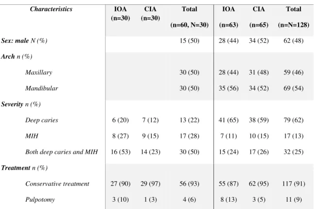
Table 1. Patient characteristics
Split-mouth RCT Parallel-arm RCT Characteristics IOA (n=30) CIA (n=30) Total (n=60, N=30) IOA (n=63) CIA (n=65) Total (n=N=128) Sex: male N (%) 15 (50) 28 (44) 34 (52) 62 (48) Arch n (%) Maxillary Mandibular 30 (50) 30 (50) 28 (44) 35 (56) 31 (48) 34 (52) 59 (46) 69 (54) Severity n (%) Deep caries 6 (20) 7 (12) 13 (22) 41 (65) 38 (59) 79 (62) MIH 8 (27) 9 (15) 17 (28) 7 (11) 10 (15) 17 (13)
Both deep caries and MIH 16 (53) 14 (23) 30 (50) 15 (24) 17 (26) 32 (25)
Treatment n (%) Conservative treatment Pulpotomy 27 (90) 3 (10) 29 (97) 1 (3) 56 (93) 4 (6) 55 (87) 8 (13) 62 (95) 3 (5) 117 (91) 11 (9) RCT: randomized controlled trial; IOA: intraosseous anaesthesia; CIA: conventional infiltration anaesthesia; MIH: molar-incisor hypomineralisation, moderate to high severity lesion; N: number of patients; n: number of teeth
Accepted
Article
Table 2. Main results for split-mouth RCT and parallel-arm RCT
Split-mouth RCT Parallel-arm RCT Mean±SD Mean difference (95% CI) p value Mean±SD Mean difference (95% CI) p value
Mean VAS score during anaesthesia, cm IOA: 0.73 ± 1.31 CIA: 1.43 ± 1.45 -0.70 (-1.44 to 0.04) 0.06 IOA: 1.17 ± 1.40 CIA: 1.86 ± 1.81 -0.69 (-1.25 to -0.12) 0.02 Latency, min IOA: 1.07 ± 0.25 CIA: 2.83 ± 2.64 -1.77 (-2.77 to -0.77) 0.001 IOA: 1.63 ± 0.97 CIA: 3.08 ± 2.11 -1.44 (-2.02 to -0.87) <0.0001 Need for additional anaesthesia during treatment IOA: 2 cases CIA: 2 cases 1.00* (0.15 to 6.74) 1.00 IOA: 7 cases CIA: 11 cases 0.61* (0.22 to 1.70) 0.34
Mean VAS score during treatment, cm IOA: 1.07 ± 1.76 CIA: 0.53 ± 0.82 0.53 (-0.19 to 1.25) 0.14 IOA: 0.90 ± 1.51 CIA: 0.88 ± 1.64 0.03 (-0.52 to 0.58) 0.92
RCT: randomized controlled trial IOA: intraosseous anaesthesia
CIA: conventional infiltration anaesthesia CI: confidence interval
VAS: visual analog scale * Odds ratio
Accepted
Article
Table 3. Main results for combined analysis of split-mouth and parallel-arm RCTs
Mean difference (95% CI) p value
VAS score during anaesthesia, cm -0.69 (-1.13 to -0.25) 0.002
Latency, min -1.52 (-2.02 to -1.03) <0.0001
Need for additional anaesthesia during treatment 0.68* (0.28 to 1.68) 0.41
VAS score during treatment, cm 0.22 (-0.22 to 0.65) 0.33
In the combined analysis, we combined the two effect estimates of the split-mouth and parallel-arm RCTs by using an inverse-variance weighting meta-analysis approach: for each outcome, the combined effect estimate is a weighted average of the two effect estimates, each effect estimate being weighted by the inverse of its variance
CI: confidence interval VAS: visual analog scale
*Data are mean differences for IA compared to CIA, except for the need for additional anaesthesia during treatment, the effect measure is the Odds ratio for IA compared with CIA
Accepted
Article
Figure legends
Fig. 1. Flow of participants in the trial