HAL Id: tel-03216741
https://tel.archives-ouvertes.fr/tel-03216741
Submitted on 4 May 2021HAL is a multi-disciplinary open access archive for the deposit and dissemination of sci-entific research documents, whether they are pub-lished or not. The documents may come from teaching and research institutions in France or abroad, or from public or private research centers.
L’archive ouverte pluridisciplinaire HAL, est destinée au dépôt et à la diffusion de documents scientifiques de niveau recherche, publiés ou non, émanant des établissements d’enseignement et de recherche français ou étrangers, des laboratoires publics ou privés.
Regulation of the Akt/mTORC1 Pathway by HIV
Transcriptional Activator Tat in B Cells
Burkitkan Akbay
To cite this version:
Burkitkan Akbay. Regulation of the Akt/mTORC1 Pathway by HIV Transcriptional Activator Tat in B Cells. Cancer. Université Paris-Saclay; Al-Farabi Kazakh National University (Almaty, Kaza-khstan), 2021. English. �NNT : 2021UPASL026�. �tel-03216741�
Regulation of the Akt/mTORC1 pathway by HIV
transcriptional activator Tat in B cells
Régulation de la voie Akt / mTORC1 par l'activateur
transcriptionnel du VIH Tat dans les cellules B
Thèse de doctorat en cotutelle de l'Université Paris-Saclay et
Al-Farabi Kazakh National Université préparée à l'Université
Paris-Saclay
École doctorale n°582 : cancérologie : biologie - médecine – santé (CBMS)Spécialité de doctorat: Aspects moléculaires et cellulaires de la Biologie Unité de recherche : Université Paris-Saclay, CNRS, Institut Gustave Roussy, Aspects métaboliques et systémiques de l'oncogénèse pour de nouvelles approches thérapeutiques, 94805, Villejuif, France Référent : Faculté de médecine
Thèse présentée et soutenue en Visio conférence totale, le 29 Mars
2021, par
Burkitkan AKBAY
Composition du Jury
Murat SAPARBAEV
Directeur de Recherche, CNRS, Université Paris-Saclay
Président Valery FILONENKO
Professeur, National Academy of Sciences of Ukraine
Rapporteur Isabelle VERGNE
Chargée de Recherche, CNRS Rapporteur Audrey ESCLATINE
Directrice de Recherche, CNRS, Université Paris-Saclay
Examinatrice David BOUTBOUL
Maitre de conférence, Hôpital Saint-Louis Examinateur
Direction de la thèse
Yegor VASSETZKY Directeur de Recherche, CNRS Svetlana DOKUDOVSKAYA Directrice de Recherche, CNRS Directeur de thèse Co-directrice de thèse Amangeldy BISSENBAEVProfesseur, Al-Farabi Kazakh National University Co-directeur de thèse
Thèse de
doctorat
NNT : 2 02 1U PA SL0 261
Acknowledgements
I cannot begin to express my deepest appreciation to my esteemed supervisors, Dr. Svetlana Dokudovskaya, Dr. Yegor Vassetzky and Pr. Amangeldy Bissenbaev. I am extremely grateful to them for their invaluable supervision, unwavering guidance and continuous support. They showed profound belief in my abilities. Their immense knowledge and plentiful experience have encouraged me in all the time of my thesis project. They taught me to be professional, to be critical, and to be scientific wise. I truly learned from them how to be a scientist. I’m deeply indebted to Dr Svetlana Dokudovskaya for her patience which I appreciated throughout my thesis especially during the long processes of writing papers and thesis. She transmitted to me her professional passion and her knowledge in various fields. I am very proud and satisfied to be a doctoral student in her team. I am deeply thankful to Dr Yegor Vassetzky who is the first person I contacted and gave me the opportunity to start my doctoral study life in the team. I would also like to extend my deepest gratitude to Dr. Diego Germini for his practical suggestions and helpful advices. I thank them for their wise advices and insightful suggestions in difficult times and for guiding me throughout this thesis. Without their persistent help, the goal of my thesis would not have been realized.
I would like to express my deepest appreciation to the jury members who accepted to witness and assess my work. I wish to show my gratitude to Dr. Valery Filonenko and Dr. Isabelle Vergne for their time and energy contributed to read and examine my thesis manuscript. I am extremely thankful for their constructive suggestions. I would also like to extend my sincere thanks to Dr. Murat Saparbaev who accepted to be the examinator and the president of the jury. I am also grateful to Dr. Audrey Esclatine and Dr. David Boutboul who accepted to be the examinators. Special thanks to Dr. Audrey Esclatine who was also the member of “le comité de suivi individuel du doctorant” to assess the progress of my thesis, where she gave her valuable suggestions and advices.
I also had great pleasure of working with Dr. Eugene Sheval and Dr. Yana Musinova for the fruitful collaborations which gave us the opportunity to generate the cell lines that were further validated and used throughout my thesis.
I would also express my gratitude to the French Embassy in Kazakhstan and Campus France in Paris for their support and funding. My gratitude extends to l’Etablissement Français du Sang - Hôpital Saint Louis for facilitating our study.
I would also like to express my gratitude to all the members of previous UMR 8126 and current UMR 9018 - Aspects métaboliques et systémiques de l’oncogenèse pour de nouvelles approches thérapeutiques (METSY), a research unit rich in themes and nationalities. I’m proud to be the part of this research unit. I
2
especially thank each person from the " Dynamique de la chromatine et métabolisme dans le cancer" team, most notably Alberto, Yingxing, Fabi, and Reynand, with whom I spent beautiful times in the lab which will remain as a beautiful memory forever.
Finally, I am extremely grateful to my parents. Definitely words will never be enough to express my thanks to them. Thank you for many years of unconditional support, trust, and love. Since my childhood you have given me the opportunity, at any cost, to make my dream come true, even if you were living a harsh nomadic life in the steppe. Despite all the difficulties you have always made my study life easier for me. It will never be possible for me to return everything that you have offered to me. Thank you for my sister Zhaynakhan, and my brother Kenzhebek for helping me, believing in me and always pushing me for the best, with their support, their sacrifices, and their prayers. My dear brother Kenzhebek thank you very much for looking after our parents, which I couldn’t be able to do since I am away from you all for so many years to pursue my dream of studying.
I wish to acknowledge the support and great love of my wife, Gulgina, who has been taking care of my two cute sons, Rassul and Amire. Even when I am away from you all, you stayed alone to take care of them without any complains, even though you faced a lot of difficulties. Hope we will go through life adventures together in the future. Hope our children stay healthy. We will do all to our children’s future as our parents did to us. I do believe that we have a bright future.
3
Synthèse
Malgré l’avènement de la trithérapie antirétrovirale (ARV), l'incidence des lymphomes agressifs à cellules B reste élevée chez les personnes vivantes avec le VIH-1 (PVVIH), ces lymphomes constituant ainsi une des principales causes de décès chez ces patients. Pourtant, les cellules B ne sont pas la cible d’infection par le virus et les mécanismes exacts du développement de ces lymphomes ne sont pas encore totalement connus. Plusieurs hypothèses ont cependant été proposées pour expliquer les mécanismes de la lymphomagenèse chez les PVVIH: 1) les lymphocytes T infectés par le VIH-1 et qui échappent à l'élimination dans les centres germinatifs pourraient provoquer une expansion inhabituelle des cellules B; 2) des hypermutations somatiques aberrantes sur les gènes d'immunoglobuline pourraient favoriser la survenue de translocations chromosomiques conduisant à une transformation maligne; 3) une inflammation chronique pourrait être le lit d’une lymphoprolifération. Une autre hypothèse sur le processus de lymphomagenèse B chez les PVVIH est l’implication directe des protéines du VIH-1 qui peuvent être sécrétées par les cellules infectées. Parmi elles, nous avons la protéine Tat qui, après sécrétion par les cellules infectées, peut pénétrer dans d'autres cellules non infectées, y compris les cellules B.
Tat est un activateur transcriptionnel du VIH-1. C'est l'une des premières protéines virales à être exprimée juste après l'infection. Grâce à sa taille et de ses structures telles que le domaine de transduction protéique (PTD) et le signal de localisation nucléaire (NLS), il peut pénétrer dans les cellules voisines non infectées. Par conséquent, Tat est impliqué dans l’oncogenèse de nombreux cancers incluant les cancers colorectaux, les cancers du col de l’utérus, le sarcome de Kaposi associé au SIDA et les lymphomes à cellules B. Les mécanismes exacts de l'oncogenèse induite par Tat, en particulier celle des lymphomes à cellules B, restent cependant à être élucidés. Des études antérieures de notre équipe ont montré d’une part, que dans les cellules B, Tat induisaitt une production d’espèces réactives de l’oxygène (ROS), augmentant ainsi les dommages oxydatives à l'ADN, source d’instabilité génomique. D’autre part, Tat favorisait également dans les cellules B, une proximité les loci MYC et IGH (normalement situés dans des compartiments nucléaires différents) et la surexpression du gène AICDA (activation-induced cytidine deaminase), ce qui conduit vraisemblablement à la translocation oncogènique t(8 ;14) entre MYC et IGH caractéristique du lymphome de Burkitt. Cependant, la manière dont ces événements sont liés n'est pas bien comprise.
Parmi les voies de signalisations cellulaires, la voie AKT/mTORC1 apparaît comme un intégrateur central de nombreux signaux intra et extracellulaires, y compris dans l'infection virale et les dommages à l'ADN. Des études ont montré que le VIH-1 et ses protéines Tat, Env et Nef pouvaient réguler la voie AKT/mTORC1. En revanche, à notre connaissance, aucune étude ne s’est encore intéressée à la régulation de la voie AKT / mTORC1 par Tat dans les cellules B.
4
Compte tenu de l'importance de la voie de AKT / mTORC1 dans l'oncogenèse et en combinant les résultats des travaux précédents de notre équipe sur Tat, nous avons émis l'hypothèse selon laquelle la protéine Tat du VIH-1 pourrait induire dans les cellules B les effets oncogènes sus-mentionnés en modulant la voie de signalisation AKT / mTORC1. Nous nous sommes essentiellement focalisés sur les mécanismes de régulation de l’expression du gène AICDA par Tat non seulement parce que son expression est associée à la voie mTORC1, mais aussi parce que Tat induit une expression aberrante d'AICDA dans les cellules B qui est étroitement associée à la lymphomagenèse B.
Une grande partie de ma thèse portait sur la régulation de la voie AKT / mTORC1 par Tat dans les cellules B. Nous avons utilisé différentes lignées cellulaires B : i) des cellules B primaires issues de donneurs sains traitées ou pas avec une protéine Tat purifiée ; ii) des cellules d’une lignée lymphoblastoïdes RPMI8866 traitées ou pas avec une protéine Tat purifiée ; et iii) des cellules de RPMI8866 exprimant la protéine Tat de façon stable mais inductible par la doxycycline. Cette dernière lignée a été développée avec nos collaborateurs et une procédure détaillée décrivant la construction de cette lignée cellulaire est publiée dans le Russian Journal of Developmental Biology. En utilisant ces différents modèles cellulaires, nous avons étudié en présence et en l’absence de Tat, l’activation de la voie AKT/mTORC1, l’autophagie, les dommages d’ADN et la production de ROS dans les cellules B. Nous avons ensuite analysé, en fonction du profil d’activation de la voie mTORC1 par Tat, l'expression du gène AICDA en présence et en l'absence de ses activateurs et répresseurs transcriptionnels.. Tous les résultats obtenus sont décrits en détail dans notre article publié dans le numéro spécial «mTOR Signalling network in Cell Biology and Disease» du Journal of International Molecular Sciences.
Nos résultats montrent, en présence de Tat, une activation de la voie Akt / mTORC1, conséquence des dommages à l'ADN induits par Tat via la production de ROS. mTORC1 activé va à son tour inhiber l'expression de cMYB et E2F8, des inhibiteurs transcriptionnels de l'AICDA, conduisant ainsi à sa surexpression. Tat semble aussi inhiber l'autophagie, cependant, cette régulation de l'autophagie devra être explorée plus en détail.
En conclusion, la protéine Tat libérée dans le milieu extracellulaire par les cellules infectées, pénètre dans les cellules B et y induit la production de ROS conduisant à des dommages à l'ADN, qui sont en outre détectés par la voie AKT / mTORC1. Une fois activé, mTORC1 inhibe l'expression des répresseurs transcriptionnels d’AICDA, Cmyb et E2F8, favorisant ainsi la surexpression d'AICDA. Ces perturbations peuvent finalement conduire à une instabilité génomique accrue qui constitue une des principales caractéristiques de l’oncogenèse, y compris dans les cellules B.
5
Table of Contents
Introduction ... 13 1. Viral infection ... 13 1.1. Attachment ... 14 1.2. Entry ... 141.3. Replication, Assembly and Release ... 14
2. Human immunodeficiency virus type - 1 ... 17
2.1. Structure of the HIV-1 virion ... 17
2.2. Life cycle of HIV-1 ... 18
3. HIV-1-Tat ... 21
3.1. Structure of Tat... 21
3.2. Functions of Tat ... 22
3.3. Tat in HIV-1 transcription ... 22
3.4. Tat in host cell gene expression ... 22
3.5. Tat secretion and penetration in bystander cells ... 24
4. HIV-associated cancers and related diseases ... 25
5. The mammalian target of rapamycin complex ... 28
5.1. Upstream regulation of mTORC1 ... 30
5.1.1. PI3K-AKT signaling to mTORC1 ... 30
5.1.2. Amino acid signaling to mTORC1 ... 32
5.1.3. DNA damage response and mTORC1 ... 35
5.2. Downstream targets of mTORC1 ... 39
5.3. mTORC1 and the Immune System ... 41
5.3.1. mTORC1 in the innate immunity ... 41
5.3.2. mTORC1 in adaptive immunity ... 43
5.3.2.1. mTORC1 in T lymphocytes ... 44
5.3.2.2. mTORC1 in B lymphocytes ... 45
5.4. mTORC1 and viruses ... 48
5.4.1. Regulation of mTORC1 by HIV-1 ... 49
5.4.2. Regulation of Autophagy by HIV-1 ... 51
5.4.3. mTORC1 in HIV-1 Latency ... 52
6
Materials and Methods ... 74
1. Isolation of the peripheral blood mononuclear cells ... 74
2. Purification of B lymphocytes ... 74
3. Cell lines ... 74
4. Tat treatment and expression induction ... 75
5. Inhibition of AKT/mTORC1 pathway by MK-2206 and Rapamycin treatments ... 75
6. Western Blot ... 76
7. Monitoring ROS production ... 76
8. qRT-PCR ... 77
9. DNA damage detection with immunofluorescence... 78
10. Statistical analyses ... 79
Results ... 80
1. Generation of B cell lines providing inducible and constant Tat expression ... 80
2. HIV-1 Tat activates AKT/mTORC1 pathway and AICDA expression by downregulating its transcriptional inhibitors in B cells ... 87
2.1. Increased AICDA expression by Tat is not prevented by AKT inhibition ... 103
2.2. Exogenous Tat differently regulates downstream targets of mTORC1, p70S6K and 4E-BP1 ... 104
2.3. HIV-Tat inhibits starvation-induced autophagy in B cells ... 105
Discussion ... 106
Conclusions and perspectives ... 110
Conferences and Publications ... 111
1. Conferences ... 111
2. Publications ... 111
7
List of abbreviations
ICTV International Committee on Taxonomy of Viruses
4E-BP1 Eukaryotic translation initiation factor 4E (eIF4E)-binding protein 1 5’UTR 5’ untranslated region
aa Amino acid
ADE Antibody‐dependent enhancement AICDA Activation-induced cytidine deaminase AIDS Acquired Immunodeficiency Syndrome AMPK AMP-activated protein kinase
ARL AIDS-related lymphomas
ART Antiretroviral therapy
ATF4 Activating transcription factor 4 ATG13 Autophagy-related 13
ATM Ataxia telangiectasia mutated
ATR Ataxia telangiectasia- and RAD3-related ATRIP ATR-interacting protein
BACE1-AS BACE1‐antisense transcript
BCR B cell receptor
BER Base excision repair
BL Burkitt’s lymphomas
CAD Carbamoyl-phosphate synthetase 2, aspartate transcarbamylase, dihydroorotase cART Combination antiretroviral therapy
CASTOR1 Cellular Arginine Sensor for mTORC1 1 CASTOR2 Cellular Arginine Sensor for mTORC1 2 CCR5 C-C chemokine receptor type 5
CDK Cyclin-dependent kinase
CDK9 Cycline-dependent kinase 9 Chk1 Checkpoint kinase 1 Chk2 Checkpoint kinase 2
CREB CAMP-responsive element–binding protein
CTL Cytolytic T lymphocytes
CXCR4 C-X-C chemokine receptor type 4
CycT1 Cyclin T1
DC Dendritic cell
DDR DNA damage response
DEPDC5 DEP domain-containing 5
8 DLBCL Diffuse large B cell lymphomas DNA-PK DNA-dependent protein kinase
DSB Double strand break
dsDNA Double stranded DNA E2F3 E2F transcription factor 3
EBV Epstein–Barr virus
env Envelope
ESCRT Endosomal sorting complexes required for transport FAT Focal adhesion targeting
FAT FRAP-ATM-TRRAP
FATC FAT carboxy-terminal
FIP200 Focal adhesion kinase family interacting protein of 200kDa
FLCN Folliculin
FNIP FLCN-interacting protein
FOXO Forkhead family of transcription factors
FRAP FK506-binding protein-rapamycin-associated protein
FRB FKBP-rapamycin-binding
gag Group-specific antigen GAP GTPase-activating protein
GEF Guanine nucleotide exchange factor gp120 Glycoprotein 120
gp41 Glycoprotein 41
GSK3 Glycogen synthase kinase 3 HAART Highly active antiretroviral therapy HAND HIV-associated neurocognitive disorder
HBV Hepatitis B virus
HCV Hepatitis C virus
HHV8 Human herpesvirus 8
HIV-1 Human immunodeficiency virus type-1 HIVAN HIV-associated nephropathy
HL Hodgkin’s lymphomas
HPV Human papillomavirus
HR Homologous recombination
HSC Hematopoietic stem cells
hSMG-1 Human suppressor of morphogenesis in genitalia 1 HSPGs Heparan sulfate proteoglycans
IL-1b Interleukin 1 beta IL-6 Interleukin 6
9
KS Kaposi’s sarcoma
LEL Late endosomes/lysosomes
LITAF lncRNA
Lipopolysaccharide-induced tumor necrosis factor Long non-coding RNA
LRP Lipoprotein receptor-related protein
LRS Leucyl-tRNA synthetase
LTR Long terminal repeats
MA Matrix
MCD Multicentric Castleman’s disease MDM2 Mouse double minute 2 homolog MHC Major histocompatibility complex
MIOS Meiosis regulator for oocyte development MITF Microphthalmia transcription factor mLST8 Mammalian lethal with sec-13 protein 8
MMR Mismatch repair
MRN Mre11-Rad50-Nbs1
mSin1 Mammalian stress-activated map kinase-interacting protein 1
MTHFD2 Mitochondrial tetrahydrofolate cycle enzyme methylenetetrahydrofolate dehydrogenase 2
mTOR Mammalian TOR
mTORC Mammalian target of rapamycin complex
mTORC1 mTOR complex 1
mTORC2 mTOR complex 2
NC Nucleocapsid
nef Negative regulating factor NER Nucleotide excision repair NF-κB Nuclear factor of κB
NHEJ Non-homologous end joining
NHL Non-Hodgkin’s lymphomas
NK Natural killer
NLS Nuclear localization signal
NPRL2 Nitrogen permease regulator-like 2 NPRL3 Nitrogen permease regulator-like 3 Nrf2 Nuclear factor (erythroid-derived 2)-like 2 p70S6K 70 kDa ribosomal protein S6 kinase 1
PBL Plasmablastic lymphoma
PBMC Peripheral blood mononuclear cells PCNSL Primary central nervous system lymphoma PDK1 3-phosphoinositide-dependent kinase 1
10
PEL Primary effusion lymphoma
PI3K Phosphatidylinositol 3-kinase
PIKK Phospatidylinositol 3-kinase related kinases PIP2 Phosphatidylinositol 4,5-bisphosphate PIP3 Phosphatidylinositol-3,4,5-trisphosphate
PKC Protein kinase C
pol Polymerase
PP2 Protein phosphatase 2
PR Protease
PRAS40 Proline-rich AKT substrate 40 kDa
PRD PIKK-regulatory domain
Protor Protein observed with rictor PTD Protein transduction domain PTEN Phosphatase and tensin homolog
RAFT Rapamycin and FK506-binding protein target
RAPT Rapamycin target
Raptor Regulatory-associated protein of mammalian target of rapamycin
RBD RNA binding domain
rev RNA splicing-regulator RGD Arginine-glycine-aspartic acid RHEB Ras homologue enriched in brain
Rictor Rapamycin-insensitive companion of mTOR ROS Reactive oxygen species
RT Reverse transcriptase
RTK Receptor tyrosine kinases
S6K1 Ribosomal protein S6 kinase beta-1
SAMTOR S-adenosylmethionine sensor upstream of mTORC1 SEH1L Seh1 like nucleoporin
Ser Serine
SGK1 Serum- and glucocorticoid-induced protein kinase 1
SHM Somatic hyper mutation
SMAD2 SMAD Family Member 2
SREBP1/2 Sterol regulatory element binding protein 1/2
SSB Single strand break
Superoxide
Dismutase 2 SOD2
TAR Transactivation response tat Transactivator of tanscription
11 TFE3 Transcription factor E3
TFEB Transcription factor EB Tfh Follicular helper T cells
Th T helper
TNF-β Tumor necrosis factor beta
TopBP1 Topoisomerase II Binding Protein 1
TOR1 Target of rapamycin 1
TOR2 Target of rapamycin 2
TOS TOR signaling
Treg Regulatory T cell
TRRAP Transformation/transcription associated protein TSC Tuberous Sclerosis Complex
Twist1 Twist-related protein 1
ULK1 Unc-51-like autophagy-activating kinase 1 UNG Uracil nucleoside glycosylase
UPR Unfolded protein response vif Viral infectivity factor
vpr Virus protein r
vpu Virus protein unique
vRNA Viral RNA
WDR24 WD repeat domain 24 WDR59 WD repeat domain 59
12
List of Figures
Figure 1 Basic structure of a virus
Figure 2 Viral attachment and entry into the host cells Figure 3 Viral replication, assembly and release Figure 4 Structure of HIV-1 virion
Figure 5 Life cycle of HIV-1
Figure 6 Structure of HIV-1 Tat protein
Figure 7 Tat regulates gene expression of host and bystander cells Figure 8 HIV-associated cancers and other diseases
Figure 9 Mechanisms of development of AIDS-related B cell lymphomas Figure 10 Structure of mTOR protein and its complexes
Figure 11 PI3K-AKT signaling pathway Figure 12 Activation of the small GTPases
Figure 13 Regulation of mTORC1 by growth factors, cytokines and amino acids Figure 14 DNA damage signaling
Figure 15 Regulation of mTORC1 by DNA damage Figure 16 Downstream targets of mTORC1
Figure 17 Role of mTORC1 in the innate immunity Figure 18 mTORC1 in T lymphocytes
Figure 19 Function of mTORC1 in B cells Figure 20 Regulation of mTORC1 by HIV-1
Figure 21 Implication of various HIV-1 proteins in modulation of autophagy in different cells Figure 22 HIV-1 latency
Figure 23 Treatment with AKT/mTORC1 inhibitors as a promising approach for HIV-1 therapy Figure 24 Inhibition of AKT by its specific inhibitor MK-2206
Figure 25 Inhibition of AKT was not able to prevent AICDA overexpression by Tat
Figure 26 P70S6K and 4E-BP1 are differently regulated by exogenous Tat treatment in B cells Figure 27 Tat inhibits autophagy induction by amino acid starvation
List of Tables
Table 1 AIDS-related lymphomas and associated viruses Table 2 List of primers used for qRT-PCR
13
Introduction
1.
Viral infection
Discovery of viruses goes back to late nineteenth and early twentieth century with yellow fever virus known to be the first that infect humans (1,2). Since then more than 200 human viruses have been identified with a rate of three to four each year (1) with SARS-Cov-2 being the latest one threatening the globe.
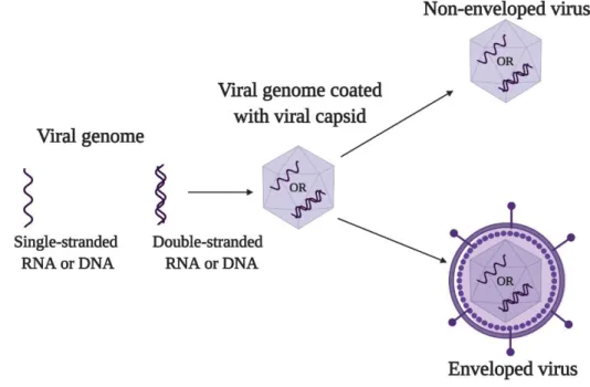
An infectious viral particle or virion is a fully assembled virus (Figure 1), which consists of genetic material (RNA or DNA) protected by a protein coat, capsid, and often (but not always) an outer membrane, envelope, which are typically derived from proteins and phospholipid membranes of the host cell, but may also contain other viral glycoproteins. Proteins constructing the viral particle are called structural proteins. The main function of capsid and envelope is protection which enables the virus to be stable in various extra-cellular environments. Capsid and envelope also help the virus to enter and deliver its genome into the host cell. There are other proteins known as non-structural or regulatory proteins that control the expression of viral genome and modulate host immune responses (3–6).
Figure 1. Basic structure of a virus: an infectious viral particle consists of viral genome (single- or double-stranded RNA or DNA) protected by the capsid, which can be non-enveloped or enveloped.
14
Viruses are named and classified by the International Committee on Taxonomy of Viruses (ICTV)
(https://talk.ictvonline.org/). Due to the extreme diversity and complexity of viruses, their classification or
grouping at different hierarchical levels of order, family, subfamily, genus, and species are based on a variety of parameters such as presence or absence of an envelope, structure of capsids, morphology, mode of replication and type of viral genome (DNA or RNA, linear or circular, single- (ss) or double-stranded (ds), positive or negative ssRNA, ssRNA requiring a DNA intermediate or dsDNA requiring an RNA intermediate) (5–8).
In general, most viral infections include attachment, entry, replication, assembly, and release cycle (Figure 2, Figure 3). Viruses must first recognize and attach to their host cells. After successful attachment viruses penetrate the cell membrane, upon which they copy their genome and make their own proteins inside the host cells. In order to survive within the host, viruses must escape from the destruction caused by host immune system so that they can infect innate immune cells or affect other cells (bystander cells).
1.1. Attachment
Many viruses use glycoproteins on the surface of the viral membrane or certain sites of viral capsid to bind to the molecules on the host cell called viral receptors (Figure 2). The receptors on cell surfaces are the molecules that normally have their own physiological functions. Viruses can have one single receptor or several receptors. As interactions between virus and receptors are specific, presence of receptors always determines which cell types can be infected. Receptors further facilitate various processes of viral entry by activating certain signaling pathways within the host cells or by inducing structural changes in the viral attachment proteins (9–11).
1.2. Entry
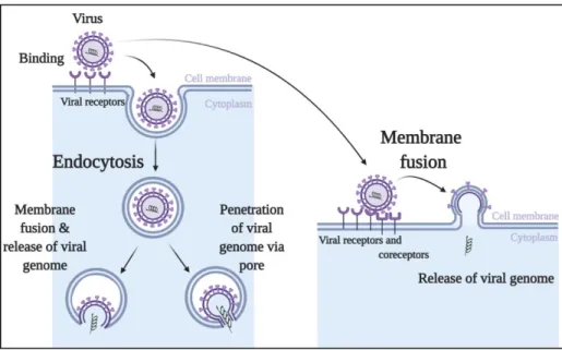
Viral entry is a process through which virus gains access to the sites of replication. Many viruses pass through the cellular membrane via either a process called endocytosis with further membrane fusion or via direct membrane fusion, while some other viruses (non-enveloped) enter the cells through a pore formed with viral capsid and cellular membrane after endocytosis (Figure 2) (12–16). Once inside of the host cells’ cytoplasm, the viral capsid is degraded. The viral genetic material become available for further trafficking in order to be replicated, transcribed, translated, thus providing conditions for production of new virions.
1.3. Replication, Assembly and Release
The replication allows the virus to generate enough copies of viral genome to make new virions (Figure 3), which is essential for viral survival and infection of new hosts. The replication mechanism is
15
dependent on the type of viral genome. Viruses keep their genome either in the form of RNA or DNA. There are two ways of replication for RNA viruses: either RNA-dependent RNA synthesis or RNA-dependent DNA synthesis. In both cases, RNA viruses must encode their own polymerases and other necessary enzymes. In the RNA-dependent RNA synthesis process, RNA viruses first make a complimentary RNA (cRNA) which is then used as template to make new viral RNA (vRNA) copies.
Viral mRNA is further transcribed with the help of viral polymerase in coordination with cellular RNA polymerase. In the RNA-dependent DNA synthesis process, virus use an enzyme called reverse transcriptase that converts viral RNA into viral DNA which is integrated into the host genome with the help of another enzyme called integrase. The integrated virus genome is then transcribed as a cellular gene. DNA viruses benefit from replication and transcription strategies of host cells.
Following replication and transcription, viral mRNA is exported to the cytoplasm to make viral proteins by the translational machinery of the host cell. Newly synthesized viral proteins with nuclear localization signal are imported to the nucleus to assist in the vRNA replication and viral mRNA transcription. (Figure 3). Some viruses replicate their genome in the nucleus while others - in the cytoplasm of the host cells. Viruses replicating in the nucleus transport their genome to the nucleus via nuclear pore complex, using host nuclear import machinery. Viruses replicating in the cytoplasm are assisted by organelle-like compartments: single/double membrane vesicles and invaginations formed by cellular membrane rearrangements (17–20).
Figure 2. Viral attachment and entry into host cells. Viruses bind the receptors and coreceptors expressed on the host cell surfaces. Successful binding allows the viruses to enter the cells through the process of endocytosis followed by either further membrane fusion or formation of membrane pore (in the case of non-enveloped viruses) and direct membrane fusion. Once inside of the host cells’ cytoplasm, the viral capsid is degraded and viral genetic material is released. The processes of viral replication, assembly and release of new virions start.
16
Replication and transcription of viral genome, followed by translation of viral mRNAs to produce proteins, provide all necessary components for assembly. Packaging signals or certain proteins that interact with viral genome with high specificity allow viral genome packaging into the capsid which in the case of enveloped viruses is further surrounded by an envelope (21–23). Assembled new virions are released from the host cells to infect new ones.
New virions are released from their specific host cells to be able to infect neighboring cells or new hosts and repeat the replication cycle (Figure 3). Some viruses are released after killing the host cells, while other viruses leave infected cells by budding through the membrane without killing the cells (20,23).
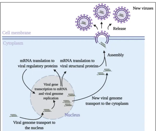
Figure 3. Viral replication, assembly and release. Following entry, viral genetic material in the cytoplasm is transported to the nucleus through host nuclear import machinery. With the help of host RNA polymerases and by synthesizing their own necessary enzymes, viruses produce new viral mRNA that is used to make structural proteins and regulatory proteins. Regulatory proteins translocate to the nucleus to further assist viral genome replication and synthesized viral genome is then transported to the cytoplasm, where the assembly process begins with viral genome packaging into viral capsid wrapped with an envelope in the case of enveloped virus. Finally, assembled new virions are released to infect new cells.
17
2.
Human immunodeficiency virus type - 1
Human immunodeficiency virus type-1 (HIV-1) is the cause of the Acquired Immunodeficiency Syndrome (AIDS). HIV-1 was first isolated by research groups from Pasteur Institute in France and National Cancer Institute in USA in 1983 (24). HIV-1 belongs to the Lentivirus genus within the family of Retroviridae and subfamily Orthoretrovirinae. HIV-1 was transmitted to the human population from Central African chimpanzees in around 1920 (25,26). Since then more than 30 million people have died of AIDS. As of 2019, around 38 million people are living with HIV-1 and 1.7 million new infections occur annually in the world (UNIAIDS, 2020), still remaining as a major health burden.
2.1. Structure of the HIV-1 virion
HIV-1 contains two positive-sense single strand RNAs encapsulated in a cone shaped capsid surrounded by an envelope. Also enclosed within the capsid are reverse transcriptase, integrase, and protease. The viral envelope contains an external surface glycoprotein (gp120) and a transmembrane glycoprotein (gp41) (Figure 4). HIV-1 genome is around 9.7 kB, flanked by a repeated sequence called long terminal repeats (LTR) at 5’ and 3’ ends. These LTRs are actively involved in the integration of viral genome into host chromosome and also serve as a promoter for the transcription of proviral DNA. HIV-1 genome has nine genes which code for the following 15 proteins (27,28) (Figure 4) :
Genes coding for structural proteins:
gag (group-specific antigen) : codes for matrix protein (MA, p17), the capsid protein (CA, p24), and the nucleocapsid proteins (NC, p6 and p7).
pol (polymerase): codes for reverse transcriptase (RT), protease (PR) and integrase (IN). env (envelope): codes for gp160, which is the precursor of the gp120 and gp41 proteins.
Genes coding for regulatory proteins:
tat (transactivator of tanscription): codes for Tat protein rev (RNA splicing-regulator): codes for Rev protein
Genes coding for accessory proteins:
nef (negative regulating factor): codes for Nef protein vif (viral infectivity factor): codes for Vif protein vpr (virus protein r): codes for Vpr protein vpu (virus protein unique): codes for Vpu protein
18
Figure 4. Structure of HIV-1 virion. HIV-1 virion consists of two single strand RNAs which are encapsulated in a capsid along with integrase, protease and reverse transcriptase. Matrix layer targets the RNA to the plasma membrane and mediates incorporation of envelope containing external surface glycoprotein (gp120) and a transmembrane glycoprotein (gp41). HIV-1 genome (9.7 kB) consists of 9 genes flanked by LTR (long terminal repeat) sequences at 5’ and 3’ ends.
2.2. Life cycle of HIV-1
Like many other viruses after infection HIV-1 viral particles proceed through the stages of attachment, entry, transcription, translation, assembly and release, with two more extra steps characteristic of the Retroviridae viruses family - reverse transcription and integration – to complete a life cycle (Figure 5).
Main targets of HIV-1 are T cells, macrophages and dendritic cells (DCs), because they express viral receptors essential for HIV-1 attachment (29,30). Envelope protein gp120 first binds to cluster of differentiation 4 (CD4) receptors, which causes a structural change in gp120 generating a binding site either for the C-C chemokine receptor type 5 (CCR5) or for the C-X-C chemokine receptor type 4 (CXCR4) co-receptors. Binding of the co-receptor causes further structural changes in gp41 protein, which triggers the
19
viral entry process (31,32). A series of conformational changes in gp41 protein caused by the trimeric complex of gp120/CD4/CCR5 or by CXCR4 expose the fusion domain on the N-terminus of gp41, which triggers the direct fusion of viral and cellular membranes and destabilizing the host cell membrane (32–34). Although main entry of HIV-1 is mediated by CD4/CCR5 or CXCR4, CD4/co-receptor independent entry into other host cells such as renal epithelial cells and astrocytes also takes place, which can occur either through direct cell-cell interaction or through certain receptors like C-type lectin DEC-205 and human mannose receptor (35–38).
After successful entry, the capsid disassembles (a process known as uncoating), single stranded viral RNA is released into the cytoplasm where it is converted into double stranded DNA (dsDNA) by the reverse transcriptase. Uncoating probably occurs in the cytoplasm in coordination with reverse transcription or at the nuclear envelope during nuclear import (39). Subsequently viral dsDNA uses the host nuclear import machinery to move to the host cell nucleus, where it integrates into the host DNA with the help of an integrase coded by HIV-1. Integrated viral DNA can also remain latent, without making new copies of HIV-1, which is also forming a barrier for successful treatment of HIV-1 infected patients (40–44). Remarkably, recent studies revealed that intact viral cores can enter to the nucleus and uncoat just before the integration to their chromosomal integration sites (39,45). Integrated dsDNA, also called proviral DNA, continues to transcribe the viral genome and encode necessary viral proteins.
Integrated proviral viral DNA uses host transcriptional machinery along with viral regulatory proteins to synthetize RNA copies of HIV-1 genome and viral mRNA, which is subsequently translated by host translational machinery into viral proteins. Transcription of the viral genome starts from 5′ LTR region of the proviral DNA that acts as promoter which has binding sites for host transcription factors. Host transcription factors, however, mostly lead to the generation of short transcripts. Therefore, HIV-1 needs a viral protein called Tat (transactivator of transcription) that binds to transactivation response (TAR) RNA element which is located at the 5’ end of all nascent viral transcripts and robustly increases the efficiency of polymerase II to make full-length viral transcripts (46).
After splicing three types of transcripts are formed: unspliced RNA, partially spliced RNA, and fully spliced RNA. Unspliced and partially spliced mRNAs are exported to the cytoplasm via Rev protein (regulator of expression of virion proteins), which binds to the unspliced intron sequences of viral mRNA and further connects with conventional mRNA export machinery, while fully spliced mRNAs are directly transported to the cytoplasm by the export machinery. In the cytoplasm, transported mRNAs are translated into viral proteins by host translational machinery. HIV-1 viral proteins can be classified as structural proteins (comprise virion particles), regulatory proteins (involved in transcription and translation process), and accessory proteins (not required for replication but critical virulence factors) (47–49).
20
Assembly of HIV-1 virion takes place at the plasma membrane, where the precursor polypeptides of Gag and Gag-Pol interact between themselves via Gag proteins to form an assembly platform. The Gag precursor has several domains namely matrix (MA), capsid (CA), nucleocapsid (NC) and p6 domains, along with two other spacer peptides SP1 and SP2. The MA domain of Gag binds to the plasma membrane and at the same time recruits Env glycoproteins while its CA domain promotes Gag multimerization and forms conical capsid. Two copies of viral mRNA are also recruited to the assembly platform via NC domain of Gag. At last, Gag p6 domain brings Vpr along with the ESCRT factors (endosomal sorting complexes required for transport) to sites of assembly which promotes membrane fission to facilitate the release of nascent viral particle, but at this stage it is an immature and noninfectious particle characterized by a thick layer of Gag and Gag-Pol precursors. Therefore, during or shortly after budding, viral protease is activated and cleaves Gag and Gag-Pol precursor , which results in mature infectious particle (virion) (50–52).
Figure 5. Life cycle of HIV-1. HIV-1 binds its receptors and co-receptors enabling viral fusion with cellular membrane and subsequent uncoating of its single strand RNA and associated enzymes (integrase, protease, and reverse transcriptase). With the help of reverse transcriptase, viral RNA is reverse transcribed into double stranded DNA that is transported into the nucleus by nuclear import machinery. In the nucleus, viral double stranded DNA is integrated into the host genome by integrase. After integration, a proviral DNA is transcribed into mRNA and transported into cytoplasm. A fraction of this mRNA pool serves as HIV-1 genomic RNA, another fraction is translated into early regulatory proteins and structural proteins. Regulatory proteins go back to the nucleus to increase the efficiency of proviral DNA transcription. Coordinated interactions between genomic RNA and all necessary HIV-1 proteins initiate the assembly of HIV-1 virion which is then released into the extracellular environment through budding process following virion maturation.
21
3.
HIV-1-Tat
HIV-1 transactivator of transcription (Tat), is a HIV-1 regulatory protein essential for viral replication, establishment of infection and virus reactivation (53–57).
3.1. Structure of Tat
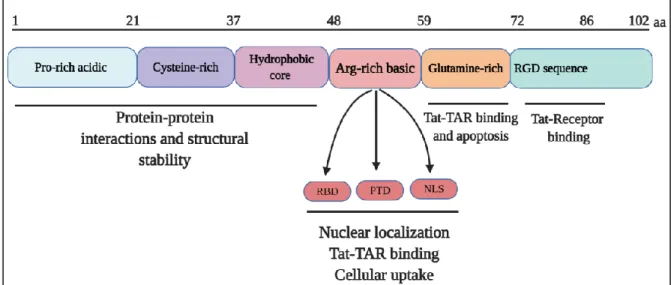
Tat is a small protein of 86-102 amino acids (aa) depending on the viral strains with a predicted molecular weight of 14-16 kDa. The 86 aa version of Tat is commonly referred to as the “full-length” Tat. Tat mRNA is composed of two exons (Figure 6). The first exon (aa 1-72) is highly conserved, while the second exon (aa 73-102) is less conserved. Tat protein has several domains which are mainly coded by the first exon. The first domain contains a proline rich acidic N-terminus (aa 1-21), a cysteine-rich region (aa 22-37), and a hydrophobic core region (aa 38-48), which are important for protein-protein interactions. Second domain is an arginine-rich basic domain (aa 49-58) comprising the protein transduction domain (PTD), the nuclear localization signal (NLS) and RNA binding domain (RBD). Glutamine-rich domain is required for Tat binding to TAR element and is important for the apoptotic function of Tat. Finally, C-terminal domain coded by the second exon contains arginine-glycine-aspartic acid (RGD) sequence (aa 73-86) necessary for Tat interaction with cell surface receptors(56,58) (Figure 6). Tat is a disordered protein, which gets more ordered structure through conformational changes following interaction with its target partners (59).
22
3.2. Functions of Tat
Tat can be active both in cytoplasm and nucleus, but its main localization is in the nucleus. Two properties of Tat, the small molecular weight and NLS sequence, allows Tat to shuttle between the nucleus and the cytoplasm (Figure 7). Its small molecular weight assures the passive diffusion through nuclear pore complexes, while NLS sequence enables its nuclear import through classical nuclear import pathway (60). Tat can also be differently distributed within the nucleus itself. When Tat is abundant it localizes into the nucleoli, otherwise it localizes in the nucleoplasm (61).
3.3. Tat in HIV-1 transcription
Efficient HIV-1 gene expression depends on Tat. In the absence of Tat, viral gene transcription starts normally, but short, abortive viral transcripts are generated due to the inefficient activity of polymerase II. In order to overcome such unproductive transcription process, Tat expression starts early. Abortive viral transcripts are subsequently spliced and translated into Tat protein. Newly synthesized Tat enters to the nucleus (Figure 7) and directly binds to cyclin T1 (CycT1) subunit of pTEFb kinase complex consisting of CycT1 and cycline-dependent kinase 9 (CDK9). pTEFB complex binds to TAR element in a Tat-dependent manner (62), CDK9 subunit of the complex phosphorylates C-terminal domain of RNA Polymerase II increasing its activity (63). pTEFB complex also recruits TATA box binding proteins to the LTR promoter while Tat recruits chromatin-modifying proteins to the promoter region, which dramatically promotes the transcription of HIV-1 RNA and stimulates the assembly of new transcription complexes (64,65).
3.4. Tat in host cell gene expression
Tat not only induces viral response, it can also generate host cell response by regulating a large number of genes coding for cytokines, cell cycle-related proteins, surface and chemokine receptors, mRNA processing factors, and proteins, involved in autophagy, DNA damage, and apoptosis (Figure 7). Tat can interact with promoter regions of hundreds of genes, involved in development, proliferation, growth, cellular organization, and intracellular signaling (66), showing its ability to significantly regulate gene expression in the host (67,68). How Tat modulates host gene expression is still not completely clear, yet several mechanisms of gene expression regulation by Tat have been suggested: i) binding to the TAR-like sequences at the 5’-untranslated regions of mRNA, ii) binding to the promoter region of target genes, iii) interacting with various transcriptional regulator proteins (56,69). These actions of Tat are ultimately favored by a variety of cellular signaling pathways.
23
Genes with TAR-like stem-loop structure on the 5’ untranslated region (5’UTR) are one of the “favorite” targets of Tat. For example, proinflammatory cytokines interleukin 1 beta (IL-1b), interleukin 6 (IL-6) and tumor necrosis factor beta (TNF-β) proteins are overexpressed, when Tat binds to corresponding 5’UTR and up-regulates promoter activity, thus further increasing their expression (66,70,71).
Tat can also act through a TAR-independent way, due to its ability to bind to the promoter region of genes. For example, Tat binds to the promoters of PTEN (phosphatase and tensin homolog) and PP2 (protein phosphatase 2), two upstream inhibitors of AKT pathway, and boosts their expressions. As a result, the activity of AKT signaling pathway is decreased.
Increased activity of key transcription factors is another proposed mechanism of Tat action (Figure 7). Tat increases the activity of NF-κB transcription factor and further promotes NF-κB signaling which leads to the increased BAG3 protein level, resulting in autophagy induction (see below) (72). Tat-activated NF-κB transcription factor is also involved in DNA damage further causing aberrant chromosomal translocations (73). Recent study showed that transcription factors Slug, Snail, Twist1 (Twist-related protein 1) and ZEB1 (Zinc finger E-box binding protein 1) were activated by Tat following the phosphorylation of SMAD2 (SMAD Family Member 2) transcriptional modulator through TGF-β and MAPK signaling pathways promoting epithelial–mesenchymal transition of mucosal epithelial cells (74). Tat up-regulates the expression of HIF-1α transcription factor and further favors its interaction with the long non-coding RNA (lncRNA) BACE1‐antisense transcript (BACE1-AS), forming a HIF-1α/lncRNABACE1-AS/BACE1 axis that resulted in the accumulation of a toxic amyloid protein which contributes to the progressions of HIV-associated neurocognitive disorders (HANDs) (75).
Not only Tat can promote the expression of transcription factors, it can also interact with them and modulate their binding to the promoter region of genes. For example, Tat interacts with Sp-family transcription factors (Sp-1, Sp-3) and alters their binding to the promoter of SOD2 (Superoxide Dismutase) gene that may lead to altered oxidative stresses (76). Tat is not always enhancing the activity of transcriptional regulators, but it also might have suppressive effects on them. Tat inhibits the activity of transcription factor Nrf2 (Nuclear factor (erythroid-derived 2)-like 2) along with its target genes (77). Expression and transcriptional activity of transcription factors like E2F3 (E2F transcription factor 3) and CREB (CAMP-responsive element–binding protein) is also hindered by Tat, due to the activation of p53 signaling pathway (78). Down-regulation of CREB activity by Tat can also happen via AKT signaling pathway (79).
24
3.5. Tat secretion and penetration in bystander cells
Tat can be secreted by the productively infected cells, accumulated in the extracellular environment in a soluble form, and enter the neighboring cells through cell surface receptors (Figure 7). Tat concentration in blood of HIV-1-infected patients can be as high as 500 ng/ml (73). Tat secreted from HIV-infected cells can perturb both HIV-infected and uninfected bystander cells in the surrounding microenvironment. The mechanism of Tat secretion is poorly understood, as it lacks secretion sequences, but it seems that Tat is released by an unconventional secretion pathway different from the conventional ER-to-Golgi secretory pathway (80,81). Several Tat secretion pathways have been described: oligomerization-mediated pore formation, spontaneous translocation, and incorporation into exosomes (82) .
Released Tat remains in a soluble form which can circulate in the blood and bind to the cell surface receptors to enter different cells. Secreted Tat can transduce many cell types (monocytes, macrophages, microglia, B cells. CD4 T lymphocytes, astrocytes, neurons, cardiomyocytes, endothelial cells) due to its protein transduction domain (Figure 7). Circulating Tat has been detected in cerebrospinal fluid, sera and tissues of HIV-infected individuals, even in patients with no detectable viral load (80,83–85). PTD of Tat can be used as a highly efficient drug delivery peptide (86,87). As it is able to cross the biological membranes, PTD is conjugated to other therapeutic molecules as cell penetrating peptide, delivering them to their difficult-to-access targets (88).
Figure 7. Tat regulates gene expression of host and bystander cells. Tat released from infected cells can go back to the nucleus of infected cells to promote HIV-1 gene transcription. Tat can reactivate latent HIV-1. Tat can enter uninfected cells through cell surface receptors. Tat regulates the expression of transcription factors and signaling pathways.
25
Tat entrance into bystander cells have been attributed to its different cell surface receptors - heparan sulfate proteoglycans (HSPGs), lipoprotein receptor-related protein (LRP), C-X-C chemokine receptor type 4 (CXCR4), dipeptidyl aminopeptidase IV (CD26), integrins (α5β1, αvβ3 and αvβ5) (Figure 7) (83,89–92). Tat interacts with these receptors through its basic region and the RGD motif, which allows Tat to enter the cells via endocytosis (93). As receptors like CD26, the LRP and HSPGs are ubiquitous, they further increase the binding range of Tat. Moreover, as Tat receptors (LRP, CXCR4, and HSPGs) are endocytic receptors, they further facilitate the internalization of Tat (94). Once inside the host cell Tat enters the nucleus due to its nuclear localization signal and regulates the expression of host genes.
All together Tat is a flexible versatile small protein which is expressed in productively infected cells, and released into the extracellular environment. Tat may affect both infected and non-infected neighboring cells of any kind, which may influence the expansion of HIV-1 infection and trigger other diseases.
4.
HIV-associated cancers and related diseases
Patients infected with HIV have a substantially elevated risk of developing so-called AIDS-defining cancers (Kaposi’s sarcoma (KS), aggressive B-cell lymphoma and invasive cervical cancer) and non-AIDS defining cancers (Hodgkin lymphoma, anal cancer, liver cancer, and lung cancer) (Figure 8). Some of these cancers are caused by co-infection with other viruses, such as Epstein–Barr virus (EBV), human herpesvirus 8 (HHV8), human papillomavirus (HPV), hepatitis B virus (HBV), and hepatitis C virus (HCV) (95,96). Other than cancers, neuronal and kidney diseases related to HIV infection (HAND, HIV-associated neuronal disorders; HIVAN, HIV-associated nephropathy) are also among the major causes of mortality in people with HIV infection (97,98). Even though the introduction cART had dramatically reduced certain HIV-associated malignancies like KS, the incidence of AIDS-related lymphomas (ARL) has remained high in HIV patients (99). Thus, it is of great importance to understand the mechanisms of the development of HIV-1-related diseases, even in patients under cART.
Patients infected with HIV-1 are at high risk of developing both non-Hodgkin’s (NHL) and Hodgkin’s (HL) lymphomas collectively called ARL that are always of B cell origin. Some lymphoma types are common to both HIV-1 uninfected and infected patients in whom the incidence is much higher, while some other lymphomas preferentially develop in the context of HIV-1 infection and frequently tend to be associated with the co-infection with other oncogenic viruses, such as EBV and HHV8 (Table 1). ARLs can be divided into several subtypes: Diffuse large B cell lymphomas (DLBCLs), Burkitt’s lymphomas (BL), PCNSL (primary central nervous system lymphoma), primary effusion lymphoma (PEL), plasmablastic lymphoma (PBL), lymphoma arising in KSHV-associated multicentric Castleman’s disease (MCD).
26 Figure 8. HIV-associated cancers and other diseases.
Though a decreased incidence of ARLs since the introduction of the HAART is reported in HIV-infected patients, ARLs still occur more frequently in HIV+ individuals than in general population. Moreover, survival rate of patients with NHL and HL is about two times lower than their HIV-uninfected counterparts (100–105).
Exact mechanisms of ARL pathogenesis are not very well understood, as B cells are not direct targets of HIV-1 and there is no evidence of B cell transformation due to HIV-1 infection. Several possible mechanisms, however, have been proposed: 1) secreted or transmitted viral proteins (Tat, p17) might have oncogenic effects on B cells (106,107); 2) aberrant somatic hypermutations of immunoglobulins might cause chromosomal translocations leading to malignant transformation (107); 3) infected T lymphocytes escaped from elimination in germinal centers could cause unusual B cell expansion (108); 4) chronic inflammation could lead to lymphoproliferation (109). These perturbations might generate an environment full of genetic abnormalities (chromosomal translocations, inactivation of p53, mutations), which might be further favored by co-infection with EBV and/or HHV8, leading to the emergence of ARL (Figure 9) (110–112).
Other possible mechanisms might involve deregulated cellular signaling pathways by HIV-1 or its viral proteins. Among many cellular signaling pathways, AKT/mTORC1 pathway plays an important role in ARL development, as various studies have shown the hyperactivation of this signaling pathway in different subgroups of ARL (106,113–118).
27
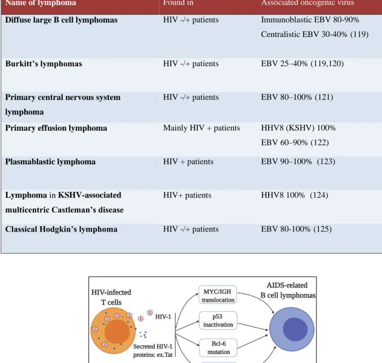
Table 1. AIDS-related lymphomas and associated oncogenic viruses
Name of lymphoma Found in Associated oncogenic virus
Diffuse large B cell lymphomas HIV -/+ patients Immunoblastic EBV 80-90% Centralistic EBV 30-40% (119)
Burkitt’s lymphomas HIV -/+ patients EBV 25–40% (119,120)
Primary central nervous system lymphoma
HIV -/+ patients EBV 80–100% (121)
Primary effusion lymphoma Mainly HIV + patients HHV8 (KSHV) 100% EBV 60–90% (122)
Plasmablastic lymphoma HIV + patients EBV 90–100% (123)
Lymphoma in KSHV-associated multicentric Castleman’s disease
HIV+ patients HHV8 100% (124)
Classical Hodgkin’s lymphoma HIV -/+ patients EBV 80-100% (125)
Figure 9. Mechanisms of development of AIDS-related B cell lymphomas. Secreted HIV-1 proteins e.g. Tat and infected T lymphocytes escaped from immune destruction cause a series of events including chromosomal translocations, inactivation of p53, mutations in Bcl6 gene, and coinfection with other viruses, which eventually lead to the development of AIDS-related B cell lymphomas.
28
5.
The mammalian target of rapamycin complex
mTOR stands for the “mammalian (or mechanistic) target of rapamycin”, and, as the name indicates, its characterization is related to the discovery of rapamycin during a Canadian expedition to Rapa Nui island in 1964. Soil samples collected from this island had led to the isolation from the bacterium Streptomyces hygroscopicus of a macrolide having antifungal activity. Thus, the name “rapamycin” was given to this molecule because of the relation to Rapa Nui island (126). Later rapamycin was also found to have immunosuppressive and antitumor effects (127,128). Its mechanism of action remained unanswered till early 1990s when Michael Hall and his colleagues identified in Saccharomyces cerevisiae yeast cells two genes related to the rapamycin resistance, TOR1 (target of rapamycin 1) and TOR2 (target of rapamycin 2) (129). In 1994 mammalian orthologue of yeast TOR was identified (130–135). Even though two TOR genes exist in yeast S.cerevisiae , there is only one TOR gene in higher eukaryotes (136). In human TOR gene is localized to the chromosome region 1p36.22. Different groups that identified TOR gave different names to this mammalian orthologue of yeast TORs: FRAP (FK506-binding protein-rapamycin-associated protein, RAFT (rapamycin and FK506-binding protein target), RAPT (rapamycin target) and mTOR (mammalian TOR). The name mammalian TOR or mTOR was prevailed in the TOR community. In 2009, however, modification 'mechanistic TOR' or MTOR was adopted by the HUGO gene nomenclature committee as the official name (137).
mTOR protein (~280 kDa) is an evolutionarily conserved serine-threonine protein kinase, which belongs to the phosphatidylinositol 3-kinase related kinase family. mTOR has several conserved structural domains: 20 tandem HEAT repeats on the N-terminus followed by focal adhesion targeting (FAT) domain, kinase domain, FKBP-rapamycin-binding (FRB) domain and FAT carboxy-terminal (FATC) domain (Figure 10). Rapamycin together with FKBP12 binds to the FRB domain of mTOR and thus inhibits its activity (135).
mTOR kinase forms two different macromolecular protein complexes mTOR complex 1 (mTORC1) and mTOR complex 2 (mTORC2) (Figure 10), which differ in their composition, downstream targets and regulation. mTOR complex 1 (mTORC1) comprised from three core components - mTOR, regulatory-associated protein of mammalian target of rapamycin (Raptor), mammalian lethal with sec-13 protein 8 (mLST8, also known as GβL) – and two inhibitory subunits: proline-rich AKT substrate 40 kDa (PRAS40) and DEP domain containing mTOR-interacting protein (DEPTOR). Raptor strongly interacts with N-terminal region of mTOR (138), but also binds S6K1 (Ribosomal protein S6 kinase beta-1) and 4E-BP1 (Eukaryotic translation initiation factor 4E (eIF4E)-binding protein 1), substrates of mTORC1, through their TOR signaling motifs that further ensures an efficient substrate phosphorylation event (139). mLST8 maintains the stability of mTOR/Raptor interaction and promotes the kinase activity of mTOR (140). Interaction of PRAS40 with mTOR is mediated by Raptor, but PRAS40 inhibits mTOR autophosphorylation
29
and further inhibits its kinase activity (141). Second inhibitory component DEPTOR through its PDZ domain binds to FAT domain of mTOR which sequesters mTORC1 away from its active pool and leads to the inhibition of mTOR kinase activity (142,143).
Figure 10. Structure of mTOR protein and its complexes. mTOR is comprised of HEAT repeat, FAT and FRB, kinase and FATC domains. FKBP12-Rapamycin complex binds mTOR through FRB domain and inhibits the activity of mTORC1. mTORC1 is comprised of Raptor, mLST8, PRAS40, and DEPTOR that interact with mTOR protein. Raptor interacts with N-terminal region and mLST8 - with kinase domain of mTOR, while interaction of the inhibitory subunit PRAS40 is mediated by Raptor and secondary inhibitory component DEPTOR interacts with FAT domain. DEPTOR, mLST8, Rictor, mSin1, and Protor 1/2 interact with mTOR to form mTORC2.
mTOR complex 2 (mTORC2) consists of six proteins: rapamycin-insensitive companion of mTOR (Rictor), mammalian stress-activated map kinase-interacting protein 1 (mSin1), protein observed with Rictor (Protor) 1/2, and mTOR, mLST8, DEPTOR commonly shared between two complexes (144–146).
mTORC1 is sensitive, while mTORC2 is much less responsive to the rapamycin (Sirolimus®), which
suppresses T and B cell activation by inhibition of the cell cycle. Various analogues of rapamycin, so called rapalogues (Everolimus®, Temsirolimus®), are also frequently used in clinics for immunosuppression. In
addition, a number of alternative mTOR inhibitors have been developed. These inhibitors block both mTORC1 and mTORC2 (pan-inhibitors or TOR-KIs, i.e., INK128) or act on mTOR kinase and another protein (dual inhibitors), most often targeting a network upstream of mTORC1/2 (147).
Rapamycin first forms a complex with FKBP12, which then directly binds to the FRB domain of mTOR (Figure 10). This interaction results in the changes in FRB domain, thereby allosterically inhibiting the kinase activity of mTOR (135,148), challenged by in vitro studies describing that rapamycin inhibits mTOR function by dissociating Raptor from mTOR but without changing its kinase activity (149), as Raptor plays essential role in mTORC1 activation (150). Recent 3.2 Å resolution structure of mTORC2 revealed that
30
C-terminal domain of RICTOR masks FRB domain in mTOR, explaining the rapamycin insensitivity of mTORC2 (151).
mTORC2 regulates many biological processes including cell survival, proliferation, metabolism, and cytoskeletal organization. Growth factors are found to be the most important upstream signals to activate mTORC2. AKT, serum- and glucocorticoid-induced protein kinase 1 (SGK1) and protein kinase C (PKC) are the best characterized substrates of mTORC2. mTORC2 regulates cell survival, proliferation and metabolism mainly through AKT and SGK1. mTORC2 phosphorylates AKT on Ser473 (Figure 11) and SGK1 on Ser422 (152–155). mTORC2 regulates FoxO transcription factors which are known substrates of AKT and SGK1 (154,156,157). Besides, mTORC2 controls cytoskeletal organization through PKC (158). Activation of AKT is also found be necessary for the induction cytoskeletal reorganization by mTORC2 (159).
mTORC1 integrates signals from many intracellular and extracellular cues: growth factors, amino acids, energy, oxygen, DNA damage and infectious agents, including viruses. Depending on the nature of the signal, its duration, cell type and many other factors, mTORC1 will “determine” the subsequent cell fate.
5.1. Upstream regulation of mTORC1
5.1.1. PI3K-AKT signaling to mTORC1
Growth factors, cytokines, immune cell receptors (e.g. TCR) and co-receptors activate mTORC1 through PI3K-AKT signaling pathway (160,161). The phosphatidylinositol 3-kinases (PI3Ks) are a family of lipid kinases that phosphorylate phosphatidylinositol and phosphoinositides to activate many intracellular signaling pathways (162). PI3K is activated by signals from growth factor receptor tyrosine kinases (RTKs). In response to growth factor stimulation and the subsequent activation of RTKs, its substrate phosphatidylinositol 4,5-bisphosphate (PIP2) at the plasma membrane is converted to phosphatidylinositol-3,4,5-trisphosphate (PIP3), which in turn activates downstream signaling pathways (Figure 11). On the other hand, the cellular level of PIP3 is tightly regulated by phosphatase and tensin homolog (PTEN), a tumor suppressor, which antagonizes PI3Ks activity by converting PIP3 back to PIP2 via its intrinsic lipid phosphatase activity (Figure 11) (163).
PIP3 acting as second messenger activates, among multiple downstream singlaing pathways, the protein serine/threonine kinase AKT (also known as PKB). Binding of PIP3 to AKT leads to its membrane recruitment. Subsequently Thr308 and Ser473 residues are phosphorylated by distinct kinases. Thr308 is phosphorylated by 3-phosphoinositide-dependent kinase 1 (PDK1), while Ser473 is phosphorylated by mTORC2. In the response to DNA damage AKT is phosphorylated at Ser473 by three major DNA damage sensors - ATM, ATR and DNA-PK kinases (see below). Once phosphorylated andactivated, AKT in its turn
31
either activates or inhibits many downstream proteins such as glycogen synthase kinase 3 (GSK3), the forkhead family of transcription factors (FOXOs), nuclear factor of κB (NF-κB), and mouse double minute 2 homolog (MDM2) as well as various signaling pathways including mTORC1 pathway (see below) (Figure 11), thereby regulating a wide range of cellular functions including protein synthesis, cell survival, proliferation, and metabolism (164).
Active AKT phosphorylates and inactivates TSC2 component of TSC (Tuberous Sclerosis Complex) consisted of TSC1, TSC2, TBC1D7 (Figure 13) (165). TSC2 has a GTPase activating protein activity (Figure 12) for the small GTPase RHEB (Ras homologue enriched in brain), converting RHEB-GTP to the inactive RHEB-GDP. In its GTP form, RHEB is an activator of mTORC1. Thus, through its GAP activity, TSC2 represses mTORC1 by inactivating RHEB. Phosphorylation of TSC2 by AKT relieves this inhibitory action on mTORC1 (166). AKT can also activate mTORC1 directly, in a TSC-independent way, by phosphorylating and inactivating the mTORC1 inhibitory component PRAS40 (Figure 13) (167).
Figure 11. PI3K-AKT signaling pathway. Class IA PI3K receives signals from growth factors via receptor tyrosine kinase (RTK). Catalytic p110 subunit converts PIP2 to PIP3 which subsequently activates AKT by phosphorylating it on Thr308 via 3-phosphoinositide-dependent kinase 1 (PDK1) and on Ser473 via mTORC2. Phosphatase and tensin homolog (PTEN) antagonizes this activity by converting PIP3 back to PIP2. Phosphorylated AKT regulates a large number of downstream targets including mTORC1, nuclear factor of κB (NF-κB), mouse double minute 2 homolog (MDM2), forkhead family of transcription factor (FOXO), and glycogen synthase kinase 3 (GSK3).