HAL Id: inserm-00986641
https://www.hal.inserm.fr/inserm-00986641
Submitted on 3 May 2014
HAL is a multi-disciplinary open access
archive for the deposit and dissemination of
sci-entific research documents, whether they are
pub-lished or not. The documents may come from
teaching and research institutions in France or
abroad, or from public or private research centers.
L’archive ouverte pluridisciplinaire HAL, est
destinée au dépôt et à la diffusion de documents
scientifiques de niveau recherche, publiés ou non,
émanant des établissements d’enseignement et de
recherche français ou étrangers, des laboratoires
publics ou privés.
Germline genes hypomethylation and expression define a
molecular signature in peripheral blood of ICF patients:
implications for diagnosis and etiology.
Guillaume Velasco, Emma Walton, Delphine Sterlin, Sabrine Hédouin,
Hirohisa Nitta, Yuya Ito, Fanny Fouyssac, André Mégarbané, Hiroyuki Sasaki,
Capucine Picard, et al.
To cite this version:
Guillaume Velasco, Emma Walton, Delphine Sterlin, Sabrine Hédouin, Hirohisa Nitta, et al.. Germline
genes hypomethylation and expression define a molecular signature in peripheral blood of ICF patients:
implications for diagnosis and etiology.. Orphanet Journal of Rare Diseases, BioMed Central, 2014, 9
(1), pp.56. �10.1186/1750-1172-9-56�. �inserm-00986641�
R E S E A R C H
Open Access
Germline genes hypomethylation and expression
define a molecular signature in peripheral blood
of ICF patients: implications for diagnosis and
etiology
Guillaume Velasco
1*, Emma L Walton
1, Delphine Sterlin
2, Sabrine Hédouin
1, Hirohisa Nitta
5, Yuya Ito
5,
Fanny Fouyssac
6, André Mégarbané
7, Hiroyuki Sasaki
5, Capucine Picard
2,3,4and Claire Francastel
1*Abstract
Background: Immunodeficiency Centromeric Instability and Facial anomalies (ICF) is a rare autosomal recessive disease characterized by reduction in serum immunoglobulins with severe recurrent infections, facial dysmorphism, and more variable symptoms including mental retardation. ICF is directly related to a genomic methylation defect that mainly affects juxtacentromeric heterochromatin regions of certain chromosomes, leading to chromosomal rearrangements that constitute a hallmark of this syndrome upon cytogenetic testing. Mutations in the de novo DNA methyltransferase DNMT3B, the protein ZBTB24 of unknown function, or loci that remain to be identified, lie at its origin. Despite unifying features, common or distinguishing molecular signatures are still missing for this disease. Method: We used the molecular signature that we identified in a mouse model for ICF1 to establish transcriptional biomarkers to facilitate diagnosis and understanding of etiology of the disease. We assayed the expression and methylation status of a set of genes whose expression is normally restricted to germ cells, directly in whole blood samples and epithelial cells of ICF patients.
Results: We report that DNA hypomethylation and expression of MAEL and SYCE1 represent robust biomarkers, easily testable directly from uncultured cells to diagnose the most prevalent sub-type of the syndrome. In addition, we identified the first unifying molecular signatures for ICF patients. Of importance, we validated the use of our biomarkers to diagnose a baby born to a family with a sick child. Finally, our analysis revealed unsuspected complex molecular signatures in two ICF patients suggestive of a novel genetic etiology for the disease.
Conclusions: Early diagnosis of ICF syndrome is crucial since early immunoglobulin supplementation can improve the course of disease. However, ICF is probably underdiagnosed, especially in patients that present with incomplete phenotype or born to families with no affected relatives. The specific and robust biomarkers identified in this study could be introduced into routine clinical immunology or neurology departments to facilitate testing of patients with suspected ICF syndrome. In addition, as exemplified by two patients with a combination of molecular defects never described before, our data support the search for new types of mutations at the origin of ICF syndrome.
Keywords: Agammaglobulinemia, DNA methylation, Heterochromatin, DNA, Satellite, Gene expression, Biological markers/diagnosis use, Biological markers/etiology, Genes, X-linked
* Correspondence:guillaume.velasco@univ-paris-diderot.fr;claire.francastel@ univ-paris-diderot.fr
1Université Paris Diderot-Paris7, CNRS UMR7216, Epigénétique et Destin
Cellulaire, Case Courrier 7042; 35, rue Hélène Brion, 75205 Paris, France Full list of author information is available at the end of the article
© 2014 Velasco et al.; licensee BioMed Central Ltd. This is an Open Access article distributed under the terms of the Creative Commons Attribution License (http://creativecommons.org/licenses/by/4.0), which permits unrestricted use, distribution, and reproduction in any medium, provided the original work is properly credited. The Creative Commons Public Domain Dedication waiver (http://creativecommons.org/publicdomain/zero/1.0/) applies to the data made available in this article, unless otherwise stated.
Background
Immunodeficiency with centromeric instability and facial anomalies (ICF; OMIM no. 242860) is a rare autosomal recessive disorder mainly characterized by primary im-munodeficiency [1]. Recurrent infections are the present-ing symptom, usually in early childhood. Other features include mild facial anomalies and variable symptoms in-cluding intellectual disability, congenital malformations and developmental delay [2].
ICF syndrome is a genetically heterogeneous disease with heterogeneous molecular defects and phenotypic variability (Reviewed in [3]). Around 60% of patients (ICF1) have mu-tations in the catalytic domain of the de novo DNA methyl-transferase (DNMT) DNMT3B [4-6] leading to reduced enzymatic activity [7,8] associated with a significant loss of DNA methylation, notably at juxtacentromeric satellite repeats on chromosome 1 and 16, and less frequently 9 (Reviewed in [9]). The remainder have either non-sense mutations in the zinc-finger and BTB domain-containing 24 (ZBTB24) gene (ICF2) [10] or no identifiable mutation in either DNMT3B or ZBTB24 coding sequences (ICFX) [11]; both ICF2 and ICFX show hypomethylation of centro-meric alpha-satellites (α-Sat) in addition to the above men-tioned repeats [12]. Hypomethylation of satellite repeats is associated with centromeric instability and constitutes an invariant molecular hallmark of ICF patients. Chromosomal anomalies are detectable by karyotype analysis of mitogen-stimulated lymphocytes and that is used to establish the diagnosis [13,14].
Data obtained in ICF lymphoblastoid cell lines (LCLs) showed that DNMT3B mutations also lead to hypome-thylation and perturbed expression of several hundred of genes involved in immune function, development and neurogenesis, being both up- and down-regulated, which probably account for the phenotypical manifestations documented in patients [15-17]. Additional molecular mechanisms acting in trans, such as perturbed nuclear organization as a result of the altered juxtacentromeric or telomeric heterochromatin organization [18-20], deregu-lated expression of small regulatory microRNAs (miRNA) [21] or changes in replication timing [22] may also have a deep impact on the deregulation of gene expression pro-grams in ICF patients. However, these perturbed profiles showed great variability, probably reflecting phenotypic variability between patients and cell culture effects, and no common or distinguishing molecular signatures could be reliably established from these studies.
Our analysis of perturbed DNA methylation patterns and expression programs in a mouse model for ICF1 re-vealed the striking over-representation of germline genes among the most upregulated genes [23]. In addition, our data suggested a new role for Dnmt3b in the protection of somatic cells against the promiscuous expression of the germ line program, playing the central role in both
the establishment and maintenance of DNA methylation profiles at these genes [24]. Although the impact of their illicit expression on ICF phenotypes remains to be solved, their inherent repressed state in all somatic cells makes them good candidate biomarkers of molecular dysfunc-tions in ICF syndrome. We tested hypomethylation and illicit expression of the thus identified germline genes in a cohort of ICF patients that included five newly enrolled patients, in order to establish a molecular signature easily testable directly from the blood of ICF patients.
Material and methods
This study was conducted in accordance with the Helsinki Declaration, with informed consent obtained from each patient or the patient’s family. The study was approved by the local ethics committee of Necker-Enfants Malades Hospital, Paris, France.
An expanded Methods section for DNA methylation and gene expression analysis can be found in Additional file 1.
Healthy donors and patients
Blood samples of six healthy volunteers, aged 35, 24, 26, 27, 29 and 41 years, respectively, were collected and were numbered from 1 to 6. Donors 3 and 6 are women.
Our cohort of patients included thirteen ICF1 patients with mutations in DNMT3B, six ICF2 patients with mu-tations in ZBTB24 and four ICFX patients with as of yet unknown mutations (Additional file 2). Most patients were described earlier [11,12,25-27] except for five newly en-rolled patients (Table 1). The ICF B-lymphoblastoid and fibroblasts named here pCor were obtained from the Coriell Cell Repositories (USA) (http://ccr.coriell.org/). Patients pG, pR, pI, pH, pC, pD, pN, pP, pS were recruited by the ICF Consortium and described together with pa-tients pG, pR, pI, pH, pC, pD, pN, pP, pS in [12]. Papa-tients pW, pT and P5 were described earlier [11,25,26]. Patients P7 and P8 were recently classified as ICF2 patients [27]. Patients pC, pS, pU and pN were classified as ICFX pa-tients since sequence analysis of DNMT3B and ZBTB24 genes performed as previously described [12,27] did not reveal any mutation in their coding sequences.
New ICF syndrome patients P1, P2, P3, P4 and pY were diagnosed according to cytogenetic analysis that revealed typical multiradial chromosome configurations with multiple arms from chromosomes 1 and 16, and clinical features, mainly primary immunodeficiency. For these patients, primary immunodeficiency and hypome-thylation at satellite repeats are detailed in Additional files 3 and 4. These patients were classified as ICF1 pa-tients (Necker Hospital, Paris, France) based on mutations found in DNMT3B using previously described sequencing methods [12,27]. Written informed consent was obtained from the parents of the patients.
Velasco et al. Orphanet Journal of Rare Diseases 2014, 9:56 Page 2 of 8 http://www.ojrd.com/content/9/1/56
Primary cells and cell lines
Primary fibroblasts (passage 7 for pG, pW and pT; passage 5 for pR; passage 6 for pI; passage 14 for pC and pP; pas-sage 9 for pCor and pS) and Lymphoblastoid cell lines (LCLs) from healthy donors and ICF patients were cultured in DMEM and RPMI 1640 respectively, supplemented with 15% FCS, glutamine and antibiotics (Invitrogen).
Methylation-sensitive restriction enzyme-coupled qPCR assay
Genomic DNA (200 ng) was digested at 37°C for 4 h with 10 U of the methylation-sensitive enzyme AciI, or NcoI(New England Biolabs) which does not have cutting sites in our regions of interest and served to normalize the data. The endonucleases were subsequently inacti-vated by incubation at 65°C for 20 min. Real-time PCR was carried out using the light cycler-DNA MasterPLUS SYBR Green I mix (Roche) supplemented with 0.5 μM specific primer pairs and with 2 μL of digested DNA. Real-time quantification PCR were run on a light cycler rapid thermal system (LightCycler®480 2.0 Real time PCR system, Roche) with 20 sec of denaturation at 95°C, 20 sec of annealing at 65°C and 20 sec of extension at 72°C for all primers, and analyzed by the comparative CT (∆CT) method according to the formula: methylation (%) = E(∆CT)× 100 where E represents PCR efficiency and ∆CT = CTsample(AciI digest) - CTsample(NcoI digest). Se-quences of primers within CpG islands at germline gene promoters are shown in Additional file 1. Each data shown on histograms is the mean result of qPCR analysis on at least three independent experiments performed on at least three independent genomic extractions.
Results and discussion
Hypomethylation and expression of MAEL and SYCE1 define molecular markers for ICF1 syndrome
Peripheral blood samples were obtained from healthy do-nors and ICF patients. We assessed profiles of gene ex-pression focusing on the germline genes transcriptional signature that we previously established in a mouse model for ICF1 [23,24].
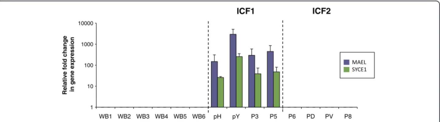
As expected from their expression primarily in germ cells (Additional file 5; and RNAseq and exon arrays data-sets on normal tissues publically available at UCSC gen-ome browser) and function in meiosis, expression of the germline genes tested was undetectable in blood cells
from healthy controls, although that of SYCE1 showed some degree of variation between healthy donors that did not seem to correlate with age or sex (Figure 1 and Additional file 6). In contrast, strong expression was de-tected for two of them in blood cells from ICF patients; among all the germline genes tested, only that of MAEL-STROM (MAEL), involved in repression of transposable elements during meiosis, and Synaptonemal Complex Central Element Protein 1 (SYCE1), involved in assembly of the synaptonemal complex during meiosis in germ cells, were transcribed in blood cells from ICF patients (Figure 1 and Additional files 6 and 7). This illegitimate expression in whole blood was very strong and remarkably specific for ICF1 patients while at significantly lower levels in ICF2 blood cells (raw data and p values in Additional file 6). Interestingly, patient P5 had only mild ICF pheno-type [25] although he showed strong expression of MAEL and SYCE1. Therefore, given their repressed state in nor-mal somatic tissues, detection of MAEL and SYCE1 ex-pression in blood cells provides a statistically significant signature for the most prevalent sub-type of the disease, regardless of the severity of the symptoms.
Since the germline genes tested in this study are known to be repressed through DNA methylation in mice [23,24,28] we analyzed their methylation status on genomic DNA isolated from fresh whole blood samples (Figure 2A) or epithelial cells obtained from buccal swabs (Figure 2B), from ICF patients and healthy donors. Consistent with their expression only in blood cells from ICF1 patients (Figure 1), analysis of methylation at MAEL and SYCE1 revealed their hypomethylated state specifically in ICF1 patients since ICF2 and ICFX patients (except ICFX patient pN; discussed below) were methylated at values comparable to healthy controls (raw data and p values in Additional file 8). Simi-larly, Solute Carrier family 25 member 31 (SLC25A31 also known as ANT4), encoding a mitochondrial ADP/ATP carrier essential during spermatogenesis, was signifi-cantly hypomethylated only in ICF1 patients (Figure 2 and Additional file 8) although this hypomethylation did not correlate with transcriptional activation (Additional file 6).
Importantly, and as mentioned above, this transcriptional signature could not have been unraveled using classically available cultured patient cells like Epstein-Barr virus-transformed lymphoblastoid cell lines (LCLs) and primary fibroblasts. Indeed, although we could easily detect MAEL
Table 1 Genetic characteristics of newly identified ICF1 patients
Patients P1 P2 P3* P4* pY
Year of birth 2010 2008 2009 2013 1996
Gender F M F F M
Amino acid changes in DNMT3B (NP_008823.1) p. S655L/ ? p. R104X/p. I721T p. G583S/p. G583S p. G583S/p. G583S p.T775I/p.T775I
Consanguinity NO NO YES YES YES
and SYCE1 transcripts in these cultured cells, this expres-sion was independent of patient genotype (Additional file 7). We also noticed that the illicit expression of MAEL in fibroblasts from ICF1 patients became un-detectable beyond passage 9 (Additional file 9), although DNA methylation levels at its promoter remained rela-tively unchanged, suggesting that extensive culture may limit findings of illegitimately expressed genes. Hypo-methylation of DNMT3B-target genes in ICF2 and ICFX cells (Additional file 7), in which DNMT3B is not mutated, could result from well-known, direct or indirect, aberrant culture-induced defects of DNA methylation [29]. Furthermore, EBV transformation and cell culture perturb DNA methylation patterns in human LCLs, in particular at satellite repeats of juxtacentromeric regions [30] that are used to diagnose ICF patients. Therefore, like in previously reported transcriptomic studies examining differential gene expression profiles between ICF patients and healthy controls [15,16], our data obtained in LCLs or fibroblasts probably reflected culture-associated defects in DNA methylation and gene expression, which have been superimposed on the intrinsic defects that affect patients, and do not allow the reliable identification of biomarkers with diagnostic value.
In contrast, the analysis that we conducted in uncul-tured cells from patients provided the first blood-based gene expression test to identify ICF1 syndrome, and the first discriminating transcriptional differences amongst ICF patients. The molecular functions of ZBTB24 remain to be identified, but the finding that the germline genes tested herein are not similarly affected by mutations in DNMT3B or ZBTB24 contradicts earlier suggestion that ZBTB24 is a mere adapter of DNMT3B function [12].
Hypomethylation signature common to patients with mutations in DNMT3B or ZBTB24
DNA methylation at the promoters of DEAD (Asp-Glu-Ala-Asp) Box Polypeptide 4 (DDX4), the homolog of
VASA proteins in Drosophila with key roles in germ cells development, and Testis Expressed 12 (TEX12), was simi-lar between patients and controls, suggesting that, in con-trast to the mouse, methylation of these genes was not affected in human DNMT3B- or ZBTB24-deficient cells (Additional file 10). Interestingly, the Synaptonemal Complex Protein 1 (SYCP1) gene showed significant hy-pomethylation in all but one ICFX patient with unknown mutation. Similarly, we observed hypomethylation of the X-linked testis-expressed gene 11 (TEX11) irrespective of ICF subtype (Figure 2; black bars), but restricted to female ICF cells (raw data and p values in Additional file 8). Whether hypomethylation at SYCP1 and TEX11 pro-moters was a direct consequence of ZBTB24 loss of integ-rity remains to be seen. However, these data identifies SYCP1and TEX11 as the first unique gene loci affected by ZBTB24 mutations and contribute to a significant pro-gress in our understanding of the etiology of ICF syn-drome. In the case of the X-linked gene TEX11, it is likely related to the global hypomethylation of the inactive X-chromosome reported in female ICF1 patients [31,32]. Therefore, the hypomethylation of SYCP1 and TEX11 (Figure 2 and Additional file 8) regardless of the ICF sub-type represents the first unifying molecular signature for ICF syndrome and suggests that hypomethylation of the inactive X-chromosome in female patients may represent an invariant feature of ICF syndrome. In addition, these data point to a putative role of ZBTB24 in establishment or maintenance of DNA methylation at the inactive X-chromosome.
A new molecular heterogeneity among ICF patients
The perturbations that we reported here represent a reli-able index of DNMT3B dysfunction in ICF1 patients, with the notable exception of patient pN. Based on our predictions of a molecular signature that varies accord-ing to ICF genotype, the germline methylation (Figure 2) and expression (Figure 1) signatures that we found in
1 10 100 1000 10000 WB1 WB2 WB3 WB4 WB5 WB6 pH pY P3 P5 P6 PD PV P8
Relative fold change in gene expression
ICF1 ICF2
Figure 1 Expression of MAEL and SYCE1 in whole blood defines specific biomarkers for ICF1 patients. Expression levels of MAEL (purple bars) and SYCE1 (green bars) were assessed by qRT-PCR, normalized to U6 snRNA levels and presented as fold change relative to that of control whole blood from healthy donors (WB). ICF subtypes 1 and 2 are indicated and separated by dotted lines. Raw data used to built this Figure can be found in Additional file 6. WB, control whole blood from healthy donors. Error bars represent standard error.
Velasco et al. Orphanet Journal of Rare Diseases 2014, 9:56 Page 4 of 8 http://www.ojrd.com/content/9/1/56
ICFX patient pN would place this patient in the ICF1 subtype. However, no mutation was previously found in the coding regions of DNMT3B for this patient [12] or in a new sequencing of DNMT3B and ZBTB24 exons. Intriguingly, the ICFX patient pN differs from other ICFX patients in that this patient had some degree of thylation at α-Sat repeats, intermediate between hypome-thylation found in ICF2 cells and almost full mehypome-thylation characteristic of normal and ICF1 cells (Additional file 4). In addition, this profile resembles that of patient P1, first classified as ICF1 because of mutations in DNMT3B ma-ternal allele but for whom no mutation could be found in the coding regions of the paternal allele. The combination
of hypomethylation at MAEL and SYCE1 germline genes, indicative of DNMT3B impaired activity, with modest but reproducible hypomethylation at α-Sat repeats, which is never observed in ICF1 patients and was instrumental in suggesting genetic heterogeneity among ICF patients, im-plies an even more complex genetic etiology for these two patients. In the absence of an identified “culprit gene” for these patients, it remains unclear whether a third locus is implicated in ICF syndrome. Alternatively, these patients could be “compound” heterozygotes for mutations in both DNMT3B and ZBTB24, though it would have to be out-side coding regions since exome sequencing did not reveal any mutation in the exons of any of the two genes. The
0% 50% 100% WB1 WB2 WB3 WB4 WB5 WB6 pG pR pW pH P1 PY P2 P3 P5 P6 pD pV P7 P8 pC pS pN Relative % DNA methylation 0% 50% 100% WB1 WB2 WB3 WB4 WB5 WB6 pG pR pW pH P1 PY P2 P3 P5 P6 pD pV P7 P8 pC pS pN Relative % DNA methylation 0% 50% 100% WB1 WB2 WB3 WB4 WB5 WB6 pG pR pW pH P1 PY P2 P3 P5 P6 pD pV P7 P8 pC pS pN Relative % DNA methylation 0% 50% 100% WB1 WB2 WB3 WB4 WB5 WB6 pG pR pW pH P1 PY P2 P3 P5 P6 pD pV P7 P8 pC pS pN Relative % DNA methylation
MAEL
SYCE1
SLC25A31
TEX11
SYCP1
A
0% 20% 40% 60% 80% 100% BS P3 P4 pH P8 Relative % DNA methylation 0% 20% 40% 60% 80% 100% BS P3 P4 pH P8 Relative % DNA methylationMAEL
SYCE1
SYCP1
0% 20% 40% 60% 80% 100% BS P3 P4 pH P8 Relative % DNA methylationSLC25A31
1 2 0% 20% 40% 60% 80% 100% BS P3 P4 pH P8 Relative % DNA methylation 0% 20% 40% 60% 80% 100% BS P3 P4 pH P8 Relative % DNA methylationTEX11
B
0% 50% 100% WB1 WB2 WB3 WB4 WB5 WB6 pG pR pW pH P1 PY P2 P3 P5 P6 pD pV P7 P8 pC pS pN Relative % DNA methylation 1 2 XFigure 2 Relative DNA methylation levels at germline gene promoters in whole blood and buccal swabs from ICF patients. Methylation analysis in whole blood (A) and buccal swabs (B) were assessed by Methylation-Sensitive Restriction Assay, followed by qRT-PCR amplification of the AciI digested product with primers flanking at least two AciI sites within the promoter CpG island. A non-cutter NcoI control digest served to normalize data that are presented as a percentage of methylation relative to the control digest. ICF subtypes 1, 2, and X are indicated and separated by dotted lines. For the X-linked gene TEX11, female patients are indicated as black bars. Raw data used to built this Figure can be found in Additional file 8. WB and BS are control whole blood DNA and buccal swabs from healthy donor, respectively. Error bars represent standard error.
genetic defects in these patients will be probably highly in-formative from a mechanistic perspective regarding the establishment and maintenance of DNA methylation pat-terns. These observations also emphasize that additional molecular markers besides the methylation of α-Sat se-quences are required to distinguish various classes of ICF patients.
Expression and methylation of germline genes to diagnose individuals with suspected ICF syndrome
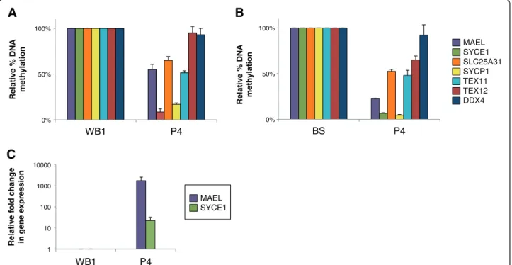
We propose that profiling of DNA methylation at MAEL and SYCE1 germline genes, combined to the detection of their illicit expression using non-invasive diagnostic techniques, could serve as a powerful, quick, and simple procedure to diagnose patients with suspected ICF1 syn-drome. Because these genes are normally repressed in somatic tissues, any diagnostic test based on their expres-sion will give an unambiguous result. To test the power of these genes for diagnostic means, we assessed methylation and expression profiles of germline genes in a child born to a consanguineous family but whose genotype at the time of testing was not known. This child, who is a sibling of patient P3, was born during the course of this study, and we were able to sample buccal epithelial cells and per-ipheral blood when she was 3 months old. Our analysis
revealed germline gene methylation and transcription pro-files typical of that of ICF1 patients tested in this study (Figure 3 and p values in Additional file 8). These observa-tions were highly suggestive of the ICF1 syndrome in this child, who we named patient P4 (Table 1). In line with our predictions, diagnosis of the ICF1 syndrome was subse-quently confirmed by sequencing of DNMT3B exons and characterization of the same mutations identified in her older sister (Table 1).
Conclusions
Correlations between genotype and phenotype of ICF patients are emerging [11], and the defining of a reliable ICF-specific molecular signatures will help to explain such differences in the clinical manifestations of ICF syndrome amongst ICF patient subtypes. In addition to providing a new contribution to the characterization of unifying and distinguishing molecular signatures between ICF patient subtypes, this first molecular signature identifiable in the peripheral blood or epithelial cells of patients could be eas-ily introduced into routine clinical immunology or neur-ology departments to facilitate testing of patients with suspected ICF syndrome. Because ICF is probably under-diagnosed, we predict that the analysis of such markers will greatly aid diagnosis and prioritize patients for mutation
1 10 100 1000 10000 WB1 P4
Relative fold change in gene expression 0% 50% 100%
WB1 P4
Relative % DNA methylation
A
C
0% 50% 100% BS P4Relative % DNA methylation
B
MAEL SYCE1 MAEL SYCE1 SLC25A31 SYCP1 TEX11 TEX12 DDX4Figure 3 Hypomethylation coupled to illegitimate expression of MAEL and SYCE1 to diagnose ICF1 syndrome. Uncultured cells from a child born to a consanguineous family with one sick children were used to test the power of these biomarkers. Methylation analysis (A and B) at germline gene promoters indicated on the right was carried out as in Figure 2, from whole blood (A) and buccal swabs (B). Expression analysis of MAEL and SYCE1 (C), the only germline genes to be transcriptionally activated upon hypomethylation, was performed from whole blood samples as in Figure 1. The newborn, a sibling of P3, is now patient P4 after the ICF syndrome was confirmed by sequencing of DNMT3B alleles. Raw data used to built this Figure can be found in Additional file 8. WB, Control Whole blood DNA from healthy donor; BS, control buccal swab from healthy donor; Errors bars represent standard error.
Velasco et al. Orphanet Journal of Rare Diseases 2014, 9:56 Page 6 of 8 http://www.ojrd.com/content/9/1/56
screening in cases where performing exome-sequencing or cytogenetics analysis may represent a challenge or when ICF should be suspected like in patients with immunodefi-ciency as the presenting factor if associated with facial anomalies [33], in patients that present an incomplete ICF phenotype or individuals from consanguineous families [25], or newborns from families where ICF cases have already been reported (this study). In addition, our analysis allowed the identification of unprecedented molecular char-acteristics in two ICF patients, suggestive of a novel type of genetic origin for the disease, stressing the need to continue the search for mutations that lead to immunodeficiency as-sociated with chromosomal instability. The hallmark of ICF being hypomethylation of heterochromatin DNA repeats, combined, these efforts will have great impact on our un-derstanding of the DNA methylation pathways.
Additional files
Additional file 1: Additional methods section for DNA methylation and gene expression analysis. Table of primers used for PCR amplification. Additional file 2: Table summarizing genetic mutations found in ICF patients already described and associated references. Additional file 3: Table showing immunological characteristics of newly identified ICF patients.
Additional file 4: Southern blot analysis of DNA methylation at centromeric and juxta-centromeric regions of ICF patients. Additional file 5: Germline genes expression and promoter methylation in control conditions including control fibroblasts as somatic cells, human testes and a cell line treated by the demethylating agent 5-azacytidine.
Additional file 6: Raw PCR data used to built expression histograms shown in Figure 1, and statistical analysis to compare healthy controls and ICF patients.
Additional file 7: (A) Additional information on expression analysis performed in cultured cells from patients, EBV-transformed lymphocytes or immortalized fibroblasts, suggesting that these cellular systems cannot provide reliable molecular markers for a disease with methylation defects. (B) Expression analysis and DNA methylation of germ line genes in lymphoblastoid cell lines from patients. (C) Expression analysis and DNA methylation of germ line genes in immortalized fibroblasts from patients.
Additional file 8: Raw PCR data used to built DNA methylation histograms shown in Figure 2, and statistical analysis to compare healthy controls and ICF patients.
Additional file 9: Control experiment showing that expression of Maelstrom decreases with the number of passages in culture. Additional file 10: DNA methylation analysis at TEX12 and DDX4 promoters showing that their dependency on DNMT3B for
methylation and silencing in murine cells is not conserved in humans.
Competing interests
The authors declare that they have no competing interest. Authors’ contributions
GV, ELW and SH carried out the molecular genetic studies. DS carried out the immunological characterization of newly enrolled ICF patients. HN, IY, HS carried out the exome sequencing of ICFX patients. FF, AM, CP collected the biological samples from ICF patients. GV and CF designed the study. GV coordinated the sample and data collection and analyzed the data. GV and
ELW drafted the manuscript. CF supervised the study and revised the manuscript. All authors read and approved the final manuscript. Acknowledgements
The authors wish to thank all patients and family members for their participation in this study. We are grateful to Evani Viegas-Péquignot and Fabienne Nigon for assistance with registries and tissue collections collected in the frame of the previous “ICF consortium”, and to Florent Hubé for critical reading of the manuscript. This work was supported by ANR (Agence Nationale pour la Recherche; ANR-09-GENO-035); ELW was supported by the French Ministry of Research and Ligue Nationale Contre le Cancer.
Author details
1
Université Paris Diderot-Paris7, CNRS UMR7216, Epigénétique et Destin Cellulaire, Case Courrier 7042; 35, rue Hélène Brion, 75205 Paris, France.
2
Center for the Study of Primary Immunodeficiencies, Assistance Publique-Hôpitaux de Paris, Necker Hospital, Paris, France.3Institut
National de la Santé et de la Recherche Médicale, Laboratory of Human Genetics of Infectious Diseases, Necker Branch, U980 Paris, France.
4
Imagine Institute, University Paris Descartes, Sorbonne Paris Cité, Paris, France.5Division of Epigenomics and Development, Department of
Molecular Genetics, Medical Institute of Bioregulation, Kyushu University, Fukuoka, Japan.6Service d'Hémato-oncologie Pédiatrique et
Transplantations Médullaires, Centre Hospitalier Universitaire de Nancy, Hôpital Brabois Enfants, Vandœuvre-lès-Nancy, France.7Unité de
Génétique Médicale et laboratoire associé, INSERM UMR S_910, Faculté de Médecine, Université Saint Joseph, Beirut, Lebanon.
Received: 7 February 2014 Accepted: 11 April 2014 Published: 17 April 2014
References
1. Maraschio P, Zuffardi O, Dalla Fior T, Tiepolo L: Immunodeficiency, centromeric heterochromatin instability of chromosomes 1, 9, and 16, and facial anomalies: the ICF syndrome. J Med Genet 1988, 25:173–180. 2. Hagleitner MM, Lankester A, Maraschio P, Hulten M, Fryns JP, Schuetz C,
Gimelli G, Davies EG, Gennery A, Belohradsky BH, de Groot R, Gerritsen EJ, Mattina T, Howard PJ, Fasth A, Reisli I, Furthner D, Slatter MA, Cant AJ, Cazzola G, van Dijken PJ, van Deuren M, de Greef JC, van der Maarel SM, Weemaes CM: Clinical spectrum of immunodeficiency, centromeric instability and facial dysmorphism (ICF syndrome). J Med Genet 2008, 45:93–99.
3. Ehrlich M, Jackson K, Weemaes C: Immunodeficiency, centromeric region instability, facial anomalies syndrome (ICF). Orphan J Rare Dis 2006, 1:2. 4. Xu GL, Bestor TH, Bourc'his D, Hsieh CL, Tommerup N, Bugge M, Hulten M,
Qu X, Russo JJ, Viegas-Pequignot E: Chromosome instability and immuno-deficiency syndrome caused by mutations in a DNA methyltransferase gene. Nature 1999, 402:187–191.
5. Hansen RS, Wijmenga C, Luo P, Stanek AM, Canfield TK, Weemaes CM, Gartler SM: The DNMT3B DNA methyltransferase gene is mutated in the ICF immunodeficiency syndrome. Proc Natl Acad Sci U S A 1999, 96:14412–14417.
6. Okano M, Bell DW, Haber DA, Li E: DNA methyltransferases Dnmt3a and Dnmt3b are essential for de novo methylation and mammalian development. Cell 1999, 99:247–257.
7. Gowher H, Jeltsch A: Molecular enzymology of the catalytic domains of the Dnmt3a and Dnmt3b DNA methyltransferases. J Biol Chem 2002, 277:20409–20414.
8. Moarefi AH, Chedin F: ICF syndrome mutations cause a broad spectrum of biochemical defects in DNMT3B-mediated de novo DNA methylation. J Mol Biol 2011, 409:758–772.
9. Ehrlich M: The ICF syndrome, a DNA methyltransferase 3B deficiency and immunodeficiency disease. Clin Immunol 2003, 109:17–28.
10. de Greef JC, Wang J, Balog J, den Dunnen JT, Frants RR, Straasheijm KR, Aytekin C, van der Burg M, Duprez L, Ferster A, Gennery AR, Gimelli G, Reisli I, Schuetz C, Schulz A, Smeets DF, Sznajer Y, Wijmenga C, van Eggermond MC, van Ostaijen-Ten Dam MM, Lankester AC, van Tol MJ, van den Elsen PJ, Weemaes CM, van der Maarel SM: Mutations in ZBTB24 are associated with immunodeficiency, centromeric instability, and facial anomalies syndrome type 2. Am J Hum Genet 2011, 88:796–804.
11. Weemaes CM, van Tol MJ, Wang J, van Ostaijen-Ten Dam MM,
van Eggermond MC, Thijssen PE, Aytekin C, Brunetti-Pierri N, van der Burg M, Graham Davies E, Ferster A, Furthner D, Gimelli G, Gennery A, Kloeckener-Gruissem B, Meyn S, Powell C, Reisli I, Schuetz C, Schulz A, Shugar A, van den Elsen PJ, van der Maarel SM: Heterogeneous clinical presentation in ICF syndrome: correlation with underlying gene defects. Eur J Hum Genet 2013, 21:1219–1225.
12. Jiang YL, Rigolet M, Bourc'his D, Nigon F, Bokesoy I, Fryns JP, Hulten M, Jonveaux P, Maraschio P, Megarbane A, Moncla A, Viegas-Péquignot E: DNMT3B mutations and DNA methylation defect define two types of ICF syndrome. Hum Mutat 2005, 25:56–63.
13. Tiepolo L, Maraschio P, Gimelli G, Cuoco C, Gargani GF, Romano C: Multibranched chromosomes 1, 9, and 16 in a patient with combined IgA and IgE deficiency. Hum Genet 1979, 51:127–137.
14. Tuck-Muller CM, Narayan A, Tsien F, Smeets DF, Sawyer J, Fiala ES, Sohn OS, Ehrlich M: DNA hypomethylation and unusual chromosome instability in cell lines from ICF syndrome patients. Cytogenet Cell Genet 2000, 89:121–128. 15. Ehrlich M, Buchanan KL, Tsien F, Jiang G, Sun B, Uicker W, Weemaes CM,
Smeets D, Sperling K, Belohradsky BH, Tommerup N, Misek DE, Rouillard JM, Kuick R, Hanash SM: DNA methyltransferase 3B mutations linked to the ICF syndrome cause dysregulation of lymphogenesis genes. Hum Mol Genet 2001, 10:2917–2931.
16. Jin B, Tao Q, Peng J, Soo HM, Wu W, Ying J, Fields CR, Delmas AL, Liu X, Qiu J, Robertson KD: DNA methyltransferase 3B (DNMT3B) mutations in ICF syndrome lead to altered epigenetic modifications and aberrant expression of genes regulating development, neurogenesis and immune function. Hum Mol Genet 2008, 17:690–709.
17. Heyn H, Vidal E, Sayols S, Sanchez-Mut JV, Moran S, Medina I, Sandoval J, Simo-Riudalbas L, Szczesna K, Huertas D, Gatto S, Matarazzo MR, Dopazo J, Esteller M: Whole-genome bisulfite DNA sequencing of a DNMT3B mutant patient. Epigenetics 2012, 7:542–550.
18. Matarazzo MR, Boyle S, D'Esposito M, Bickmore WA: Chromosome territory reorganization in a human disease with altered DNA methylation. Proc Natl Acad Sci U S A 2007, 104:16546–16551.
19. Jefferson A, Colella S, Moralli D, Wilson N, Yusuf M, Gimelli G, Ragoussis J, Volpi EV: Altered intra-nuclear organisation of heterochromatin and genes in ICF syndrome. PLoS One 2010, 5:e11364.
20. Yehezkel S, Segev Y, Viegas-Pequignot E, Skorecki K, Selig S: Hypomethylation of subtelomeric regions in ICF syndrome is associated with abnormally short telomeres and enhanced transcription from telomeric regions. Hum Mol Genet 2008, 17:2776–2789.
21. Gatto S, Della Ragione F, Cimmino A, Strazzullo M, Fabbri M, Mutarelli M, Ferraro L, Weisz A, D'Esposito M, Matarazzo MR: Epigenetic alteration of microRNAs in DNMT3B-mutated patients of ICF syndrome. Epigenetics 2010, 5:427–443.
22. Lana E, Megarbane A, Tourriere H, Sarda P, Lefranc G, Claustres M, De Sario A: DNA replication is altered in Immunodeficiency Centromeric instability Facial anomalies (ICF) cells carrying DNMT3B mutations. Eur J Hum Genet 2012, 20:1044–1050.
23. Velasco G, Hube F, Rollin J, Neuillet D, Philippe C, Bouzinba-Segard H, Galvani A, Viegas-Pequignot E, Francastel C: Dnmt3b recruitment through E2F6 transcriptional repressor mediates germ-line gene silencing in murine somatic tissues. Proc Natl Acad Sci U S A 2010, 107:9281–9286. 24. Walton EL, Francastel C, Velasco G: Maintenance of DNA methylation:
Dnmt3b joins the dance. Epigenetics 2011, 6:1373–1377.
25. Brun ME, Lana E, Rivals I, Lefranc G, Sarda P, Claustres M, Megarbane A, De Sario A: Heterochromatic genes undergo epigenetic changes and escape silencing in immunodeficiency, centromeric instability, facial anomalies (ICF) syndrome. PLoS One 2011, 6:e19464.
26. Rigolet M, Gregoire A, Lefort G, Blanchet P, Courbes C, Rodiere M, Sarda P, Viegas-Pequignot E: Early prenatal diagnosis of ICF syndrome by mutation detection. Prenat Diagn 2007, 27:1075–1078.
27. Nitta H, Unoki M, Ichiyanagi K, Kosho T, Shigemura T, Takahashi H, Velasco G, Francastel C, Picard C, Kubota T, Sasaki H: Three novel ZBTB24 mutations identified in Japanese and Cape Verdean type 2 ICF syndrome patients. J Hum Genet 2013, 58:455–460.
28. Maatouk DM, Kellam LD, Mann MR, Lei H, Li E, Bartolomei MS, Resnick JL: DNA methylation is a primary mechanism for silencing postmigratory primordial germ cell genes in both germ cell and somatic cell lineages. Development 2006, 133:3411–3418.
29. Antequera F, Boyes J, Bird A: High levels of de novo methylation and altered chromatin structure at CpG islands in cell lines. Cell 1990, 62:503–514.
30. Vilain A, Bernardino J, Gerbault-Seureau M, Vogt N, Niveleau A, Lefrancois D, Malfoy B, Dutrillaux B: DNA methylation and chromosome instability in lymphoblastoid cell lines. Cytogenet Cell Genet 2000, 90:93–101. 31. Miniou P, Jeanpierre M, Blanquet V, Sibella V, Bonneau D, Herbelin C, Fischer A,
Niveleau A, Viegas-Pequignot E: Abnormal methylation pattern in constitutive and facultative (X inactive chromosome) heterochromatin of ICF patients. Hum Mol Genet 1994, 3:2093–2102.
32. Hansen RS, Stoger R, Wijmenga C, Stanek AM, Canfield TK, Luo P, Matarazzo MR, D'Esposito M, Feil R, Gimelli G, Weemaes CM, Laird CD, Gartler SM: Escape from gene silencing in ICF syndrome: evidence for advanced replication time as a major determinant. Hum Mol Genet 2000, 9:2575–2587.
33. Kaya N, Al-Muhsen S, Al-Saud B, Al-Bakheet A, Colak D, Al-Ghonaium A, Al-Dhekri H, Al-Mousa H, Arnaout R, Al-Owain M, Iqbal M: ICF syndrome in Saudi Arabia: immunological, cytogenetic and molecular analysis. J Clin Immunol 2011, 31:245–252.
doi:10.1186/1750-1172-9-56
Cite this article as: Velasco et al.: Germline genes hypomethylation and expression define a molecular signature in peripheral blood of ICF patients: implications for diagnosis and etiology. Orphanet Journal of Rare Diseases 2014 9:56.
Submit your next manuscript to BioMed Central and take full advantage of:
• Convenient online submission
• Thorough peer review
• No space constraints or color figure charges
• Immediate publication on acceptance
• Inclusion in PubMed, CAS, Scopus and Google Scholar
• Research which is freely available for redistribution
Submit your manuscript at www.biomedcentral.com/submit
Velasco et al. Orphanet Journal of Rare Diseases 2014, 9:56 Page 8 of 8 http://www.ojrd.com/content/9/1/56