HAL Id: hal-01537094
https://hal.archives-ouvertes.fr/hal-01537094
Submitted on 12 Jun 2017HAL is a multi-disciplinary open access archive for the deposit and dissemination of sci-entific research documents, whether they are pub-lished or not. The documents may come from teaching and research institutions in France or abroad, or from public or private research centers.
L’archive ouverte pluridisciplinaire HAL, est destinée au dépôt et à la diffusion de documents scientifiques de niveau recherche, publiés ou non, émanant des établissements d’enseignement et de recherche français ou étrangers, des laboratoires publics ou privés.
Adrien Marchand, Dominika Strzelecka, Valérie Gabelica
To cite this version:
Adrien Marchand, Dominika Strzelecka, Valérie Gabelica. Selective and Cooperative Ligand Binding to Antiparallel Human Telomeric DNA G-Quadruplexes. Chemistry - A European Journal, Wiley-VCH Verlag, 2016, 22 (28), pp.9551-9555. �10.1002/chem.201601937�. �hal-01537094�
Selective and Cooperative Ligand Binding
to Antiparallel Human Telomeric DNA G-quadruplexes
A. Marchand[a,b,c], D. Strzelecka[a,b,d], V. Gabelica[a,b,c]*
[a] A. Marchand, D. Strzelecka, Dr. V. Gabelica* Univ. Bordeaux, IECB, ARNA Laboratory Pessac, France
E-mail: valerie.gabelica@inserm.fr
[b] A. Marchand, D. Strzelecka, Dr. V. Gabelica* Inserm, U1212, ARNA Laboratory
Bordeaux, France
[c] A. Marchand, Dr. V. Gabelica*
CNRS, UMR 5320, ARNA Laboratory Talence, France
[d] Current affiliation: Division of Biophysics, Institute of Experimental Physics, Faculty of Physics, University of Warsaw.
Abstract:
The quest for ligands that bind specifically to particular G-quadruplex nucleic acid structures is particularly important to conceive molecules with specific effects on gene expression or
telomere maintenance, or conceive structure-specific molecular probes. Here, using electrospray mass spectrometry in native conditions, we reveal a highly cooperative and selective 2:1 binding of Cu(II)-tolylterpyridine complexes to human telomeric G-quadruplexes. Circular dichroism and comparisons of affinities for different sequences reveal a marked preference for antiparallel structures with diagonal loops and/or wide-medium-narrow-medium groove width order. The cooperativity is attributed to conformational changes in the polymorphic telomeric G-quadruplex sequences, which convert preferably to an antiparallel, 3-quartet
G-quadruplex structures formed by guanine-rich DNA and RNA sequences are stabilized by Hoogsteen hydrogen bonding between four guanines (forming a G-quartet), by cations coordination between the quartets, and by base stacking. G-quadruplexes in key regions of the genome can influence the DNA replication processes[1] or the telomere structure.[2] G-quadruplex
binding ligands could influence oncogene expression and telomere maintenance, and are therefore considered as potential anti-cancer therapeutics.[3]
Several families of ligands were reported to date.[4] Metal complexes of planar geometry can
target G-quadruplexes: the positive core interacts favourably with the external quartets and the organic moiety stabilizes the G-quadruplex by π-π stacking.[5] Good selectivity for G-quadruplexes
versus other DNA structures was reported, and planar complexes using copper, platinum and palladium show good affinity.[6] Copper is a good G-quadruplex stabilizer when bound to a
salphen[5c] or tolylterpyridine ttpy (Figure 1).[5a, 7] Metal cations were also used in porphyrin
complexes, which showed telomerase[8] or oncogene expression inhibition.[9]
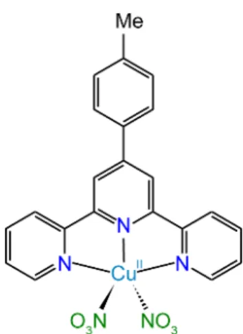
Although specificity for quadruplexes vs. other DNA structures has been achieved by several ligand families, a good selectivity between G-quadruplex families has been much more rarely reported. We studied here the binding of the Cu(II)-4'-(p-tolyl)-2,2':6',2''-terpyridine (Cu-ttpy) ligand (Figure 1) and of some derivatives to several oligonucleotide sequences (listed in ESI Table S1). The Cu, Zn, Pt and Pd complexes were synthesized in-house by mixing the ligand and the metal salt (Cl- or NO
3-)[5a, 7] (ESI Section 2 and Figures S1-S10). We report herein that the binding
of Cu-ttpy derivatives is very specific for telomeric DNA G-quadruplexes, and that the ligand binding to telomeric sequences is accompanied by a striking 2:1 (ligand:DNA) binding cooperativity, which is correlated to changes of the telomeric structure to an antiparallel conformation with 3 G-quartets.
Figure 1. Structure of the ligand Cu-ttpy (dinitrate salt).
Electrospray mass spectrometry (ESI-MS) in non- denaturing conditions was key to study the stoichiometries of the complexes and determine their individual KD values. Previous studies by
G-
quadruplexes were prepared here in ESI-compatible 1-mM potassium conditions using trimethylammonium acetate (TMAA).[10] To determine the K
D values (Eq 1-2), we used the peak
areas of the 5- charge state of free DNA and DNA bound with 1 or 2 ligands, assuming that the response factors are equal for all the species (the verifications that validate this hypothesis are presented in Figure S11). Although this sounds surprising, equal response seems the rule rather than the exception in DNA-ligand complexes: the electrospray response depends more on the total charge than on how the individual charges are distributed over the molecule.[11]
KD1 = [DNA][L]/[DNA•L] (Eq. 1)
KD2 = [DNA•L][L]/[DNA•L2] (Eq. 2)
Figure 2 shows the ESI-MS titrations of two G-quadruplexes by the Cu-ttpy ligand. The curves of DNA concentration vs total ligand concentration were fitted using the DynaFit software[12] to obtain KD1 and KD2. The DynaFit software works by iteration on the two KD values in order to fit all the
concentrations of the three stoichiometries as a function of total ligand added. The inputs are the concentrations of each species obtained by MS (0:1, 1:1 and 2:1 L:DNA stoichiometries) and the output consist in the values of the KD and the fits. We observed ligand binding, without nitrate
counter-ions, to both sequences. We quantified the complexes of each stoichiometry and fitted the titration data for the human telomeric G-quadruplex 22GT (dGGG(TTAGGG)3T),[13] for the
parallel G-quadruplex from the c-myc promoter Pu24,[14] (Figure 2), and for the bimolecular
antiparallel G-quadruplex [dG4T4G4]2[15] (Figure S12 for full titration). For the other sequences, the KD values were estimated using single-concentration measurements.[11a] In such case, the ligand
free concentration is deduced using the total ligand concentration and the concentration of all complexes.
Figure 2. Top, MS titrations of 10 µM 22GT or Pu24 by Cu-ttpy in 100 mM TMAA + 1 mM KCl. Zooms on the 5- charge state. Bottom, quantification and fitting.
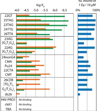
Equilibrium dissociation constants for 21 sequences including 8 human telomeric sequences, 11 other G-quadruplexes (both parallel and antiparallel), one hairpin and one single strand (see ESI Table S1 for the full list of sequences) are reported in Figure 3 (see ESI Table S3 for the values). The product KD1×KD2 represents the global constant for the binding of two ligands. The
total amount of ligand bound was also calculated, and the percentage ligand bound in the 10µM:10µM (ligand:DNA) mixtures is shown in Figure 3. We observed a very good binding of
Cu-ttpy to G-quadruplexes, with a high affinity for all the human telomeric ones. Remarkably, the
amount of bound Cu-ttpy on telomeric sequences (>90%) is similar to what is obtained for some of the most potent G-quadruplex ligands reported to date (360A, Phen-DC3 or pyridostatin, see ESI Figure S15). Cu-ttpy binds with low affinity to the single stranded 24nonG4 and the hairpin ds26. This result confirms the selectivity in favour of G-quadruplexes, and a particular selectivity for all telomeric sequence derivatives.
Indeed, even not all pre-formed G-quadruplexes are targets for this ligand. For example, the sequence ckit1 binds the ligand exclusively without trapping any potassium ion (the fraction unfolded in solution), but the quadruplex form (which contains 2 potassium ions) does not bind the ligand (Figure S16A). The HIV-PRO1, a 2-quartet antiparallel G-quadruplex, is not folded in our MS conditions (the 0-K+ form predominates). When one ligand bind to the sequence, although
the amount of 1-K+ complex is a little increased, the ligand binds mostly to the 0-K+ species (Figure
S16B). Cu-ttpy did not bind either to the thrombin binding aptamer TBA (Figure S16C), an antiparallel G-quadruplex with only two G-quartets.
Figure 3. Comparison of the log KD1 (orange) and log KD2 (green) and the percentage of ligand
bound at 1 equivalent (10 µM) for the 21 sequences for Cu-ttpy. The sequences are ranked according to their KD1/KD2 ratio (Table S3).
For the telomeric sequences only, the distribution of ligand binding stoichiometries is strongly biased towards the 2:1 complex. Upon titration the 1:1 (Ligand:DNA) complex is almost not populated, in contrast with the 2:1 complex (see Figure 2). The ligand binding is therefore highly cooperative. We also tested other electrolyte conditions (100 mM NH4OAc) to probe whether the
low potassium conditions were responsible for the cooperativity, and the results were very similar to those obtained in TMAA + KCl (Figure S17).
We ranked the sequences according to their ratio KD1/KD2, which indicates cooperativity
(Table S3),[11a] and found a very high cooperativity (KD1/KD2 > 5) for almost all human telomeric
sequences (22GT, 25TAG, 23TAG, 24TTG and 26TTA) (Figure 3). The highest cooperativity is obtained for 22GT, KD1 being almost 2 orders of magnitude larger than KD2. Only the two human
telomeric sequences beginning with an adenine (22AG and 23AG) were binding Cu-ttpy less cooperatively (5>KD1/KD2>0.7). No cooperativity is observed for any of the other sequences, for
example all derivatives forming parallel G-quadruplexes (c-myc promoter sequences Pu24, CMA and CMT), the [dTG4T]4 tetramolecular parallel G-quadruplex and 26CEB, a parallel G-quadruplex
To probe which ligand properties are essential to the cooperativity, we synthetized two derivatives of the Cu-complexes (Scheme S1): Cu-tpy (lacking the tolyl group) and Cu-Clptp (Chlorophenyterpyridine, the methyl group being replaced by a chlorine, see ESI section 2).
Cu-tpy was previously described as a poor binder[5a] and Cu-Clptp is reported here for the first time.
ESI-MS confirmed the poor binding affinity of the Cu-tpy complex (Figure S14), and the small amount of ligand that binds does so non-cooperatively (Figure S18 and Table S3). In contrast, Cu-Clptp behaves exactly as Cu-ttpy, although being a little bit less affine for G-quadruplexes (see Figure S13 and Table S3 for the KD values). The presence of the fourth aromatic moiety
therefore seems essential for both the high affinity and high cooperativity of ligand binding. Next, to test the effect of the metal centre, we replaced copper by Zn, Pd or Pt (Figure S18). As known already from past studies,[5a] Zn-ttpy is not a good ligand (it binds with poor affinity to all DNA
sequences), due to the trigonal bipyramidal geometry adopted by Zn(II), which leads to steric hindrance. Pt- and Pd-complexes bind to the G-quadruplex[7] but without any cooperativity: only
a 1:1 complex is forming. For the Pt derivatives, the ligand first binds non-covalently, then loses chlorine and binds covalently to the DNA, as described previously.[7, 16] Changing the metal core
of the ligand abolished the cooperative behaviour. We also synthesized and tested the chloride salt of the ligands. Like for NO3-, the counter ion was not observed directly bound (the ligand binds
as CuL2+). The nature of the anion did not influence the binding affinity or cooperativity of those
ligands. In conclusion, both the extended aromatic moiety and the preferred square pyramidal penta-coordination mode enabled by Cu(II) are essential both for the high binding affinity[6a] and
for the binding cooperativity.
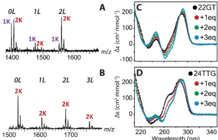
In order to get information on possible conformational changes induced by the ligand, we examined the number of potassium ions bound, as detected using ESI-MS, on the complexes with and without ligands (Figure 4A). In our conditions, the free G-quadruplex of 22GT is formed with both 1 and 2 potassium ions. The minor 1:1 complex presents a K+ distribution close to that
of the free G-quadruplex. In contrast, the 2:1 complex is different: the 2-K+ stoichiometry is
preferred. We previously reported potassium ions ejection from the human telomeric G-quadruplexes upon 1:1 complex formation with ligands 360A, Phen-DC3 or pyridostatin.[17] Here
with Cu-ttpy we observe the complete opposite. For all human telomeric sequences, the 2:1 complex is binding 2-K+ even when the free sequence did not contain purely 2-K+: 23AG, 22AG
and 22CTA (ESI Figure S19). Interestingly, even if 22CTA[18] binds the ligand in a non-cooperative
manner on the population level (total 2:1 complex vs. total 1:1 complex), the second ligand binding event also induces the capture of one more potassium ion. Those hints of structural changes were observed only for the human telomeric sequence.
Circular dichroism (CD) was also performed on the 22GT and 24TTG sequences to study the ligand-induced changes in preferential base-stacking (Figure 4C-D). The CD spectra indeed display different signatures according to the stacking of the guanines involved in G-quartets: a positive peak at 260 nm is indicative of parallel G-quadruplexes, a negative peak at 260 nm and a positive at 290 nm of antiparallel G-quadruplexes, and two positive peaks at 270 and 290 nm indicate hybrid G-quadruplexes. For 22GT, the starting structure has a CD spectrum close to the typical antiparallel one. Addition of Cu-ttpy influences the CD signature, making the negative band at 260 nm more negative. The presence of isodrichroic points at 254 and 286 nm indicates a
single transition between two species, i.e. the free G-quadruplex and the 2:1 complex. Following the same trend, the CD titration of 24TTG shows a transition from the hybrid structure to a more antiparallel one. On the other hand, the CD spectra of Pu24, a parallel G4, and [dG4T4G4]2, an
antiparallel one, are not influenced by the binding of Cu-ttpy (Figure S20). Indeed, no change in the potassium stoichiometry was observed. Despite a small intensity decrease for Pu24’s CD signal, the fact that there is no change in the CD spectrum upon binding also rules out possible ligand-induced CD signal that could affect the CD signature.
The structural preference for the antiparallel human telomeric G-quadruplex has been observed before for triarylpyridine-platinum complexes[16, 19] or a dinuclear Ru-complex.[20] Other
ligands were previously shown to alter the human telomeric G-quadruplexes, as for example TMPyP4, inducing the formation of more hybrid G-quadruplexes.[21] Another example is the
N-methyl mesoporphyrin IX that binds preferentially to parallel G-quadruplexes.[22] Cu-ttpy is
however the first example of a ligand for which this structural preference is accompanied by the cooperative binding of two ligands.
Figure 4. Topology characterization of the binding of Cu-ttpy. MS and determination of the number of potassium in the complexes for one Cu-ttpy equivalent A, for 22GT and B, for 24TTG. CD of C, 22GT and D, 24TTG with increasing amount of ligand.
In order assess whether the ligands are bound to proximal or distant sites, we prepared an equimolar mixture of Cu-ttpy and Cu-Clptp ligands, which was added to the 22GT G-quadruplex (Figure S21). We found a maintained cooperativity, but a purely statistical distribution of each ligand, suggesting that the nature of one ligand bound does not condition the nature of the second ligand bound. The result of this competition experiment therefore suggests that the ligands do not interact with the quadruplex as pre-formed dimers.
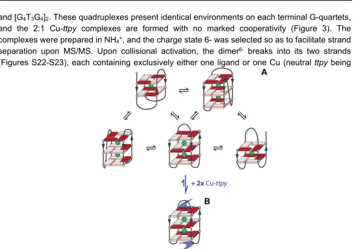
A rather common ligand binding mode in G-quadruplexes, compatible with the 2:1 stoichiometry, is stacking of one ligand to each terminal G-quartet. To test that structural hypothesis easily with mass spectrometry, we used the bimolecular G-quadruplexes [dG4T4G4]2
and [G4T3G4]2. These quadruplexes present identical environments on each terminal G-quartets,
and the 2:1 Cu-ttpy complexes are formed with no marked cooperativity (Figure 3). The complexes were prepared in NH4+, and the charge state 6- was selected so as to facilitate strand
separation upon MS/MS. Upon collisional activation, the dimer6- breaks into its two strands
(Figures S22-S23), each containing exclusively either one ligand or one Cu (neutral ttpy being
ejected from the complex). This strongly suggests that the two ligands are bound at distal positions, so the ligands presumably bind to each of the two external G-quartets.
Figure 5. A) The various topologies that the human telomeric sequence can adopt in solution. B) One of the possible preferred topology of the 2:1 complex with Cu-ttpy: an antiparallel structure with 3 G-quartets.
In summary, for the intramolecular telomeric G-quadruplexes, the preferred binding of
Cu-ttpy is therefore to an antiparallel, three-quartet structure, presumably on each terminal G-quartet
(Figure 5B). The ligand is also selective among several possible antiparallel structures. Indeed, the binding to the antiparallel quadruplexes HIV-PRO1 and TBA is extremely poor. These G-quadruplexes are characterized by lateral-lateral-lateral loop arrangement and a wide-narrow-wide-narrow groove width order. This is also in line with the higher binding affinity for [dG4T4G4]2
and dG4(T4G4)3 as compared to [dG4T3G4]2. Among antiparallel structures, the ligand Cu-ttpy
apparently prefers structures with diagonal loops and/or wide-medium-narrow-medium groove width order. In the human telomeric sequences, the preferred quadruplex fold would therefore be the antiparallel with lateral-diagonal-lateral loops, i.e. the same fold as formed in Na+ by 22AG.
Interestingly, past results showed a higher FRET-Tm when 5 eq. Cu-ttpy are added to the human
telomeric sequence F21T in NaCl (+15.3°C)[5a] compared to in KCl (+6°C).[7]
The binding of the two ligands to the human telomeric G-quadruplexes is accompanied by a conformational change. This conformational change could occur after the binding of the ligands:
this mechanism is called induced fit.[23] For ligands prone to π-π stack on external G-quartets, this
scenario is very unlikely because ligand binding should then be followed by an unfolding of the G-quadruplex. Indeed, to form the antiparallel, 3-quartet G-quadruplex starting from any of the hybrid topologies, all G-quartets must first be broken to release at least one strand,[24] and if
starting from hybrid I or II, to allow the groove widths to rearrange in order to be compatible with the antiparallel strand arrangement. We therefore favor a conformational selection mechanism (Figure 5), in which the antiparallel, 3-quartet structure, although minor in the ligand-free form, becomes predominant upon binding of two ligands which stack on the top and bottom quartet.
In conclusion, Cu(II)-tolylterpyridinium complex derivatives bind all the human telomeric G-quadruplex variants with high affinity, high specificity, and high cooperativity to form 2:1 complexes. The cooperative ligand binding is accompanied by a conformational change to an antiparallel structure with three G-quartets. The cooperativity is due to a combined effect of (1) allosteric changes in the G-quadruplex upon ligand binding, (2) conformational preference of the ligand, and (3) the fact that one ligand is not sufficient to promote the conformational equilibrium displacement. This ligand binding mechanism points to a structural selectivity for one particular fold, and the ability to switch the conformational equilibria of all telomeric sequence variants to this fold.
Acknowledgements
This work was financially supported by the Inserm (ATIP-Avenir Grant no. R12086GS), the Conseil Régional Aquitaine (Grant no. 20121304005), and the EU (FP7-PEOPLE-2012-CIG-333611 and ERC-2013-CoG-616551). D.S. benefited from an Erasmus+ traineeship from the University of Warsaw to the University of Bordeaux. The authors thank Dr. Eric Largy and Mélanie Lenfant (M1 student, U. Bordeaux) for contributing preliminary experiments, Dr. Céline Douat (CBMN laboratory) for her assistance with the synthesis of the Zn, Pt and Pd complexes and Dr. Stéphane Massip (Structural biophysicochemistry platform, IECB) for the crystal structures of the ligands.
Keywords: mass spectrometry • G-quadruplex • ligand • cooperativity • affinity
[1] a) L. A. Cahoon and H. S. Seifert, Science 2009, 325, 764-767; b) S. Kumari, A. Bugaut, J. L. Huppert and S. Balasubramanian, Nature Chem. Biol. 2007, 3, 218-221; c) K. Paeschke, J. A. Capra and V. A. Zakian, Cell 2011, 145, 678-691; d) A. Siddiqui-Jain, C. L. Grand, D. J. Bearss and L. H. Hurley, Proc. Natl. Acad. Sci. USA 2002, 99, 11593-11598.
[2] a) J.-L. Mergny, J.-F. Riou, P. Mailliet, M.-P. Teulade-Fichou and E. Gilson, Nucleic Acids Res. 2002, 30, 839-865; b) A. Rizzo, E. Salvati, M. Porru, C. D'Angelo, M. F. Stevens, M. D'Incalci, C. Leonetti, E. Gilson, G. Zupi and A. Biroccio, Nucleic Acids Res. 2009, 37, 5353-5364; c) Y. Wang and D. J. Patel, Biochemistry 1992, 31, 8112-8119.
[3] T.-M. Ou, Y.-J. Lu, J.-H. Tan, Z.-S. Huang, K.-Y. Wong and L.-Q. Gu, ChemMedChem 2008,
3, 690-713.
[5] a) H. Bertrand, D. Monchaud, A. De Cian, R. Guillot, J.-L. Mergny and M.-P. Teulade-Fichou,
Org. Biomol. Chem. 2007, 5, 2555-2555; b) N. H. Campbell, N. H. A. Karim, G. N. Parkinson,
M. Gunaratnam, V. Petrucci, A. K. Todd, R. Vilar and S. Neidle, J. Med. Chem. 2012, 55, 209-222; c) J. E. Reed, A. A. Arnal, S. Neidle and R. Vilar, J. Am. Chem. Soc. 2006, 128, 5992-5993.
[6] a) A. Arola-Arnal, J. Benet-Buchholz, S. Neidle and R. N. Vilar, Inorg. Chem. 2008, 47, 11910-11919; b) S. E. Evans, M. A. Mendez, K. B. Turner, L. R. Keating, R. T. Grimes, S. Melchoir and V. A. Szalai, J. Biol. Inorg. Chem. 2007, 12, 1235-1249; c) J. Pan and S. Zhang, J. Biol.
Inorg. Chem. 2009, 14, 401-407.
[7] E. Largy, F. Hamon, F. Rosu, V. Gabelica, E. De Pauw, A. Guédin, J.-L. Mergny and M.-P. Teulade-Fichou, Chem. Eur. J. 2011, 17, 13274-13283.
[8] a) E. Izbicka, R. T. Wheelhouse, E. Raymond, K. K. Davidson, R. A. Lawrence, D. Sun, B. E. Windle, L. H. Hurley and D. D. Von Hoff, Cancer Res. 1999, 59, 639-644; b) D.-F. Shi, R. T. Wheelhouse, D. Sun and L. H. Hurley, J. Med. Chem. 2001, 44, 4509-4523.
[9] a) J. Alzeer, B. Vummidi, P. Roth and N. W. Luedtke, Angew. Chem. Int. Edit. 2009, 48, 9362-9365; b) A. Membrino, M. Paramasivam, S. Cogoi, J. Alzeer, N. W. Luedtke and L. E. Xodo,
Chem. Commun. 2010, 46, 625-627.
[10] A. Marchand and V. Gabelica, J. Am. Soc. Mass Spectr. 2014, 25, 1146-1154.
[11] a) F. Rosu, E. De Pauw and V. Gabelica, Biochimie 2008, 90, 1074-1087; b) V. Gabelica, F. Rosu and E. De Pauw, Anal. Chem. 2009, 81, 6708-6715.
[12] P. Kuzmic, Anal. Biochem. 1996, 237, 260-273.
[13] K. W. Lim, S. Amrane, S. Bouaziz, W. Xu, Y. Mu, D. J. Patel, K. N. Luu and A. T. Phan, J.
Am. Chem. Soc. 2009, 131, 4301-4309.
[14] A. T. Phan, V. Kuryavyi, H. Y. Gaw and D. J. Patel, Nat Chem Biol 2005, 1, 167-173. [15] P. Schultze, F. W. Smith and J. Feigon, Structure 1994, 2, 221-233.
[16] H. Bertrand, S. Bombard, D. Monchaud, E. Talbot, A. Guédin, J.-L. Mergny, R. Grünert, P. J. Bednarski and M.-P. Teulade-Fichou, Org. Biomol. Chem. 2009, 7, 2864-2864.
[17] A. Marchand, A. Granzhan, K. Iida, Y. Tsushima, Y. Ma, K. Nagasawa, M.-P. Teulade-Fichou and V. Gabelica, J. Am. Chem. Soc. 2015, 137, 750-756.
[18] K. W. Lim, P. Alberti, A. Guédin, L. Lacroix, J. F. Riou, N. J. Royle, J. L. Mergny and A. T. Phan, Nucleic Acids Res. 2009, 37, 6239-6248.
[19] J.-T. Wang, Y. Li, J.-H. Tan, L.-N. Ji and Z.-W. Mao, Dalton Trans. 2011, 40, 564-566. [20] T. Wilson, P. J. Costa, V. Félix, M. P. Williamson and J. A. Thomas, J. Med. Chem. 2013, 56,
8674-8683.
[21] R. D. Gray, J. Li and J. B. Chaires, J. Phys. Chem. B 2009, 113, 2676-2683.
[22] J. M. Nicoludis, S. P. Barrett, J.-L. L. Mergny and L. A. Yatsunyk, Nucleic Acids Res. 2012,
40, 5432-5447.
[23] S. Gianni, J. Dogan and P. Jemth, Biophys Chem 2014, 189, 33-39.
[24] P. Stadlbauer, M. Krepl, T. E. Cheatham, 3rd, J. Koca and J. Sponer, Nucleic Acids Res 2013, 41, 7128-7143.