Structural analyses of the aquaporin super‐family
3
0
0
Texte intégral
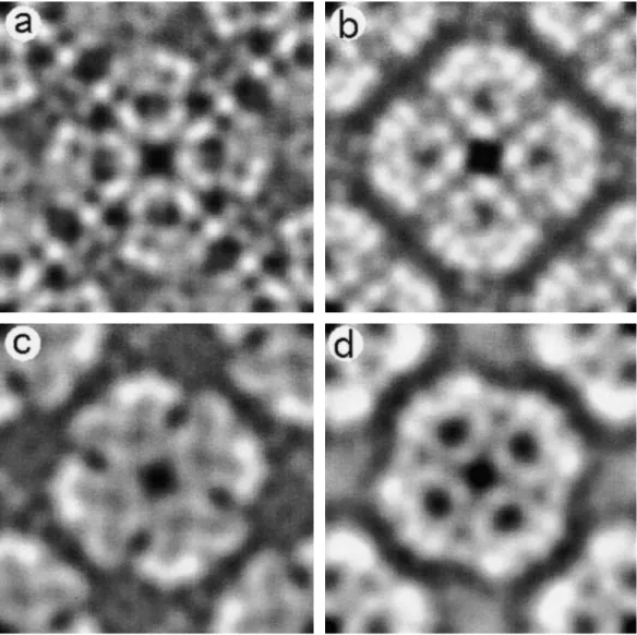
(2) 24. A. Engel. ˚ Fig. 1. Projection maps of water channel proteins acquired by cryo-electron microscopy. (a) The red cell water channel, AQP1, at 3.5 A ˚ . (b) The lens fibre cell water resolution. AQP1 packs into arrays with P42 2 symmetry, housing two tetramers per unit cell of size 96 A 1 ˚ resolution [14]. AQP0 ˚ side length. The area channel, AQP0, at 5.7 A packs into P4 arrays with a single tetramer per unit cell of 64 A ˚ resolution [10]. AqpZ is packed in an up-and-down shown comprises two unit cells. (c) The bacterial water channel, AqpZ, at 8 A ˚ width. (d ) The bacterial glycerol diffusion facilitator, GlpF, rendered here at 7 A ˚ resolution orientation as AQP1 into unit cells of 94 A ˚ width, but the crystals exhibit a P4 symmetry. [11]. Two GlpF tetramers are packed in an up-and-down orientation into unit cells of 104 A All projections are viewed from the cytoplasmic side.. density, which consists of two V-shaped regions touching one another in the centre of the AQP1 monomer ˚ resolution, the to form the density ‘X’ [8]. At 4.5 A central density is now resolved as two short helices projecting outwards from the centre of the monomer, connected to adjacent helices by loop regions [17].. visualized and its functional implication to be unravelled. Froger et al. [18] have proposed to distinguish the AQPs and the GLPs based on five particular amino acid residues. Further clues are found in the more recent sequence analysis by Heymann and Engel [19], who have identified two critical conserved hydrophobic residues in the middle of helices 1 and 4.. Major differences between AQPs and GLPs Perspectives The similarity of projection maps (Figure 1) suggests that the architecture of these proteins, namely AQP1, AQP0, AqpZ and GlpF, is similar within the membrane core which houses the six helices and two functional loops. The major difference between AQPs and GLPs is that loop E is longer by ~10–15 residues in the latter, but this difference still needs to be. The atomic structures of members of the two subfamilies AQP and GLP of the aquaporin super-family are now required in order to understand the function of these ubiquitous channels. Such knowledge is important, since it ultimately may be used to develop therapeutic agents for important clinical disorders. 3D.
(3) Structural analyses of the aquaporin super-family. maps from electron crystallographic analyses now approach a sufficient resolution to build an atomic model, and highly ordered 2D crystals of AQP0, AQP1, AqpZ, a-TIP [20] and GlpF [11] are now available. This progress promises that this goal should be reached soon.. References 1. Gorin MB, Yancey SB, Cline J, Revel J-P, Horwitz J. The major intrinsic protein (MIP) of the bovine lens fiber membrane: characterization and structure based on cDNA cloning. Cell 1984; 39: 49–59 2. Preston GM, Agre P. Isolation of the cDNA for erythrocyte integral membrane protein of 28 kilodaltons: member of an ancient channel family. Proc Natl Acad Sci USA 1991; 88: 11110–11114 3. Preston GM, Carroll TP, Guggino WB, Agre P. Appearance of water channels in Xenopus oocytes expressing red cell CHIP28 protein. Science 1992; 256: 385–387 4. Heymann J, Engel A. Aquaporins: phylogeny, structure and physiology of water channels. News Physiol Sci 1999; 14: 187–193 5. Jung J, Preston G, Smith B, Guggino W, Agre P. Molecular structure of the water channel through aquaporin CHIP. The hourglass model. J Biol Chem 1994; 269: 14648–14654 6. Smith BL, Agre P. Erythrocyte M 28,000 transmembrane r protein exists as a multisubunit oligomer similar to channel proteins. J Biol Chem 1991; 266: 6407–6415 7. Verbavatz J-M, Brown D, Sabolic I et al. Tetrameric assembly of CHIP28 water channels in liposomes and cell membranes: a freeze-fracture study. J Cell Biol 1993; 123: 605–618 ˚ three-dimensional 8. Walz T, Hirai T, Murata K et al. The 6 A structure of aquaporin-1. Nature 1997; 387: 624–627. 25 9. Hasler L, Walz T, Tittmann P, Gross H, Kistler J, Engel A. Purified lens major intrinsic protein (MIP) forms highly ordered tetragonal two-dimensional arrays by reconstitution. J Mol Biol 1998; 279: 855–864 10. Ringler P, Borgnia MJ, Stahlberg H, Agre P, Engel A. Structure of the water channel AqpZ from Escherichia coli revealed by electron crystallography. J Mol Biol 1999; 291: 1181–1190 11. Braun T, Philippsen A, Wirtz S et al. Projection structure of the ˚ resolution. EMBO Rep 2000; 1: glycerol facilitator at 3.5 A 183–189 12. Jap BK, Li H. Structure of the osmo-regulated H O-channel, ˚ resolution. J Mol2 Biol 1995; AQP-CHIP, in projection at 3.5 A 251: 413–420 13. Walz T, Smith B, Agre P, Engel A. The three-dimensional structure of human erythrocyte aquaporin CHIP. EMBO J 1994; 13: 2985–2993 14. Fotiadis D, Hasler L, Mu¨ ller DJ, Stahlberg H, Kistler J, Engel A. The surface topography of lens MIP supports dual functions. J Mol Biol 2000; 300: 779–789 15. Li H, Lee S, Jap BK. Molecular design of aquaporin-1 water channel as revealed by electron crystallography. Nature Struct Biol 1997; 4: 263–265 16. Cheng A, van Hoek AN, Yeager M, Verkman AS, Mitra AK. Three-dimensional organization of a human water channel. Nature 1997; 387: 627–630 17. Mitsuoka K, Murata K, Walz T et al. Short-helices in hourglass pore-forming domains of AQP1 water channel protein visualized ˚ . J Struct Biol 1999; 128: 34–43 at 4.5 A 18. Froger A, Tallur B, Thomas D, Delamarche C. Prediction of functional residues in water channels and related proteins. Protein Sci 1998; 7: 1458–1468 19. Heymann JB, Engel A. Structural clues in the sequences of the aquaporins. J Mol Biol 2000; 295: 1039–1053 20. Daniels MJ, Chrispeels MJ, Yeager M. 2D crystallization of a plant vacuole membrane aquaporin and determination of its projection structure by electron crystallography. J Mol Biol 1999; 294: 1337–1349.
(4)
Figure
Documents relatifs
describe their experiments at a large-scale comparison of timbre and cultural similarity measures, on a large database on songs (8772). The ex- periments notably focus on the
With the last statement we can now implement the strategy of Theorem 5.20 in [13] in order to prove the continuity of the curtain coupling Curt.. The proof relies on the fact
is the disjoint union of a twisted cubic and a line, so this case gives an irreducible family, and the general element belongs to E 12.. Let us follow the strategy
Second, we provide empirical evidence that the choice of an appropriate family of models is often more important—and sometimes much more important, especially when the size of
This study is focused around the extracellular domain of two single-pass transmembrane receptors of the Roundabout and UNC5 protein families that are majorly involved
L’archive ouverte pluridisciplinaire HAL, est destinée au dépôt et à la diffusion de documents scientifiques de niveau recherche, publiés ou non, émanant des
However, this logic (if sense) must not be reduced to the Manichean opposition between the quan- titative actual and qualitative virtual. The difference between the difference in
Key Words: Zeros of Entire Functions, Exponential Polynomials, Almost- Periodic Functions, Partial Sums of the Riemann Zeta Function..
