Direct evidence for a functional role of
HLA-DRB1 and -DRB3 gene products in the
recognition of Dermatophagoides spp.
(house dust mite) by helper T lymphocytes
Robyn E. O'Hehir, Bernard Mach1, Christine Berte1, Roseanna Greenlaw,
Jean-Marie Tiercy1, Vineeta Bal, Robert I. Lechler, John Trowsdale2, and
Jonathan R. Lamb
Department of Immunology, Royal Postgraduate Medical School, Hammersmith Hospital, Du Cane Road, London W12 ONN, UK
'Department of Microbiology, University of Geneva Medical School, 1211 Geneva 4, Switzerland imperial Cancer Research Fund, Lincoln's Inn Fields, London WC2A 3PX, UK
Key words: HLA class II, B3 gene products, T cell recognition, house dust mite, Dermatophagoides spp.
Abstract
The contribution of the HLA-DRB1, -B3, and -BS gene products in the recognition of
Dermatophagoides spp. (house dust mite) by helper T cells Isolated from an atopic individual
(HLA-DRw12, DR7; DRw52b) with perennial rhinitis was investigated. Using a panel of
histocompatlble and histoincompatible accessory cells, the restriction specificity obtained for a long term T cell suggested that a component of the dust mite reactive repertoire recognized antigen in association with DRB3 gene products. Ollgonucleotide DNA typing of the presenting cell panel demonstrated a correlation between the DRw52b allele and T cell responsiveness. Murine fibroblasts expressing DRw52b, but not DRw52a or -c molecules, presented antigen to both the T cell line and cloned T cells (DE26) derived from the line, Indicating that the supertypic specificity DRw52b was able to restrict recognition of dust mite antigens. Additional T cell clones (DE9 and DE41) also isolated from the line were restricted by the products of the B1 gene locus (DRw12B1) as determined by murine fibroblasts transfected with the appropriate HLA-DR genes. Clone DE9 was degenerate in Its restriction specificity, also recognizing dust mite presented by accessory cells expressing the DR2 subtypes. Presentation by fibroblasts transfected with DRw12B1, DR2Dw2B5 genes and EBV-transformed B cell lines expressing DR2Dw21B1 and -B5 indicated that the functional site restricting recognition may be associated with residues 70 and 71 of the DR/3 chain helical wall of the antigen combining site. Furthermore, we have recently demonstrated that both T cell clones DE9 and DE26 induce allergen dependent IgE synthesis
in vitro. Thus these results demonstrate directly that the DRB1, -B3, and -B5 gene products are
functional in the restriction of T cell recognition of dust mite antigens. Introduction
Immune responses are initiated following the interaction of the antigen specific receptor of CD4+ T lymphocytes with molecular complexes formed between peptide fragments of antigen and MHC class II proteins (1-4). In man, MHC class II genes encode three major families of molecules, HLA-DR, -DQ, and -DP, all of which appear to function in the regulation of immune responsiveness (5-7). Furthermore, the DR locus on most haplotypes contains two expressed B genes, the products of which pair with the same DRa chain to give rise to two DR
Correspondence to' R. E. O'Hehir Transmitting editor: T. Sasazuk)
molecules, DRa/3l and DRo^lll (or DRa/3IV or DRajSV), the major DR and the supertypic specificities respectively, both of which are polymorphic and able to restrict T cell antigen recognition (8-10).
Immune recognition of the aeroallergen Dermatophagoides spp. (house dust mite) induces immunoglobulin E (IgE) synthesis and symptomatic allergic disease in 10 - 1 5 % of the population. Detailed analysis of the specificity and effector function of IgE in mite allergy is well documented (11-13). Although IgE
synthesis is T cell dependent, only limited information is available on the antigen and MHC class II restriction specificity of the T cell repertoire activated in atopic individuals after exposure to house dust mite. Investigation of polyclonal responses to only the group I allergen of Dermatophagoides pteronyssinus, Der
p I, indicated that T cells from atopic but not control non-atopic
subjects proliferated to allergen (14). In addition, the results of experiments using anti-HLA class II antibodies to inhibit the antigen-dependent response of D. pteronyssinus reactive T cell clones and lines indicated that HLA-DR molecules restricted recognition of house dust mite but failed to identify the HLA-D region locus encoding the restriction elements used (15).
In previous studies we examined the HLA class II restriction of D. farinae reactive T cell clones isolated from the peripheral blood of an atopic individual using serological inhibition and allogeneic presenting cell panels (16,17). Complex patterns of restriction specificities were obtained suggesting that the product of locus DRAB3, in addition to the DRAB1 gene products, may function as restriction elements in T cell recognition. This prompted us to investigate the functional role of DRw52 class II molecules in the allergic response by correlating the patterns of reactivity of dust mite specific T cells with the results of DRB3 allele-specific hybridization with oligonucleotide probes in panel studies. Additionally, T cell clones capable of supporting allergen dependent IgE synthesis in vitro (18) were isolated from the T cell line and their restriction specificities examined using homozygous Epstein - Barr virus (EBV) transformed B cells and munne fibroblast transfectants expressing specific HLA-DR genes as antigen presenting cells (APC). The results of these experiments indicate that HLA-DRB1, -B3, and -B5 gene products are able to restrict the recognition of house dust mite and have allowed the mapping of functional sites on these HLA class II proteins.
Methods
Antigens
Lyophilized extracts of D. farinae were obtained from Pharmacia Ltd, Uppsala, Sweden.
Isolation of antigen reactive T cell line and clones
The isolation of the T cell line and clones used in these experiments has been described in detail elsewhere (16,17). Briefly, peripheral blood mononuclear leukocytes (PBMC; 2.5 x 10s/ml) were stimulated with an optimal concentration of
D. farinae [103 Biological Units (BU)/ml] for 7 days in RPMI 1640 (Flow Laboratories, Irvine, Ayrshire, UK) medium supplemented with 2 mM i-glutamine, 100 U/ml penicillin/strepto-mycin, and 5% v/v screened, heat-inactivated human A+ serum (National Blood Transfusion Centre, Edinburgh, UK). Lympho-blasts enriched on Ficoll-Paque (Pharmacia) were maintained as a long term line in the presence of irradiated (2500 rad) autologous PBMC, D. farinae, and interleukin 2 (IL-2, 10% v/v; Lymphocult T, Biotest Folex, Frankfurt, FRG), or cloned by limiting dilutjon from the line. For cloning, viable cells (0.3 cells/well) were plated in Microtest II trays together with irradiated autologous PBMC (5 x 10⁵/ml), D. farinae, and IL-2. After 7 days, growing clones were transferred to 96-well flat-bottom microtitre trays and subsequently to 24-well trays. At each transfer the clones received
filler cells, antigen, and IL-2. The clones and the line were main-tained and expanded by the addition of IL-2 every 3 - 4 days and antigen together with filler cells every 7 days. Prior to their use in proliferation assays, the T cell clones and the line were rested for 6 - 8 days after the last addition of filler cells and antigen.
Antigen specificity of the T cell line and clones
The T cell line (DX) recognizes both major species of house dust mite (D. farinae and D. pteronyssinus) but the predominant specificity was for the group II allergen {Der f II; 12.5 kd) of
D. farinae as determined using nitrocellulose immunoblots of
fractionated antigen (16) T cell clones DE9 and DE26 responded to both dust mite species, with the determinant recognized by DE9 being further mapped to the group I allergens (26 kd). The T cell clone DE41 was only stimulated by D. farinae preparations.
Cell populations used as antigen presenting cells
Autologous and allogeneic PBMC, homozygous EBV trans-formed B cells, and murine fibroblasts transfected with HLA-D region genes were used as antigen presenting cells. The HLA-typed antigen presenting cell panel was generously provided by Dr D. Eckels, Blood Center of Southeastern Wisconsin, Milwaukee, Wl. The cloning of the DR1Dw1, DRw2Dw2B1 (DRB1*1501), DR2Dw2B5, DRw12B1, DR7Dw17, and DRw52a, -b, and -c genes and their co-transfection with the DRA gene into the Ltk- fibroblast cell line (DAP3) has been described elsewhere (19-25).
T lymphocyte proliferation assays
T cells of the long term line (DX) and clones (DE9, DE26, and DE41; 5 x lO^/ml) were cultured with antigen in the presence of irradiated HLA typed histocompatible and histoincompatible PBMC (1.25 x 10⁵/ml), EBV transformed B cells (10s/ml), or mitomycin C treated transfected murine fibroblacts (lO^/ml) in a total volume of 200 ^1 of complete medium in 96-well U-bottom plates. Assays in which fibroblasts were used as presenting cells were performed in 96-well flat-bottom plates. After 72 h of incubation, tritiated methyl thymidine (1 jiCi/well, PHVTdR; Amersham International Inc., Amersham, UK) was added to cultures which were harvested onto g!a&s fibre filters, 8 - 1 6 h later. Proliferation as correlated with [3H]TdR incorporation was measured by liquid scintillation spectroscopy. The results are expressed as mean counts per minute (c p.m.) + % error of the mean for triplicate cultures and were < 2 0 % in all experiments.
Mitomycin C treatment of murine fibroblasts
After trypsinization, cells were washed in serum free medium. Cells, up to 107/ml, were suspended in serum free medium and mitomycin C (Sigma) was added to a final concentration of 50/xg/ml. Cells were incubated at 37°C for 45 mm, washed extensively in A+ supplemented medium, and used in proliferation assays (20).
Oligonucleotide DNA typing
DNA was prepared from the presenting cell panel and probed with oligonucleotides labelled with [^-^PJdATP as described previously (21). The oligonucleotide 52a (GGAGCT-GCGTAAGTCTGAG) probe is complementary to the nucleotide sequence residues 1 1 - 2 9 of DRw52a which encode for
amino acid residues 8 - 1 4 of the /31 domain of the DRw52a allele. Oligonucleotide 52b (GTTCCTGGAGAGACACTTCC) is complementary to the nucleotide sequence residues 6 2 - 8 1 of the DRw52b allele and oligonucleotide 52c (GTTCCTGGA-GAGATACTTCC) is complementary to the nucleotide sequence 6 2 - 8 1 of the DRw52c allele (9). Nucleotide sequence residues 6 2 - 8 1 encode for amino acid residues 2 5 - 3 1 of the 01 domain of DRw52b and DRw52c.
Results
Identification of Dermatophagoides reactive T cell restricted by DRw52 associated determinants by oligonucleotide hybridization
The restriction specificity of a long term T cell line (DX) reactive with D. fannae was examined using a panel of histocompatibte and histoincompatiWe PBMC, including RM (DRw12, DR7; DRw52), the autologous control, as accessory cells (Table 1). In each of the experiments examining the restriction specificity of the T cell line the complete panel of accessory cells was used and identical protocols were followed. The pattern of restriction was complex and revealed no clear correlation with the serologically defined B1 gene products. All of the accessory cells expressing DR7 were able to present D. fannae with the exception of 005G, which may reflect polymorphism in the DR7 alleie. No proliferation was observed in the control cultures containing the T cell line and accessory cells in the absence of antigen. Although certain panel members had apparently identical specificity as determined serologically (150G and 814G, 857D and 826E), they differed in their efficiency as presenting cells. This may reflect mcroheterogeneity in the class II alleles that may not be detected unless the individual alleles are sequenced. Furthermore, a component of the long term T cell line recognized antigen
presented by all the DRw11+ and DRw12+ accessory cells tested. Based on the sequence variation in the DR5 subtypes (DRw11 and DRw12), the pattern of proliferation suggests that the DRB3 gene product DRw52, as well as the DRB1 gene product, may also be restricting T cell recognition.
The supertypic specificity DRw52 is associated serologically with HLA-DR3, -5, -w13, and -w14. It was found by transfection studies to be encoded by the DRB3 locus (22) and sequence analysis has revealed at least three alleles at that locus (8). The restriction specificity of tetanus toxoid T cell clones has suggested a functional role for products of the DRB3 locus (10). To investi-gate further the possibility that DRB3 gene products were restrict-ing T cell recognition of house dust mite antigens, the panel of presenting cells was typed using oligonucleotide probes specific for the DRw52a, -b, and -c alleles. The results demonstrated a complete correlation between the DRw52b oligonucleotide hybridization pattern in those cells expressing DRw52 (not donors 305G and 239G) and the induction of antigen dependent proliferation (Table 1). In contrast, the T cells failed to recognize
D. farinae presented by accessory cells expressing DRw52a
and -c.
Antigen presentation by murine fibroblasts expressing DRw52b class II molecules
To confirm the functional role of DRw52 in the recognition of
D farinae, murine fibroblasts transfected with DRw52a, -b,
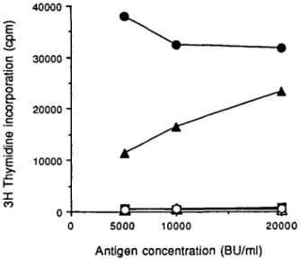
or -c genes were used as antigen presenting cells Only those fibroblasts expressing DRw52b, but not DRw52a and -c, were able to present antigen in a dose dependent manner to T cells of the long term line DX (Fig. 1). Comparison of the primary sequence reveals that amino acid substitutions at positions 30, 37, 38, 51, 57, and 60 of the /S chain distinguish between DRw52b and the other two subtypes, DRw52a and -c (Table 2).
Table 1 . Restriction specificity of the house dust mite reactive
T cell line
40000 -i
Presenting cell Haplotype
RM 088G 150G 814G 056G 857 D 826E 005G 305G 239G 161E 855E 001C 1009 0O4C 8O4E DR DRw 52 7,w12 1,5 4,w12 4,w12 5,-5,6 5,6 1,7 2,7 7,9 2,9 2,4 2.-3,8 3,-3,6 b b b b b/b b b a a a/c 53 Response (A c.p.m.) 28,501 2484 11,855 2694 6802 18,074 6424 271 15,320 4499 202 173 798 285 877 482
The long term T cell line was stimulated with D fannae in the presence of a panel of histocompatible and histoincompatible irradiated PBMC as a source of APCs. Background responses to APCs in the absence of antigen have been subtracted in each case (<1000 c.p.m.). Culture conditions and proliferation were determined as described in Methods.
I
c 30000I
o
e8 20000 -<0 c 'E 10000 2: CO
-D=
5000 10000 20000Antigen concentration (BU/ml)
Fig. 1. Proliferate response of the T cell line DX to D. fannae presented by murine fibroblasts expressing the DRw52 alleles. T cells (10%nl) were cultured with D. farinae ( 5 - 2 0 x 103 BU/ml) in the presence of
autologous PBMC (1.25 x 10%nl, • ) or mitomycm C-treated murine fibroblasts (10%nl) expressing DRw52a ( • ) , DRw52b (A), DRw52c (A). DR1Dw1 ( • ) , and Ltk- cells (O). Background responses of T cells to accessory cells in the absence of antigen were <500 c.p.m. Culture conditions and proliferation were determined as described in Methods.
No proliferation was obtained when antigen was presented by control fibroblasts expressing DR1. The failure of L cell transfectants to present house dust mite components in the experiments described in this paper could not be attributed to their lack of functional activity as they supported antigen presentation in other models of T cell antigen recognition (20,24,
20000 -]
E
8;
cg
IE
oe-8
CD C >. 10000-- i t
5000 10000 20000Antigen concentration (BU/ml)
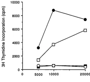
Fig. 2. Recognition of D. fannae by T cell clone DE26 is restricted by the DRw52b specificity Cloned T cells (105/ml) were cultured with D
fannae ( 5 - 2 0 x 103 BU/ml) in the presence of autologous PBMC
(1.25 x 10⁵/ml, • ) or mitomycin C-treated murine fibroblasts (1 OS/ml)
expressing DRw52a ( • ) , DRw52b (A), DRw52c (A), DR1Dw1 ( • ) , DR7Dw17(Y), and Ltk- cells (O). Background responses of T cells to accessory cells in the absence of antigen were <350 c p.m. Culture conditions and proliferation were determined as described in Methods.
personal communications). Stimulation of T cells by the house dust mite was markedly greater in the presence of autologous PBMC. The absence of accessory molecule interactions and the potential for differential processing by murine fibroblasts may account for the less efficient antigen presentation.
In order to examine the relative contribution of the DRB1 and B3 gene products in the recognition of house dust mite antigens the T cell line was cloned by limiting dilution The patterns of proliferation obtained with T cell clones DD11, DE12, and DE26 isolated in this way were similar to that observed for the T cell line as regards their response to antigen presented by DR5 + (DRw11+ and DRw12+) cells (17). Consequently, the restriction specificity of T cells of clone DE26 was examined using DRw52 transfected murine fibroblasts. Antigen dependent proliferation was obtained only in the presence of fibroblasts expressing DRw52b (Fig. 2). Neither the DRw52a nor the DRw52c transfectants were able to present antigen to DE26. Similarly, in control cultures containing DR1+, DR7 + , and Ltk- fibroblasts no proliferation was observed. Thus allergen reactive T cells restricted by the DRAB3 gene product (DRw52b) could be identified at both the oligoclonal and monoclonal levels.
DRAB 1-and DRAB5-restricted T cell recognition of Dermato-phagoides
From the pattern of T cell proliferation of the long term line observed using the allogeneic presenting cell panel (Table 1) and from the results of previous experiments (17), it appeared that DRAB1 gene products also contributed to the recognition of house dust mite allergen by T cells of this individual (Table 1). The restriction specificities of the T cell clones DE9 and DE41, derived from the T cell line, were examined in detail.
T cells of clone DE9 were stimulated by antigen in association with some DR2 + , DR5+, and DR8+ accessory cells (17).
30000 -i 80000 -i
5000 10000 20000
Antigen concentration (BU/ml)
Fig. 3. EBV transformed B cells expressing DR2 or DR5 class II protans restrict the recognition of D. fannae by the T cell clone DE9 Cloned T cells of DE9 (1 OS/ml) were cultured with D. fannae ( 5 - 2 0 x 103 BU/ml)
in the presence of autologous PBMC (1 25 x lOS/ml; • ) , or EBV-transformed B cells (lOS/ml) expressing DRw2Dw2 ( • ) , DR2Dw21 ( • ) , DRw12 (O), or DR7Dw17(A). Background responses of T cells to accessory ceils in the absence of antigen were <1400 c.p m. Culture conditions and proliferation were determined as described in Methods.
5000 10000 20000
Antigen concentration (BU/ml)
R g . 4. Mapping the fine restriction specificity of T cell clone DE9 with murine fibroblasts expressing the DRw12 and DR2Dw2 specificities as presenting cells. Cloned T cells of DE9 (lOWml) were cultured with D. fannae ( 5 - 2 0 x 103 BU/ml) in the presence of autologous PBMC
(1 25 x 1 C^/ml; • ) or mitomycin C-treated munne fibroblasts (lOWml) expressing DRw12B1 ( • ) , DR2Dw2B5 (O), DR2Dw2B1 ( • ) , and DR1Dw1 (A) gene products. Background responses of T cells to accessory cells in the absence of antigen were <940 c p.m. Culture conditions and proliferation were determined as described in Methods.
Homozygous EBV-transformed B cells expressing DR2Dw2 or DR2Dw21 molecules were able to present mite antigens to the T cell clone (Fig. 3). However, their capacity to present antigen was quantitatively different, with DR2Dw2 EBV B cells being markedly more effective. Nevertheless, presentation of the DR2Dw21 + B cells was always >4-fold that of the background accessory cells alone and antigen presented by dass II molecules of unrelated specificity. Similarly, the parental allele DRw12+ but not the control DR7+ EBV B cells induced antigen dependent proliferation of T cell clone DE9 (Fig. 3).
To determine the contributions of DRB1 and DRB3 gene products to the presentation of antigen by DRw12, DR2Dw2, and DR2Dw21 expressing EBV B cell lines, murine fibroblasts transfected with the appropriate HLA class II genes were used as presenting cells. Fibroblasts expressing the DRw12B1 or DR2Dw2B5 gene products were able to present antigen to done DE9 in a dose dependent manner (Fig. 4). In contrast, the DR2Dw2B 1 gene expressed in fibroblasts failed to induce T cell proliferation. In the absence of antigen or with control DR1 Dw1 + transfectants no activation of the T cells was observed. From the results of previous experiments T cell clone DE41 appeared to be restricted by DRw12+ (DR5) accessory cells (17). Indeed, murine fibroblasts transfected with the DRw12B1 gene were able to present Dermatophagoides to the T cell clone DE41 (Fig 5). However, the efficiency of presentation by these cells was reduced as compared with autologous PBMC.
Discussion
In this paper the HLA class II restriction specificity of T cell recognition of the house dust mite, Dermatophagoides spp., is examined using T cell clones isolated from the peripheral Wood
10000 -\
i (cp
m
poratio
r
8
lin
e
i
E SI _ CO 80006000 4000 - 2000-5000 10000 20000Antigen concentration (BU/ml)
Fig. 5. Recognition of D. faunae by T cell clone DE41 is restricted by the DRw12 class II specificity T cells of clone DE41 (lOS/ml) were cultured with D. farinae ( 5 - 2 0 x 103 BU/ml) in the presence of
autologous PBMC (1.25 x IC^/ml; • ) or mitomycin C-treated murine fibroblasts (1 OS/ml) expressing DRw12B1 (O), DR1Dw1 ( • ) , and Ltk-cells (O). Background responses of T Ltk-cells to accessory Ltk-cells in the absence of antigen were <410 cp.m. Culture conditions and prolrferation were determined as described in Methods.
of an atopic individual with perennial rhinitis (16,17). To investigate the heterogeneity of the HLA class II alleles regulating the responsivenss to house dust mite allergens a panel of HLA-DR histocompatible and histoincompatibte PBMC was used to present antigen to an ohgoclonal T cell population. The T cell line responded to both major species of the house dust mite, although the dominant specificity was for component(s) of the group II allergen of D. farinae. Although T cell recognition was HLA class II restricted, the pattern of the proliferative responses did not correspond to serdogically defined DRB1 gene products. However, correlation between the functional studies and oligonucleotide typing of the presenting cell panel strongly suggest that the B3 gene products were able to restrict recogni-tion of components of house dust mite. Although DRw52a restricted T cell recognition of tetanus toxoid has been reported, unlike the response to dust mite (17), the major component of the tetanus toxcxd reactive repertoire is not restricted by B3 gene products (10). Furthermore, using murine fibroblasts expressing each of the three alleles of DRw52 (10) as presenting cells, the restriction determinants were assigned to the DRw52b molecule. Similarly, cloned T cells reactive with a shared determinant in both species of dust mite (DE26) were isolated from the T cell line and showed identical restriction specificity for DRw52b expressed on murine fibroblasts. These findings demonstrate directly that the product of locus DRB3 is functional in antigen presentation and that it contributes to the recognition of house dust mite allergens. The magnitude of the proliferative response induced in the T cell line when D. farinae was presented by DRw52b+ fibroblasts indicated that this was a major restriction element. This result is somewhat surprising for, although DR proteins, as opposed to DQ and DP (26), are the main restric-tion elements used in T cell recognirestric-tion, this can be mapped to the B1 gene products in most cases. Quantitation of mRNA for class II proteins suggests that the B3 gene products are expressed at 2- to 5-fold lower levels than the B1 gene products (27) Thus, the dominance of the DRw52 allele as a restricton element in the recognition of the house dust mite by this individual is independent of the differential expression of the B1 and B3 gene products and may reflect differences in the binding of house dust mite peptides or in the T cell repertoire activated in the allergic immune response, with a very different pattern of allelic diversity observed among the DRB3 alleles than among the DRB1 alleles (10). As the specific T cell response of other house dust mite allergic individuals is inhibitable by an antibody reactive with DRw52b, this indicates that the contribution of the B3 gene products in the recognition of Dermatophagoides is not limited. The immunodominance of allele 52b in the case of the house dust mite is also of interest in view of the wide distribution of that allele.
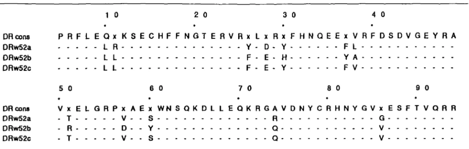
From comparison of the primary ammo acid sequence of the first domain of the /3 chain of the DRw52 alleles, differences in positions 30, 37, 38, 50, 57, and 60 distinguish DRw52b from the other two DRw52 alleles (Table 2). Based on the three dimensional structure of HLA-A2 (28) and the sequence homologies between class I and class II histocompatibihty proteins, a hypothetical model of the antigen combining site of MHC dass II molecules has been generated (29). From their proposed location in the combining site, the amino acids at positions 30, 37, 57, and 60 may influence T cell recognition. Residues at positions 30 and 37 are located on the central strands
Table 2. The primary amino acid sequence of the B1 domains of supertypic specificities DRw52a, -b, and -c 1 0 2 0 3 0 4 0 DRcons P R F L E Q x K S E C H F F N Q T E R V R x L x R x F H N Q E E x V R F D S D V G E Y R A DRw52a L R Y - D - Y F L DRw52b L L F - E - H Y A DRw52c L L F - E - Y F V 50 60 70 80 90 DR cons V x E L G R P x A E x W N S Q K D L L E Q K R G A V D N Y C R H N Y G V x E S F T V Q R R DRw52a - T V - - S R G DRw52b - R D - - Y Q V DRw52c - T V - - S Q V
Table 3. The primary ammo acid sequence of the B1 domains of DRw12B1, DR2Dw2B1, -Dw2B5, -Dw21B1, and -Dw21B5 MHC
class II proteins 1 0 2 0 3 0 4 0 DR cons P R F L E Q x K x E C H F F N G T E R V R F L x R Y F Y N Q E E Y V R F D S D V G E Y R A 0RWI2BI Y S T G Y L E H H L L F -DR2DW2B5 Q - 0 • V H - D l D L DR2DW2BI W P R D S V F DR2DW2IB5 C Q D Y H G I N V -DR2DW21B1 W - P - R 0 S V 5 0 6 0 7 0 8 0 9 0 DR cons V T E L G R P D A E Y W N S Q K D L L E Q x R A A V D T Y C R H N Y G V G E S F T V Q R R DRW12B1 V S I D R A V -0R2DW2B5 F - - D R DR2DW2B1 I A V DR2DW21B5 I A A V DR2DW21B1 F - - D R
of the j3 pleated sheets forming the floor of the combining site, whereas positions 57 and 60 would be located at one end of the a helical wall of the cleft. All four residues are remarkably clustered on the DR molecule. Substitutions at any of these positions may affect antigen binding and T cell recognition. Similarly, the residue at position 38 may influence the conforma-tion of the a helical wall and thus restrict the ability of certain peptides to occupy the antigen combining site. By site directed mutagenesis it has been demonstrated that a single amino acid substitution at position 29 in the floor of the antigen combining site could determine T cell recognition of pigeon cytochrome c (30). This would imply that amino acid variation at position 30 may be critical for T cell recognition of house dust mite peptides restricted by the DRw52 molecule. Likewise, residue differences at position 57 alone in the DQ/3 chain have been reported to influence the susceptibility to insulin dependent diabetes (31). To extend this analysis on the contribution of the polymorphic residues in DRw52 on T cell recognition of dust mite, defined T cell epitopes are required. However, this is hampered by a
lack of structural information on the components of mites, with only the group I allergens of D. pteronyssinus having been cloned and sequenced. Nevertheless, the ability of unfractionated house dust mite allergen to competitively inhibit the binding of peptides to fibrotdasts expressing DRw52 supports the role of the B3 gene products as the major restriction elements in the recognition of
Dermatophagades (R E. O'Hehir and J. R. Lamb, unpublished
results).
Population analysis of the immunogenetic basis of atopy has indicated associations between the HLA-DR phenotype and the induction of specific IgE for several purified allergens (32-34). The isolation of T cell clones from the long term line also allowed the functional sites on the B1 gene products capable of restricting recognition of house dust mite determinants to be mapped. The restriction specificity of the T cell clone DE9, which recognizes an invariant sequence in the group I allergens of the house dust mite, was degenerate in that antigen was recognized in association with the parental allele DRw12 as well as DR2Dw2 and DR2Dw21. This is in contrast to the D. farinae reactive T
cell clone DE41, which is restricted by DRw12 but shows no degeneracy on the DR2Dw2 or DR2Dw21 proteins. Interestingly, the presence of serum IgE specific for the pollen antigens, Amb
a V and Amb a VI, are DR2Dw2 and DR5 (DRw11) associated,
respectively (32,33). Amino acid sequence comparison of the 01 domains of DRw12B1 (DRB1 * 1201) and DR2Dw2B5 reveals extensive differences in the N-terminal half of the domains (Table 3), the region predicted to compose the floor of the antigen combining site (29). In contrast, the C-terminal half of these domains, predicted to form the a helix running across the floor of anti-parallel strands, is similar for these two DR molecules, with identity at positions 70 and 71. The DR2Dw2B1 and DR2Dw21 B5 a helical sequences differ from DRw12B1 and DR2Dw2B1 at these positions, however, the sequence of the DR2Dw21 B1 gene product is identical. These observations imply that the HLA class II restriction of this T cell clone is determined by allelically poly-morphic residues in the a helix of the /31 domain, irrespective of sequence differences in the floor (35). Similar patterns of MHC restriction have been described for the influenza haemagglutinin-specific, DR1Dw1-restricted T cell clone, HA1.7 (36), and for multiple murine T cell clones (1,35) Another implication of the results of these experiments is that, although the B1 gene product is the restriction element on the DRw12 and DR2Dw21 haplotypes, it is probable that the B5 gene product is used in the case of the DR2Dw2-expressmg cells. This can be inferred from the sequence identity of these molecules at positions 70 and 71. Similar flexibility of MHC restriction may account for the DR3/DR5 association of the allergic immune response to the rye allergen, Lolp III (34), since these alleles have different sequences but share the same B3 gene sequence. This may be the molecular basis for DR3/DR5 restricted recognition of Lot p III as opposed to conserved residues (ammo acids 9-13) in the /? strands of the floor of the antigen combining site (34).
The findings reported here demonstrate the heterogeneity of MHC class II restriction of T cell recognition of house dust mite antigens within an individual in terms of the use of the B1 and B3 gene products. Furthermore, both clones DE9 and DE26, although restricted by B1 and B3 gene products respectively, are able to provide help for IgE synthesis in an allergen dependent in vitro B cell assay (17,18). It is surprising that the major component of the T cell repertoire is restricted by the B3 gene products in view of their reduced expression relative to the B1 locus products (27). Contrary to the B1 gene products, where the polymorphism is restricted to three variable domains, in the DRw52 alleles sequence differences are scattered throughout the /31 domain (Table 2). This may regulate T cell recognition qualitatively by restricting the diversity of peptides capable of occupying the antigen combining site (37), in this case DRw52b, and the subsequent activation of T cells whch requires co-recognition of MHC class II and peptide. The immunological advantage of B1, B3, and B5 gene products functioning as restriction elements may be to increase the diversity of the T cell repertoire activated in response to the house dust mite, although in some individuals sensitized with allergen the population of T cells activated may induce an aberrant immune response.
The heterogeneity of the antigen and restriction specificity of T cell recognition of the house dust mite is of practical importance for the development of therapy based on immunological inter-vention, such as the induction of T cell tolerance or competitive inhibition of MHC class II proteins (38).
Acknowledgements
The authors wish to thank Professors A. B Kay and J. R. Batchetor for helpful discussions. J. R Lamb is a member of the External Scientific Staff of the Medical Research Council and R. E. O'Hehir is supported by a Wellcome Senior Research Fellowship in Clinical Science.
Abbreviations APC BU EBV IL-2 PBMC TdR
antigen presenting cells biological units Epstein-Barr virus interieukin 2
penpheral blood mononuclear leukocytes methyl thymidine
References
1 Schwartz, R. H. 1985. T lymphocyte recognition of antigen in association with gene products of the major histocompatibility complex Annu. Rev Immunol 3 205
2 Babbitt, B , Allen, P M , Matsueda, G., Haber, E., and Unanue, E. 1985. The binding of immunogenic peptides to la histocompatibility molecules Nature 317 359
3 Buus, S., Sette, A., Colon, S , Jems, D , and Grey, H 1986. Isolation and characterisation of antigen-la complexes in T cell recognition Cell 47.1071.
4 Moller, G., ed. 1987. Antigen requirements for activation of MHC restricted responses Immunol. Rev 98.1.
5 Bergholtz, B., Thoresen, A B , and Thorsby, E. 1990. HLA-D/DR restriction of macrophage dependent antigen activation of immune T lymphocytes' cross-reacting allogeneic HLA-D/DR may partly substitute for self HLA-D/DR. Scand. J Immunol. 11:541 6 Eckels, D , Lake, P , Lamb, J. R., Woody, J. N., Johnson, A. H., and
Hartzman, R J. 1982 SB restncted presentation of influenza and herpes simplex virus antigens to human T lymphocyte clones. Nature 301.313
7 Qvigstad, E , Moen, T., and Thorsby, E. 1984. T cell clones with similar antigen specificity may be restncted by DR, MT(DC), or SB class II HLA molecules Immunogenetics 19:455
8 Gorski, J. and Mach, B. 1986 Polymorphisms of human la antigens: gene conversion between two DR/3 loci results in a new HLA-D/DR specificity. Nature 322:67
9 Tiercy, J -M , Gorski, J , Jeannet, M , and Mach, B 1988. Identification and distribution of three serologically undetected alleles of HLA-DR by oligonucleotide DNA typing analysis. Proc Natl. Acad. Sci. USA 85.198
10 Irle, C, Jaques, D., Tiercy, J.-M., Fuggle, S V., Gorski, J., Termijteien, A., Jeannet, M , and Mach, B. 1988. Functional polymorphisms of each of the two HLA-DR 0 chain loci demonstrated with antigen specific DR3- and DRw52-restricted T cell clones. J. Exp. Med. 167:853.
11 Heymann, P W., Chapman, M D., and Platts-Mills, T. A. E. 1986 Antigen Der f I from the dust mite Dermatophagoides farinae structure comparison with Der p I from D. pteronyssinus and epitope specificity of murine IgG and human IgE antibodies. J. Immunol. 137:2841 12 Ishizaka, K 1984. Regulation of IgE synthesis. Annu. Rev. Immunol.
2159
13 Platts-Mills, T A. E. and Chapman, M. D. 1987. Dust mites: immunology, allergic disease, and environmental control J Allergy Clin. Immunol. 80:755
14 Rawle, F. C , Mitchell, E. B., and Platts-Mills, T. A. E. 1984. T cell responses to the major allergen from the house dust mite Dermatophagoides pteronyssinus, antigen PV a comparison of patients with asthma, atopic dermatitis and perennial rhinitis. J. Immunol. 133:195.
15 Lanzavecchia, A., Santini, P., Maggi, E., Del Prete, G. F., Falagiani, P., Romagnani, S , and Ferrarmi, M 1983. In vitro selective expansion of allergen specific T cells from atopic patients. Clin. Exp. Immunol. 5221.
human T lymphocytes reactive with Dermatophagotdes fannae (house dust mite) a comparison of T- and B-cell antigen recognition. Immunology 62:635
17 O'Hehir, R. E., Eckels, D D , Frew, A. J., Kay, A B , and Lamb, J. R 1988. MHC class II restriction spectficrty of cloned human T lymphocytes reactive with Dermatophagoides fannae (house dust mite). Immunology 64:627.
18 O'Hehir, R E., Bal, V , Quint, D , Moqbel, R., Kay, A. B., Zanders, E. D., and Lamb, J. R. 1989. An in vitro model of allergen dependent IgE synthesis by human B cells: comparison of the response of an atopc and a non-atopic individual to Dermatophagoides spp. Immunology 66:499.
19 Rabourdin-Combe, C and Mach, B. 1983 Expression of HLA-DR antigens at the surface of munne L cells co-transfected with cloned human genes Nature 303:670.
20 Rothbard, J. B., Lechler, R. I., Howland, K., Bal, V , Eckels, D. D , Sekaly, R., Long, E. O., Taylor, W R., and Lamb, J R. 1988. Structural model of HLA-DR1 restricted T cell recognition. Cell 52:515 21 Angelmi, G., de Preval, C , Gorski, J., and Mach, B. 1986
High-resolution analysis of the human HLA-DR polymorphism by hybridiza-tion with sequence-specific digonucleotide probes Proc. Nat). Acad Sci. 834489
22 Gorski, J , Tosi, R , Strubin, M , Rabourdin-Combe, C , and Mach, B. 1985 Serological and immunochemical analysis of the products of a single HLA-DRa and DR/3 chan expressed in a mouse cell line after DNA-mediated cotransfection reveals that the /3 chain carries a known supertypic specificity J. Exp Med. 162-105.
23 Tonnelle, C , Demars, R , and Long, E 0.1985 DO/3 a new/3 chain gene in HLA-D with a distinct recognition of expression. EMBO J. 42847
24 Wilkinson, D., de Vries, R R. P., Madrigal, J A., Lock, C. B., Morgenstern, J P., Trowsdale, J., and Altmann, D. M. 1988. Analysis of HLA-DR glycoproteins by DNA-mediated gene transfer definition of DR20 gene products and antigen presentation to T cell clones from leprosy patients. J. Exp. Med 167.1442
25 Tieber, V. L , Abruzzini, L F., Didier, D. K., Schwartz, D. B., and Rotwein, P 1986. Complete characterization and sequence of an HLA class II DR/3 chain cDNA from the DR5 haplotype J. Bid Chem 261-2738.
26 Ottenhoff, T. H M., Elfermk, D. G., Hermans, J , and de Vries, R R. P. 1985 HLA class II restriction repertoire of antigen-specific T cells. I The main restriction determinants for antigen presentation are associated with HLA-D/DR and not with DP and DQ. Hum. Immunol. 13.105
27 Berdoz, J , Gorski, J , Termijtelen, A , Dayer, J. M , Irle, C, Schendel, D., and Mach, B 1987. Constitutive and induced expression of the individual HLA-DR/3 and a chan loa in different cell types J Immunol. 139-1336
28 Bprkman, P., Saper, M., Samraoui, B., Bennett, W., Strominger, J , and Wiley, D 1987. The foreign antigen binding site and T cell recognition regions of class I histocompatibdity antigens. Nature 329.512.
29 Brown, J , Jardetzky, T., Saper, M., Samraoui, B , Bjorkman, P , and Wiley, D 1988 A hypothetical model of the foreign antigen combining site of class II histocompatibility molecules Nature 332.845. 30 Ronchese, F., Schwartz, R. H., and German, R. N. 1987. Functionally
distinct subsites on a major histocompatibility complex molecule. Nature 329 254.
31 Todd, J A , Bell, J I., and McDevitt, H. O 1987. HLA-DQ/3 gene contributes to susceptibility and resistance to insulin-dependent diabetes mellitus. Nature 329 599.
32 Marsh, D. G., Freidhoff, L. R , Ehriich-Kautzky, E., Bias, W B., and Roebber, M 1987. Immune responsiveness to Ambrosa artemisiifolia (short ragweed) pollen antigen Amb a VI (Ra6) is associated with HLA-DR5 in allergic humans Immunogenetics 26 230.
33 Marsh, D. G , Hsu, H. S., Roebber, M., Eriich-Kautzky, E., Freidhoff, L. R., Meyers, D. A , Pollard, M K., and Bias, W. B. 1982. HLA-Dw2. a genetic marker for human immune response to short ragweed pollen antigen Ra5. I Response resulting primarily from natural antigenic exposure J Exp Med. 155.1439.
34 Ansan, A , Freidhoff, L R , and Marsh, D G. 1989. Molecular genetics of human responsiveness to Lolium perenne (rye) allergen, Lol p\\\ Int. Arch. Allergy Immunol. 83164.
35 Ronchese, F., Brown, M., and Germain, R N 1987 Structure-function analysis of the A/S**"12 mutation using
site-directed mutagenesis and DNA-mediated gene transfer J. Immunol. 139 629.
36 Rothbard, J B., Busch, R., Lechler, R I., Trowsdale, J , and Lamb, J. R. 1989. Recognition of the HLA class ll-peptide complex byT-cell receptor: reversal of major histocompatibility complex restriction of a T cell clone by a point mutation in the peptide determinant. Phil Trans. R. Soc. Lond B 323553.
37 Guillet, J , Ming, Z L., Bnner, T J., Smith, J. A , and Gefter, M. L 1986 Interaction of peptide antigens and class II mapr histo-compatibility complex antigens. Nature 324260.
38 Adonni, L., Muller, S., Cardinaux, F., Lehmann, P. V., Falcioni, F., and Nagy, Z A. 1988 In vivo competition between self peptides and foreign antigens in T cell activation. Nature 334623.