Molecular characterization of Neospora caninum MAG1,
a dense granule protein secreted into the parasitophorous
vacuole, and associated with the cyst wall and the cyst matrix
CHRISTOPHE GUIONAUD1, ANDREW HEMPHILL2*, MEIKE MEVISSEN1
and FERIAL ALAEDDINE1* 1
Institute of Veterinary Pharmacology and Toxicology, Vetsuisse Faculty, University of Berne, La¨nggass-Strasse 124, CH-3012 Berne, Switzerland
2
Institute of Parasitology, Vetsuisse Faculty, University of Berne, La¨nggass-Strasse 122, CH-3012 Berne, Switzerland (Received 9 December 2009; revised 2 February 2010; accepted 17 February 2010; first published online 6 May 2010)
S U M M A R Y
In Neospora caninum and Toxoplasma gondii, the parasitophorous vacuole (PV) is synthesized at the time of infection. During tachyzoite-to-bradyzoite stage conversion, the PV is later transformed into a tissue cyst that allows parasites to survive in their host for extended periods of time. We report on the characterization of NcMAG1, the N. caninum orthologue of T. gondii MAG1 (matrix antigen 1 ; TgMAG1). The 456 amino acid predicted NcMAG1 protein is 54 % identical to TgMAG1. By immunoblotting, a rabbit antiserum raised against recombinant NcMAG1 detected a major product ofy67 kDa in extracts of N. caninum tachyzoite-infected Vero cells, which was stained more prominently in extracts of infected Vero cells treated to induce in vitro bradyzoite conversion. Immunofluorescence and TEM localized the protein mainly within the cyst wall and the cyst matrix. In both tachyzoites and bradyzoites, NcMAG1 was associated with the parasite dense granules. Comparison between NcMAG1 and TgMAG1 amino acid sequences revealed that the C-terminal conserved regions exhibit 66 % identity, while the N-terminal variable regions exhibit only 32 % identity. Antibodies against NcMAG1-conserved region cross-reacted with the orthologuous protein in T. gondii but those against the variable region did not. This indicates that the variable region possesses unique antigenic charac-teristics.
Key words : Neospora caninum, cyst matrix, cyst wall, parasitophorous vacuole, matrix antigen 1 (MAG1).
I N T R O D U C T I O N
Neospora caninum is an intracellular apicomplexan parasite that is closely related to Toxoplasma gondii, and represents a major cause of abortion in cattle and neuromuscular disease in dogs worldwide (Dubey et al. 2006 ; Hemphill et al. 2006). Transplacental transmission is regarded as the major source of in-fection. The life cycle of N. caninum consists of 3 principal stages : (1) tachyzoites, which rapidly replicate within the parasitophorous vacuole (PV), and disseminate in the host during the acute phase, (2) bradyzoites, slowly dividing parasites, which are enclosed within a tissue cyst that develops particu-larly in the brain during the chronic phase of the infection (Dubey et al. 2006), and (3) oocysts which represent the product of a sexual process taking place within the intestine of the definitive hosts (dog and
coyote), and upon fecal shedding, sporulate and form sporozoites (McAllister et al. 1998 ; Gondim et al. 2004). There is evidence that tachyzoites differentiate into bradyzoites as a reaction to the immune response occurring in immune-competent hosts (Dubey et al. 2006 ; Hemphill et al. 2006).
Intracellularly, all invasive stages of N. caninum are located within a parasitophorous vacuole (PV), separated from the host cell cytoplasm through the parasitophorous vacuole membrane (PVM). The PVM is derived from the host cell cytoplasmic membrane during host cell invasion, and sub-sequently undergoes important modifications. While proteins secreted by the parasite are incorporated into the PVM, those of the host cell are selectively eliminated and the PV becomes resistant to fusion with lysosomes (Beyer et al. 2002). The PV of T. gondii transforms into a tissue cyst very early after parasite penetration into brain cells, and the PVM progressively matures into a cyst wall that protects bradyzoites from the immunological and physio-logical responses on part of the host (Beyer et al., 2002). Intact tissue cysts containing bradyzoites of both N. caninum and T. gondii can persist in the host, usually without being harmful or causing an
* Corresponding author : Institute of Veterinary Phar-macology and Toxicology, Vetsuisse Faculty, University
of Berne, La¨nggass-Strasse 124, CH-3012 Berne,
Switzerland. Tel : +41 31 631 22 41. Fax: +41 31 631 26 30. E-mail : ferial.alaeddine@vpi.unibe.ch ; E-mail : hemphill@ipa.unibe.ch
inflammatory response (Dubey et al. 1988 ; Innes et al. 2002).
The proteins involved in tachyzoite-bradyzoite interconversion could represent important targets for vaccination or chemotherapy in order to prevent or reduce the development of intra-cerebral cysts. Stage-specifically expressed proteins in T. gondii and N. caninum have been identified (McAllister et al. 1996 ; Fuchs et al. 1998 ; Ferguson, 2004 ; Risco-Castillo et al. 2007 ; Aguado-Martinez et al. 2009), while others are expressed in both stages, but differ in their localization (Vonlaufen et al. 2004). In this context, T. gondii MAG1 was first reported as a bradyzoite-specific antigen localized in the cyst matrix (Parmley et al. 1994). Subsequently, the protein was also detected in the parasitophorous vacuoles containing intracellular tachyzoites and in tachyzoite lysates (Ferguson and Parmley, 2002). Besides its importance as a marker for cerebral in-fection with T. gondii in AIDS patients (Contini et al. 2002 ; Pfrepper et al. 2005), TgMAG1 represents a potentially useful tool for the immunodiagnosis of patients with toxoplasmosis (Di Cristina et al. 2004 ; Pfrepper et al. 2005). In addition, TgMAG1 has been considered as a promising candidate in vacci-nation trials (Parmley et al. 2002 ; Nielsen et al. 2006). We have recently shown that intraperitoneal vaccination of mice with recombinant NcMAG1 expressed in E. coli confers 50 % protection against experimental N. caninum infection in a murine acute disease model (Debache et al. 2009). In this study, we report on the molecular characterization, localization and antigenic properties of NcMAG1.
M A T E R I A L S A N D M E T H O D S Parasite cultures
Parasites were cultured as previously described (Barber et al. 1995 ; Hemphill and Gottstein, 1996). Briefly, N. caninum NC-Liverpool or T. gondii ME49 tachyzoites were maintained in Vero cells at 37 xC under 5 % CO2 in RPMI 1640 medium
containing 10 % FCS (Gibco-Invitrogen, Carlsbad, CA, USA), 2 mML-glutamine, 50 IU/ml penicillin, and 50mg/ml streptomycin. N. caninum grown in keratinocytes were cultured as previously described (Vonlaufen et al. 2002).
Tachyzoite-to-bradyzoite stage conversion was performed in vitro by 10 days treatment of NC-Liverpool-infected Vero cells with 17mM sodium nitroprusside (SNP ; Sigma, St Louis, MO, USA), using a procedure adapted from Vonlaufen et al. (2004) and by 4 days treatment of T. gondii-infected Vero cells with 100mMSNP as previously described (Bohne et al. 1994 ; Weiss et al. 1998).
For purification, parasites were mechanically re-leased from infected cells by passing through a 25G, 5/8 needle, washed in ice-cold medium, and run on
PD-10 columns (GE Healthcare, Piscataway, NJ, USA) as described by Hemphill et al. (1996).
Oligonucleotides
N. caninum MAG1-specific primers were designed based on the ApiDots (http://www.cbil.upenn.edu/ apidots/ ; (Li et al. 2003, 2004)) DT.92484705 tran-script. All primers were purchased from MWG (Ebersberg, Germany).
RNA isolation and first strand cDNA synthesis Total RNA was isolated from 3r106 purified
N. caninum Liverpool tachyzoites using Trizol re-agent (Invitrogen, Carlsbad, CA, USA) according to the manufacturer’s instructions. First strand cDNA synthesis reactions were performed for 1 h at 42 xC in a 20ml reaction mixture containing 2 mg of total RNA, 1r first-strand buffer (Invitrogen), 0.5 mMof each dNTP (Endotell, Allschwil, Switzerland), 10 mM dithiothreitol, 20 U of RNasin RNase in-hibitor (Promega, Madison, WI, USA), 200 U of SuperScript II reverse transcriptase (Invitrogen). Primers (20 pmol) were either MAG1-R1, 5 k-CT-GTCCCTTACACCTACACT-3k, for the 5k-RACE (Rapid Amplification of cDNA Ends) and to gener-ate the recombinant NcMAG1 protein, or T-primer
3,
5k-AAGCAGTGGTATCAACGCAGAGTA-C(T)30VN-3k (V=A, C or G; N=any nucleotide),
for the 3k-RACE (Matz et al. 1999).
RACE procedures
A manual hot-start PCR (Chou et al. 1992) was used in order to reduce background amplifications in both RACE reactions.
For 5k-RACE, the first strand cDNA primed with MAG1-R1 primer was ultrafiltrated 3 times against water on a Microcon YM-100 filter unit (Millipore, Billerica, MA, USA). The cDNA in the whole retentate was dA-tailed for 20 min at 37 xC in 50ml of 1r terminal deoxynucleotidyl transferase (TdT) buffer (Roche, Basel, Switzerland) using 25 U of TdT (Roche) and 0.2 mM dATP (Endotell). The reaction was terminated by heating at 95 xC for 5 min. Two rounds of amplification were performed to generate the 5k-RACE product. The first round was carried out in a 50ml reaction volume containing 5ml (1/10th) of the (dA)-tailed product, 5 pmol of each T-primer 3 and MAG1-R2 (5k-GCATT-ACCAACTTCGTCCTC-3k) primers, 0.2 mM of each dNTP and 2.5 U of Pfu DNA polymerase (Promega) in the supplied reaction buffer. Ten PCR cycles were performed with an annealing tempera-ture of 55 xC. The second round of amplification was carried out in 50ml containing 20 ml of the first step
amplification reaction, 15 pmol of Heel-carrier (5k- CTAATACGACTCACTATAGGGCAAGCAG-TGGTATCAACGCAGAGT-3k) and MAG1-R2 primers, 0.2 mM of each dNTP, and 2.5 U of Pfu DNA polymerase (Promega) in the supplied reaction buffer. PCR was performed for 30 cycles with an annealing temperature of 58 xC.
The 3k-RACE reaction was carried out in a 100 ml reaction mixture containing 2ml (1/10th) of the first strand cDNA prepared with T-primer 3, 4 pmol of Heel-carrier primer, 20 pmol of each MAG1-F
(5
k-TGAACAACCCTATGAACAAACAGACGC-3k), and Heel-specific (5k-CTAATACGACTCA-CTATAGGGC-3k) primers, 0.2 mMof each dNTP and 2.5 U of Pfu DNA polymerase in the supplied reaction buffer. PCR was performed for 35 cycles with an annealing temperature of 55 xC.
Sequencing and sequence analyses
RACE products were cloned into pCR Blunt II TOPO (Invitrogen) and sequenced using a primer walking approach. All expression constructs were verified by sequencing of the insert. Sequencing re-actions were carried out using BigDye v3.1 fluor-escent dye terminators and run on an ABI PRISM 3100 Genetic Analyzer (Applied Biosystems, Foster City, CA, USA). Raw sequencing data were as-sembled and edited with the Staden package (Bonfield et al. 1995). Sequence data reported in this paper are available in the GenBank database under the Accession number EF580924.
Homology searches were done using BLAST (blastp) program (http://www.ncbi.nlm.nih.gov/ blast/ ; (Altschul et al. 1990)) and the Conserved Domain Database CD-Search (http://www.ncbi. nlm.nih.gov/Structure/cdd/wrpsb.cgi) with default settings. Proteins were aligned on the Muscle server (http://www.drive5.com/muscle/ (Edgar, 2004)), minimally edited, and formatted with GeneDoc (Nicholas et al. 1997). Residue grouping and shading was according to the MM5 reduced amino acid alphabet of Melo and Marti-Renom (2006). Potential signal peptide cleavage sites were identified with SignalP 3.0 (http://www.cbs.dtu.dk/services/ SignalP/ (Bendtsen et al. 2004)) and potential trans-membrane regions were checked with the ProtScale tool on the Expasy server (http://expasy.org/tools/ protscale.html).
Expression of recombinant N. caninum MAG1 proteins
A portion of NcMAG1, thereafter referred to as recNcMAG1, encoding aa 31-394 (numbering ac-cording to the precursor) was amplified by RT-PCR using MAG1-BamHI-F (5k-GGATCCCAAAGG-GTGCCTCGCTACCC-3k) and MAG1-SmaI-R
(5k-CCCGGGTTATTCCTCCACTATTTCGT-CCGC-3k) primers; BamHI and SmaI restriction sites underlined. The PCR product was cloned into pCR blunt II TOPO (Invitrogen) and verified by sequencing. The BamHI/SmaI MAG1 insert was then subcloned into the (His)6-tag pQE-30
ex-pression vector (Qiagen, Hilden, Germany), result-ing in pQE-30-NcMAG1. We also expressed portions of the conserved (C) and variable (V) re-gions within NcMAG1 (Fig. 2). In order to monitor their expression and to facilitate their purification, the two NcMAG1 C and V regions were expressed as (His)6-GFP (green fluorescent protein) fusion
pro-teins in E. coli. The portion of NcMAG1 central to the C region (aa 241–386, numbering according to the precursor) was amplified using MAG1-C-XmaI-F (5k-CTGCGTCCCGGGGGACCGACGGTT-TCGACTCG-3k) and MAG1-C-PstI-R (5k-CT- GCGTCTGCAGGGTCCCCACGAATTGTCT-CG-3k) primers; XmaI and PstI restriction sites underlined. The portion of NcMAG1 central to the V region (aa 31–162) was amplified using
MAG1-V-XmaI-F
(5k-CTGCGTCCCGGGCAAAGGGT-GCCTCGCTACCC-3k) and MAG1-V-PstI-R
(5k-CTGC
GTCTGCAGCGACGTGGAAAGT-GGTAGCG-3k) primers; XmaI and PstI restric-tion sites underlined. The pQE-30-NcMAG1 vector was used as template for PCR amplifications. Re-stricted PCR products were inserted into the XmaI/ PstI-cut pQE-GFP vector (C.G. and F.A., un-published ; sequence available upon request), resulting in MAG1-C and pQE-GFP-MAG1-V.
Expression vectors were used to transform E. coli BL21 (Novagen-EMD Biosciences, Madison, WI) harbouring the pREP4 repressor plasmid (Qiagen). To express the recombinant proteins, 1 l of 2r YT medium (per litre : 16 g bacto tryptone, 10 g bacto yeast extract, and 5 g NaCl), pre-warmed to 37 xC and supplemented with carbenicillin and kanamycin (Sigma ; 100 and 25 mg/ml, respectively), was in-oculated with 10 ml of an overnight starter culture grown in the same medium. When cultures reached an OD of 0.5 at l=600 nm, isopropyl b-D-1-thiogalactopyranoside (Sigma) was added to 1 mM and expression was carried on for 3 h at 37 xC. Soluble recNcMAG1 and GFP-MAG1-V proteins were purified by nickel chelate chromatography on Protino Ni-IDA columns (Macherey-Nagel, Du¨ ren, Germany), as recommended by the manufacturer. Due to its extreme sensitivity to proteolysis after bacterial lysis, GFP-MAG1-C could not be purified by chromatography. It was therefore run on a large format SDS-PAGE gel after boiling over-expressing bacteria in SDS sample buffer. The whole gel was transferred onto a nitrocellulose membrane and the major band was excised and used for the purification of anti-GFP-MAG1-C antibodies.
Production and purification of antibodies
Antisera were obtained after immunization of 2 female white New Zealand rabbits, with 150mg of recNcMAG1 antigen per injection, using a stan-dard 10-week immunization protocol (Institut fu¨ r Labortierkunde, Zurich, Switzerland). Antibodies specific to either the whole recNcMAG1, the C or the V region were affinity-purified using recNcMAG1, GFP-MAG1-C or GFP-MAG1-V bound to nitro-cellulose membranes, respectively (Robinson et al. 1988).
Preparation of protein extracts
For preparation of total protein extracts from in-fected or uninin-fected Vero cells, cultures in a T-75 culture flask were washed twice with PBS (137 mM NaCl, 2.7 mM KCl, 4.3 mM Na2HPO4, 1.47 mM
KH2PO4, pH 7.4) and proteins were extracted with
1 ml of ice-cold RIPA buffer (Pierce-Thermo Fisher Scientific, Waltham, MA, USA) supplemented with 10ml/ml Halt protease inhibitor cocktail (Pierce-Thermo Fisher Scientific). Protein extracts were sonicated, clarified by centrifugation, and the protein concentration in the supernatant was measured using the DC Protein Assay Kit (Bio-Rad, Hercules, CA, USA). For SDS-PAGE, samples of protein extracts from infected cultures were adjusted to contain the required parasite number using data obtained by quantitative real-time PCR.
Protein extracts from free parasites were prepared using 100ml of RIPA buffer per 5r106 purified
parasites.
Quantitation of parasites
To determine the number of parasites in infected Vero cells, a quantitative real-time PCR (LightCycler, Roche Diagnostics, Basel, Switzerland) was used. DNA was purified from Neospora- or Toxoplasma-infected cells using the High Pure PCR Purification kit (Roche Diagnostics) according to the manufacturer’s recommendations. DNA concentrations were measured by Hoechst 33258 (Sigma) fluorimetry (Ausubel et al. 1997) on a Synergy HT plate reader (Biotek Instruments, Winooski, VT, USA). Experimental procedures described by Mu¨ ller et al. (2002) for Neospora and Reischl et al. (2003) for Toxoplasma were followed, using 200 ng DNA per sample. Free parasites were directly quantified by counting using a Neubauer chamber.
SDS-polyacrylamide gel electrophoresis (SDS-PAGE) and Western blot
Protein samples were mixed with reducing, de-naturing sample buffer, boiled for 5 min and protein
separation was conducted by SDS-PAGE. Proteins were transferred onto PVDF membranes (Bio-Rad) for 1 h at 100 V using a wet transfer apparatus (Bio-Rad). Membranes were blocked by incubation in a blocking buffer containing 5 % non-fat milk in TBST (20 mMTris, 150 mMNaCl, 0.1 % (v/v) Tween 20) and then incubated for 2 h at room temperature or overnight at 4 xC with affinity-purified anti-NcMAG1 antibodies at a dilution of 1 : 3000. Antibodies against the NcMAG1-conserved region and against the NcMAG1-variable region were ap-plied at a dilution of 1 : 400. Detection was performed with a donkey anti-rabbit alkaline phosphatase-conjugated secondary antibody (Pierce-Thermo Fisher Scientific) at a dilution of 1 : 10 000 and NBT / BCIP (nitro blue tetrazolium/bromochloroindolyl phosphate ; Sigma) (Ausubel et al. 1997).
Immunofluorescence (IF ) analysis
IF staining was performed on purified parasites or on parasite-infected Vero cells grown on poly-L lysine-coated glass cover-slips. For intracellular brady-zoites, cover-slips were seeded (105 infected cells
per well) on the sixth day following addition of SNP to the culture medium and the treatment carried on until day 9 post-infection. Purified parasites were fixed using 4 % paraformaldehyde in PBS (20 min at RT). Cover-slips were rinsed 3 times in PBS, immersed into pre-cooled (x20 xC) methanol/ acetone (v :v), and incubated atx20 xC for 30 min. The cover-slips were washed 3 times with PBS, 5 min each, and incubated in blocking buffer (PBS/ 3 % BSA) for 2 h. The following primary antibodies, diluted in PBS/0.3 % BSA, were applied : (a) affinity-purified polyclonal rabbit anti-NcMAG1 antibody at a 1 : 2500 dilution, (b) rabbit anti N. caninum anti-serum (Hemphill et al. 1996) at a 1 : 2500 dilution, (c) CC2, a rat mAb reacting with a T. gondii cyst wall antigen (Gross et al. 1995), diluted at 1 : 300, and (d) rabbit anti-BAG1 polyclonal antibody, directed against a bradyzoite-specific antigen (McAllister et al. 1996), diluted at 1 : 300. Affinity-purified anti-bodies against conserved and NcMAG1-variable regions were used at 1 : 200 to assess their cross-reactivity on T. gondii.
Incubations with primary antibodies were per-formed for 1 h, followed by 3 washes in PBS for 5 min each. Cells were then incubated for 45 min with the appropriate Alexa Fluor (AF)-labelled sec-ondary antibodies (Invitrogen) at a dilution of 1 : 3000 (AF-488-, AF-546) or 1 : 300 (AF-350) in PBS/0.3 % BSA. For multiple detection, antibodies were applied sequentially, without a given order. Finally, the preparations were washed in PBS 3 times for 5 min each. Cover-slips with infected cells were incubated for 2 min in PBS containing DAPI at 1mg/ ml to stain nuclei, rinsed briefly in PBS, and mounted in ProLong Gold antifade reagent (Invitrogen).
Images were acquired on an Axioskop 2 microscope equipped with an AxioCam CCD camera (Carl Zeiss, Oberkochen, Germany) or on a Nikon Eclipse 80i microscope (Kanagawa, Japan) equipped with a QImaging cooled CCD camera (Retiga 2000R Fast 1394 ; Surrey, BC, Canada). Images were processed with ImageJ 1.38 software (http://rsb.info.nih.gov/ ij/ ; (Abramoff et al. 2004)).
Immunogold-labelling and transmission electron microscopy (TEM)
LR-White embedding and on-section labelling of N. caninum cultures were performed essentially as previously described (Hemphill and Croft, 1997 ; Vonlaufen et al. 2002, 2004). Sections were loaded onto formvar-carbon coated grids and non-specific binding sites were blocked for 2 h in PBS/1 % BSA. They were then incubated in affinity-purified anti-NcMAG1 antibodies, diluted 1 : 100 in PBS/ 0.1 % BSA for 1 h. After washing in 5 changes of PBS (2 min each), the goat anti-rabbit antibody conjugated to 10 nm diameter gold particles (Amersham) was applied at a dilution of 1 : 5 in PBS/ 0.1 % BSA. After extensive washing in PBS, grids were air-dried and stained with lead citrate and uranyl acetate. Specimens were viewed on a Philips 400 TEM (Philips Electronics, Eindhoven, The Netherlands) operating at 80 kV.
R E S U L T S
Cloning and analysis of a cDNA encoding Neospora caninum MAG1
A sequence homology search against N. caninum expressed sequence tags (ESTs) in the ApiDots database (Li et al. 2003, 2004) using T. gondii MAG1 cDNA (Parmley et al. 1994) resulted in a single hit, a 2886 NT assembly of 21 ESTs : DT.92484705. This consensus sequence possesses an open reading frame (ORF) potentially encoding a protein sharing over 50 % identity with T. gondii MAG1. To confirm the DT.92484705 data, we amplified NcMAG1 cDNA by RACE, using a first strand cDNA pre-pared from N. caninum Liverpool tachyzoites as a template. The 5k-RACE reaction produced a single amplicon ofy1.5 kb, consistent with the sequence of the five 5k-RACE clones that we analysed. On the contrary, agarose gel electrophoresis of 3k-RACE products revealed a more complex banding pattern, with discrete fragments ranging from 0.6 to over 1.5 kb (data not shown). After bulk cloning and sequencing of twelve 3k-RACE products, we found that differences between clones were limited to their 3k-end, immediately preceding the poly(A) tail, suggesting an alternative polyadenylation site usage.
The unique 5k- and the longest 3k-RACE products, overlapping by 345 NT, were used to build a 2288 NT NcMAG1 cDNA sequence (GenBank Accession no. EF580924 ; Fig 1). The consensus cDNA possesses a 1371 nucleotide ORF (including the TGA stop codon), potentially encoding a 456 amino acid protein identical to that predicted to be encoded by the DT. 92484705 transcript. The putative ATG initiation codon is part of a sequence (AGCA-CAATGG) matching 8 positions of the previously described T. gondii translation initiation consensus sequence (gNCAAaATGg) (Seeber, 1997). An ad-ditional, out-of-frame, ATG triplet was found closer to the NcMAG1 cDNA 5k-end (NT 52–54); how-ever, in a context apparently less favourable for translation initiation.
The predicted 5k-UTR is 128 NT long, 76 NT shorter than in the ApiDots transcript. The twelve 3k-RACE clones sequenced had a 3k-UTR ranging from 304 to 789 NT and a poly(A) tail, but electro-phoretic analysis of 3k-RACE products suggested that polyadenylation of the NcMAG1 transcript could occur even further downstream, consistent with the longer 3k-UTR present on the DT.92484705 transcript. We did not observe any consensus eu-karyotic polyadenylation signal (AATAAA) down-stream of the coding sequence. However, performing a less stringent search, 6 out of the 12 analysed transcripts appeared to be terminated in close vicinity (within the generally accepted 10–30 NT window) to upstream sequences differing at most by 1 mismatch from the consensus AATAAA hexamer. An imperfect 84/90 NT direct repeat was identified (NT 1934–2017 and 2019–2108) in the 3k-UTR (Fig. 1).
The predicted NcMAG1 precursor is 54 % identical to TgMAG1 (Fig. 2) but does not share significant similarity to any other protein in GenBank nor in the Conserved Domain Database (CDD v2.17, October 2009). A typical signal peptide with a predicted signal peptidase cleavage site be-tween Gly30and Gln31was identified by SignalP 3.0
with the hidden Markov models and the neural networks prediction methods (Bendtsen et al. 2004). The position of the predicted cleavage site was conserved in TgMAG1, but only with the hidden Markov models method (Fig. 2). The pre-dicted MWs of NcMAG1 are 52.9 and 50.5 kDa, for the precursor and the mature protein, respect-ively.
We did not identify a hydrophobic region that could represent an internal transmembrane domain. The predicted NcMAG1 and TgMAG1 mature proteins share an overall 53 % identity but sequence conservation is not homogeneously distributed along the proteins. The C-terminal region (aa189–456) is much more conserved than the N-terminal region (aa 31–188), with 66 % and 32 % sequence identity, respectively (Fig. 2).
Expression and immunolocalization of NcMAG1 in N. caninum tachyzoites and bradyzoites
A recombinant protein (recNcMAG1), comprising aa 31–394, was expressed as a (His)6-tagged fusion
protein in E. coli and used to immunize rabbits. All intermediate and final bleeds had a high titre against the purified recombinant protein as determined by Western blot and ELISA (data not shown). Sera from the third bleed were affinity-purified against recNcMAG1 and used for all further experiments.
In Western blots of protein extracts from purified extracellular N. caninum, anti-NcMAG1 antibodies
detected a y67 kDa band. The band was more prominent in lysates from SNP-treated cultures (containing predominantly bradyzoites) than in tachyzoite lysates (Fig. 3, lanes 1 and 2). Similarly, the protein was much more abundant in lysates of SNP-treated Neospora-infected Vero cells compared to untreated, infected cells (Fig. 3, lanes 3 and 4). The antibodies did not show any reactivity with uninfected Vero cell lysates (Fig. 3, lanes 5 and 6).
Affinity-purified antibodies were then used to localize NcMAG1 by IF and TEM. IF detection of MAG1 in extracellular parasites showed that the protein was distributed in a punctuated pattern
Fig. 1. The cDNA encoding NcMAG1. The largest cDNA clone (GenBank Accession no. EF580924, bottom) is represented together with the deduced MAG1 precursor protein (GenBank Accession no. ABQ52425, top). The predicted signal peptide cleavage site is indicated by a filled arrowhead. The 5k- and 3k-UTRs are in lowercase with the two nucleotides differing from the ApiDots DT.92484705 transcript underlined. Nucleotide residues at which polyadenylation was observed in other cloned NcMAG1 cDNAs are shaded in grey. Sequences differing at most by 1 nucleotide from the consensus polyadenylation signal (AATAAA) are boxed. Tandem repeat units (NT 1931–2016 and 2017–2107) present in the 3k-UTR are shown as horizontal arrows above the nucleotide sequence. Mismatches and gaps created to align the two repeat units are displayed as ‘ x ’.
within the cytoplasm. These punctuations were more apparent in bradyzoites (Fig. 4A). Staining of intra-cellular tachyzoites detected the protein mostly within the lumen of the parasitophorous vacuole (PV) ; the PV periphery was also faintly labelled. In contrast, after triggering tachyzoite-to-bradyzoite conversion with SNP, a more pronounced staining of the cyst wall and the cyst matrix was observed (Fig. 4B and C). The efficiency of the in vitro stage conversion using SNP treatment was confirmed using the bradyzoite-specific marker BAG1 on Neospora-infected cells (Fig. 4B) and on purified parasites (Fig. 4D). In a double-labelling experiment with anti-recNcMAG1 and mAbCC2, a monoclonal antibody directed against a cyst-specific antigen, both antigens appeared to largely co-localize (Fig. 4C)
By immunogold-TEM, performed on keratino-cytes infected with N. caninum tachyzoites, minor labelling was observed in the intra-parasitic space, the lumen of the PV, and within the dense granule organelles of the tachyzoites (Fig. 5A). Staining of cell cultures undergoing SNP-treatment and
hence tachyzoite-to-bradyzoite stage conversion (Fig. 5B–D) exhibited a much more pronounced immunogold staining. Gold particles were abundant within the cyst matrix and, in many cysts, also on the cyst wall. In addition, dense granule (DG) organelles were intensely labelled (Fig. 5B–D), indicating an increased expression of NcMAG1 during stage conversion, or an accumulation of the protein due to reduced degradation.
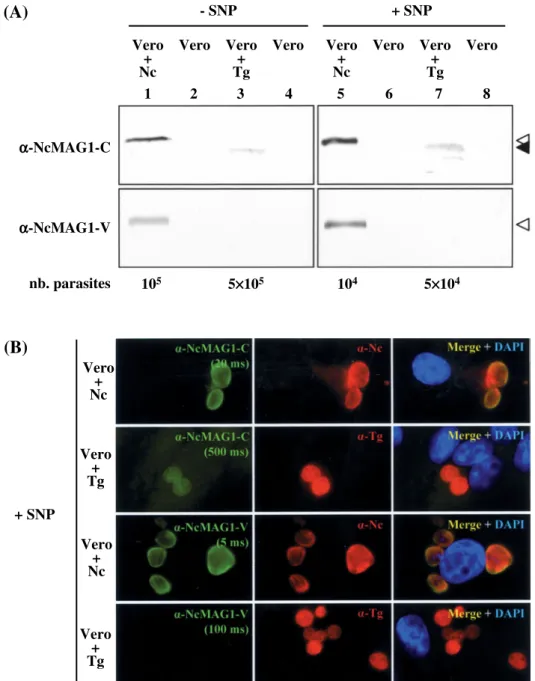
Antibodies against the NcMAG1 conserved region, but not the variable region, cross-react with TgMAG1
Since NcMAG1 and TgMAG1 are orthologous proteins, we investigated the potential antibody-cross-reactivity of these antigens. Immunoblot and immunofluorescence using affinity-purified anti-bodies against the recombinant NcMAG1 resulted in a weak, but distinct, cross-reactivity with T. gondii, no matter whether cultures had been treated with 100mMSNP or not (data not shown). We thus de-termined, which region (conserved or variable) could
Fig. 2. Protein sequence alignment of deduced MAG1 precursors. Neospora caninum (Nc ; GenBank Accession no. ABQ52425, this work) and Toxoplasma gondii (Tg ; GenBank Accession no. AAC46484) MAG1 predicted protein sequences (single letter code) were aligned with MUSCLE and shaded according to the MM5 reduced, 5-letter amino acid alphabet. Residue grouping is as follows : [C] ; [FILMVWY] ; [AG] ; [H] ; [DEKNPQRST]. Identical residues are indicated by colons, similar residues are marked by dots. The putative signal peptide was identified with SignalP 3.0. Boundaries between the predicted signal peptide, the conserved (C) C-terminal region (66 % identity, aa 189–456) and the less conserved (V) N-terminal region of NcMAG1 (32 % identity, aa 31–188) are indicated by filled arrowheads. Antibodies were raised against a large portion of a recombinant NcMAG1 (aa 31–394, not shown). Amino acid stretches within the V and C regions, which were used for affinity-purification of antibodies specific to these regions are boxed.
be responsible for this cross-reactivity. Western blots were performed, using extracts of non-treated or SNP-treated Neospora- and Toxoplasma-infected cells. In order to enhance the detection of any weak cross-reactivity, the samples loaded from Toxoplasma-infected cells contained 5 times more parasites than those loaded from Neospora-infected cells (non-treated cells : 105 Neospora and 5r105
Toxoplasma ; SNP-treated cells : 104 Neospora and
5r104 Toxoplasma). Even though the Toxoplasma
samples were more concentrated in terms of parasite numbers, the 65 kDa TgMAG1 was only faintly detected by the antibodies affinity-purified on the conserved region of NcMAG1 in both non-treated and SNP-treated cultures (Fig. 6A, upper panel, lanes 3 and 7). In contrast, no cross-reactivity in extracts containing Toxoplasma was observed when antibodies affinity-purified on the variable region were used (Fig. 6A, lower panel, lanes 3 and 7). Note that the 67 kDa NcMAG1 was recognized by both affinity-purified antibodies in Neospora-infected cells (Fig. 6A, lanes 1 and 5). These results were largely confirmed by IF performed on infected cells after treatment with SNP (Fig. 6B) or without treatment (not shown). Antibodies directed against
the variable region of NcMAG1 failed to detect T. gondii in spite of an exposure time 20 times longer than what was used for the detection of Neospora.
D I S C U S S I O N
The apicomplexan EST database (Li et al. 2003, 2004) allowed retrieval of the NcMAG1 sequence and amplification of the corresponding cDNA by RACE. The 3k-ends of the NcMAG1 transcripts were highly heterogeneous in size, strongly suggest-ing that polyadenylation occurs at multiple sites. Interestingly, a number of NcMAG1 mRNA pro-perties were reminiscent of T. gondii dihydrofolate reductase (DHFR) mRNA. Common features in-clude the heterogeneity of the 3k-end lengths, the absence of a consensus AATAAA polyadenylation signal downstream of the coding sequence, and the presence of a direct repeat in the 3k-UTR (Matrajt et al. 2004). Half of the NcMAG1 transcripts sequenced were polyadenylated within 10–30 NT downstream of sequences differing at most by 1 NT from the consensus AATAAA hexamer. This sug-gests that apicomplexan parasites, similarly to mammals, may use, in addition to the consensus AATAAA, some of its single NT sequence variants. In the other half of the transcripts that we analysed, there was no evident sequence reminiscent of a consensus hexamer within 10–30 NT upstream of the polyadenylation site, suggesting that, similar to that observed in yeast, a variety of other positioning elements might also be used (Guo and Sherman, 1995). In the DHFR transcript, the direct repeat in the 3k-UTR was found to exert a slight stimulatory effect on chloramphenicol acetyltransferase (CAT) expression in a CAT reporter assay (Matrajt et al. 2004). In this context, it would be of interest to assess whether the 84/90 NT direct repeat present in NcMAG1 3k-UTR would produce a similar effect.
The deduced amino acid sequences of T. gondii and N. caninum MAG1 share 54 % identity and each protein contains a single 30 aa hydrophobic domain, which is located at the N-terminus and represents a predicted signal peptide. Since TgMAG1 and NcMAG1 do not exhibit any significant similarity with other proteins, it remains rather difficult to postulate a biological function for these proteins at present. Given their relative abundance in the tissue cysts of T. gondii and N. caninum, they could possibly exert a scaffolding role. In addition, the rather con-served C-terminal region of MAG1 in the two closely related parasites suggests that this part of the protein could be involved in common functional activities.
To date, information concerning the composition of N. caninum tissue cysts is limited, since they have not been really amenable to experimental pro-cedures. A protocol based on the SNP treatment described by Vonlaufen et al. (2004) was used to produce N. caninum tissue cysts in vitro. In this way,
Fig. 3. Western blot analysis of NcMAG1 expression in Neospora caninum. Affinity-purified antibodies raised against a recombinant NcMAG1 were used to detect the protein in lysates obtained from free parasites (Nc), Neospora-infected Vero cells (Vero+Nc), and uninfected Vero cells (Vero). Cultures were either left non-treated (x) or treated for 9 days with SNP (+). Lanes 1 and 2: lysates from 2r105
free N. caninum ; lanes 3 and 4 : N. caninum-infected Vero cells lysates containing 5r104 parasites. Total protein concentrations in control lanes 5 and 6 were adjusted to fit those in lanes 3 and 4, respectively. M : pre-stained protein marker ; apparent MWs in kDa are indicated. The open arrowhead indicates the position of the 67 kDa NcMAG1 band.
- SNP + SNP - SNP + SNP - SNP + SNP - SNP + SNP D C A B
Fig. 4. Immunofluorescence analysis of NcMAG1 expression. Neospora caninum were cultured in non-treated (xSNP) or in SNP-treated Vero cells (+SNP), and analysed by indirect IF, either within infected host cells (B-C) or after purification from host cells (A and D). The primary antibodies were : anti-NcMAG1 affinity-purified polyclonal antibodies (MAG1), a polyclonal antibody directed against the bradyzoite-specific marker BAG1 (BAG1), the
monoclonal antibody CC2 directed against a cyst-specific antigen (CC2), and an antiserum directed against N. caninum crude extract (Nc). The following secondary Alexa Fluor (AF)-labelled antibodies were used : AF-350 (blue), AF-488 (green) and AF-568 (red). An overlay image with DAPI staining of nuclei (B-C, blue) or with AF-350 staining of whole parasites (A, blue) is presented on the right panel. Scale bars=5, 10 and 20 mm in panels (A), (B-C), and (D),
we could achieve up to 70 % tachyzoite-to-bradyzoite conversion, as demonstrated by staining with anti-bodies directed against the bradyzoite-specific BAG1 marker (McAllister et al. 1996). The high percentage of stage conversion we achieved corro-borates the validity of the comparison of NcMAG1 expression levels between tachyzoites and brady-zoites.
Immunoblotting of lysates from purified parasites of either tachyzoite or bradyzoite-converted cultures showed a similar 67 kDa band reacting with anti-NcMAG1 antibodies. This sharply contrasted with the predicted MW of the mature NcMAG1 (50.5 kDa). A difference between the predicted and
the apparent MW of the protein was also observed in TgMAG1 (Parmley et al. 1994). This difference could, at least partially, be explained by post-translational modifications such as extensive glyco-sylations and/or phosphorylations. However, since the recombinant NcMAG1 expressed in E. coli also displayed an aberrant apparent MW in SDS-PAGE (65 kDa instead of the predicted 41 kDa), it is very likely that the main clue to the electrophoretic mobility shift lies within the protein sequence itself. The 67 kDa NcMAG1 product was more prominent in purified bradyzoites than in tachyzoites, indicating an increase in expression and/or a decrease of the protein turnover (degradation) resulting in a net
Fig. 5. Immunolocalization of NcMAG1 by immunogold EM. TEM on Neospora caninum-infected mouse epidermal keratinocytes using affinity-purified anti-NcMAG1 primary antibodies. (A) Parasitophorous vacuole containing tachyzoites. Note the relatively marginal staining of the tachyzoite dense granules (DG) and the vacuolar space between the parasites. PVM, Parasitophorous vacuole membrane. (B–D) Cyst-like vacuoles obtained after SNP-treatment. Cysts containing either a single parasite (B) or several parasites (C and D). Note the massive immunogold labelling within the cyst matrix (CM), the dense granules (DG) and the cyst wall (CW). N depicts the nucleus, which remains virtually unlabelled. Scale bars=0.25mm in (A) and (D) and 0.4mm in (B) and (C). Inserts in (A), (C) and (D) represent the corresponding low-magnification views.
accumulation of NcMAG1 in the developing cyst matrix and wall.
The higher steady-state level of NcMAG1 in bradyzoites was also confirmed by IF analysis. Simi-larly to TgMAG1 (Ferguson and Parmley, 2002), NcMAG1 was highly abundant within tissue cysts.
The increase of NcMAG1 staining during stage conversion was paralleled by that of BAG1 within the parasite cytoplasm. The staining obtained with mAb CC2 and anti-NcMAG1 antibody also simul-taneously increased during stage conversion, and the two antigens appeared to largely co-localize. The
- SNP + SNP + SNP Vero + Nc Vero + Nc Vero + Tg Vero + Tg Vero Vero Vero + Nc Vero + Nc Vero + Tg Vero + Tg Vero Vero 1 2 3 4 5 6 7 8 α-NcMAG1-C α-NcMAG1-V nb. parasites 105 5×10⁵ 104 5×10⁴
(A)
(B)
Fig. 6. Cross-reactivity of antibodies directed to the conserved or the variable region of NcMAG1. Polyclonal anti-NcMAG1 antibodies were affinity-purified on recombinant proteins corresponding to either the conserved (C) or the variable (V) region of NcMAG1. The cross-reactivity of the resulting antibodies (a-NcMAG1-C and a-NcMAG1-V) was assessed on Neospora caninum- (Vero+Nc) and Toxoplasma gondii-infected (Vero+Tg) Vero cells. (A) Western blot analysis. Infected cell cultures were either left non-treated (xSNP) or treated with SNP to trigger tachyzoite to bradyzoite conversion (+SNP). The number of parasites present in infected cell lysates is indicated. Total protein concentrations in uninfected control extracts (Vero ; even lanes), were adjusted to match those in the corresponding infected cell extracts (odd lanes). The open and filled arrowheads indicate the positions of the 67 kDa NcMAG1 and the 65 kDa TgMAG1 bands, respectively. (B) Immunofluorescence analysis. N. caninum or T. gondii were cultured in SNP-treated Vero cells and analysed by indirect IF usinga-NcMAG1-C or a-NcMAG1-V (green) and an antiserum directed against either N. caninum (a-Nc) or T. gondii (a-Tg) crude extracts (red). Overlay images with DAPI staining of nuclei (blue) are presented on the right. Green channel photographs were not enhanced post-acquisition ; the respective exposure times are indicated.
antigen detected by mAb CC2 in cysts has not yet been identified, but it is nevertheless unlikely that the co-localization we observed was due to the binding of mAb CC2 to MAG1. Indeed, mAb CC2 recognizes a 115 kDa glycosylated cyst antigen (Gross et al. 1995), a MW, which does not fit with the apparent MW of neither NcMAG1 (y67 kDa) nor TgMAG1 (65 kDa) in bradyzoites. Therefore, NcMAG1 rep-resents a reliable marker for the detection of tachyzoite-to-bradyzoite stage conversion.
Immunogold TEM confirmed the IF results and revealed the presence of NcMAG1 within the cyst wall and the cyst matrix, which was much more pronounced compared to the labelling of the PV of tachyzoite-infected cells. Importantly, the ultra-structural study revealed that NcMAG1 was also localized within DG. Taken together, our results suggest that NcMAG1 is targeted to the DG and subsequently released into the parasitophorous vacuole in a process that is upregulated during tachyzoite-bradyzoite stage conversion and cyst formation.
The fact that MAG1 is targeted to the cyst wall and the cyst matrix following secretion from the DG organelles was not surprising since the incorporation of DG proteins into the cyst wall during cyst bio-genesis has already been reported for other proteins such as NcGRA1, NcGRA2 and NcGRA7, as well as for the antigen recognized by mAb CC2, (Fuchs et al. 1998 ; Vonlaufen et al. 2002, 2004). With the ex-ception of a few DG proteins, for which the function is known (such as NTPases, cyclophilins and serine protease inhibitors) the majority of these GRA pro-teins, including T. gondii GRA1-10 and N. caninum GRA1, 2, 3, 6, and 7, do not exhibit sequence simi-larities with other known proteins (Adjogble et al. 2004 ; Ahn et al. 2005 ; Mercier et al. 2005). Moreover, their function remains largely unknown, except for TgGRA7, which participates in seques-tering host cell lysosomes into the vacuolar space by acting like a garrote. However, this process does apparently not occur in N. caninum (Coppens et al. 2006).
GRA proteins share a few common features, in-cluding a signal peptide that appears to be sufficient to target the proteins to DG organelles and, being ubiquitous (Mercier et al. 2005) was predicted in the NcMAG1 primary sequence. An additional charac-teristic of GRA proteins that was also observed in MAG1 is the intriguing difference between the MW predicted from the protein primary sequence and the higher MW observed on SDS-PAGE gels (Mercier et al. 2005). A trans-membrane domain present in most (TgGRA3-10), but not all GRA proteins (Ahn et al. 2005 ; Mercier et al. 2005) was, however, not found in NcMAG1.
Interestingly, even though MAG1 is more abun-dant in bradyzoites and accumulates into cysts that define the chronic stage of infection, it was described
as an antigen detected promptly after infection with T. gondii. Di Cristina et al. (2004) showed that a fragment of MAG1 (aa 28–126, therefore within the region we refer to as ‘ variable ’) is highly im-munogenic and triggers a B-cell response in 73 % of healthy individuals. The immune response against MAG1 is rapid, appearing as early as 1 month after infection (Di Cristina et al. 2004). Recombinant TgMAG1 was also tested, with encouraging results, in order to improve current serodiagnostic methods of acute toxoplasmosis during pregnancy (Pfrepper et al. 2005 ; Holec et al. 2007).
Two studies have so far reported that an efficient, although partial, protection against challenge with T. gondii cysts could be obtained, as evidenced by a marked reduction in the number of cerebral cysts following immunization with TgMAG1 alone (Parmley et al. 2002) or combined with a bradyzoite-specific antigen (Nielsen et al. 2006). In the first study, Parmley et al. demonstrated that vaccination with the 79-aa C-terminal part of TgMAG1 signifi-cantly increased the survival rate of animals and re-duced the number of cerebral cysts by as much as about 3-fold. In the second study, Nielsen et al. ob-served an effective protection after DNA vaccination with a mixture of 2 plasmid vectors encoding TgMAG1 and the bradyzoite-specific TgBAG1. A preferential induction of antibodies of the IgG2a subclass directed against the immunizing antigens was also observed (Nielsen et al. 2006). Altogether, these results indicate that MAG1 could be a useful antigen for diagnosis, as well as a promising candi-date for vaccination trials in T. gondii.
Recently, the use of NcMAG1 as a vaccine was assessed in an acute disease mouse model for neosporosis (Debache et al. 2009). In this exper-iment, mice were vaccinated either intraperitoneally or intranasally and, following challenge infection with N. caninum tachyzoites, a partial (50 %) protec-tion was observed for the intraperitoneally vacci-nated animals, while only 10 % of animals survived the challenge following intranasal vaccination. However, application of NcMAG1 as a vaccine will make more sense in the context of re-activation of neosporosis in chronically infected animals har-bouring tissue cysts, or the challenge should ideally be performed by oral administration of tissue cysts. This procedure appeared to be difficult to carry out since, at present, only a limited number of studies have demonstrated the possibility to produce N. caninum cysts in immunosuppressed mice (McGuire et al. 1997 ; Rettigner et al. 2004). Thus, a more suitable experimental model than the mouse, such animals possessing the ability to develop cerebral tissue cysts, should preferentially be used.
Even though the MAG1 primary sequences in T. gondii and N. caninum share a high degree (66 %) of identity at their C-termini, only limited, but still clear, antibody cross-reactivity was observed in
Toxoplasma when anti-NcMAG1 antibodies affinity-purified on the relatively conserved C-terminal re-gion were tested. No cross-reactivity was found when antibodies affinity-purified on the variable region were assessed. Both affinity-purified antibodies re-acted with N. caninum tachyzoites and bradyzoites. This indicates that only the variable but not the conserved region of the protein could be an ideal tool for distinguishing between Toxoplasma and Neospora species, and corresponding investigations are cur-rently being carried out to investigate the sero-diagnostic potential of the NcMAG1 variable region.
A C K N O W L E D G E M E N T S
D. Williams and A. J. Trees (University of Liverpool) are gratefully acknowledged for providing the Nc-Liverpool isolate. We are indebted to W. Bohne and U. Gross (University of Go¨tttingen) for providing mAbCC2, and to M. McAllister (University of Illinois) for the gift of anti-BAG1 antiserum.
F I N A N C I A L S U P P O R T
This study was supported by the Vetsuisse Faculty (University of Bern), and the Swiss National Science Foundation (grants No. 3100A0-112532/1 and 31003A-127374/1).
R E F E R E N C E S
Abramoff, M. D., Magelhaes, P. J. and Ram, S. J. (2004). Image processing with Image J. Biophotonics International11, 36–42.
Adjogble, K. D., Mercier, C., Dubremetz, J. F., Hucke, C., Mackenzie, C. R., Cesbron-Delauw, M. F. and Daubener, W. (2004). GRA9, a new Toxoplasma gondii dense granule protein associated with the intravacuolar network of tubular membranes. International Journal for Parasitology34, 1255–1264. Aguado-Martinez, A., Alvarez-Garcia, G.,
Fernandez-Garcia, A., Risco-Castillo, V.,
Marugan-Hernandez, V. and Ortega-Mora, L. M. (2009). Failure of a vaccine using immunogenic recombinant proteins rNcSAG4 and rNcGRA7 against neosporosis in mice. Vaccine27, 7331–7338. doi : 10.1016/S0264-410X(09)01384-X.
Ahn, H. J., Kim, S. and Nam, H. W. (2005). Host cell binding of GRA10, a novel, constitutively secreted dense granular protein from Toxoplasma gondii. Biochemical and Biophysical Research Communications331, 614–620. Altschul, S. F., Gish, W., Miller, W., Myers, E. W. and Lipman, D. J. (1990). Basic local alignment search tool. Journal of Molecular Biology215, 403–410.
Ausubel, F. M., Brent, R., Kingston, R. E., Moore, D. D., Seidman, J. G., Smith, J. A. and Struhl, K. (1997). Current Protocols in Molecular Biology. John Wiley & Sons, Somerset, UK.
Barber, J. S., Holmdahl, O. J., Owen, M. R., Guy, F., Uggla, A. and Trees, A. J. (1995). Characterization of the first European isolate of Neospora caninum (Dubey, Carpenter, Speer, Topper and Uggla). Parasitology111, 563–568.
Bendtsen, J. D., Nielsen, H., von Heijne, G. and Brunak, S. (2004). Improved prediction of signal peptides : SignalP 3.0. Journal of Molecular Biology 340, 783–795.
Beyer, T. V., Svezhova, N. V., Radchenko, A. I. and Sidorenko, N. V. (2002). Parasitophorous vacuole : morphofunctional diversity in different coccidian genera (a short insight into the problem). Cell Biology
International26, 861–871.
Bohne, W., Heesemann, J. and Gross, U. (1994). Reduced replication of Toxoplasma gondii is necessary for induction of bradyzoite-specific antigens : a possible role for nitric oxide in triggering stage conversion. Infection and Immunity62, 1761–1767.
Bonfield, J. K., Smith, K. and Staden, R. (1995). A new DNA sequence assembly program. Nucleic Acids Research23, 4992–4999.
Chou, Q., Russell, M., Birch, D. E., Raymond, J. and Bloch, W. (1992). Prevention of pre-PCR mis-priming and primer dimerization improves low-copy-number amplifications. Nucleic Acids Research20, 1717–1723. Contini, C., Cultrera, R., Seraceni, S., Segala, D.,
Romani, R., Fainardi, E., Cinque, P., Lazzarin, A. and Delia, S. (2002). The role of stage-specific oligonucleotide primers in providing effective laboratory support for the molecular diagnosis of reactivated Toxoplasma gondii encephalitis in patients with AIDS. Journal of Medical Microbiology51, 879–890.
Coppens, I., Dunn, J. D., Romano, J. D., Pypaert, M., Zhang, H., Boothroyd, J. C. and Joiner, K. A. (2006). Toxoplasma gondii sequesters lysosomes from
mammalian hosts in the vacuolar space. Cell125, 261–274.
Debache, K., Guionaud, C., Alaeddine, F. and Hemphill, A. (2009). Intraperitoneal and intra-nasal vaccination of mice with three distinct recombinant Neospora caninum antigens results in differential effects with regard to protection against experimental challenge with Neospora caninum tachyzoites. Parasitology137, 229–240. doi : 10.1017/S0031182009991259.
Di Cristina, M., Del Porto, P., Buffolano, W., Beghetto, E., Spadoni, A., Guglietta, S., Piccolella, E., Felici, F. and Gargano, N. (2004). The Toxoplasma gondii bradyzoite antigens BAG1 and MAG1 induce early humoral and cell-mediated immune responses upon human infection. Microbes and Infection6, 164–171. Dubey, J. P., Buxton, D. and Wouda, W. (2006).
Pathogenesis of bovine neosporosis. Journal of Comparative Pathology134, 267–289.
Dubey, J. P., Carpenter, J. L., Speer, C. A., Topper, M. J. and Uggla, A. (1988). Newly recognized fatal protozoan disease of dogs. Journal of the American Veterinary Medical Association192, 1269–1285. Edgar, R. C. (2004). MUSCLE : multiple sequence
alignment with high accuracy and high throughput. Nucleic Acids Research32, 1792–1797.
Ferguson, D. J. (2004). Use of molecular and
ultrastructural markers to evaluate stage conversion of Toxoplasma gondii in both the intermediate and definitive host. International Journal for Parasitology 34, 347–360.
Ferguson, D. J., and Parmley, S. F. (2002). Toxoplasma gondii MAG1 protein expression. Trends in Parasitology 18, 482.
Fuchs, N., Sonda, S., Gottstein, B. and Hemphill, A. (1998). Differential expression of cell surface- and dense granule-associated Neospora caninum proteins in tachyzoites and bradyzoites. Journal of Parasitology84, 753–758.
Gondim, L. F., McAllister, M. M., Pitt, W. C. and Zemlicka, D. E. (2004). Coyotes (Canis latrans) are definitive hosts of Neospora caninum. International Journal for Parasitology34, 159–161.
Gross, U., Bormuth, H., Gaissmaier, C., Dittrich, C., Krenn, V., Bohne, W. and Ferguson, D. J. (1995). Monoclonal rat antibodies directed against Toxoplasma gondii suitable for studying tachyzoite-bradyzoite interconversion in vivo. Clinical and Diagnostic Laboratory Immunology2, 542–548.
Guo, Z. and Sherman, F. (1995). 3’-end-forming signals of yeast mRNA. Molecular and Cellular Biology 15, 5983–5990.
Hemphill, A. and Gottstein, B. (1996). Identification of a major surface protein on Neospora caninum tachyzoites. Parasitology Research82, 497–504.
Hemphill, A. and Croft, S. L. (1997). Electron microscopy of parasites. In Analytical Parasitology (ed. Rogan, M. and Graig, P.), pp. 227–268. Springer Verlag, Heidelberg, Germany.
Hemphill, A., Gottstein, B. and Kaufmann, H. (1996). Adhesion and invasion of bovine endothelial cells by Neospora caninum. Parasitology112, 183–197.
Hemphill, A., Vonlaufen, N. and Naguleswaran, A. (2006). Cellular and immunological basis of the host-parasite relationship during infection with Neospora caninum. Parasitology133, 261–278. doi : 10.1017/ S0031182006000485.
Holec, L., Hiszczynska-Sawicka, E., Gasior, A., Brillowska-Dabrowska, A. and Kur, J. (2007). Use of MAG1 recombinant antigen for detection of Toxoplasma gondii infection in human. Clinical and Vaccine Immunology14, 220–225.
Innes, E. A., Andrianarivo, A. G., Bjorkman, C., Williams, D. J. and Conrad, P. A. (2002). Immune responses to Neospora caninum and prospects for vaccination. Trends in Parasitology18, 497–504. Li, L., Crabtree, J., Fischer, S., Pinney, D., Stoeckert,
C. J., Jr., Sibley, L. D. and Roos, D. S. (2004). ApiEST-DB : analyzing clustered EST data of the apicomplexan parasites. Nucleic Acids Research32, D326–328.
Li, L., Brunk, B. P., Kissinger, J. C., Pape, D., Tang, K., Cole, R. H., Martin, J., Wylie, T., Dante, M., Fogarty, S. J., Howe, D. K., Liberator, P., Diaz, C., Anderson, J., White, M., Jerome, M. E., Johnson, E. A., Radke, J. A., Stoeckert, C. J., Jr., Waterston, R. H., Clifton, S. W., Roos, D. S. and Sibley, L. D. (2003). Gene discovery in the apicomplexa as revealed by EST sequencing and assembly of a comparative gene database. Genome Research13, 443–454.
Matrajt, M., Platt, C. D., Sagar, A. D., Lindsay, A., Moulton, C. and Roos, D. S. (2004). Transcript initiation, polyadenylation, and functional promoter mapping for the dihydrofolate reductase-thymidylate synthase gene of Toxoplasma gondii. Molecular and Biochemical Parasitology137, 229–238.
Matz, M., Shagin, D., Bogdanova, E., Britanova, O., Lukyanov, S., Diatchenko, L. and Chenchik, A.
(1999). Amplification of cDNA ends based on template-switching effect and step-out PCR. Nucleic Acids Research27, 1558–1560.
McAllister, M. M., Parmley, S. F., Weiss, L. M., Welch, V. J. and McGuire, A. M. (1996). An immunohistochemical method for detecting bradyzoite antigen (BAG5) in Toxoplasma gondii-infected tissues cross-reacts with a Neospora caninum bradyzoite antigen. Journal of Parasitology82, 354–355.
McAllister, M. M., Dubey, J. P., Lindsay, D. S., Jolley, W. R., Wills, R. A. and McGuire, A. M. (1998). Dogs are definitive hosts of Neospora caninum. International Journal for Parasitology28, 1473–1478. McGuire, A. M., McAllister, M. M., Jolley, W. R.
and Anderson-Sprecher, R. C. (1997). A protocol for the production of Neospora caninum tissue cysts in mice. Journal of Parasitology83, 647–651.
Melo, F. and Marti-Renom, M. A. (2006). Accuracy of sequence alignment and fold assessment using reduced amino acid alphabets. Proteins63, 986–995. Mercier, C., Adjogble, K. D., Daubener, W. and
Delauw, M. F. (2005). Dense granules : are they key organelles to help understand the parasitophorous vacuole of all apicomplexa parasites ? International Journal for Parasitology35, 829–849.
Mu¨ ller, N., Vonlaufen, N., Gianinazzi, C., Leib, S. L. and Hemphill, A. (2002). Application of real-time fluorescent PCR for quantitative assessment of Neospora caninum infections in organotypic slice cultures of rat central nervous system tissue. Journal of Clinical Microbiology40, 252–255.
Nicholas, K. B., Nicholas, H. B. J. and Deerfield, D. W. I. (1997). GeneDoc : analysis and visualization of genetic variation. EMBNEW News4.
Nielsen, H. V., Di Cristina, M., Beghetto, E., Spadoni, A., Petersen, E. and Gargano, N. (2006). Toxoplasma gondii : DNA vaccination with bradyzoite antigens induces protective immunity in mice against oral infection with parasite cysts. Experimental Parasitology112, 274–279.
Parmley, S., Slifer, T. and Araujo, F. (2002). Protective effects of immunization with a recombinant cyst antigen in mouse models of infection with Toxoplasma gondii tissue cysts. Journal of Infectious Diseases185 (Suppl 1), S90–S95
Parmley, S. F., Yang, S., Harth, G., Sibley, L. D., Sucharczuk, A. and Remington, J. S. (1994). Molecular characterization of a 65-kilodalton Toxoplasma gondii antigen expressed abundantly in the matrix of tissue cysts. Molecular and Biochemical Parasitology66, 283–296.
Pfrepper, K. I., Enders, G., Gohl, M., Krczal, D., Hlobil, H., Wassenberg, D. and Soutschek, E. (2005). Seroreactivity to and avidity for recombinant antigens in toxoplasmosis. Clinical and Diagnostic Laboratory Immunology12, 977–982.
Reischl, U., Bretagne, S., Kruger, D., Ernault, P. and Costa, J. M. (2003). Comparison of two DNA targets for the diagnosis of Toxoplasmosis by real-time PCR using fluorescence resonance energy transfer hybridization probes. BMC Infectious Diseases3, 7. Rettigner, C., Leclipteux, T., De Meerschman, F.,
Focant, C. and Losson, B. (2004). Survival, immune responses and tissue cyst production in outbred
(Swiss white) and inbred (CBA/Ca) strains of mice experimentally infected with Neospora caninum tachyzoites. Veterinary Research35, 225–232.
Risco-Castillo, V., Fernandez-Garcia, A., Zaballos, A., Aguado-Martinez, A., Hemphill, A., Rodriguez-Bertos, A., Alvarez-Garcia, G. and Ortega-Mora, L. M. (2007). Molecular characterisation of BSR4, a novel bradyzoite-specific gene from Neospora caninum. International Journal for Parasitology37, 887–896. doi : 10.1016/S0020-7519(07)00046-X.
Robinson, P. A., Anderton, B. H. and Loviny, T. L. (1988). Nitrocellulose-bound antigen repeatedly used for the affinity purification of specific polyclonal antibodies for screening DNA expression libraries. Journal of Immunological Methods108, 115–122. Seeber, F. (1997). Consensus sequence of translational
initiation sites from Toxoplasma gondii genes. Parasitology Research83, 309–311.
Vonlaufen, N., Guetg, N., Naguleswaran, A., Mu¨ ller, N., Bjorkman, C., Schares, G., von Blumroeder, D., Ellis, J. and Hemphill, A. (2004). In vitro induction of Neospora caninum bradyzoites in vero cells reveals differential antigen expression, localization, and host-cell recognition of tachyzoites and bradyzoites. Infection and Immunity72, 576–583.
Vonlaufen, N., Mu¨ ller, N., Keller, N., Naguleswaran, A., Bohne, W., McAllister, M. M., Bjorkman, C., Mu¨ ller, E., Caldelari, R. and Hemphill, A. (2002). Exogenous nitric oxide triggers Neospora caninum tachyzoite-to-bradyzoite stage conversion in murine epidermal keratinocyte cell cultures. International Journal for Parasitology32, 1253–1265.
Weiss, L. M., Ma, Y. F., Takvorian, P. M., Tanowitz, H. B. and Wittner, M. (1998). Bradyzoite development in Toxoplasma gondii and the hsp70 stress response. Infection and Immunity66, 3295–3302.