Abstract Malignant mixed Müllerian tumours (malignant
mixed mesodermal tumours, MMMT) of the uterus are metaplastic carcinomas with a sarcomatous component and thus they are also called carcinosarcomas. It has now been accepted that the sarcomatous component is derived from epithelial elements that have undergone metaplasia. The process that produces this metaplasia is epithelial to mesenchymal transition (EMT), which has recently been described as a neoplasia-associated programme shared with embryonic development and enabling neoplastic cells to move and metastasise. The ubiquitin proteasome system (UPS) regulates the turnover and functions of hundreds of cellular proteins. It plays important roles in EMT by being involved in the regulation of several pathways participating in the execution of this metastasis-associated programme. In this review the specifi c role of UPS in EMT of MMMT is discussed and therapeutic opportunities from UPS ma-nipulations are proposed.
Keywords Malignant mixed Müllerian tumours · Malignant
mixed mesodermal tumours · Endometrial carcinosarcoma · Ubiquitin proteasome system · Epithelial to mesenchymal transition
Introduction: malignant mixed Müllerian tumour as a metaplastic carcinoma
Malignant mixed Müllerian tumours (MMMTs), also re-ferred to as endometrial carcinosarcomas or malignant mixed mesodermal tumours, are neoplasms with a debated histogenesis in the past. Currently evidence from various studies points to an epithelial histogenesis with extensive metaplasia that is seen histologically as a sarcomatoid ele-ment. Most commonly the epithelial component is serous followed by endometrioid while the most common sarco-matoid component is stromal and less commonly leiomyo-sarcoma or heterologous elements such as rhabdomyosar-coma, osteo-, chondro- or liposarcoma [1].
The presence of two different elements, epithelial and sarcomatous, had initially prompted various theories for its pathogenesis. The collision theory put forward the pres-ence of two concomitant distinct malignancies while two other non-mutually exclusive theories, the stem cell (also called combination theory) and the metaplastic monoclonal theory (also called conversion theory), argued for a com-mon origin of the two components, either from a pluri-potent endometrial stem cell that can create both or from the transformation of part of the epithelial component to morphologically sarcomatous areas. The fact that in some cases the sarcomatous element is heterologous suggests that the metaplastic process is associated with the neoplas-tic process and is not part of the reactivation of a pluripo-tent popluripo-tential in a normal resident stem cell. This may be more consistent with the metaplastic theory rather than the stem cell theory. Nevertheless it remains possible that the initiating lesion takes place in an epithelial stem cell. CD133+ stem cells have been identifi ed in MMMTs [2]. In view of the role of EMT in MMMT (see next section), it is I.A. Voutsadakis (쾷)
Centre Pluridisciplinaire d’Oncologie Centre Hospitalier Universitaire Vaudois Bugnon 46
Lausanne 1011, Switzerland e-mail: ivoutsadakis@yahoo.com DOI 10.1007/s12094-012-0792-4
E D U C A T I O N A L S E R I E S B l u e S e r i e s
Epithelial to mesenchymal transition in the pathogenesis of uterine
malignant mixed Müllerian tumours: the role of ubiquitin proteasome
system and therapeutic opportunities
Ioannis A. Voutsadakis
Received: 24 December 2011 / Accepted: 20 January 2012
important to note that the stem cell phenotype is associated with EMT [3].
Stem cell and/or metaplastic origin of MMMT are sup-ported by experimental data arguing for a common initiat-ing source of the two components. Intuitively this is also more probable whereas two neoplasms in the same patient and organ would be unlikely. Epidemiologic data show that MMMT shares risk factors such as obesity and nulliparity with endometrial carcinoma [4], a fact that may imply a common pathogenesis. MMMT natural history with initial metastases through lymph nodes mimics carcinoma. In contrast, uterine sarcomas spread through haematogenous metastases, commonly to the lung [5, 6].
The common origin of the two components is supported by various experimental studies. An in vitro evaluation of a MMMT cell line and clones derived from it showed stable expression of epithelial markers as well as vimentin and electron microscopy showed that even spindle shaped cells retained some epithelial characteristics [7]. In a study of epithelial and sarcomatous clones of a MMMT cell line, only the epithelial clone could produce both morphologies in culture while the sarcomatous clone could give rise to only sarcomatous elements [8]. Similarly experiments of this epithelial clone of MMMT cells xeno-transplanted to mice showed that these cells could give rise to tumours with both components in vivo [8]. Metaplastic carcinomas of other sites such as the breast are also of monoclonal ori-gin [9, 10].
Epithelial to mesenchymal transition in MMMT
Epithelial to mesenchymal transition (EMT) is a phenom-enon that takes place in normal development where cells of epithelial layers lose their attachment to their neighbouring cells, acquire a fibroblast-like morphology and become motile to populate other structures during organogenesis and histogenesis in the foetus. In carcinogenesis, a patho-logic EMT allows cancer cells to become invasive at the tissue level and subsequently metastasise [11].
Evidence that MMMT represents a metaplastic carci-noma of the endometrium with extensive EMT is comple-mented by morphologic observations in a sub-set of en-dometrial carcinomas mainly of low grade, which, at the periphery, possess glands of a particular morphology called MELF (microcystic, elongated and fragmented) [12, 13]. These glands, as their name denotes, are deformed, elon-gated and fragmented, and display microcystic elements probably secondary to fragmentation, which creates non-communicating lumens resembling small cysts. Immuno-histochemically they partially lose expression of ß-catenin and hormonal receptors for oestrogens and progesterone, while they retain cytokeratin AE1/AE3 and CK7 expres-sion. Their morphology and immunohistochemical profi le suggest that MELF represent a form of EMT. It is of inter-est that some adenocarcinomas of uterine endocervix are
characterised by a similar phenomenon, with attenuated glands, sparse clusters of cells or single cells at the periph-ery showing decreased ß-catenin and E-cadherin staining [14].
Endometrial cancer cells extracted from clinical sam-ples exhibit sub-populations with stem cell-like properties that are called side populations and have the ability to effl ux the fl uorescent dye Hoechst 33342 due to their ex-pression of transporter protein ABCG (also called BCRP1, breast cancer resistance protein 1) [15, 16]. Sub-popula-tions with the same properties derived from an endometrial cancer cell line have the ability to grow more aggressively, are more mobile than the rest of the cells, and form tu-mours in mice that display both epithelial and sarcomatous components staining for mesenchymal markers vimentin and -smooth muscle actin and arguing for a potential for dual differentiation in neoplastic epithelial endometrial stem cells [17]. Mesenchymal cells were confi rmed to be derived from the xenograft through an EMT and produce their own stroma. Isoforms of the stem cell marker CD44, the hyaluronan receptor, are over-expressed already in endometrioid carcinomas compared with normal endome-trium [18].
Thus it appears that EMT is already present in low-grade endometrioid endometrial carcinomas [19] and be-comes more prominent in higher grade, where the glands of normal morphology disappear, with MMMT represent-ing the most advanced grade of the spectrum presentrepresent-ing frank areas of sarcomatous morphology.
The ubiquitin proteasome system and EMT
Post-translational modifi cations of cellular proteins such as phosphorylation, methylation and acetylation play an important role in cellular functions. Ubiquitination is a post-translational protein modifi cation referring to the at-tachment of the small protein ubiquitin to a lysine residue of a target protein. This modifi cation is more versatile than other post-translational modifi cations because sev-eral ubiquitin molecules can be added to the fi rst, creating chains of ubiquitin on the target proteins that modify the signal [20]. In addition, the ubiquitin molecule possesses seven lysine residues, all of which can serve as attach-ment sites for this addition of subsequent ubiquitins. Resulting chains have different conformations and lead to various outcomes [21]. A prominent place among these outcomes is occupied by recognition of the ubiquitinated protein by the proteasome and degradation. Recognition by the proteasome is effectuated after a chain of at least four ubiquitins have been attached to the target protein through lysine 48. Sometimes chains of lysine 11-at-tached ubiquitin may also be recognised by the protea-some [22]. In contrast, ubiquitin attached through lysine 63 plays roles in other functions such as endocytosis, transcription, DNA repair and degradation through the
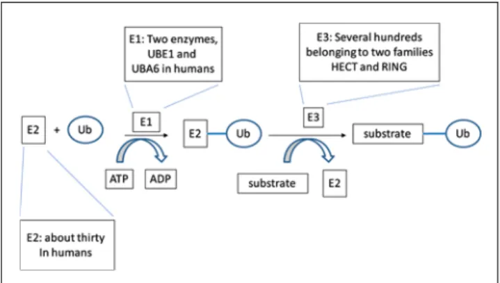
lysosome. Ubiquitination is executed in a well described cascade of enzymatic reactions mediated by three types of enzymes (Fig. 1): ubiquitin activating enzymes or E1s (two known enzymes in humans), ubiquitin conjugat-ing enzymes or E2s (about 30 in humans) and ubiquitin ligases or E3s (several hundred in human cells). Specifi c pairs of E2s and E3s decide what type of ubiquitin attach-ment will be executed [23]. Ubiquitination, similarly to other post-translational modifi cations, is reversible and there are fi ve families of de-ubiquitinating enzymes that perform this function [24].
The proteasome, a barrel-shaped multi-protein structure with a lumen residing both in the cytoplasm and the nucle-us, has two distinct parts: a central part called 20S protea-some or core particle (CP), where the enzymatic activities executing proteolysis reside, and a peripheral part covering one or both ends of the CP, called 19S proteasome or regu-latory particle (RP). RP sub-units function in ubiquitinated target protein recognition, denaturing, de-ubiquitination and transfer to the CP for degradation. Three enzymatic functions (trypsin-like, chymotrypsin-like and caspase-like) residing in distinct sub-units of CP result in the production of fragments of 4–14 amino acids [25].
The ubiquitin proteasome system (UPS) regulates all processes that are involved in carcinogenesis, among them invasion and metastasis. EMT, the process that endows neoplastic cells with invasive and metastatic potential, is intimately interwoven with other neoplastic processes, being served by several common pathways, several of which are regulated by UPS [26]. In the next section EMT pathways pertaining to MMMT pathogenesis will be dis-cussed.
Common molecular lesions in MMMT: role in EMT induction and regulation by UPS
Epithelial endometrial carcinomas are divided into two general types, called I and II or endometrioid and non-en-dometrioid respectively [27]. Type II carcinomas are most commonly of papillary serous or clear cell histology and some authors argue that poorly differentiated endometrioid carcinomas should be included with type II because they display similar immunohistochemistry and similar prog-nosis [28]. Endometrioid or type I endometrial carcinomas commonly display activating mutations of ß-catenin and disabled PTEN and less commonly K-ras activation (15– 30%), Her2 activation (10–20%) and p53 loss of function (10–20%). Type II or non-endometrioid endometrial can-cers are most commonly of serous or clear cell histology and have p53 disabling in 90% of cases. Her2 activation is present in a similar percentage of cases compared with type I carcinomas but PTEN inactivation and K-ras activation or ß-catenin mutations are rare. MMMTs present lesions in common with both type I and II endometrial carcinomas, with p53 inactivation and ß-catenin pathway activation being the most common [29, 30]. In addition, C-myc gene amplifi cation or polysomy of chromosome 8q appears to be a common lesion in uterine MMMTs and ovarian carci-nosarcomas [31]. Two other genes frequently amplifi ed in MMMTs are transforming growth factor ß1 (TGFß1) and kinase Akt2 at chromosome 19 [32]. All these lesions must provide, in a sub-set of MMMT cells, the co-operative ac-tion that allows epithelial cells to undergo an EMT and give rise to the sarcomatous component.
ß-Catenin activation plays a signifi cant role in EMT regulation by two mechanisms (Fig. 2). As a transcription factor, ß-catenin participates in the induction of
transcrip-Fig. 1 The ubiquitination cascade. E1 or ubiquitin-activating enzyme binds ubiquitin (Ub) in an ATP-dependent manner and transfers it to E2 or ubiquitin-conjugating enzyme as a thioester. Then E2-linked ubiquitin is transferred to the target protein with the aid of E3 ligase. There are two E1 enzymes, UBE1 and UBA6, in humans. The lat-ter also serves as the ligase for the ubiquitin-like molecule FAT10. There are 30–40 E2s and more than 500 E3s in the human genome. E3s belong to two families, the HECT (Homologous to HPV E6 Car-boxyterminal domain) and the RING (Really Interesting New Gene) family. A third family, the U-box containing ligases, is considered a sub-family of RING ligases due to the conformational similarity of the U-box and RING domains
Fig. 2 Cytoplasmic β-catenin constitutes the pool for both nuclear entry to act as a transcription factor and cytoplasmic membrane localisation to act as a component of adherens junctions. If Wnt sig-nalling is inactive, β-catenin is phosphorylated and ubiquitinated to be degraded by the proteasome. Among the target genes of β-catenin-promoted transcription, Slug suppresses E-cadherin transcription and thus promotes junction dissolution and favours β-catenin entry to the nucleus in a positive feedback loop that promotes EMT. Arrows de-note activation and inverse ┬ signs inhibition
tional modulator Slug (Snail2), which is a repressor of E-cadherin. ß-Catenin has another function, as a compo-nent of adherens junctions together with E-cadherin and
-catenin. Total cellular ß-catenin amounts are in
equilib-rium between adherens junctions and nuclear transcription function while cytoplasmic ß-catenin represents the pool that feeds both functions, or alternatively it is phosphory-lated and ubiquitinated to be degraded by the proteasome. In MMMT, immunohistochemistry for nuclear ß-catenin has been found to correlate with phosphorylated Akt kinase and Slug, and inversely with expression of E-cadherin [30]. Phosphorylated Akt is activated and phosphorylates, in its turn, multiple substrate proteins, including kinase gluco-gen synthase kinase 3ß (GSK3ß). The same effect may be obtained by activating mutations or amplifi cation of Akt2. Phosphorylated GSK3ß is inhibited and prevented from phosphorylating ß-catenin, which is then stabilised to either enter the nucleus and act as a transcription factor promoting the transcription of Slug or move to the plasma membrane and stabilise adherens junctions through interaction with E-cadherin. In this model Akt can regulate the same pool of ß-catenin that is activated by signals through the canonical Wnt pathway. Of interest in other situations, it appears that inhibition of GSK3ß by Akt or Wnt signalling activates dif-ferent pools of ß-catenin, an Axin-independent pool in the former case and an Axin-complexed in the latter [33]. Ac-tivation of Slug by ß-catenin may provide a feed-forward loop for the establishment of EMT, given that Slug-induced repression of E-cadherin would dissolve adherens junctions and further favour the movement of ß-catenin to the nucle-us for transcription activity as long as GSK3ß is inhibited. Thus, Akt activation in MMMT is a strong stimulus for the promotion of EMT. An additional down-stream effector of Akt with a role in EMT is kinase IKK. Activation of IKK by Akt leads to phosphorylation followed by ubiquitina-tion of protein IB and fi nally to activation of transcription factor NF-B. NF-B is an EMT promoter by induction of Snail and ßHLH (basic helix-loop-helix) transcription factor Twist. Nevertheless, in histochemical evaluation of MMMT specimens, no correlation was found between the NF-B family factor p65 and phosphorylated Akt or Slug [34], probably refl ecting the fact that these pathways are not linear but there is a multiplicity of regulations function-ing in conjunction.
p53, a tumour-suppressing transcription factor and one of the most common mutated proteins in cancer, is com-monly mutated in MMMT. Its function is activated after DNA damage and other stress signals and leads to cell cycle arrest or apoptosis if damage is too severe to be re-paired [35]. Post-transcriptional modifi cations, availability of co-factors and duration of triggering signal are among the factors that infl uence the fi nal outcome of p53 activa-tion. Due to these functions that allow the cell to repair reversible DNA damage or promote cell death if damage is irreparable in order to avoid inheritance of mutations in progeny, p53 is named the guardian of the genome. In addi-tion it plays a prime role in counteracting several pathways
of EMT and thus it can be considered a guardian of metas-tasis. p53 inactivation, either directly through mutations of its gene or indirectly through lesions interfering with its regulation, not only inhibits apoptosis, promotes the cell cycle and genomic instability, but also promotes invasion and metastasis. Mutant p53 protein often acquires gain of function properties promoting carcinogenesis in general and invasion in particular [36]. EMT is an invasion-pro-moting process that is inhibited by normal p53 in several ways. p53 directly induces microRNAs of the miR-200 family, miR-200c and miR-141 [37]. As a result, transla-tion of ZEB1 mRNA, which is a target of miR-200c, is suppressed. miR-200c induction concomitantly suppresses polycomb group member BMI1 (B lymphoma mouse Moloney leukaemia virus Insertion region 1) and transcrip-tion factor KLF4, both important in the maintenance of stem cell phenotype. Thus p53, through miR-200c, sup-presses stemness. This denotes that the two conditions, EMT and stem cell phenotype, may be served by tightly interwoven networks, as is evident also in development [3].
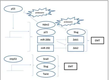
E3 ligase mdm2 (mouse double minute 2, also called Hdm2 in humans) is a transcriptional target of p53. Besides targeting p53 in a negative feedback loop, mdm2 mediates ubiquitination of Slug, being an additional effector of p53-induced EMT suppression (Fig. 3).
p53 down-regulates Slug through promotion of its mdm2-mediated ubiquitination and proteasome degrada-tion, which leads to E-cadherin expression [38]. CDK inhibitor p21, a p53 transcription target, has been found to decrease EMT of breast cancer cells induced by Ras and C-myc [39]. In contrast, cancer-associated mutant p53 pro-motes Snail, Slug and Twist induction and EMT [40–42].
Fig. 3 Wild-type p53 (upper part of the fi gure) suppresses EMT by inducing transcription of cyclin-dependent kinase inhibitor p21 and micro-RNAs miR-200c and miR-192. It also induces E3 ligase mdm2 (also called Hdm2 in humans), which acts as a negative feedback loop by ubiquitinating p53 for proteasomal degradation, but also as an EMT suppressor by ubiquitinating EMT inducer Slug. In contrast, mutant p53 (lower part of the fi gure) promotes EMT by favouring transcription of Snail, Slug and Twist. Arrows denote activation and inverse ┬ signs inhibition
A main regulation of p53 is executed by the UPS. Tight regulation of its availability and activation is of paramount importance as an importunate activation could lead to un-timely cell demise. Under normal non-stress conditions p53 has a short half-life because it is ubiquitinated with the help of E3 ligase mdm2 and degraded by the proteasome. Other E3 ligases, such as Pirh2, ARF-BP1/Mule, COP1 and CHIP, are also implicated in p53 regulation in various conditions [43–46].
p53 immunohistochemical staining, which is usually equivalent with the presence of a mutant form unable to be degraded and as a result having a longer half-life, is pres-ent in both epithelial and sarcomatous componpres-ents of most MMMTs [47–50]. p53 positivity has been reported in the in situ epithelial component of MMMTs, suggesting that p53 mutations represent an early molecular lesion at least in a percentage of cases [51].
C-myc is a basic helix-loop-helix leucine zipper family transcription factor with a role in neoplastic transforma-tion and in stem cell maintenance. It heterodimerises with protein Max to bind DNA on specifi c sequences called E-boxes and trigger recruitment of the transcription machin-ery [52]. When activated, C-myc promotes both prolifera-tion and apoptosis by up-regulating p53 activator p14ARF. p14ARF activates p53 by binding and inhibiting ligase mdm2. As a result, C-myc is an effi cient transformation ef-fector in cells with a disabled p14ARF/mdm2/p53 pathway. In addition, p14ARF inhibits C-myc directly in a negative feedback loop. The same relationship may also exist be-tween ß-catenin and p53 because, at least in some endome-trial cell lines, p14ARF is a target of ß-catenin transcription [53] and thus the p14ARF/mdm2/p53 pathway needs to be disabled for effi cient transformation in this instance too.
C-myc is regulated by the UPS after ubiquitination with the aid of two E3 ligases. Ubiquitination by E3 ligase Skp2 complex promotes both C-myc transcriptional function and turnover, although ubiquitination is not required for transcription [52]. This is a general theme in transcription where ubiquitination of transcription factors promotes the process by allowing the recruitment of new transcription factor molecules to access the DNA binding site if activat-ing signals persist, in order for transcription to continue. Another ligase, Fbwx7, promotes C-myc degradation in-dependently of promoter binding but dependent on previ-ous phosphorylation by kinases ERK and GSK3 [54, 55]. Phosphorylation of C-myc at a different site by IKK ki-nases leads to an opposite outcome protecting C-myc from ubiquitination and degradation [56].
C-myc over-expression promotes EMT in cancer cells in vitro and in vivo [57]. C-myc over-expressing cells dis-play Snail up-regulation due to both increased transcription and decreased ubiquitination and proteasome degradation, E-cadherin down-regulation and a fi broblast-like confi gura-tion [58]. C-myc further destabilises intercellular adhesions through induction of micro-RNA miR-9, which suppresses translation of both E-cadherin and -catenin [59]. In ad-dition, it suppresses expression of endometrial
differen-tiation, promoting F box transcription factor FOXO1 [60]. Dissolution of adherens junctions promotes ß-catenin transcription function as ß-catenin is shifted to the nucleus if the GSK3ß/axin/APC destruction complex is inhibited. As C-myc is a ß-catenin target gene, a feed-forward EMT-promoting loop is completed [61]. In a series of MMMTs and ovarian carcinosarcomas C-myc amplifi cation by FISH was detected in the majority of cases [31].
Oestrogen and TGFß crosstalk in MMMTs
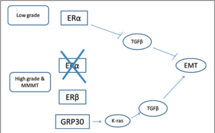
Low-grade endometrioid endometrial carcinomas express the sub-type of oestrogen receptor (ER) and are oestro-gen responsive. High oestrooestro-gen exposure is a risk factor for these carcinomas. ERß sub-type is expressed normally in uterus together with ER during development where it antagonises ER effects, but its expression decreases in normal adult endometrium [62]. This minor role of ERß in endometrial physiology is supported by studies of knock-out mice [63]. As grade increases and in serous carcinomas and MMMTs, ER expression decreases and expression of receptor ERß increases [64–66]. Activation of this sub-type is not effi cient in stimulating proliferation in the endome-trium [67] and thus high-grade carcinomas and MMMTs become oestrogen-independent and unresponsive. Alterna-tively spliced forms of ERß incapable of binding the ligand oestradiol are expressed in endometrial carcinomas of all grades and in carcinomas of higher grade, where ER is not present, and may contribute to oestrogen refractoriness [68]. In low-grade ER-positive endometrial cancer cell line, Ishikawa, oestrogen stimulation has been found to promote EMT [69]. ER expression in low-grade endometrioid en-dometrial carcinoma may promote EMT independently of its genomic/transcription factor action by inhibiting TGFß signalling [70]. This action is mediated by facilitation of degradation by the proteasome of Smad2 and Smad3, which are receptor-type intracellular transducers of TGFß signalling cascade [70]. Ubiquitination is executed with the aid of E3 ligases Smurf1 and Smurf2. Reciprocally Smad4, another transducer in the TGFß cascade, in complex with Smad3 may inhibit ERß transcription in breast cancer cells [71]. In contrast, in high-grade endometrial cancer cells HEC-1-A TGFß signalling is important for survival and migration [72]. A dominant negative TGFß receptor II inhibited growth and EMT of these cells. Given the well known dependence of TGFß signalling on the stage of malignancy, with an anti-neoplastic effect predominating in early carcinogenesis and a pro-carcinogenic effect in more advanced stages where other pathways such as K-ras are concomitantly activated [73], it is conceivable that in Ishikawa cells representing a low-grade, early carcinogen-esis step, TGFß signalling is anti-carcinogenic and thus its inhibition by ER promotes instead of inhibits EMT. In contrast, in most advanced grades, as represented by the HEC-1-A cell line, TGFß may promote EMT by
interact-ing with Snail and suppressinteract-ing transcription of E-cadherin and other junctional components such as Occludin and Claudin-3 [74]. In higher-grade histologies and MMMTs where ER is down-regulated, TGFß is left unopposed and at the same time it becomes pro-carcinogenic and EMT-promoting (Fig. 4).
An additional layer of complexity is conferred because non-genomic actions of oestrogens in MMMTs can be me-diated by a membrane-associated ER, G-coupled protein receptor GPR30 (also known as GPER, G-coupled protein oestrogen receptor), which is expressed in this malignancy in parallel with ERß and increases as stage advances [75]. GPR30 over-expression detected by immunohistochemis-try has been proposed as an adverse prognostic factor in endometrial carcinomas [76]. GPR30 is a seven transmem-brane domain receptor and signals through activation of MAPK and PI3K pathways [77] and downstream media-tors and transcription facmedia-tors [78]. Its physiologic ligand is oestradiol and it can also be activated by tamoxifen and fulvestrant but has a much lower affi nity for estrone and estriol and does not bind progesterone, glucocorticoster-oids or testosterone. In a breast cancer cell line expressing also ER, GPR30 activation by oestradiol resulted in TGFß signalling inhibition [79], but in another cell line lacking ER and ERß, GPR30 activation by oestradiol promoted proliferation and invasion [80]. In clinical breast cancer samples, GPR30 expression was associated with tumour resistance to tamoxifen [81]. In addition, in endometrial cancer, GPR30 activation promoted proliferation and inva-sion of both ER-positive and -negative cells [82]. A role for tamoxifen in promoting MMMT development in some breast cancer patients has been reported [83] and may be mediated by GPR30.
Ubiquitination and the UPS have essential and complex roles in transcription, as also discussed in the section on
c-myc, and nuclear receptors and ER in particular are no exception. The complexity of the regulation is outlined by the fact that proteasome inhibition enhances some but suppresses the expression of other ER target genes [84]. Proteasome function is required for ER transcriptional activity by promoting mono-ubiquitination of histone H2B and facilitating transcription elongation in breast cancer cells [85]. Interestingly, knocking-down of the E3 ligase involved in histone H2B ubiquitination, RNF40, led to oestrogen-independent cell proliferation and activation of the PI3K/Akt and MAPK pathways, providing an ad-ditional link between ER and K-ras-initiated signalling. Deubiquitination also plays a role in ER transcription. The deubiquitinating enzyme of the OTU (ovarian tumour) family OTUB1 is part of the ER transcription complex and acts in this deubiquitination of the receptor, which results in modulating transcription of target genes and the stability of ER itself [86]. In addition, ubiquitination is involved in endocytosis of G-protein coupled receptors that may lead either to lysosomal degradation or re-cycling to the surface [87] and this has been shown specifi cally for GPR30 [88].
TGFß signalling is also regulated by the UPS in mul-tiple levels [89]. Several E3 ligases such as Smurf1 and 2 and NEDD4 of the HECT domain family, and ßTrCP and Fbwx1 of the RING family, among others, regulate the sta-bility of proteins participating in TGFß signalling [90–92].
In conclusion, and despite unresolved issues in an evolving fi eld, oestrogen signalling, which may promote proliferation and EMT in lower-grade endometrial carcino-ma through inhibition of TGFß anti-neoplastic signalling, becomes down-regulated in higher-grade carcinomas and MMMTs where ER is not expressed and ERß is probably less effective in counteracting TGFß signalling. In these cases TGFß signalling is transformed to a invasion pro-gramme due to concomitant MAPK and Akt activation by GPR30 and other receptors, and promotes EMT (Fig. 4).
EMT inducing transcription factors ZEB, Snail and Twist in MMMT
EMT is promoted by a set of core transcription factors such as ZEB (Zinc fi nger E-box Binding homeodomain), Snail, Slug and Twist that result in down-regulation of key com-ponents of intercellular adhesions, which is a prerequisite for the acquisition of motility.
The zinc fi nger homeodomain factor ZEB1 is an EMT inducer. It has been reported to be expressed in the nor-mal adult endometrium and its expression is oestrogen-dependent [93]. ZEB1 is over-expressed in the stroma of low-grade endometrial carcinomas [94] but its gene is deleted in the low-grade epithelial cancer component [93]. In grade 3 endometrioid endometrial carcinomas, papillary serous endometrial carcinomas and MMMTs, ZEB1 is of-ten expressed in the epithelial neoplastic cells and becomes oestrogen-independent [93]. Moreover, forced expression
Fig. 4 In low-grade endometrial carcinomas (upper part of the fi gure) ERα is expressed and promotes EMT by inhibiting TGFβ, which on this occasion is an EMT inhibitor. In higher-grade endometrioid carcinomas, carcinomas of other histologies and MMMTs, ERα is not expressed, replaced by ERβ (including alternatively spliced forms) and GPR30 (lower part of the fi gure). GPR30 activates K-ras, which transforms TGFβ to an EMT promoting signaling. Arrows denote ac-tivation and inverse ┬ signs inhibition
of ZEB1 in endometrial cancer cell lines representative of low-grade carcinomas promotes their migratory potential in an in vitro wound healing assay and reduces the expression of E-cadherin [95]. Conversely, partial silencing of ZEB1 by shRNA in a high-grade endometrial cancer cell line re-duced its in vitro migratory potential although no increase in E-cadherin expression was noticed [95].
High expression of ßHLH transcription factor Twist has been associated with decreased E-cadherin expres-sion and deeper myometrial invaexpres-sion and with decreased survival in endometrioid endometrial carcinomas [96]. An endometrial cell line undergoing irradiation showed Twist up-regulation and EMT morphologic changes in parallel with an increased migratory potential [97]. Knock-down of Twist with siRNA reversed the increased irradiation-induced migration. Another endometrial cell line did not show prominent EMT changes under the same experi-mental conditions. These fi ndings support the role of an additional EMT inducer, Twist, in the aggressiveness of endometrial carcinoma. Although no histologic features of MMMT were observed in the cases with high Twist expres-sion in vivo, the in vitro study suggests that, in some cases, mesenchymal morphologic changes are present.
The homeobox gene HoxA10 is a specific differen-tiation gene for the endometrium and is induced in the secretory phase of the endometrial cycle by progesterone. HoxA10 is down-regulated as the grade of endometrioid endometrial cancer increases, being most suppressed in grade 3 endometrioid as well as in papillary serous cancers [98]. HoxA10 is a suppressor of Snail and thus its down-regulation through promoter methylation in high-grade endometrial cancers leads to E-cadherin down-regulation, providing a link between increasing grade and EMT/inva-sion [98]. An association of Snail with higher grade and reduced E-cadherin expression was observed in metastatic lesions of endometrial carcinomas [99]. The related tran-scription factor Slug (also called Snail2) is up-regulated by ß-catenin signalling and suppressed E-cadherin in MMMT, as already discussed [30].
microRNAs constitute physiologic important post-transcriptional regulators of protein expression and play a role in carcinogenesis. In MMMT a specifi c microRNA signature has been revealed in the mesenchymal compo-nent compared with the epithelial part of the tumour [100]. Prominent in this signature is the 200 family of miR-NAs, which is down-regulated in the mesenchymal compo-nent. The miR-200 family is composed of fi ve members, miR-200a, miR-200b, miR-200c, miR-141 and miR-429, which are post-transcriptional repressors of ZEBs and, as a result, their down-regulation allows ZEBs to suppress E-cadherin transcription. Indeed, E-E-cadherin expression was completely lost and p120 cadherin decreased in the mesen-chymal component of MMMTs, while mesenmesen-chymal mark-ers vimentin, SPARC (secreted protein acidic and rich in cysteine) and fascin were up-regulated. Also up-regulated were EMT inducers TGFß1 and TGFß2. TGFß1 induces EMT in endometrial cell lines and up-regulates Slug,
ad-hesion molecule L1CAM and vimentin [101]. L1CAM is associated in histochemical evaluation of endometrial car-cinomas with ER and progesterone receptor (PR) negativ-ity and E-cadherin negativnegativ-ity and is sometimes observed in the invasive front of tumours. Other relevant EMT targets of miR-200c which are suppressed in aggressive endome-trial cancer cell lines include fi bronectin, moesin, an actin cytoskeleton connector to the plasma membrane, and the receptor tyrosine kinase TrkB [102]. In addition, when compared with endometrioid endometrial cancers, the epi-thelial component of MMMTs displays higher expression of several mesenchymal markers and lower expression of E-cadherin [100], arguing for the presence of a predispo-sition or early stages of EMT already in this component. Another miRNA, miR-194, which did not sort out in the above signature, was found to down-regulate the polycomb group protein BMI-1 and to reduce EMT and invasion po-tential in vitro in endometrial cell lines [103]. BMI-1 is a promoter of invasion and metastasis from various primaries and also a target of miR-200c, as previously discussed.
Collectively these data paint a picture in which tran-scription factors of the core EMT circuitry are up-regulated in MMMT while differentiation promoting factor HoxA10 and miRNAs normally counter-acting them are down-reg-ulated with increasing grade as the balance shifts towards the EMT/metastatic phenotype (Fig. 5). The UPS modu-lates the stability of all core EMT transcription factors sometimes in a co-ordinated manner. This is the example of core EMT transcription factors Snail, Slug, Twist and Zeb2, which are all regulated by ubiquitination with the aid of a Skp ligase complex having the F-box protein Ppa
Fig. 5 Schematic representation of key EMT transcription factor con-centrations in relationship to important regulators. When β-catenin shifts from membrane localisation as an adherens junction compo-nent to the nucleus, it promotes transcription of Slug. Suppression of HoxA10 due to promoter methylation de-represses transcription of Snail. Down-regulation of miR-200 family members promotes translation of ZEB transcription factors. Co-ordinate increase in the concentrations of these transcription factors triggers EMT. The UPS is involved in the regulation of these factors at multiple points and may tip the balance in either direction, as discussed in the text
(Partner of paired or FBXL14 in humans) as the substrate recognising sub-unit [104–106].
Intercellular adhesions and cell-matrix adhesions
Intercellular adhesions are important defining elements of epitheliums and need to be dissolved during EMT in order for cells to move. Adherens junctions are made of E-cadherin molecules that span the cytoplasmic membrane and have both extracellular and intracellular domains [107, 108]. With their extracellular domains, E-cadherins make homotypic contacts with E-cadherin molecules of neigh-bouring cells. The intracellular domain associates together with -catenin and ß-catenin to the actin cytoskeleton. Other proteins such as p120 catenin, EPLIN (epithelial protein lost in neoplasm), ZO1 (Zonula Occludens 1), Afa-din, Vinculin, Paxillin and -actinin may participate in ini-tiating and strengthening the interactions of catenins with the actin cytoskeleton. Adherens junctions are dynamic and E-cadherin molecules are continuously incorporated in cell membranes and removed by clathrin-mediated endocytosis. Endocytosis of E-cadherin is triggered after phosphoryla-tion by c-src kinase, which is followed by ubiquitinaphosphoryla-tion with the aid of c-cbl family E3 ligase Hakai [109]. p120 catenin prevents E-cadherin endocytosis by masking Hakai interaction sites in the juxta-membrane area of its mol-ecule. Hakai over-expression in epithelial cells results in Paxillin down-regulation and decreased cell-matrix adhe-sion [110].
E-cadherin is a central target of EMT-inducing tran-scription regulators and is down-regulated by ZEB and Snail transcription factors as well as ß-catenin in high-grade uterine carcinomas and MMMTs [111, 112]. In addi-tion ß-catenin, as menaddi-tioned, participates in a feed-forward loop by becoming available to act as a transcription factor when adherens junctions are resolved.
Adherens junction components P-cadherin, E-cadherin, p120 and ß-catenin have been investigated as prognostic markers in a histochemical study of all endometrial carci-nomas diagnosed in a Norwegian region between 1981 and 1990 [113]. Decreased expression of E-cadherin and p120 was more commonly seen in higher-grade and clear cell carcinomas while mesenchymal P-cadherin high expres-sion was more commonly seen in these types [113]. High E-cadherin and low P-cadherin expression were associated with a better prognosis in endometrial carcinomas.
EMT is promoted by up-regulation of matrix metallo-proteinase MMP-3 (Stromelysin 1) in endometrial cancer, which results in E-cadherin degradation and is associated with vascular invasion and more aggressive tumours [114]. MMP-7 (matrilysin), another matrix metalloproteinase, is expressed in the epithelial component of 70% of cases of MMMTs examined, while expression is lost in correspond-ing sarcomatous components [115]. Thus, it appears that, after intercellular adhesion dissolution, motion of epithelial
cells acquiring EMT phenotype and properties is accompa-nied by changes in the profi le of expressed matrix-modifying enzymes with up-regulation of some MMP-3 and down-reg-ulation of others. MMP-3 and MMP-7 production is stimu-lated by MAPK cascade activation and may, as a result, be modulated by the UPS given that several proteins taking part in this cascade are regulated by the UPS [116, 117].
Therapeutic opportunities
MMMT can be considered a high-grade epithelial endome-trial cancer with extensive metaplasia due to lesions acti-vating EMT pathways. UPS plays a key role in regulating these pathways as discussed in the previous sections. This patho-physiological insight offers opportunities to reverse or counteract EMT by modulating UPS function. Given that EMT is served in many instances by overlapping path-ways with other carcinogenesis-enabling properties, re-versal of EMT through UPS modifying interventions may have more global anti-neoplastic effects.
Inhibition of the proteasome is already used in can-cer therapy. Bortezomib, a specifi c inhibitor of the chy-motrypsin-like proteasome activity, constitutes a well established treatment for multiple myeloma and sub-types of non-Hodgkin lymphoma [118]. Newer inhibitors of the enzymatic cleavage activity of proteasome such as carfi l-zomib and NPI-0052 are under development [119, 120]. Inhibition of the deubiquitinising activity of 19S RP is an-other way of inhibiting the proteasome and such inhibitors are in earlier study [121]. Proteasome inhibition, despite the specifi city of the enzymatic molecular reaction that is involved, has broad effects in cellular homeostasis and fi nally produces non-specifi c cytotoxicity due to perturba-tions of hundreds of proteins regulated by the proteasome. Nevertheless malignancies with specifi c molecular lesions could be particularly sensitive to this broad inhibition due to dependence of the lesions on proteasome function or to triggering of different down-stream pathways. For exam-ple, cells defi cient in p130Cas, an adaptor protein of cel-lular adhesions, are resistant to apoptosis after bortezomib treatment and trigger autophagy, while cells expressing this protein are sensitive to proteasome inhibition [122]. Thus p130Cas expression may serve as a marker of bortezomib sensitivity. On the other hand, given that p130Cas-defi cient cells are concomitantly resistant to doxorubicin, it remains possible that this resistance is generalised and related to adhesion destabilisation and EMT and may not represent a marker of resistance to particular treatments. Indeed EMT is associated with drug resistance related to the aforemen-tioned intertwining of its pathways, with pathways involved in apoptosis prevention and self-sufficiency of survival signals in neoplastic cells [123]. Nevertheless a sub-set of cancer patients could remain sensitive to particular strate-gies such as proteasome inhibition even in the presence of EMT. In the case of MMMT, a particular patient could be
sensitive to proteasome inhibition if it possesses a mutant C-myc with gain of function but still able to be degraded by the proteasome and concomitantly a wild-type p53 that is functionally inhibited due to instability from increased degradation, e.g., secondary to mdm2 amplification or stabilisation. In this example EMT related to C-myc hyper-activity and p53 disabling could be reversed by proteasome inhibition, which would inhibit C-myc transcription func-tion, producing c-myc turnover stalling on DNA and p53 activity promotion through reversal of its instability.
An alternative to proteasome inhibition strategy is to in-terfere with other points of the UPS that would offer great-er specifi city. An example of such an altgreat-ernative strategy is inhibition of NEDDylation, which refers to the ligation of a target protein with the ubiquitin-like small protein NEDD8 (Neural precursor cells-Expressed Developmentally Down-regulated 8). This is a post-translational modifi cation that often takes plays in the Cullin component of the SCF type RING ligases and helps the E2 enzyme binding to the li-gase complex in the ubiquitination cascade. MLN4924, a small molecule inhibitor of the NEDD8 activating enzyme (NAE, the E1 for NEDD8) is in early clinical development [124]. Nevertheless, the SCF ligase family is extensive and many proteins involved in EMT of MMMT (ß-catenin,
C-myc, NF-B, Snail) are regulated by ligases of this fam-ily. In addition, NEDDylation has other targets, such as p53 and the core apoptosis effectors caspases [125, 126]. Thus NEDDylation inhibition could interfere with multiple points in EMT progression and the net result of the inhibi-tion, and whether there are sub-sets of MMMT patients that could benefi t from it remains to be investigated.
Inhibiting E3 ligases could offer greater specifi city. For example, inhibition of mdm2 could be a strategy to explore [127, 128], although in MMMT it would be expected to be effective only in the sub-set with wild-type p53. In patients with mutant p53 these inhibitors may not only be inac-tive but could have deleterious effects due to inhibition of degradation of Slug. This fact underlines the importance of correctly delineating the sub-set of patients that could ben-efi t from a certain drug.
Defi ning sub-sets of neoplasms that, due to particular pathogenic lesions, are sensitive to particular drugs is a major challenge in oncology but one that would provide signifi cant advancement in personalised treatment of can-cer patients.
Confl ict of interest The authors declare that they have no confl ict of interest relating to the publication of this manuscript.
References
1. El-Nashar SA, Mariani A (2011) Uterine carcino-sarcoma. Clin Obstet Gynecol 54:292–304 2. Choijamts B, Jimi S, Kondo T et al (2011)
CD133+ cancer stem cell-like cells derived from uterine carcinosarcoma (malignant mixed Mül-lerian tumor). Stem Cells 29:1485–1495 3. Mani SA, Guo W, Liao M-J et al (2008) The
epithelial-mesenchymal transition generates cells with properties of stem cells. Cell 133:704–715 4. D’Angelo E, Prat J (2011) Pathology of mixed
Müllerian tumours. Best Practice Res Clin Obstet Gynaecol 25:705–718
5. McCluggage WG (2002) Malignant biphasic uter-ine tumours: carcinosarcomas or metaplastic car-cinomas? J Clin Pathol 55:321–325
6. Sreenan JJ, Hart WR (1995) Carcinosarcomas of the female genital tract. A pathologic study of 29 metastatic tumors: further evidence for the dominant role of the epithelial component and the conversion theory of histogenesis. Am J Surg Pathol 19:666–674
7. Gorai I, Yanagibashi T, Taki A et al (1997) Uterine carcinosarcoma is derived from a single stem cell: an in vitro study. Int J Cancer 72:821–827 8. Emoto M, Iwasaki H, Kikuchi M, Shirakawa K
(1993) Characteristics of cloned cells of mixed Müllerian tumor of the human uterus. Cancer 71: 3065–3075
9. Lien HC, Lin CW, Mao TL et al (2004) p53 over-expression and mutation in metaplastic carcinoma of the breast: genetic evidence for a monoclonal origin of both the carcinomatous and the het-erogeneous sarcomatous components. J Pathol 204:131–139
10. Van Deurzen CHM, Lee AHS, Gill MS et al (2011) Metaplastic breast carcinoma: tumour his-togenesis or dedifferentiation? J Pathol 224:434– 437
11. Kalluri R, Weinberg RA (2009) The basics of epithelial–mesenchymal transition. J Clin Invest 119:1420–1428
12. Stewart CJR, Little L (2009) Immunophenotypic features of MELF pattern invasion in endometrial
adenocarcinoma: evidence for epithelial–mesen-chymal transition. Histopathology 55:91–101 13. Murray SK, Young RH, Scully RE (2003) Unusual
epithelial and stromal changes in myoinvasive endo-metrioid adenocarcinomas: a study of their frequen-cy, associated diagnostic problems, and prognostic signifi cance. Int J Gynecol Pathol 22:324–333 14. Stewart CJR, Crook ML, Little L, Louwen K
(2011) Correlation between invasive pattern and immunophenotypic alterations in endocervical adenocarcinoma. Histopathology 58:720–728 15. Götte M (2010) Endometrial cells get
side-tracked. Side population cells promote epithelial-mesenchymal transition in endometrial carcinoma. Am J Pathol 176:25–28
16. Zhou S, Schuetz SD, Bunting KD et al (2001) The ABC transporter Brcp1/ABCG2 is expressed in a wide variety of stem cells and is a molecular determinant of the side-population phenotype. Nat Med 7:1028–1034
17. Kato K, Takao T, Kuboyama A et al (2010) Endo-metrial cancer side-population cells show promi-nent migration and have a potential to differentiate into the mesenchymal cell lineage. Am J Pathol 176:381–392
18. Leblanc M, Poncelet C, Soriano D et al (2001) Alteration of CD44 and cadherins expression: possible association with augmented aggressive-ness and invasiveaggressive-ness of endometrial carcinoma. Virchows Arch 438:78–85
19. Montserrat N, Mozos A, Llobet D et al (2011) Ep-ithelial to mesenchymal transition in early stage endometrioid endometrial carcinoma. Hum Pathol doi: 10.1016/j.humpath.2011.06.021
20. Voutsadakis IA (2010) Ubiquitin, ubiquitination and the ubiquitin-proteasome system in cancer. Atlas Genet Cytogen Oncol Haematol. January. www.AtlasGeneticsOncology.org/Deep/Ubiquit-inCancerID20083.html
21. Kirkin V, Dikic I (2011) Ubiquitin networks in cancer. Curr Opin Genet Dev 21:21–28
22. Behrends C, Harper JW (2011) Constructing and decoding unconventional ubiquitin chains. Nat Struct Mol Biol 18:520–528
23. van Wijk SJL, Timmers HTM (2010) The family of ubiquitin-conjugating enzymes (E2s):
decid-ing between life and death of proteins. FASEB J 24:981–993
24. Amerik AY, Hochstrasser M (2004) Mechanism and function of deubiquitinating enzymes. Bio-chim Biophys Acta 1695:189–207
25. Voutsadakis IA (2007) Pathogenesis of colorectal carcinoma and therapeutic implications: the roles of the ubiquitin-proteasome system and Cox-2. J Cell Mol Med 11:252–285
26. Soond SM, Chantry A (2011) How ubiquitina-tion regulates the TGF-ß signaling pathway: new insights and new players. Bioessays 33:749–758 27. Okuda T, Sekizawa A, Purwosunu Y et al (2010)
Genetics of endometrial cancers. Obstet Gynecol Int, article id 984013
28. Voss MA, Ganesan R, Ludeman L et al (2012) Should grade 3 endometrioid endometrial carci-noma be considered a type 2 cancer: a clinical and pathological evaluation. Gynecol Oncol 124:15– 20
29. Lax SF (2007) Molecular genetic changes in epithelial, stromal and mixed neoplasms of the endometrium. Pathology 39:46–54
30. Saegusa M, Hashimura M, Kuwata T, Okayasu I (2009) Requirement of the akt/ß-catenin pathway for uterine carcinosarcoma genesis, modulating E-cadherin expression through the transactivation of Slug. Am J Pathol 174:2107–2115
31. Schipf A, Mayr D, Kirchner T, Diebold J (2008) Molecular genetic aberrations of ovarian and uter-ine carcinosarcomas: a CGH and FISH study. Virchows Arch 452:259–268
32. Chiyoda T, Tsuda H, Tanaka H et al (2012) Ex-pression profi les of carcinosarcoma of the uterine corpus: are these similar to carcinoma or sar-coma? Genes Chrom Cancer 51:229–239 33. Ng SS, Mahmoudi T, Danenberg E et al (2009)
Phosphatidylinositol 3-kinase signaling does not activate the Wnt cascade. J Biol Chem 284:35308– 35313
34. Saegusa M, Hashimura M, Kuwata T et al (2007) Crosstalk between NF-B/p65 and ß-catenin/ TCF4/p300 signalling pathways through altera-tions in GSK-3ß expression during trans-differ-entiation of endometrial carcinoma cells. J Pathol 213:35–45
35. Dai C, Gu W (2010) p53 post-translational modi-fi cation: deregulated in tumorigenesis. Trends Mol Med 16:528–536
36. Solomon H, Madar S, Rotter V (2011) Mutant p53 gain of function is interwoven into the hallmarks of cancer. J Pathol 225:475–478
37. Chang CJ, Chao CH, Xia W et al (2011) p53 regulates epithelial-mesenchymal transition and stem cell properties through modulating miRNAs. Nat Cell Biol 13:317–323
38. Wang S-P, Wang W-L, Chang Y-L et al (2009) p53 controls cancer cell invasion by inducing the MDM2-mediated degradation of Slug. Nature Cell Biol 11:694–704
39. Liu M, Casimiro MC, Wang C et al (2009) p21CIP1 attenuates Ras- and c-Myc-dependent breast tumor epithelial mesenchymal transition and cancer stem cell-like gene expression in vivo. Proc Natl Acad Sci U S A 106:19035–19039 40. Zhang Y, Yan W, Chen X (2011) Mutant p53
dis-rupts MCF-10A cell polarity in three-dimensional culture via epithelial-to-mesenchymal transitions. J Biol Chem 286:16218–16228
41. Kogan-Sakin I, Tabach Y, Buganim Y et al (2011) Mutant p53R175H upregulates Twist1 expression and promotes epithelial-mesenchymal transition in immortalized prostate cells. Cell Death Diff 18:271–281
42. Ohashi S, Natsuizaka M, Wong GS et al (2010) Epidermal growth factor receptor and mutant p53 expand an esophageal cellular subpopulation capable of epithelial-to-mesenchymal transition through ZEB transcription factors. Cancer Res 70: 4174–4184
43. Leng RP, Lin Y, Ma W et al (2003) Pirh2, a p53-induced ubiquitin-protein ligase, promotes p53 degradation. Cell 112:779–791
44. Dornan D, Wertz I, Shimizu H et al (2004) The ubiquitin ligase COP1 is a critical negative regula-tor of p53. Nature 429:86–92
45. Chen D, Kon N, Li M et al (2005) ARF-BP1/Mule is a critical mediator of the ARF tumor suppressor. Cell 121:1071–1083
46. Esser C, Scheffner M, Hohfeld J (2005) The chaperone-associated ubiquitin ligase CHIP is able to target p53 for proteasomal degradation. J Biol Chem 280:27443–27448
47. Buza N, Tavassoli FA (2009) Comparative analy-sis of p16 and p53 expression in uterine malignant mixed Mullerian tumors. Int J Gynecol Pathol 28:514–521
48. Taylor NP, Zighelboim I, Huettner PC et al (2006) DNA mismatch repair and TP53 defects are early events in uterine carcinosarcoma tumorigenesis. Mod Pathol 19:1333–1338
49. Lee S-J, Kim HS, Kim HS et al (2007) Immu-nohistochemical study of DNA topoisomerase I, p53, and Ki-67 in uterine carcinosarcomas. Hum Pathol 38:1226–1231
50. Kanthan R, Senger J-LB, Diudea D (2010) Malig-nant mixed Mullerian tumors of the uterus: histo-pathological evaluation of cell cycle and apoptotic regulatory proteins. World J Surg Oncol 8:60 51. Keeling L, Taraporewalla D, Perunovic B, Smith
JHF (2011) Uterine carcinosarcoma with p53-positive intraepithelial component. Histopathol-ogy 59:1277–1278
52. Cowling VH, Cole MD (2006) Mechanism of transcriptional activation by the Myc oncopro-teins. Semin Cancer Biol 16:242–252
53. Saegusa M, Hashimura M, Kuwata T et al (2004) ß-catenin simultaneously induces activation of the p53-p21WAF1 pathway and overexpression of cyclin D1 during squamous differentiation of endometrial carcinoma cells. Am J Pathol 164: 1739–1749
54. Yada M, Hatakeyama S, Kamura T et al (2004) Phosphorylation-dependent degradation of c-Myc is mediated by the F-box protein Fbw7. EMBO J 23:2116–2125
55. Amati B (2004) Myc degradation: dancing with ubiquitin ligases. Proc Natl Acad Sci U S A 101: 8843–8844
56. Yeh P-Y, Lu Y-S, Ou D-L, Cheng A-L (2011) IB kinases increase Myc protein stability and enhance progression of breast cancer cells. Mol Cancer 10:53
57. Trimboli AJ, Fukino K, de Bruin A et al (2008) Direct evidence for epithelial-mesenchymal tran-sitions in breast cancer. Cancer Res 68:937–945 58. Cho KB, Cho MK, Lee WY, Kang KW (2010)
Overexpression of c-myc induces epithelial mes-enchymal transition in mammary epithelial cells. Cancer Lett 293:230–239
59. Ma L, Young J, Prabhala H et al (2010) miR-9, a MYC/MYCN-activated microRNA, regulates E-cadherin and cancer metastasis. Nature Cell Biol 12:247–256
60. Myatt SS, Wang J, Monteiro LJ et al (2010) Defi -nition of microRNAs that repress expression of the tumor suppressor gene FOXO1 in endometrial cancer. Cancer Res 70:367–377
61. Khew-Goodall Y, Goodall GJ (2010) Myc-modu-lated miR-9 makes more metastases. Nature Cell Biol 12:209–211
62. Weihua Z, Saji S, Mäkinen S et al (2000) Estro-gen receptor (ER) ß, a modulator of ER in the uterus. Proc Natl Acad Sci U S A 97:5936–5941 63. Harris HA (2007) Estrogen receptor-ß: recent
les-sons from in vivo studies. Mol Endocrinol 21:1–13 64. Huang GS, Arend RC, Li M et al (2009) Tis-sue microarray analysis of hormonal signalling pathways in uterine carcinosarcoma. Am J Obstet Gynecol 200:457.e1–457.e5
65. Shabani N, Mylonas I, Jeschke U et al (2007) Expression of estrogen receptors and ß, and progesterone receptors A and B in human muci-nous carcinoma of the endometrium. Anticancer Res 27:2027–2034
66. Wu W, Slomovitz BM, Celestino J et al (2003) Coordinate expression of cdc25B and ER- is frequent in low-grade endometrioid endometrial carcinoma but uncommon in high-grade endo-metrioid and nonendoendo-metrioid carcinomas. Can-cer Res 63:6195–6199
67. Nilsson S, Koehler KF, Gustafsson J-Å (2011) Development of subtype-elective oestrogen recep-tor-based therapeutics. Nat Rev Drug Discov 10: 778–792
68. Collins F, MacPherson S, Brown P et al (2009) Expression of oestrogen receptors, ERalpha, ER-beta, and ERbeta variants, in endometrial cancers and evidence that prostaglandin F may play a role in regulating expression of ERalpha. BMC Cancer 16:330
69. Chen Y-J, Li H-Y, Huang C-H et al (2010) Oestro-gen-induced epithelial-mesenchymal transition of endometrial epithelial cells contributes to the de-velopment of adenomyosis. J Pathol 222:261–270 70. Ito I, Hanyu A, Wayama M et al (2010) Estrogen
inhibits transforming growth factor ß signalling by promoting Smad2/3 degradation. J Biol Chem 285:14747–14755
71. Ren Y, Wu L, Frost AR et al (2009) Dual effects of TGF-ß on ER-mediated estrogenic transcrip-tional activity in breast cancer. Mol Cancer 8:111 72. Lei XF, Wang L, Yang J, Sun L-Z (2009) TGFß
signaling supports survival and metastasis of endo-metrial cancer cells. Cancer Manag Res 1:15–24 73. Massagué J (2008) TGFß in cancer. Cell 134:215–
230
74. Vincent T, Neve EPA, Johnson JR et al (2009) A SNAIL1-SMAD3/4 transcriptional repressor com-plex promotes TGF-ß mediated epithelial-mesen-chymal transition. Nat Cell Biol 11:943–950 75. Huang GS, Gunter MJ, Arend RC et al (2010)
Co-expression of GPR30 and ERß and their as-sociation with disease progression in uterine car-cinosarcoma. Am J Obstet Gynecol 203:242.e1–5 76. Smith HO, Leslie KK, Singh M et al (2007)
GPR30: a novel indicator of poor survival for en-dometrial carcinoma. Am J Obstet Gynecol 196: 386.e1–386.e11
77. Wang D, Hu L, Zhang G et al (2010) G protein-coupled receptor 30 in tumor development. Endo-crine 38:29–37
78. Vivacqua A, Romeo E, De Marco P et al (2011) GPER mediates the Egr-1 expression induced by 17ß-estradiol and 4-hydroxitamoxifen in breast and endometrial cancer cells. Breast Cancer Res Treat DOI 10.1007/s10549-011-1901-8 79. Kleuser B, Malek D, Gust R et al (2008)
17-ß-Estradiol inhibits transforming growth factor-ß signaling and function in breast cancer cells via activation of extracellular signal-regulated kinase through the G protein-coupled receptor 30. Mol Pharmacol 74:1533–1543
80. Pandey DP, Lappano R, Albanito L et al (2009) Estrogenic GPR30 signalling induces prolifera-tion and migraprolifera-tion of breast cancer cells through CTGF. EMBO J 28:523–532
81. Ignatov A, Ignatov T, Weißenborn C et al (2011) G-protein-coupled estrogen receptor GPR30 and tamoxifen resistance in breast cancer. Breast Can-cer Res Treat 128:457–466
82. He Y-Y, Cai B, Yang Y-X et al (2009) Estrogenic G protecoupled receptor 30 signaling is in-volved in regulation of endometrial carcinoma by promoting proliferation, invasion potential, and interleukin-6 secretion via the MEK/ERK mitogen-activated protein kinase pathway. Cancer Sci 100:1051–1061
83. Leung F, Terzibachian J-J, Govyadovskiy A et al (2009) Tamoxifen in the adjuvant setting for breast cancer: refl ections about the risk of uterine carcinosarcoma. Gyn Obstet Fertil 37:447–451 84. Fan M, Nakshatri H, Nephew KP (2004)
Inhibit-ing proteasomal proteolysis sustains estrogen receptor- activation. Mol Endocrinol 18:2603– 2615
85. Prenzel T, Begus-Nahrmann Y, Kramer F et al (2011) Estrogen-dependent gene transcription in human breast cancer cells relies upon proteasome-dependent monoubiquitination of histone H2B. Cancer Res 71:5739–5753
86. Stanišić V, Malovannaya A, Qin J et al (2009) OTU domain-containing ubiquitin aldehyde-bind-ing protein 1 (OTUB1) deubiquitinates estrogen receptor (ER) and affects ER transcriptional activity. J Biol Chem 284:16135–16145 87. Hislop JN, von Zastrow M (2011) Role of
ubiq-uitination in endocytic traffi cking of G-protein-coupled receptors. Traffi c 12:137–148
88. Cheng S-B, Quinn JA, Graeber CT, Filardo EJ (2011) Down-modulation of the G-protein-cou-pled Estrogen Receptor, GPER, from the cell sur-face occurs via a trans-Golgi-proteasome pathway. J Biol Chem 286:22441–22455
89. Inoue Y, Imamura T (2008) Regulation of TGF-ß family signaling by E3 ubiquitin ligases. Cancer Sci 99:2107–2112
90. Lin X, Liang M, Feng XH (2000) Smurf2 is a ubiquitin E3 ligase mediating proteasome-de-pendent degradation of Smad2 in transforming growth factor-beta signaling. J Biol Chem 275: 36818–36822
91. Bengoechea-Alonso MT, Ericsson J (2010) Tumor suppressor Fbw7 regulates TGFß signaling by targeting TGIF1 for degradation. Oncogene 29: 5322–5328
92. Wan M, Tang Y, Tytler EM et al (2004) Smad4 protein stability is regulated by ubiquitin ligase SCF beta-TrCP1. J Biol Chem 279:14484–14487 93. Hurt EM, Saykally JN, Anose BM et al (2008)
Expression of the ZEB1 (EF1) transcription factor in human: additional insights. Mol Cell Biochem 318:89–99
94. Spoelstra NS, Manning NG, Higashi Y et al (2006) The transcription factor ZEB1 is aber-rantly expressed in aggressive uterine cancers. Cancer Res 66:3893–3902
95. Singh M, Spoelstra NS, Jean A et al (2008) ZEB1 expression in type I vs type II endometrial can-cers: a marker of aggressive disease. Mod Pathol 21:912–923
96. Kyo S, Sakaguchi J, Ohno S et al (2006) High Twist expression is involved in infi ltrative endo-metrial cancer and affects patient survival. Hum Pathol 37:431–438
97. Tsukamoto H, Shibata K, Kajiyama H et al (2007) Irradiation-induced epithelial-mesenchymal transi-tion (EMT) related to invasive potential in endome-trial carcinoma cells. Gynecol Oncol 107:500–504 98. Yoshida H, Broaddus R, Cheng W et al (2006)
Deregulation of the HOXA10 homeobox gene in endometrial carcinoma: role in epithelial-mesen-chymal transition. Cancer Res 66:889–897 99. Blechschmidt K, Kremmer E, Hollweck R et al
(2007) The E-cadherin repressor Snail plays a role in tumor progression of endometrioid adenocarci-nomas. Diagn Mol Pathol 16:222–228
100. Castilla MÁ, Moreno-Bueno G, Romero-Pérez L et al (2010) Micro-RNA signature of the epitheli-al-mesenchymal transition in endometrial carcino-sarcoma. J Pathol 223:72–80
101. Huszar M, Pfeifer M, Schirmer U et al (2010) Up-regulation of L1CAM is linked to loss of hormone receptors and E-cadherin in aggressive subtypes of endometrial carcinomas. J Pathol 220:551–561 102. Howe EN, Cochrane DR, Richer JK (2011) Targets
of miR-200c mediate suppression of cell motility and anoikis resistance. Breast Cancer Res 13:R45 103. Dong P, Kaneuchi M, Watari H et al (2011)
Mi-croRNA-194 inhibits epithelial to mesenchymal transition of endometrial cancer cells by targeting oncogene BMI-1. Mol Cancer 10:99
104. Lander R, Nordin K, LaBonne C (2011) The F-box protein Ppa is a common regulator of core EMT factors Twist, Snail, Slug, and Sip1. J Cell Biol 194:17–25
105. Vernon AE, LaBonne C (2006) Slug stability is dynamically regulated during neural crest devel-opment by the F-box protein Ppa. Develdevel-opment 133:3359–3370
106. Viñas-Castells R, Beltran M, Valls G et al (2010) The hypoxia-controlled FBXL14 ubiquitin ligase targets SNAIL1 for proteasome degradation. J Biol Chem 285:3794–3805
107. Harris TJC, Tepass U (2010) Adherens junctions: from molecules to morphogenesis. Nat Rev Mol Cell Biol 11:502–514
108. Meng W, Takeichi M (2009) Adherens junc-tion: molecular architecture and regulation. Cold Spring Harb Perspect Biol 1:a002899
109. Fujita Y, Krause G, Scheffner M et al (2002) Hakai, a c-Cbl-like protein, ubiquitinates and in-duces endocytosis of the E-cadherin complex. Nat Cell Biol 4:222–231
110. Rodríguez-Rigueiro T, Valladares-Ayerbes M, Haz-Conde M et al (2011) Hakai reduces cell-substratum adhesion and increases epithelial cell invasion. BMC Cancer 11:474
111. Shih H-C, Shiozawa T, Miyamoto T et al (2004) Immunohistochemical expression of E-cadherin and ß-catenin in the normal and malignant human endometrium: an inverse correlation between E-cadherin and nuclear ß-catenin expression. Anti-cancer Res 24:3843–3850
112. Nishimura I, Ohishi Y, Oda Y et al (2011) Ex-pression and localization of E-cadherin and ß-catenin in uterine carcinosarcoma. Virchows Arch 458:85–94
113. Stefansson IM, Salvesen HB, Akslen LA (2004) Prognostic impact of alterations in P-cadherin expression and related cell adhesion markers in endometrial cancer. J Clin Oncol 22:1242– 1252
114. Mannelqvist M, Stefansson IM, Bredholt G et al (2011) Gene expression patterns related to vascu-lar invasion and aggressive features in endometrial cancer. Am J Pathol 178:861–871
115. Tanimoto H, Shigemasa K, Sasaki M et al (2000) Differential expression of matrix metallopro-tease-7 in each component of uterine carcinosar-coma. Oncol Rep 7:1209–1212
116. Haga A, Funasaka T, Deyashiki Y, Raz A (2008) Autocrine motility factor stimulates the invasive-ness of malignant cells as well as up-regulation of matrix metalloproteinase-3 expression via a MAPK pathway. FEBS Lett 582:1877–1882 117. Hirota Y, Osuga Y, Hirata T et al (2005) Evidence
for the presence of protease-activated receptor 2 and its possible implication in remodeling of
human endometrium. J Clin Endocrinol Metab 90:1662–1669
118. Tobinai K (2007) Proteasome inhibitor, bort-ezomib, for myeloma and lymphoma. Int J Clin Oncol 12:318–326
119. Jain S, Diefenbach C, Zain J, O’Connor OA (2011) Emerging role of carfi lzomib in treatment of relapsed and refractory lymphoid neoplasms and multiple myeloma. Core Evidence 6:43–57 120. Driscoll JJ, DeChowdhury R (2010)
Therapeuti-cally targeting the SUMOylation, ubiquitination and proteasome pathways as a novel anticancer strategy. Targ Oncol 5:281–289
121. D’Arcy P, Brnjic S, Hägg Olofsson M et al (2011) Inhibition of proteasome deubiquitinating activity as a new cancer therapy. Nat Med 17:1636–1640 122. Zhao M, Vuori K (2011) The docking protein
p130Cas regulates cell sensitivity to proteasome inhibition. BMC Biol 9:73
123. Voutsadakis IA (2011) Molecular predictors of gemcitabine response in pancreatic cancer. World J Gastrointest Oncol 3:153–164
124. Wang M, Medeiros BC, Erba HP et al (2011) Targeting protein neddylation: a novel therapeutic strategy for the treatment of cancer. Expert Opin Ther Targets 15:253–264
125. Liu G, Xirodimas DP (2010) NUB1 promotes cy-toplasmic localization of p53 through cooperation of the NEDD8 and ubiquitin pathways. Oncogene 29:2252–2261
126. Broemer M, Tenev T, Rigbolt KTG et al (2010) Systematic in vivo RNAi analysis identifi es IAPs as NEDD8-E3 ligases. Mol Cell 40:810–822 127. Dickens MP, Fitzgerald R, Fischer PM (2010)
Small-molecule inhibitors of MDM2 as a new an-ticancer therapeutics. Semin Cancer Biol 20:10– 18
128. Zhuang C, Miao Z, Zhu L et al (2011) Synthesis and biological evaluation of thio-benzodiazepines as novel small molecule inhibitors of the p53-MDM2 protein–protein interaction. Eur J Med Chem 46:5654–5661